Embed Size (px)
Citation preview
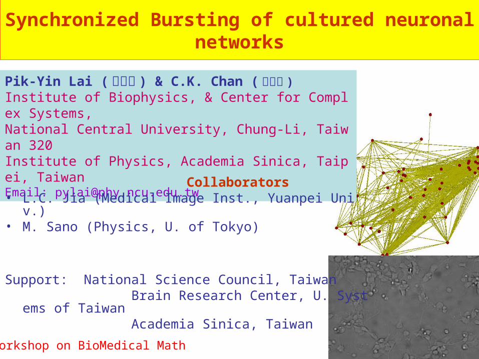
Synchronized Bursting of cultured neuronal networks
Pik-Yin Lai (黎璧賢 ) & C.K. Chan (陳志強 )Institute of Biophysics, & Center for Complex Systems, National Central University, Chung-Li, Taiwan 320Institute of Physics, Academia Sinica, Taipei, TaiwanEmail: [email protected]
Collaborators • L.C. Jia (Medical Image Inst., Yuanpei Univ.)• M. Sano (Physics, U. of Tokyo)
Support: National Science Council, Taiwan Brain Research Center, U. Systems of Taiwan Academia Sinica, Taiwan
Workshop on BioMedical Math
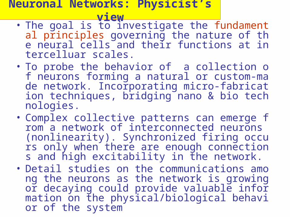
• The goal is to investigate the fundamental principles governing the nature of the neural cells and their functions at intercelluar scales.
• To probe the behavior of a collection of neurons forming a natural or custom-made network. Incorporating micro-fabrication techniques, bridging nano & bio technologies.
• Complex collective patterns can emerge from a network of interconnected neurons (nonlinearity). Synchronized firing occurs only when there are enough connections and high excitability in the network.
• Detail studies on the communications among the neurons as the network is growing or decaying could provide valuable information on the physical/biological behavior of the system
Neuronal Networks: Physicist’s view
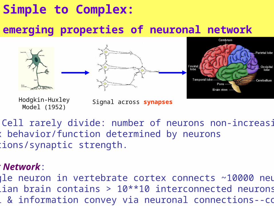
Simple to Complex:
emerging properties of neuronal network
Hodgkin-Huxley Model (1952)
Signal across synapses
Neuron Cell rarely divide: number of neurons non-increasing.Complex behavior/function determined by neurons connections/synaptic strength.
Complex Network: •A single neuron in vertebrate cortex connects ~10000 neurons•Mammalian brain contains > 10**10 interconnected neurons•Signal & information convey via neuronal connections--coding
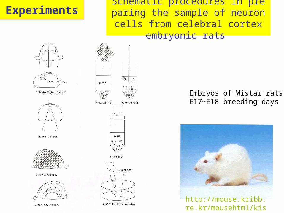
Schematic procedures in preparing the sample of neuron cells from celebra
l cortex embryonic rats
Embryos of Wistar ratsE17~E18 breeding days
http://mouse.kribb.re.kr/mousehtml/kistwistar.htm
Experiments
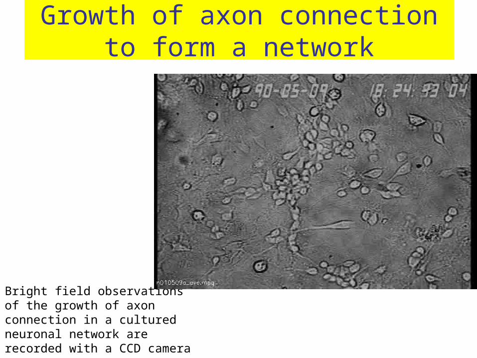
Growth of axon connection to form a network
Bright field observations of the growth of axon connection in a cultured neuronal network are recorded with a CCD camera
fluorescence microscopy
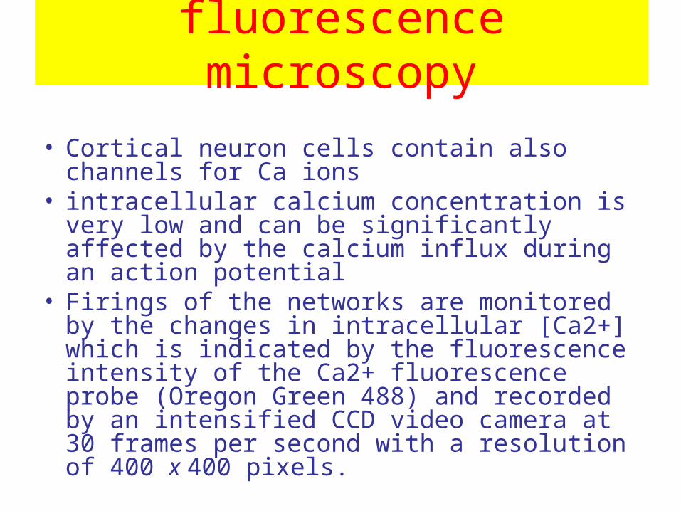
• Cortical neuron cells contain also channels for Ca ions
• intracellular calcium concentration is very low and can be significantly affected by the calcium influx during an action potential
• Firings of the networks are monitored by the changes in intracellular [Ca2+] which is indicated by the fluorescence intensity of the Ca2+ fluorescence probe (Oregon Green 488) and recorded by an intensified CCD video camera at 30 frames per second with a resolution of 400 x 400 pixels.
control the network connectivity
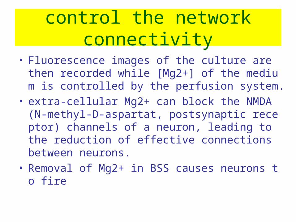
• Fluorescence images of the culture are then recorded while [Mg2+] of the medium is controlled by the perfusion system.
• extra-cellular Mg2+ can block the NMDA (N-methyl-D-aspartat, postsynaptic receptor) channels of a neuron, leading to the reduction of effective connections between neurons.
• Removal of Mg2+ in BSS causes neurons to fire
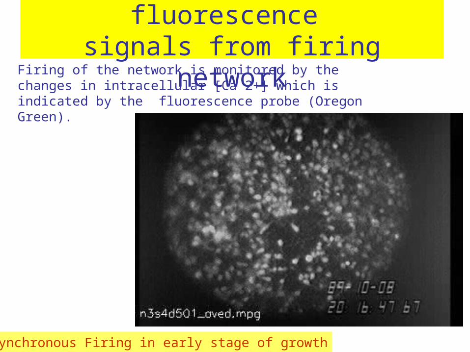
Optical recording of fluorescence signals from firing network
Firing of the network is monitored by the changes in intracellular [Ca 2+] which is indicated by the fluorescence probe (Oregon Green).
Non-synchronous Firing in early stage of growth

Synchronized Firing of Neuronal Network
Spontaneous firing of the cultures are inducedby reducing [Mg2+] in the Buffered salt solution
Synchronized Firing at later stage of growth
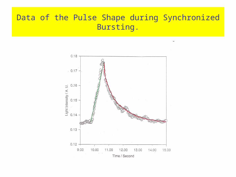
Data of the Pulse Shape during Synchronized Bursting.
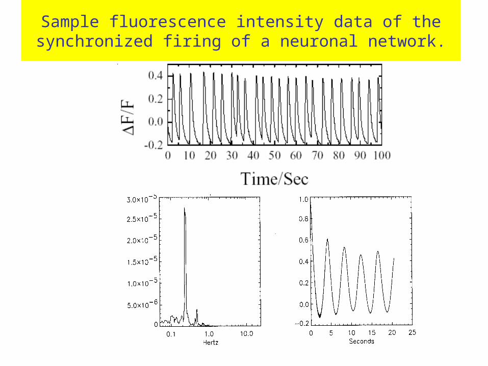
Sample fluorescence intensity data of the synchronized firing of a neuronal network.
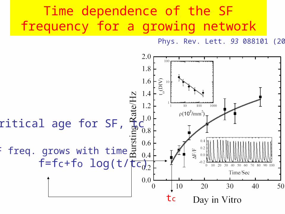
Time dependence of the SF frequency for a growing network
•Critical age for SF, tc
•SF freq. grows with time f=fc+fo log(t/tc)
tc
Phys. Rev. Lett. 93 088101 (2004)
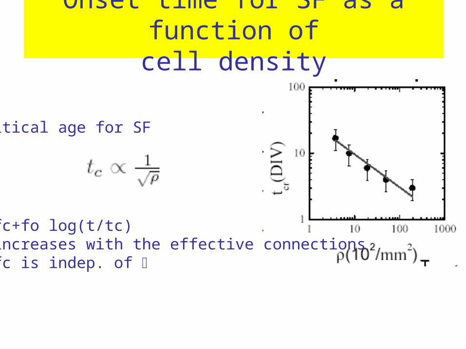
Onset time for SF as a function ofcell density
•Critical age for SF
•f=fc+fo log(t/tc)•f increases with the effective connections• fc is indep. of
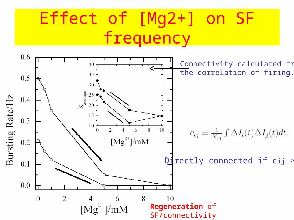
Effect of [Mg2+] on SF frequency
Regeneration of SF/connectivity
Connectivity calculated fromthe correlation of firing.
Directly connected if cij >0.5
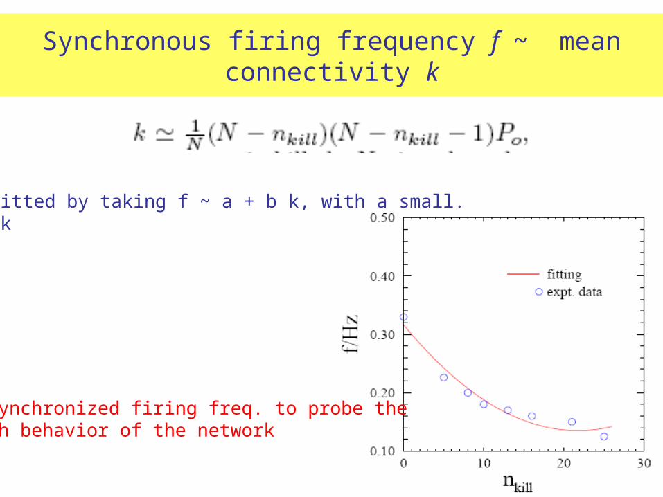
Synchronous firing frequency f ~ mean connectivity k
• Well fitted by taking f ~ a + b k, with a small. • f ~ k
Use synchronized firing freq. to probe theGrowth behavior of the network
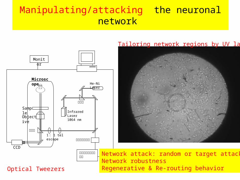
Manipulating/attacking the neuronal network
Monitor
CCD雙軸微步進馬達控制器
雙軸微步進馬達
分光鏡
1 : 1 telescope
Infrared Laser1064 nm
He-Ni Laser
分光鏡
Objective
Sample
Microscope
Optical Tweezers
Tailoring network regions by UV lasers
Network attack: random or target attackNetwork robustness Regenerative & Re-routing behavior
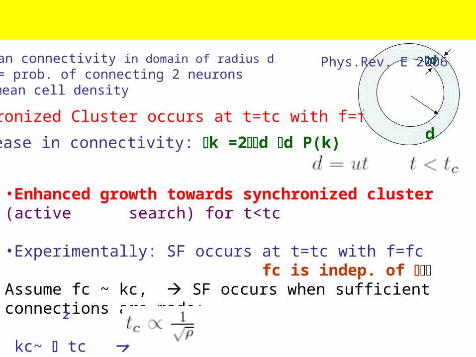
•Enhanced growth towards synchronized cluster (active search) for t<tc
•Experimentally: SF occurs at t=tc with f=fc fc is indep. of Assume fc ~ kc, SF occurs when sufficient connections are made: kc~ tc
2
Synchronized Cluster occurs at t=tc with f=fc
d
dIncrease in connectivity: k =2d d P(k)
Model for Neuronal Network Growth Phys.Rev. E 2006
k=mean connectivity in domain of radius d P(k)= prob. of connecting 2 neurons =mean cell density
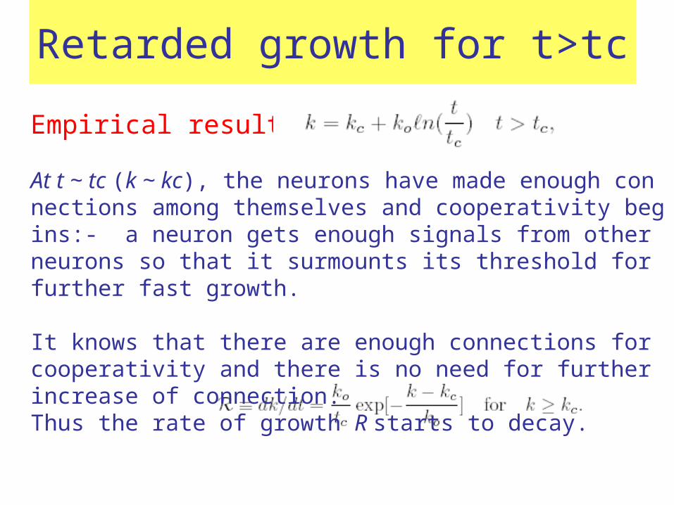
Retarded growth for t>tc
Empirical result : (f~k)
At t ~ tc (k ~ kc), the neurons have made enough connections among themselves and cooperativity begins:- a neuron gets enough signals from other neurons so that it surmounts its threshold for further fast growth.
It knows that there are enough connections for cooperativity and there is no need for further increase of connection. Thus the rate of growth R starts to decay.

Slowing down for t>tc
Using diffusive search model:
P(k)=Pc exp[-(k-kc)/ko] k>kc
Retarded growth for t>tc:
k =2d d P(k)
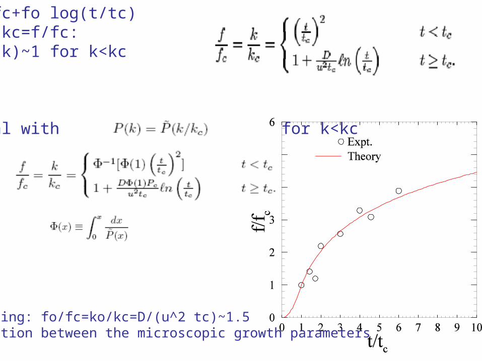
Expt: f=fc+fo log(t/tc)Assume k/kc=f/fc:Assume P(k)~1 for k<kc
In general with for k<kc
Fitting: fo/fc=ko/kc=D/(u^2 tc)~1.5relation between the microscopic growth parameters
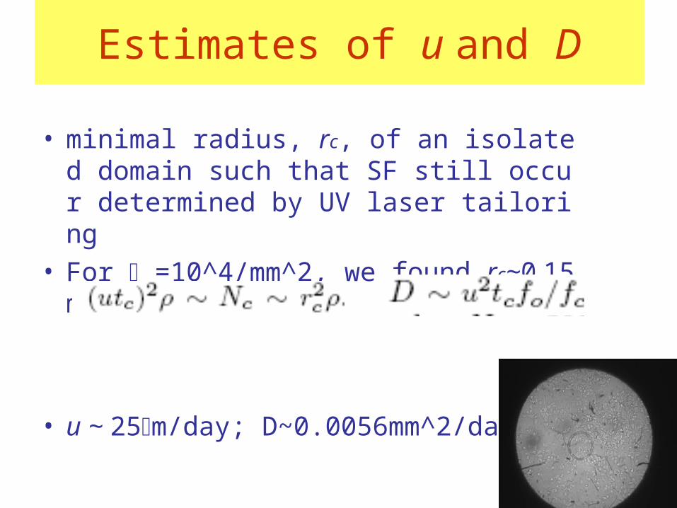
Estimates of u and D
• minimal radius, rc, of an isolated domain such that SF still occur determined by UV laser tailoring
• For =10^4/mm^2, we found rc~0.15mm
• u ~ 25m/day; D~0.0056mm^2/day
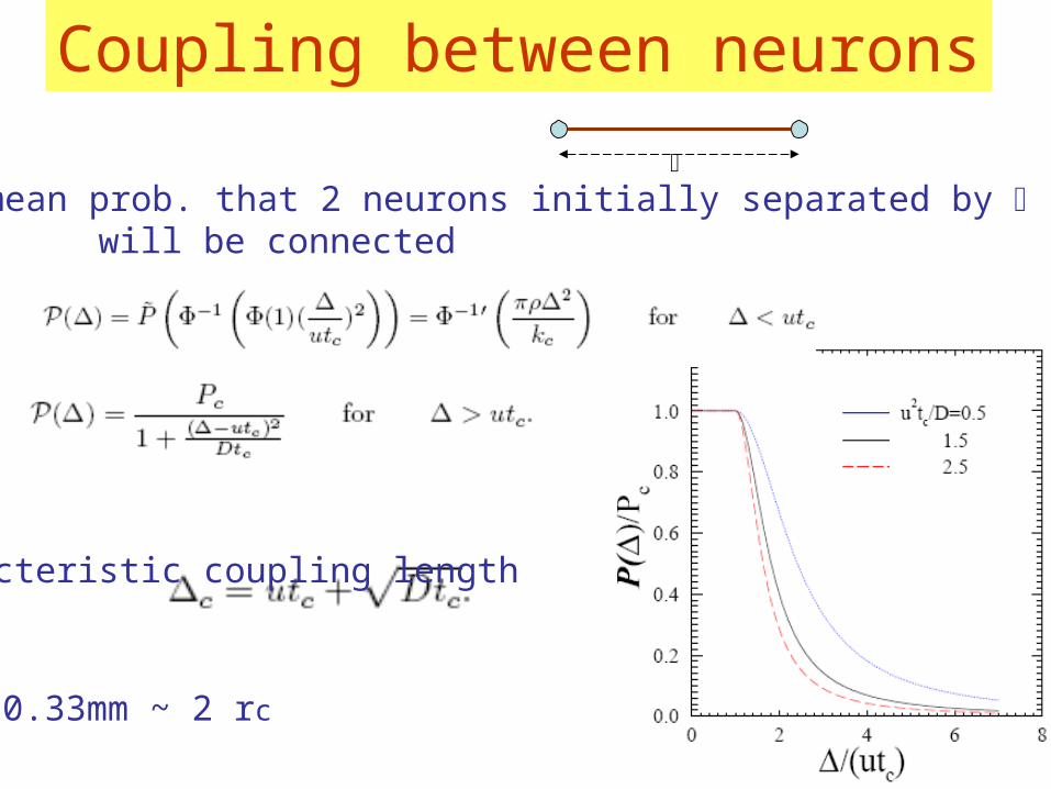
Coupling between neurons
•PP()=mean prob. that 2 neurons initially separated by will be connected
•Characteristic coupling length
• c ~ 0.33mm ~ 2 rc
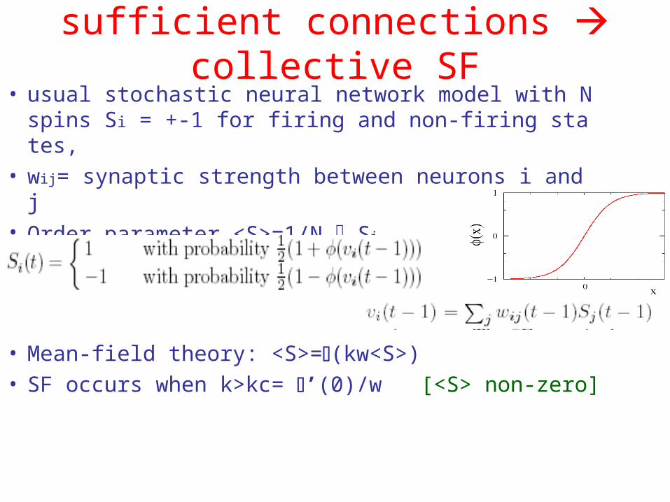
sufficient connections collective SF• usual stochastic neural network model with N spi
ns Si = +-1 for firing and non-firing states,• wij= synaptic strength between neurons i and j• Order parameter <S>=1/N Si
• Mean-field theory: <S>=(kw<S>)• SF occurs when k>kc= ’(0)/w [<S> non-zero]
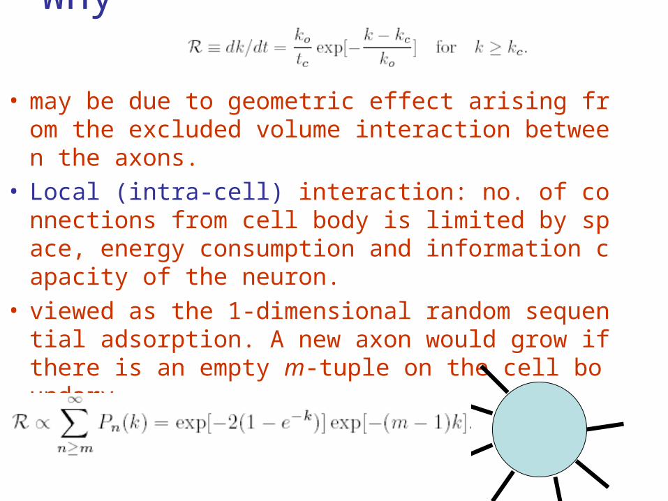
Why ?
• may be due to geometric effect arising from the excluded volume interaction between the axons.
• Local (intra-cell) interaction: no. of connections from cell body is limited by space, energy consumption and information capacity of the neuron.
• viewed as the 1-dimensional random sequential adsorption. A new axon would grow if there is an empty m-tuple on the cell boundary,
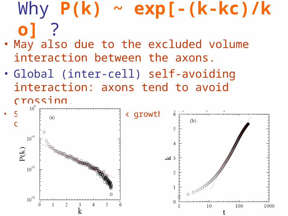
Why P(k) ~ exp[-(k-kc)/ko] ?
• May also due to the excluded volume interaction between the axons.
• Global (inter-cell) self-avoiding interaction: axons tend to avoid crossing.
• Simulation of 2D network growth with no bond crossing constraint:
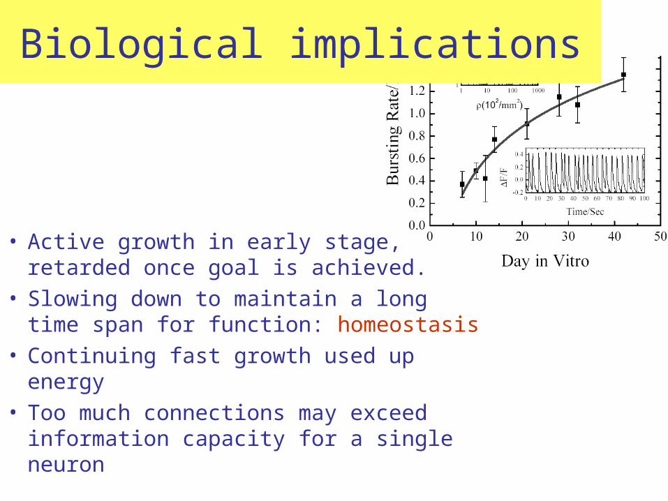
Biological implications
• Active growth in early stage, retarded once goal is achieved.
• Slowing down to maintain a long time span for function: homeostasis
• Continuing fast growth used up energy • Too much connections may exceed
information capacity for a single neuron

Physiological Homeostasis Mechanisms for self-adjusting Automatically resists changes (by negative feedback)
Walter B. CannonThe Wisdom of the Body (1939)
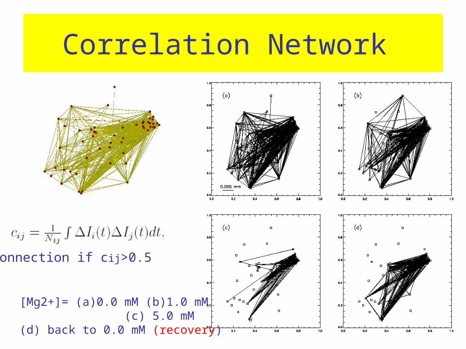
Correlation Network
[Mg2+]= (a)0.0 mM (b)1.0 mM (c) 5.0 mM (d) back to 0.0 mM (recovery)
Connection if cij>0.5
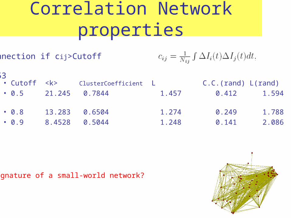
Correlation Network properties
• Cutoff <k> ClusterCoefficient L C.C.(rand) L(rand) • 0.5 21.245 0.7844 1.457 0.412 1.594 • 0.8 13.283 0.6504 1.274 0.249 1.788• 0.9 8.4528 0.5044 1.248 0.141 2.086
Connection if cij>Cutoff
N=53
Signature of a small-world network?
Some open questions• the basic mechanism of spontaneous SF is still unknown. • Detail realistic dynamics of the neurons must be added.• possible source of the induction of SF is the noise in the sy
stem. It is known that properly coupled excitable systems can be driven to synchronized states which oscillate with a well defined frequency by noise (coherence resonance).
• Heterogeneity of the elements in networks, can increase basic synchronization properties of the system.
• Effect of noise: growing network connectivity seems to be providing the needed increase in noise level.
• Robustness, plasticity & re-routing of the network.• Role of glia cells.
•Neuronal network growth is probed by synchronized firing frequencies.•Model of early stage of active search followed by diffusive search after sufficient connections can explain the experimental observations.•May provide some fundamental understanding on models of brains in the early developmental stages and learning rules. •Biological experimental system for complex network theory.
SummarySummary
Postdoc & student(M.S. Ph.D.) needed
• Theoretical/Computational research on bio-molecules, cells, neuronal/cardiac physics, biological networks.
Please contact 黎璧賢 : [email protected]
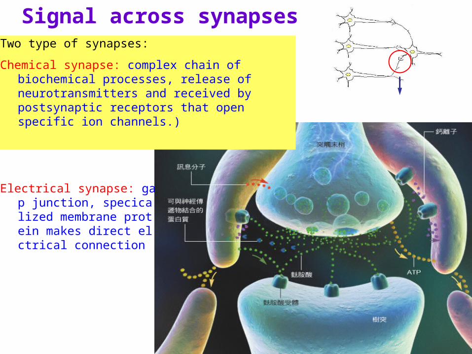
Signal across synapsesTwo type of synapses:
Chemical synapse: complex chain of biochemical processes, release of neurotransmitters and received by postsynaptic receptors that open specific ion channels.)
Electrical synapse: gap junction, specicalized membrane protein makes direct elctrical connection
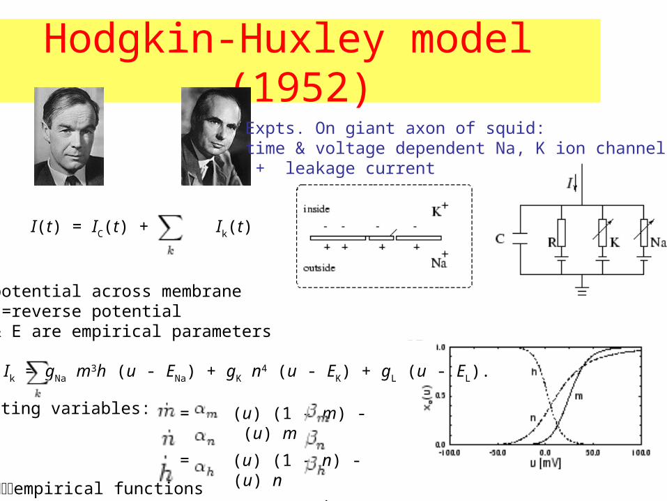
Hodgkin-Huxley model (1952)Expts. On giant axon of squid: time & voltage dependent Na, K ion channels + leakage current
I(t) = IC(t) + Ik(t)
Ik = gNa m3h (u - ENa) + gK n4 (u - EK) + gL (u - EL).
= (u) (1 - m) - (u) m
= (u) (1 - n) - (u) n
= (u) (1 - h) - (u) h
gating variables:
empirical functions
u=potential across membraneE’s=reverse potentialg & E are empirical parameters
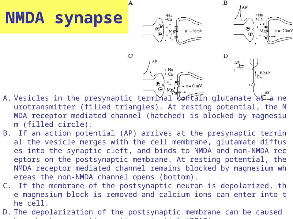
NMDA synapse
A. Vesicles in the presynaptic terminal contain glutamate as a neurotransmitter (filled triangles). At resting potential, the NMDA receptor mediated channel (hatched) is blocked by magnesium (filled circle).
B. If an action potential (AP) arrives at the presynaptic terminal the vesicle merges with the cell membrane, glutamate diffuses into the synaptic cleft, and binds to NMDA and non-NMDA receptors on the postsynaptic membrane. At resting potential, the NMDA receptor mediated channel remains blocked by magnesium whereas the non-NMDA channel opens (bottom).
C. If the membrane of the postsynaptic neuron is depolarized, the magnesium block is removed and calcium ions can enter into the cell.
D. The depolarization of the postsynaptic membrane can be caused by a back propagating action potential (BPAP).
Preparation of Neuronal Cultures
• isolated neurons from the cortex of embryos are prepared first in the form of a cell suspension in buffer solution with a concentration from 100 to 100000 cells/mm^ 2.
• Cultures are then prepared by plating a volume of 300l of the cell suspension onto the bottom of a Petri dish which has been pre-coated with polyetheylenimine.
• After the cells are allowed to adhere to the bottom of the Petri-dish, samples are subsequently filled with 2l of culture medium, and maintained in a 37C incubator with 5% CO2.
• Half of the medium is renewed twice a week. Samples can be typically maintained for up to a month.
fluorescence microscopy
• Observations are carried out while the cultures are kept in buffered salt solution. Spontaneous firing of the cultures are induced by reducing [Mg2+] in the BSS.
• Cultures are first loaded with Oregon Green for 30 min at 37C. The cultures are then washed by a Mg2+ free medium several times and then placed in a perfusion chamber on the microscope which is temperature controlled at 37C.
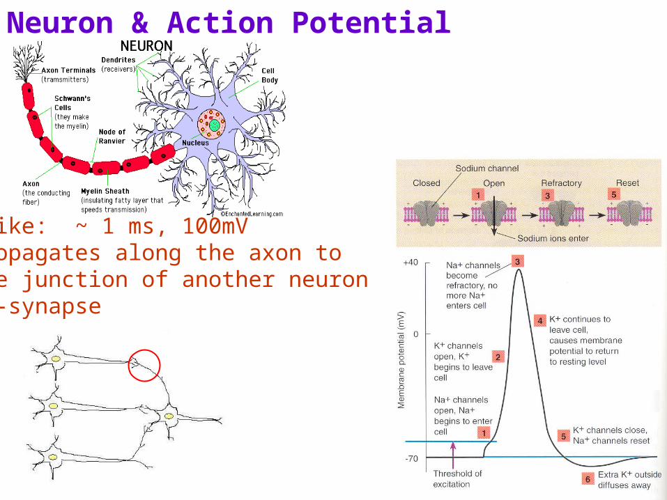
Neuron & Action Potential
Spike: ~ 1 ms, 100mVPropagates along the axon to the junction of another neuron---synapse