Embed Size (px)
Citation preview
1Muscles, Ligaments and Tendons Journal 2012; 2 (1): 1-9
Review article
Syndecans in skeletal muscle development, regeneration and homeostasis
Addolorata Pisconti*Jennifer D. BernetBradley B. Olwin
Department of Molecular, Cellular and Developmental Biology, University of Colorado, Boulder, CO
Corresponding author: Addolorata PiscontiMCD-Biology, 347 UCBBoulder, CO 80309, USAe-mail: [email protected]
*Current address:Department of Biochemistry and Cell BiologyInstitute of Integrative Biology, University of LiverpoolCrown Street, Liverpool L69 72B,UKe-mail: [email protected]
Summary
Skeletal muscle is a highly dynamic tissue that can change in size in response to physiological demands and undergo successful regeneration even upon exten-sive injury. A population of resident stem cells, termed satellite cells, accounts for skeletal muscle plasticity, maintenance and regeneration. Mammalian satellite cells, generated from muscle precursor cells during development, are maintained quiescent in the muscu-lature throughout a lifespan, but ready to activate, pro-liferate and differentiate into myocytes upon demand. Syndecans are transmembrane heparan sulfate proteo-glycans expressed in muscle precursors during embry-onic development and in satellite cells during postnatal life. In the last decades a number of crucial functions for syndecans in myogenesis and muscle disease have been described. Here we review the current knowledge of the multiple roles played by syndecans in the skel-etal muscle of several animal models and explore future perspectives for human muscle health, with a focus on muscle aging and muscular dystrophy.
Key words: syndecans, satellite cells, myogenesis, muscu-lar dystrophy, aging, muscle regeneration.
Introduction
Skeletal muscle fibers (myofibers) are large syncytial cells
derived from the fusion of hundreds of progenitor cells during development (1). These muscle precursor cells (myoblasts) originate from the epaxial somite where, during mouse em-bryonic development, undifferentiated progenitors delami-nate from the somite and migrate into the limb bud. Initially these progenitors proliferate and then terminally differenti-ate into myocytes prior to fusing with one another to form embryonic muscle fibers (2-5). A subset of these proliferat-ing muscle progenitors are thought to be “set aside” during muscle development for the generation of satellite cells dur-ing the late stages of embryonic development (6). Satellite cells, first described in frog muscle preparations (7), are the skeletal muscle stem cells (8,9) in all vertebrates, including humans (10). Satellite cells spend the vast ma-jority of their lifespan mitotically quiescent, located within a specialized anatomic niche between the plasma membrane of the myofiber and the surrounding basal lamina (7). Each myofiber harbors 7-27 satellite cells, depending on the fiber type (11). In response to stimuli such as exercise or injury, satellite cells are activated, express the myogenic master gene MyoD and re-enter the cell cycle; activated and pro-liferating MyoD+ satellite cells are termed myoblasts. After one or more rounds of proliferation, myoblasts exit the cell cycle and terminally differentiate into myocytes, which ex-press muscle contractile proteins and fuse either one to another to form new myofibers or to pre-existing damaged myofibers to repair them (12). During embryonic development and in postnatal life, a fam-ily of transmembrane heparan sulfate proteoglycans (HSP-Gs) called syndecans have emerged as key regulators of skeletal muscle formation and maintenance. In this review we discuss the role played by syndecans in skeletal muscle development, maintenance and regeneration in healthy and diseased or aging organisms. We will then highlight future perspective for human muscle health that can be inferred based on studies carried out on animal models.
Syndecan structure
Syndecans are transmembrane HSPGs, complex mol-ecules comprising a core protein that covalently links one or more long, linear carbohydrate chains, the glycosamino-glycan (GAG) chains (13). Syndecans are conserved in all metazoans (14). The core protein structure is shared by all syndecans across large evolutionary distances, from the single syndecan expressed in invertebrates to the four dif-ferent syndecans expressed in vertebrate organisms. How-ever, the specific sequence can vary considerably across gene homologues and across species (14). The ectodomain is the most variable region of the syndecan core protein containing a N-terminal signal peptide and sev-eral attachment sites for heparan sulfate chains. Addition-ally, syndecan-1 and syndecan-3 also contain attachment

A. Pisconti et al.
2 Muscles, Ligaments and Tendons Journal 2012; 2 (1): 1-9
sites for chondroitin sulfate chains (15). The syndecan ectodomain also contains at least one proteolytic cleavage site close to the transmembrane domain that is recognized by metalloproteinases (16). Syndecan shedding has an im-portant regulatory function since shed, soluble ectodomains can function as paracrine or autocrine effectors or com-petitors (16). Moreover, ectodomain shedding is a way to quickly stop the processes that transmembrane syndecans take part in (16).The transmembrane domain is a conserved single hydro-phobic region, while the short intracellular domain contains three regions where a variable intermediate region (V) sep-arates two highly conserved regions, C1 and C2, with C1 being essentially identical in all syndecans (17).Heparan sulfate (HS) contains a linear backbone com-posed by repeating sequences of glucuronic acid and N-acetyl-glucosamine disaccharide units. In HSPGs, each HS chain is attached through a xylose-galactose-galactose-uronic acid tetrasaccharide linker to serine residues on the core protein (15). HS is synthesized in the Golgi where a complex set of enzymes catalyzes not only the addition of the linker and each alternating saccharide unit, but also sub-sequent sugar modifications, which include C-5 epimeriza-tion of glucuronic acid that yields iduronic acid, replacement of N-acetylation with N-sulfation at GlcNAc residues and three different O-sulfations: 2-O-sulfation, 3-O-sulfation and 6-O-sulfation (13). HS contains a variable number of disac-charide units (up to 200) with highly sulfated domains alter-nating with less sulfated domains. It appears that specificity of heparan sulfate for its interactors is determined mainly within the highly sulfated domains. Moreover, it has been shown that one single HS chain can bind multiple interac-tors simultaneously, thus yielding complex supramolecular structures such as in the case of FGF and FGF receptors (18). The highly variable number of repeating disaccharide units together with the large number and assortment of sac-charide modifications yields an incredibly high number of possible “sequences” of functional units, which is why HS is considered the biomolecule with the highest degree of diversity (19).Chondroitin sulfate (CS) chains have a backbone com-posed by repeating glucuronic acid and N-acetyl-galac-tosamine disaccharide units attached to the core protein through the same tetrasaccharide linker that connects HS to the core protein. As opposed to HS, CS chains contain a less diverse range of modifications and these are more equally distributed along the chain (13).
Syndecans in skeletal muscle development
Syndecan involvement in skeletal muscle development has been investigated in flies, turkeys and mice (20-23).During Drosophila development, the single syndecan is expressed in muscle fibers and appears to be involved in motor-axon guidance by acting as a receptor for the neu-ral receptor tyrosine phosphatase (RPTP) LAR (22). Thus, Drosophila syndecan controls muscle innervation during development and therefore regulates the onset of muscle
functional maturation. Whether Drosophila syndecan is also involved directly in regulating embryonic myofiber forma-tion, is unknown.The role of syndecans in vertebrate muscle development has been studied in mice and birds (20,24). Developing mouse muscles express syndecan-1, syndecan-3 and syndecan-4 with similar topological distributions, but dif-ferent temporal regulation (20,21). Northern and Western blot analyses of syndecan-1, syndecan-3 and syndecan-4 mRNA and protein, respectively, show that syndecan-1 protein peaks prior to other syndecans, around E12.5, then rapidly decreases and is completely absent by P2 (20). In contrast, syndecan-3 and syndecan-4 peak around E14.5 and E13.5 respectively, but then decrease much more slow-ly and are still expressed in newborn and adult mice (20,25). Expression of syndecan-1, syndecan-3 and syndecan-4 in embryonic muscle is localized to both myoblasts and myofi-bers. While syndecan-1 is not detected in postnatal muscle, syndecan-3 and syndecan-4 proteins are restricted to satel-lite cells and possibly vascular cells (21).In embryonic turkey muscle, distribution of syndecan ex-pression between E14 and E24 is regulated in a similar pat-tern as in mice, peaking between E14 (syndecan-3), E16 (syndecan-2) and E18 (syndecan-4), followed by a decline at later time points (E22-E24). Syndecan-2, 3 and 4 expres-sion is presumably restricted to satellite cells in postnatal turkey muscle (23). Important roles for syndecans in muscle development were confirmed in turkey embryonic pectoralis major muscle at different developmental stages (E14 - E24) derived from ei-ther a high body weight genetically selected line (F line) or a low body weight line (RBC2 line). In this study, Liu et al., found that the F line (high body weight) turkey muscle has higher levels of syndecan-2, syndecan-3 and syndecan-4 than the RBC2 line (low body weight) turkey muscle, sup-porting a key role for syndecans in the regulation of muscle development and size (23).
Syndecans in skeletal muscle maintenance and regeneration
The hypothesis that HSPGs, such as syndecans, are in-volved in myogenesis could have been already inferred when a key role for HS in growth factor signaling in myo-blasts was described (26,27). Subsequent studies from Brandan and colleagues showed a role for specific HSPGs in myogenic differentiation using the C2C12 myoblast cell line (28-33). Shortly after this group showed that gene ex-pression levels and protein levels of a number of HSPGs were regulated in vivo during injury-induced regeneration in mouse limb muscles (34). The only two syndecans identi-fied by Casar et al. that appeared to be expressed in re-generating muscle were syndecan-3 and syndecan-4 (34). Indeed, Cornelison et al. had previously shown that syn-decan-3 and syndecan-4 are the only two syndecans de-tectable by immunofluorescence in postnatal mouse skel-etal muscle, co-localizing with markers of satellite cells (21). The time course of syndecan-3 and syndecan-4 expression
Syndecans in skeletal muscle development, regeneration and homeostasis
3Muscles, Ligaments and Tendons Journal 2012; 2 (1): 1-9
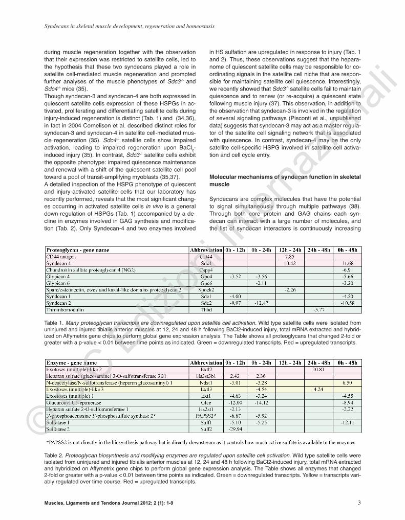
during muscle regeneration together with the observation that their expression was restricted to satellite cells, led to the hypothesis that these two syndecans played a role in satellite cell-mediated muscle regeneration and prompted further analyses of the muscle phenotypes of Sdc3-/- and Sdc4-/- mice (35). Though syndecan-3 and syndecan-4 are both expressed in quiescent satellite cells expression of these HSPGs in ac-tivated, proliferating and differentiating satellite cells during injury-induced regeneration is distinct (Tab. 1) and (34,36), in fact in 2004 Cornelison et al. described distinct roles for syndecan-3 and syndecan-4 in satellite cell-mediated mus-cle regeneration (35). Sdc4-/- satellite cells show impaired activation, leading to impaired regeneration upon BaCl2-induced injury (35). In contrast, Sdc3-/- satellite cells exhibit the opposite phenotype: impaired quiescence maintenance and renewal with a shift of the quiescent satellite cell pool toward a pool of transit-amplifying myoblasts (35,37).A detailed inspection of the HSPG phenotype of quiescent and injury-activated satellite cells that our laboratory has recently performed, reveals that the most significant chang-es occurring in activated satellite cells in vivo is a general down-regulation of HSPGs (Tab. 1) accompanied by a de-cline in enzymes involved in GAG synthesis and modifica-tion (Tab. 2). Only Syndecan-4 and two enzymes involved
in HS sulfation are upregulated in response to injury (Tab. 1 and 2). Thus, these observations suggest that the hepara-nome of quiescent satellite cells may be responsible for co-ordinating signals in the satellite cell niche that are respon-sible for maintaining satellite cell quiescence. Interestingly, we recently showed that Sdc3-/- satellite cells fail to maintain quiescence and to renew (or re-acquire) a quiescent state following muscle injury (37). This observation, in addition to the observation that syndecan-3 is involved in the regulation of several signaling pathways (Pisconti et al., unpublished data) suggests that syndecan-3 may act as a master regula-tor of the satellite cell signaling network that is associated with quiescence. In contrast, syndecan-4 may be the only satellite cell-specific HSPG involved in satellite cell activa-tion and cell cycle entry.
Molecular mechanisms of syndecan function in skeletal muscle
Syndecans are complex molecules that have the potential to signal simultaneously through multiple pathways (38). Through both core protein and GAG chains each syn-decan can interact with a large number of molecules, and the list of syndecan interactors is continuously increasing
Table 1. Many proteoglycan transcripts are downregulated upon satellite cell activation. Wild type satellite cells were isolated from uninjured and injured tibialis anterior muscles at 12, 24 and 48 h following BaCl2-induced injury, total mRNA extracted and hybrid-ized on Affymetrix gene chips to perform global gene expression analysis. The Table shows all proteoglycans that changed 2-fold or greater with a p-value < 0.01 between time points as indicated. Green = downregulated transcripts. Red = upregulated transcripts.
Table 2. Proteoglycan biosynthesis and modifying enzymes are regulated upon satellite cell activation. Wild type satellite cells were isolated from uninjured and injured tibialis anterior muscles at 12, 24 and 48 h following BaCl2-induced injury, total mRNA extracted and hybridized on Affymetrix gene chips to perform global gene expression analysis. The Table shows all enzymes that changed 2-fold or greater with a p-value < 0.01 between time points as indicated. Green = downregulated transcripts. Yellow = transcripts vari-ably regulated over time course. Red = upregulated transcripts.

A. Pisconti et al.
4 Muscles, Ligaments and Tendons Journal 2012; 2 (1): 1-9
(38). Decoration of HSPG core proteins with GAG chains and subsequent saccharide modifications may be more cell type-specific than core protein-specific. In other words, the same core protein tends to receive different GAG chains when expressed in different cell types, or under different physiological states, with this depending mainly on the set of GAG biosynthetic enzymes expressed in each cell type (39,40). The opposite case, that different core proteins re-ceive similar GAG chains when expressed in the same cell type has been proposed in the context of fibroblast adhe-sion, where different membrane bound HSPGs bear GAG chains that are different in length but similar in sulfation pattern and capability to bind fibronectin (41). However, Tu-mova et al. did observe subtle differences in HS structure present on different core proteins expressed in the same cell type and the fact that the only function tested by Tu-mova et al., fibronectin binding, did not change significantly across the different core proteins examined, does not ex-clude that other functions that were not tested (e.g. growth factor binding) could change accordingly with the difference in HS structure observed. In support of this hypothesis it has been shown that even small differences in HS structure can dramatically affect FGF binding (42) and function (43). Thus, the possibility that different core proteins receive dif-ferent GAG chains with different biological functions when expressed in the same cell type is still open.In postnatal mammalian skeletal muscle the only two syn-decan proteins detected are syndecan-3 and syndecan-4 (21), which are expressed in satellite cells and appear to control muscle homeostasis through distinct mechanisms (35,37). Syndecan-3 is the largest syndecan in mammals, harbor-ing both HS and CS chains (17,44,45). In mouse satellite cells, syndecan-3 is found in a complex with Notch1 and promotes TACE-mediated cleavage of Notch, allowing for Notch signal transduction into satellite cells (37). In the ab-sence of syndecan-3, Notch processing upon ligand binding is dramatically reduced as is generation of the Notch intra-cellular domain and subsequent induction of Notch target genes (37). As a consequence of reduced Notch signaling, Sdc3-/- satellite cells proliferate more slowly than wild type cells and fail to maintain or to return to a quiescent state (37). The resulting phenotype is intriguing: Sdc3-/- injured muscles retain full regenerative capacity and undergo pro-gressive myofiber size increase over time despite showing a dramatic loss of satellite cells (37). A possible hypothesis to explain this paradoxical phenotype has been proposed (37): loss of syndecan-3 impairs satellite cell capacity to enter a quiescent state without affecting their ability to dif-ferentiate. Thus, a shift in the satellite cell population from a quiescent pool to an activated, proliferating and differen-tiating pool over time leads to myofiber hypertrophy and depletion of the quiescent satellite cell pool (37). Although loss of quiescence but not differentiation can be explained by loss of Notch signaling, this cannot explain how Sdc3-/- myoblasts remain proliferative for a long time. It is possible that other signaling pathways regulated by syndecan-3 in satellite cells compensate for loss of Notch signaling to maintain Sdc3-/- myoblasts in a proliferative cycle. Indeed,
syndecan-3 also regulates FGF and HGF signaling, though the molecular mechanisms involved are unknown (30,35). In Sdc3-/- satellite cells Notch signaling is decreased while FGF and HGF signaling are increased (35) and may ac-count for the observed maintenance of a population of pro-liferating myoblasts. Whether the core protein or the GAG chains of syndecan-3 are the main mediators of syndecan-3 function in satellite cells and myoblasts is unknown, how-ever both are required to rescue the Notch signaling pheno-type in Sdc3-/- myoblasts (37).Syndecan-4 is the smallest syndecan, but the best stud-ied in myoblasts as well as in other systems (46). In mouse muscle, syndecan-4 plays a key role in mediating satellite cell activation in response to injury (35). Though the molec-ular mechanisms underlying syndecan-4 function in mouse satellite cells are largely unknown, it has been hypothesized that syndecan-4 HS chains are involved in the regulation of FGF and HGF signaling in proliferating satellite cells (35). In the absence of syndecan-4, both FGF and HGF signal-ing are impaired, but can be rescued by heparin treatment (35). However, a direct mechanistic analysis of syndecan-4 function in mouse satellite cells is missing. In contrast, a sig-nificant effort has been made to understand whether a func-tional interaction between syndecan-4 and FGF2 exists in turkey satellite cells (47,48). This hypothesis is reasonable, since (a) syndecan-4 is involved in FGF2 signaling in other systems outside the musculature, (b) both FGF2 treatment and syndecan-4 ectopic expression in primary satellite cells lead to differentiation inhibition (47). However, genetic anal-ysis revealed that in turkey satellite cells syndecan-4, with or without GAG chains, promotes proliferation and inhibits differentiation in an FGF2-independent manner (47-49). Syndecan-4 is also expressed in young myotubes prior to myofiber growth and final maturation (21). An intriguing recent finding shows that syndecan-4 protein, along with β1-integrin, localizes in costamers of cultured rat myotubes and is regulated by electrical activity (50). Denervation of rat tibialis anterior muscles or treatment of cultured myo-tubes with tetrodotoxin induce syndecan-4 and β1-integrin downregulation and are associated with reduced myotube adhesion (50). Lastly, a role for syndecan-4 in myoblast migration has been hypothesized, however a detailed analysis is missing.Heparan sulfate and chondroitin sulfate are the two types of GAG chains covalently attached to syndecan core pro-teins (15). The role of HS and CS in muscle function and myogenesis has been studied irrespectively of which core proteins were attached to them (21,26,51-58). Understand-ing how GAGs function and signal is complicated and fas-cinating since: (1) the saccharide sequence of GAG chains is not template driven as is the amino acid sequence of proteins, but is the final result of multiple enzymes active simultaneously in a cell; (2) the signal mediated through GAG chains appears to be an “analog” signal where the entire pattern of saccharide and sulfated domains present at a given time in a given microdomain of the cell, can af-fect multiple functions simultaneously, as opposed to the “digital” type of signal driven by canonical protein-protein interactions such as ligand-receptor, kinase-substrate, etc;
Syndecans in skeletal muscle development, regeneration and homeostasis
5Muscles, Ligaments and Tendons Journal 2012; 2 (1): 1-9
and, (3) despite its generally “analog” nature, GAG interac-tors often exhibit high specificity for distinct oligosaccharide sequences. Both HS and CS are involved in muscle precursor prolif-eration and differentiation, with highly sulfated HS generally promoting proliferation and CS generally promoting differ-entiation (26,34,51,52,55,57,59,60), though exceptions to this general trend have been described (58,61). Unexpect-edly, we have recently determined that satellite cell activa-tion induces downregulation of several proteoglycans (PGs) and GAG biosynthesis enzymes, suggesting a key role for PGs present in the satellite cell niche in maintaining satellite cells in a quiescent state (Tab. 1 and Tab. 2).Interesting results have recently arisen from the study of knockout mice lacking expression of one or more enzymes involved in GAG biosynthesis. For example the satellite cell phenotype observed in Sulf1-/-;Sulf2-/- double knockout mice appears the opposite of the phenotype observed in Sdc3-/- mice (37,57). Sulfs are extracellular enzymes that remove sulfate groups from HSPGs (heparan sulfate endosulfatas-es) (62). Based on these results it is plausible to hypothe-size that loss of one HSPG (such as syndecan-3) leads to a general rearrangement of the satellite cell glycocalyx result-ing in an overall reduction in HS on the satellite cell surface. Vice-versa, loss of two extracellular sulfatases is expected to cause a general increase in sulfated HS on the satel-lite cell surface, which may explain why the Sdc3-/- muscle satellite cell phenotype appears opposite when compared to the Sulf1-/-;Sulf2-/- phenotype (37,57). Moreover, loss of one HSPG may also indirectly affect the level of decoration and amount of sulfation of other HSPGs by disrupting the normal distribution of GAG biosynthesis enzymes across several core proteins and by altering the normal balance between positive and negative feedback loops in each bio-synthetic pathway.
Syndecans in aged and diseased skeletal muscle
Muscular dystrophy is a family of genetic disorders char-acterized by muscle weakness, chronic inflammation, fibro-sis and eventually muscle loss (63). Although mutations in more than 20 different genes have been found that cause a clinical phenotype classified as muscular dystrophy, some histopathological features are shared by the vast majority of muscular dystrophies, including alterations to the satellite cell niche that are associated with exhaustion of satellite cell regenerative capacity (63). The level of HSPGs in muscles of dystrophic patients or animals is generally increased (33,54,64-68), suggesting a pathogenic role for HS and HSPGs in muscular dystro-phy. In particular, syndecan-3 was augmented in Duchenne patients (33) and this finding, together with the finding that mdx satellite cells have increased levels of HS and CS and increased responsiveness to FGF (54), provided impetus to study the role of syndecan-3 in dystrophinopathies such as Duchenne muscular dystrophy (DMD). Indeed, our labora-tory has recently observed a possible pathogenic role for syndecan-3 in a mouse model of DMD, although this work
is still in progress as we write.Aging of human subjects is often associated with frailty, sarcopenia and impaired muscle regeneration, represent-ing a major public health problem in modern societies where the average lifespan has increased (69). As in muscular dystrophy, also in aging the prevailing hypothesis to explain loss of regenerative capacity is the exhaustion of satellite cell numbers or function, although the underlying cellular and molecular mechanisms are a matter of debate (70-75). If progressive impairment of satellite cell regenerative ca-pacity is a major cause of age-related muscle weakness and loss, it is reasonable to hypothesize a key pathogenic role for the aging satellite cell niche, which is characterized by reduced vascularization and increased fibrosis and adi-pogenesis (76,77). Several signaling pathways have been found altered in aging satellite cells in vivo (71,78,79). Moreover, it has been shown through parabiosis experi-ments that a “young environment” can rescue age-related muscle regeneration defect in mice (72). Our laboratory has recently shown that when young satellite cells are trans-planted into young hosts together with their native niche (the myofiber), the transplanted muscle retains full regen-erative capacity as the recipient animal ages as opposed to its non-transplanted contralateral, which undergoes the normal process of age-related loss of muscle mass and function (80). This prevention of muscle aging observed in transplanted muscles is entirely supported by donor-de-rived satellite cells, which remain viable in the host muscle throughout the mouse lifespan (80). When the donor cells (myofiber + associated satellite cells) where isolated from Sdc4-/- mice, this anti-aging effect was not observed, point-ing out to syndecan-4 as a crucial component of the satellite cell niche (80).
Perspective for human health
Only in the last decade have the HSPG and muscle biol-ogy communities begun to appreciate the importance of syndecans in skeletal muscle development and regenera-tion and therefore it is not surprising that only a few studies in humans are yet available. However, studies in mice and other model organisms show promising results that will cer-tainly inspire more human research. Of particular interest are the findings concerning muscle injury, muscular dystrophy and aging. While there is no in-formation on the role of syndecans during muscle injury and aging in humans, it has been shown that expression levels of some proteoglycans, including syndecan-3, is augment-ed in Duchenne muscular dystrophy patients (33,64,67). This observation, in conjunction with our recent observa-tions made in Sdc3-/- and dystrophic mice suggests that syn-decans may be promising therapeutic targets. The satellite cell niche is altered in dystrophic muscles, pos-sibly due to continuous myofiber damage and leakage, myo-fiber necrosis and chronic inflammation, which in turn lead to extracellular matrix remodeling (81). In this context, target-ing specific components of the niche, such as syndecans, may represent a potential therapeutic strategy for enhanc-
A. Pisconti et al.
6 Muscles, Ligaments and Tendons Journal 2012; 2 (1): 1-9
ing muscle regeneration and slowing disease progression. Although a therapy that enhances muscle regeneration is not expected to be curative for muscular dystrophy, it is rea-sonable to hypothesize that enhancing regeneration would greatly improve the lifestyle of dystrophic patients (82). Ad-ditionally, therapies aimed at improving muscle regenera-tion are also expected to increase the efficacy of stem cell and gene therapies, either by promoting exogenous stem cell contribution to host myofiber or by favoring contribution from transduced endogenous satellite cells.Finally, a potential role for syndecans in human muscle health that has not been sufficiently explored is the use of syndecans as viral receptors for gene therapy. HS is involved in many viral infection processes acting as a re-ceptor or co-receptor for viral particles (83). For example, infection of muscle fibers with herpes simplex virus type 1 (HSV-1) is mediated by HS, although inhibited by other unidentified ECM components (84). This is a field that has the potential to yield interesting results in the future, as the unique expression of syndecan-3 and syndecan-4 is satel-lite cells could be used, for example, to target viral vectors specifically to satellite cells.
In the last century the study of HSPGs in the musculoskel-etal and other systems has produced a whole new level of understanding of cell adhesion, cell signaling and cell dif-ferentiation and provided essential tools for the protection of human health. For example, heparin, a highly sulfated heparan sulfate, is one of the most widely used therapeutic agents worldwide. Future studies aimed at identifying roles for syndecans in human healthy and diseased muscle in conjunction with a detailed characterization of the signaling pathways and molecular networks controlled by syndecans, are expected to contribute significantly to our understanding of muscle biology and our ability to treat muscle disorders.
Acknowledgments
The authors wish to thank Dr. Kathleen Tanaka and Dr. Dawn Cornelisosn for her contribution to the preparation and analysis of the microarray datasets shown in Table 1 and 2, Dr. Florian Bentzinger for critical reading of the man-uscript and Prof. Jerry Turnbull for insightful discussions on HS structure and function.
References
1. Schultz E, McCormick KM. Skeletal muscle satellite cells. Rev Physiol Biochem Pharmacol 1994;123:213-57.
2. Ben-Yair R, Kalcheim C. Lineage analysis of the avian dermomyotome sheet reveals the existence of single cells with both dermal and muscle progenitor fates. Develop-ment 2005, Feb;132(4):689-701.
3. Gros J, Manceau M, Thomé V, Marcelle C. A common so-mitic origin for embryonic muscle progenitors and satellite cells. Nature 2005, Jun 16;435(7044):954-958.
4. Kassar-Duchossoy L, Giacone E, Gayraud-Morel B, Jory A, Gomès D, Tajbakhsh S. Pax3/pax7 mark a novel popu-lation of primitive myogenic cells during development. Genes Dev 2005, Jun 15;19(12):1426-1431.
5. Relaix F, Rocancourt D, Mansouri A, Buckingham M. A pax3/pax7-dependent population of skeletal muscle pro-genitor cells. Nature 2005, Jun 16;435(7044):948-953.
6. Vasyutina E, Lenhard DC, Birchmeier C. Notch function in myogenesis. Cell Cycle 2007, Jun 15;6(12):1451-1454.
7. Mauro A. Satellite cell of skeletal muscle fibers. J Biophys Biochem Cytol 1961, Feb;9:493-495.
8. Collins CA, Olsen I, Zammit PS, Heslop L, Petrie A, Par-tridge TA, Morgan JE. Stem cell function, self-renewal, and behavioral heterogeneity of cells from the adult muscle sat-ellite cell niche. Cell 2005, Jul 29;122(2):289-301.
9. Murphy MM, Lawson JA, Mathew SJ, Hutcheson DA, Kar-don G. Satellite cells, connective tissue fibroblasts and their interactions are crucial for muscle regeneration. De-velopment 2011, Sep;138(17):3625-3637.
10. Péault B, Rudnicki M, Torrente Y, Cossu G, Tremblay JP, Partridge T, et al. Stem and progenitor cells in skeletal muscle development, maintenance, and therapy. Mol Ther 2007, May;15(5):867-877.
11. Zammit PS, Heslop L, Hudon V, Rosenblatt JD, Tajbakhsh S, Buckingham ME, et al. Kinetics of myoblast prolifera-tion show that resident satellite cells are competent to fully regenerate skeletal muscle fibers. Exp Cell Res 2002, Nov 15;281(1):39-49.
12. Olguín HC, Pisconti A. Marking the tempo for myogenesis: Pax7 and the regulation of muscle stem cell fate decisions. J Cell Mol Med 2011, May 26.
13. Bernfield M, Götte M, Park PW, Reizes O, Fitzgerald ML, Lincecum J, Zako M. Functions of cell surface heparan sul-fate proteoglycans. Annu Rev Biochem 1999;68:729-777.
14. Chakravarti R, Adams JC. Comparative genomics of the syndecans defines an ancestral genomic context associat-ed with matrilins in vertebrates. BMC Genomics 2006;7:83.
15. Carey DJ, Conner K, Asundi VK, O’Mahony DJ, Stahl RC, Showalter L, et al. Cdna cloning, genomic organization, and in vivo expression of rat n-syndecan. J Biol Chem 1997, Jan 31;272(5):2873-2879.
16. Manon-Jensen T, Itoh Y, Couchman JR. Proteoglycans in health and disease: The multiple roles of syndecan shed-ding. Febs J 2010, Oct;277(19):3876-3889.
17. Couchman JR. Transmembrane signaling proteoglycans. Annu Rev Cell Dev Biol 2010, Nov 10;26:89-114.
18. Rapraeger AC. In the clutches of proteoglycans: How does heparan sulfate regulate FGF binding? Chem Biol 1995, Oct;2(10):645-649.
19. Turnbull JE. Heparan sulfate glycomics: Towards sys-tems biology strategies. Biochem Soc Trans 2010, Oct;38(5):1356-1360.
20. Olguin H, Brandan E. Expression and localization of pro-teoglycans during limb myogenic activation. Dev Dyn 2001, May;221(1):106-115.
21. Cornelison DD, Filla MS, Stanley HM, Rapraeger AC, Ol-win BB. Syndecan-3 and syndecan-4 specifically mark skeletal muscle satellite cells and are implicated in satellite
Syndecans in skeletal muscle development, regeneration and homeostasis
7Muscles, Ligaments and Tendons Journal 2012; 2 (1): 1-9
cell maintenance and muscle regeneration. Dev Biol 2001, Nov 1;239(1):79-94.
22. Fox AN, Zinn K. The heparan sulfate proteoglycan syn-decan is an in vivo ligand for the drosophila LAR receptor tyrosine phosphatase. Curr Biol 2005, Oct 11;15(19):1701-1711.
23. Liu C, McFarland DC, Nestor KE, Velleman SG. Differential expression of membrane-associated heparan sulfate pro-teoglycans in the skeletal muscle of turkeys with different growth rates. Poult Sci 2006, Mar;85(3):422-428.
24. Cornelison DD, Filla MS, Stanley HM, Rapraeger AC, Ol-win BB. Syndecan-3 and syndecan-4 specifically mark skeletal muscle satellite cells and are implicated in satellite cell maintenance and muscle regeneration. Dev Biol 2001, Nov 1;239(1):79-94.
25. Cornelison DD, Filla MS, Stanley HM, Rapraeger AC, Ol-win BB. Syndecan-3 and syndecan-4 specifically mark skeletal muscle satellite cells and are implicated in satellite cell maintenance and muscle regeneration. Dev Biol 2001, Nov 1;239(1):79-94.
26. Rapraeger AC, Krufka A, Olwin BB. Requirement of hepa-ran sulfate for bfgf-mediated fibroblast growth and myoblast differentiation. Science 1991, Jun 21;252(5013):1705-1708.
27. Yayon A, Klagsbrun M, Esko JD, Leder P, Ornitz DM. Cell surface, heparin-like molecules are required for binding of basic fibroblast growth factor to its high affinity receptor. Cell 1991, Feb 22;64(4):841-848.
28. Larraín J, Alvarez J, Hassell JR, Brandan E. Expression of perlecan, a proteoglycan that binds myogenic inhibitory basic fibroblast growth factor, is down regulated during skeletal muscle differentiation. Exp Cell Res 1997, Aug 1;234(2):405-412.
29. Villar MJ, Hassell JR, Brandan E. Interaction of skeletal muscle cells with collagen type IV is mediated by perlecan associated with the cell surface. J Cell Biochem 1999, Dec 15;75(4):665-674.
30. Fuentealba L, Carey DJ, Brandan E. Antisense inhibition of syndecan-3 expression during skeletal muscle differen-tiation accelerates myogenesis through a basic fibroblast growth factor-dependent mechanism. J Biol Chem 1999, Dec 31;274(53):37876-84.
31. Riquelme C, Larrain J, Schonherr E, Henriquez JP, Kresse H, Brandan E. Antisense inhibition of decorin expression in myoblasts decreases cell responsiveness to transforming growth factor beta and accelerates skeletal muscle differ-entiation. J Biol Chem 2001, Feb 2;276(5):3589-3596.
32. Henriquez JP, Casar JC, Fuentealba L, Carey DJ, Bran-dan E. Extracellular matrix histone H1 binds to perlecan, is present in regenerating skeletal muscle and stimulates myoblast proliferation. J Cell Sci 2002, May 15;115(Pt 10):2041-2051.
33. Alvarez K, Fadic R, Brandan E. Augmented synthesis and differential localization of heparan sulfate proteogly-cans in duchenne muscular dystrophy. J Cell Biochem 2002;85(4):703-713.
34. Casar JC, Cabello-Verrugio C, Olguin H, Aldunate R, In-estrosa NC, Brandan E. Heparan sulfate proteoglycans
are increased during skeletal muscle regeneration: Re-quirement of syndecan-3 for successful fiber formation. J Cell Sci 2004, Jan 1;117(Pt 1):73-84.
35. Cornelison DD, Wilcox-Adelman SA, Goetinck PF, Rau-vala H, Rapraeger AC, Olwin BB. Essential and separable roles for syndecan-3 and syndecan-4 in skeletal muscle development and regeneration. Genes Dev 2004, Sep 15;18(18):2231-2236.
36. Tanaka KK, Hall JK, Troy AA, Cornelison DD, Majka SM, Olwin BB. Syndecan-4-Expressing muscle progenitor cells in the SP engraft as satellite cells during muscle regenera-tion. Cell Stem Cell 2009, Mar 6;4(3):217-225.
37. Pisconti A, Cornelison DD, Olguín HC, Antwine TL, Olwin BB. Syndecan-3 and notch cooperate in regulating adult myogenesis. J Cell Biol 2010, Aug 9;190(3):427-441.
38. Tkachenko E, Rhodes JM, Simons M. Syndecans: New kids on the signaling block. Circ Res 2005, Mar 18;96(5):488-500.
39. Sanderson RD, Turnbull JE, Gallagher JT, Lander AD. Fine structure of heparan sulfate regulates syndecan-1 function and cell behavior. J Biol Chem 1994, May 6;269(18):13100-13106.
40. Kato M, Wang H, Bernfield M, Gallagher JT, Turnbull JE. Cell surface syndecan-1 on distinct cell types differs in fine structure and ligand binding of its heparan sulfate chains. J Biol Chem 1994, Jul 22;269(29):18881-18890.
41. Tumova S, Woods A, Couchman JR. Heparan sulfate chains from glypican and syndecans bind the hep II do-main of fibronectin similarly despite minor structural differ-ences. J Biol Chem 2000, Mar 31; 275 (13): 9410-9417.
42. Brickman YG, Ford MD, Gallagher JT, Nurcombe V, Bartlett PF, Turnbull JE. Structural modification of fibroblast growth factor - binding heparan sulfate at a determinative stage of neural development. J Biol Chem 1998, Feb 20; 273(8): 4350-4359.
43. Brickman YG, Nurcombe V, Ford MD, Gallagher JT, Bartlett PF, Turnbull JE. Structural comparison of fibroblast growth factor-specific heparan sulfates derived from a growing or differentiating neuroepithelial cell line. Glycobiology 1998, May;8(5):463-471.
44. Gould SE, Upholt WB, Kosher RA. Syndecan 3: A member of the syndecan family of membrane-intercalated proteo-glycans that is expressed in high amounts at the onset of chicken limb cartilage differentiation. Proc Natl Acad Sci U S A 1992, Apr 15;89(8):3271-3275.
45. Berndt C, Casaroli-Marano RP, Vilaró S, Reina M. Clon-ing and characterization of human syndecan-3. J Cell Bio-chem 2001;82(2):246-259.
46. Multhaupt HA, Yoneda A, Whiteford JR, Oh ES, Lee W, Couchman JR. Syndecan signaling: When, where and why? J Physiol Pharmacol 2009, Oct;60 Suppl 4:31-38.
47. Velleman SG, Coy CS, McFarland DC. Effect of syndecan-1, syndecan-4, and glypican-1 on turkey muscle satellite cell proliferation, differentiation, and responsiveness to fibro-blast growth factor 2. Poult Sci 2007, Jul;86(7):1406-1413.
48. Zhang X, Nestor KE, McFarland DC, Velleman SG. The role of syndecan-4 and attached glycosaminoglycan chains on myogenic satellite cell growth. Matrix Biol 2008,
A. Pisconti et al.
8 Muscles, Ligaments and Tendons Journal 2012; 2 (1): 1-9
Sep; 27(7): 619-630.49. Song Y, McFarland DC, Velleman SG. Role of syndecan-4
side chains in turkey satellite cell growth and development. Dev Growth Differ 2011, Jan; 53(1): 97-109.
50. Ugarte G, Santander C, Brandan E. Syndecan-4 and beta1 integrin are regulated by electrical activity in skeletal muscle: Implications for cell adhesion. Matrix Biol 2010, Jun;29(5):383-392.
51. Kardami E, Spector D, Strohman RC. Heparin inhibits skel-etal muscle growth in vitro. Dev Biol 1988, Mar;126(1):19-28.
52. Olwin BB, Rapraeger A. Repression of myogenic differen-tiation by afgf, bfgf, and K-FGF is dependent on cellular heparan sulfate. J Cell Biol 1992, Aug; 118(3): 631-639.
53. Guimond S, Maccarana M, Olwin BB, Lindahl U, Rapraeger AC. Activating and inhibitory heparin sequences for FGF-2 (basic FGF). Distinct requirements for FGF-1, FGF-2, and FGF-4. J Biol Chem 1993, Nov 15; 268(32): 23906-23914.
54. Crisona NJ, Allen KD, Strohman RC. Muscle satellite cells from dystrophic (mdx) mice have elevated levels of hepa-ran sulphate proteoglycan receptors for fibroblast growth factor. J Muscle Res Cell Motil 1998, Jan; 19(1): 43-51.
55. Bink RJ, Habuchi H, Lele Z, Dolk E, Joore J, Rauch GJ, et al. Heparan sulfate 6-o-sulfotransferase is essential for muscle development in zebrafish. J Biol Chem 2003, Aug 15; 278(33): 31118-31127.
56. Jenniskens GJ, Veerkamp JH, van Kuppevelt TH. Heparan sulfates in skeletal muscle development and physiology. J Cell Physiol 2006, Feb; 206 (2): 283 - 294.
57. Langsdorf A, Do AT, Kusche-Gullberg M, Emerson CP, Ai X. Sulfs are regulators of growth factor signaling for satel-lite cell differentiation and muscle regeneration. Dev Biol 2007, Nov 15;311(2):464-477.
58. Sangaj N, Kyriakakis P, Yang D, Chang CW, Arya G, Var-ghese S. Heparin mimicking polymer promotes myogenic differentiation of muscle progenitor cells. Biomacromol-ecules 2010, Dec 13; 11(12): 3294-3300.
59. Hutchison CJ, Yasin R. Developmental changes in sulpha-tion of chondroitin sulphate proteoglycan during myogen-esis of human muscle cultures. Dev Biol 1986, May; 115(1): 78-83.
60. Gill R, Hitchins L, Fletcher F, Dhoot GK. Sulf1A and HGF regulate satellite - cell growth. J Cell Sci 2010, Jun 1; 123 (Pt 11): 1873-1883.
61. Carrino DA, Sorrell JM, Caplan AI. Dynamic expression of proteoglycans during chicken skeletal muscle develop-ment and maturation. Poult Sci 1999, May; 78(5): 769-777.
62. Dhoot GK, Gustafsson MK, Ai X, Sun W, Standiford DM, Emerson CP. Regulation of wnt signaling and embryo pat-terning by an extracellular sulfatase. Science 2001, Aug 31;293(5535):1663-1666.
63. Cohn RD, Campbell KP. Molecular basis of muscular dys-trophies. Muscle Nerve 2000, Oct;23(10):1456-1471.
64. Hutchison CJ, Yasin R. Altered secretion of chondroitin sulfate proteoglycan in duchenne muscular dystrophy cul-tures. J Neurol Sci 1987, Jun;79(1-2):77-81.
65. Bowe MA, Mendis DB, Fallon JR. The small leucine-rich repeat proteoglycan biglycan binds to alpha-dystroglycan
and is upregulated in dystrophic muscle. J Cell Biol 2000, Feb 21;148(4):801-810.
66. Abe S, Hirose D, Kado S, Iwanuma O, Saka H, Yanagi-sawa N, Ide Y. Increased expression of decorin during the regeneration stage of mdx mouse. Anat Sci Int 2009, Apr 1.
67. Fadic R, Mezzano V, Alvarez K, Cabrera D, Holmgren J, Brandan E. Increase in decorin and biglycan in duchenne muscular dystrophy: Role of fibroblasts as cell source of these proteoglycans in the disease. J Cell Mol Med 2006;10(3):758-769.
68. Zanotti S, Saredi S, Ruggieri A, Fabbri M, Blasevich F, Romaggi S, et al. Altered extracellular matrix transcript expression and protein modulation in primary duch-enne muscular dystrophy myotubes. Matrix Biol 2007, Oct;26(8):615-624.
69. Janssen I, Shepard DS, Katzmarzyk PT, Roubenoff R. The healthcare costs of sarcopenia in the united states. J Am Geriatr Soc 2004, Jan;52(1):80-85.
70. Bockhold KJ, Rosenblatt JD, Partridge TA. Aging normal and dystrophic mouse muscle: Analysis of myogenic-ity in cultures of living single fibers. Muscle Nerve 1998, Feb;21(2):173-183.
71. Conboy IM, Conboy MJ, Smythe GM, Rando TA. Notch-Mediated restoration of regenerative potential to aged muscle. Science 2003, Nov 28;302(5650):1575-1577.
72. Conboy IM, Conboy MJ, Wagers AJ, Girma ER, Weissman IL, Rando TA. Rejuvenation of aged progenitor cells by exposure to a young systemic environment. Nature 2005, Feb 17;433(7027):760-764.
73. Shefer G, Van de Mark DP, Richardson JB, Yablonka-Re-uveni Z. Satellite-Cell pool size does matter: Defining the myogenic potency of aging skeletal muscle. Dev Biol 2006, Jun 1; 294(1): 50-66.
74. Collins CA, Zammit PS, Ruiz AP, Morgan JE, Partridge TA. A population of myogenic stem cells that survives skeletal muscle aging. Stem Cells 2007, Apr; 25(4): 885-894.
75. Day K, Shefer G, Shearer A, Yablonka-Reuveni Z. The depletion of skeletal muscle satellite cells with age is con-comitant with reduced capacity of single progenitors to produce reserve progeny. Dev Biol 2010, Apr 15; 340(2): 330-343.
76. Rogers MA, Evans WJ. Changes in skeletal muscle with ag-ing: Effects of exercise training. Exerc Sport Sci Rev 1993; 21: 65-102.
77. Cree MG, Newcomer BR, Katsanos CS, Sheffield-Moore M, Chinkes D, Aarsland A, et al. Intramuscular and liver triglyc-erides are increased in the elderly. J Clin Endocrinol Metab 2004, Aug; 89(8): 3864-3871.
78. Brack AS, Conboy MJ, Roy S, Lee M, Kuo CJ, Keller C, Rando TA. Increased wnt signaling during aging alters muscle stem cell fate and increases fibrosis. Science 2007, Aug 10;317(5839):807-810.
79. Carlson ME, Hsu M, Conboy IM. Imbalance between ps-mad3 and notch induces CDK inhibitors in old muscle stem cells. Nature 2008, Jun 15.
80. Hall JK, Banks GB, Chamberlain JS, Olwin BB. Prevention of muscle aging by myofiber-associated satellite cell trans-plantation. Sci Transl Med 2010, Nov 10; 2(57): 57ra83.
Syndecans in skeletal muscle development, regeneration and homeostasis
9Muscles, Ligaments and Tendons Journal 2012; 2 (1): 1-9
81. Serrano AL, Muñoz-Cánoves P. Regulation and dysregula-tion of fibrosis in skeletal muscle. Exp Cell Res 2010, Nov 1; 316(18): 3050-3058.
82. Oexle K, Kohlschütter A. Cause of progression in duch-enne muscular dystrophy: Impaired differentiation more probable than replicative aging. Neuropediatrics 2001, Jun;32(3):123-129.
83. Vivès RR, Lortat-Jacob H, Fender P. Heparan sulphate proteoglycans and viral vectors : Ally or foe? Curr Gene Ther 2006, Feb;6(1):35-44.
84. Huard J, Feero WG, Watkins SC, Hoffman EP, Rosenblatt DJ, Glorioso JC. The basal lamina is a physical barrier to herpes simplex virus-mediated gene delivery to mature muscle fibers. J Virol 1996, Nov;70(11):8117-8123.