Embed Size (px)
Citation preview
SYNTHESIS AND APPLICATIONS OF BHC-DIOL: A NEW PHOTOREMOVABLE
PROTECTING GROUP
by
MIN LU
(Under the Direction of Timothy M. Dore)
ABSTRACT
Photoremovable protecting groups have been used to study cell physiology. When
covalently linked to biologically active molecules, they inactivate or “cage” these messengers.
Reactivation or “uncaging” of physiological activity can be achieved with a flash of light. By
multiphoton photolysis, the release of the messenger can be controlled in time, location, and
amplitude. Caged compounds could be used in drug delivery. Because of a lack of
physiologically useful caging groups for ketones and aldehydes, functional groups that are found
in many biologically active molecules, such as steroids, a new photolabile protecting group
based on a coumarin derivative, 6-bromo-4-(1,2-dihydroxyethyl)-7-hydroxycoumarin (Bhc-diol),
was synthesized, and its photochemistry analyzed. Bhc-diol possesses sufficient one-photon
quantum efficiency and two-photon uncaging cross-section for biological use. When conjugated
to mifepristone, Bhc-diol has the promise to be a good photo-mediator of gene expression. Bhc-
diol-mifepristone has been synthesized and its photochemical properties tested.
INDEX WORDS: caged compounds, Bhc-diol, photoremovable protecting groups, coumarin
derivatives, multiphoton photolysis

SYNTHESIS AND APPLICATIONS OF BHC-DIOL: A NEW PHOTOREMOVABLE
PROTECTING GROUP
by
MIN LU
B. S., Jilin University, China, 1999
A Thesis Submitted to the Graduate Faculty of The University of Georgia in Partial Fulfillment
of the Requirements for the Degree
MASTER OF SCIENCE
ATHENS, GEORGIA
2004

© 2004
Min Lu
All Rights Reserved
SYNTHESIS AND APPLICATIONS OF BHC-DIOL: A NEW PHOTOREMOVABLE
PROTECTING GROUP
by
MIN LU
Major Professor: Timothy M. Dore
Committee: George Majetich Robert Philips
Electronic Version Approved: Maureen Grasso Dean of the Graduate School The University of Georgia May 2004

iv
ACKNOWLEDGEMENTS
I would like to express my gratitude and appreciation to Dr. Timothy M. Dore for his
support, guidance, and kindness throughout my graduate career at the University of Georgia. I
also wish to thank Dr. George Majetich and Dr. Robert Philips for serving on my committee.
I would also like to thank Dr. Olesya D. Fedoryak for her guidance in techniques as well
as all of my lab colleagues: Brent R. Moister, Yue Zhu, Khaliah Reddie, Chetan Darne, Phanneth
Som, Asher Newsome and Brandi Villarreal for all of their patience, support, input and
wonderful friendship that I would always carry with me. It was nice to be part of a group that
enjoyed spending time together.
I’d like to express all my love and appreciation to my parents and husband for their
never-ending love and encouragement.

v
TABLE OF CONTENTS
Page
ACKNOWLEDGEMENTS........................................................................................................... iv
LIST OF TABLES......................................................................................................................... vi
LIST OF FIGURES ...................................................................................................................... vii
LIST OF SCHEMES.................................................................................................................... viii
CHAPTER
1 INTRODUCTION .........................................................................................................1
Caged Compounds ....................................................................................................1
Light-switchable Gene Expression............................................................................9
2 RESULTS AND DISCUSSIONS................................................................................13
Development of a New Photoremovable Protecting Group for Aldehydes and
Ketones ....................................................................................................................13
Light-switchable Gene Expression System.............................................................25
3 EXPERIMENTAL SECTION.....................................................................................33
REFERENCES........................................................................................................68

vi
LIST OF TABLES
Page
Table 1: Photochemical Properties of Bhc-ketals or acetals..........................................................21
Table 2: HPLC Conditions of Single- and Two-photon Photolysis. .............................................64

vii
LIST OF FIGURES
Page
Figure 1: How Caged Compounds Work.........................................................................................1
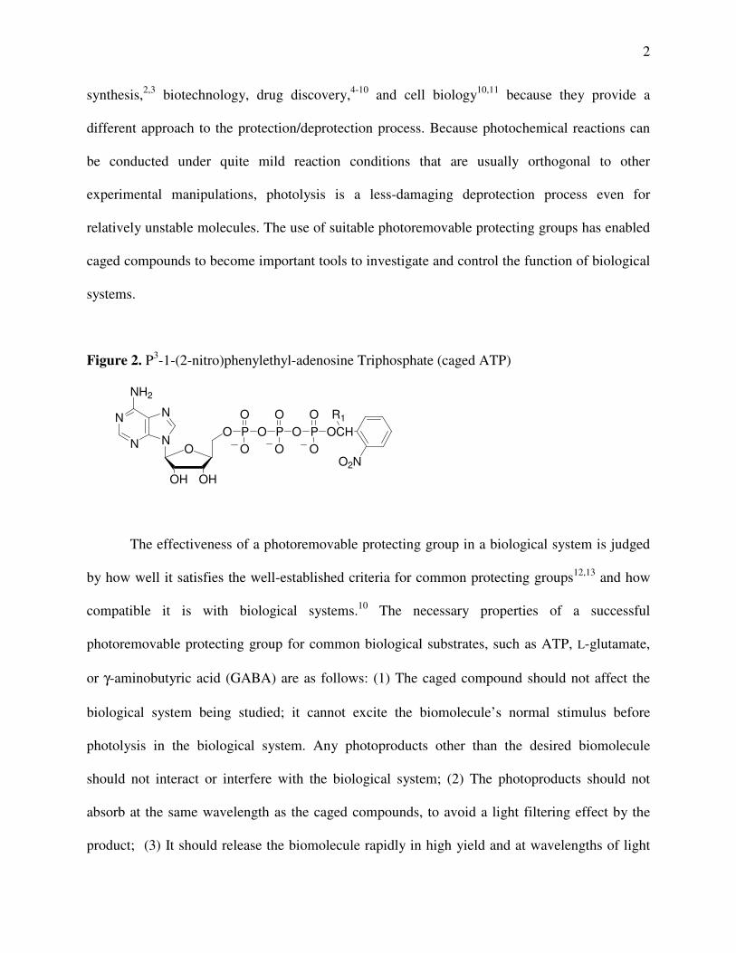
Figure 2: P3-1-(2-nitro)phenylethyl-adenosine Triphosphate (caged ATP) ....................................2
Figure 3: Common Photoremovable Protecting Groups..................................................................4
Figure 4: The Three-dimensional Spatial Selectivity of Single vs. Two-photon Excitation...........6
Figure 5: Photoactivated Gene Expression Based on GeneSwitchTM............................................12
Figure 6: Coumarin Based Photoremovable Protecting Groups and Bhc with Biological
Utility ..............................................................................................................................14
Figure 7: Time Courses of Spontaneous Hydrolysis of Bhc-diol-protected Aldehydes and
Ketones in the Dark under Simulated Physiological Condition....................................19
Figure 8: The Time Courses of One-photon Photolysis of Bhc-diol-protected Aldehydes and
Ketones ..........................................................................................................................20
Figure 9: Apparatus for the Measurement of Two-photon Uncaging Cross-sections ...................22
Figure 10: The Time Courses of Two-photon Photolysis of Bhc-diol-protected Aldehydes and
Ketones ..........................................................................................................................23
Figure 11: Mifepristone and Bhc-diol-mifepristone ......................................................................25
Figure 12: The Time Courses of One-photon Photolysis of Bhc-diol-mifepristone .....................28
Figure 13: Desmethylmifepristone and Bhc-desmethylmifepristone ............................................29
viii
LIST OF SCHEMES
Page
Scheme 1: o-Nitrophenylethylene Glycol as Photoremovable Protecting Groups for Aldehydes
and Ketones ...................................................................................................................15
Scheme 2: Photolysis of Bhc-diol-Protected Aldehydes and Ketones ..........................................16
Scheme 3: Synthesis of Bhc-diol ...................................................................................................17
Scheme 4: Synthesis of Bhc-diol-protected Aldehydes and Ketones. ...........................................18
Scheme 5: Synthesis of Bhc-diol-mifepristone..............................................................................27
Scheme 6: Formation of Trimethyl Ammonium Salts of Bhc-diol-mifepristone..........................29
Scheme 7: Synthesis of (6α- and 6β-)hydroxymifepristone..........................................................30
Scheme 8: Synthesis of Bhc-mifepristone carbonate.....................................................................30
Scheme 9: Synthesis of Bhc-mifepristone carbamate....................................................................31

1
CHAPTER 1
INTRODUCTION
PART 1. CAGED COMPOUNDS
Caged compounds are biologically inactive molecules that can liberate bioactive
molecules of interest by a flash of light. The popular term “caged” refers to molecules whose
biological recognition or activity has been disabled by chemical modification, especially by
covalently linking them to photoremovable protecting groups (Figure 1). This term was first used
in 1978 to describe photo-releaseable derivatives of natural molecules such as adenosine
triphosphate (ATP) (Figure 2).1 Covalent bond formation masks some important features for
biological recognition and photochemical cleavage of the bond releases the bioactive compound.
The rapid release of the bioactive molecule causes a sharp local increase in its concentration.
Figure 1. How Caged Compounds Work.
+hν
caged compound
(inactive messenger)
caging remnant
activemessenger
The key component of a “caged” compound is the photoremovable protecting group.
Photoremovable protecting groups for various functional groups are useful in organic
2
synthesis,2,3 biotechnology, drug discovery,4-10 and cell biology10,11 because they provide a
different approach to the protection/deprotection process. Because photochemical reactions can
be conducted under quite mild reaction conditions that are usually orthogonal to other
experimental manipulations, photolysis is a less-damaging deprotection process even for
relatively unstable molecules. The use of suitable photoremovable protecting groups has enabled
caged compounds to become important tools to investigate and control the function of biological
systems.
Figure 2. P3-1-(2-nitro)phenylethyl-adenosine Triphosphate (caged ATP)
N
N
N
NO
OHOH
O
NH2
P O P O P OCH
O2N
R1O O O
OO O
The effectiveness of a photoremovable protecting group in a biological system is judged
by how well it satisfies the well-established criteria for common protecting groups12,13 and how
compatible it is with biological systems.10 The necessary properties of a successful
photoremovable protecting group for common biological substrates, such as ATP, L-glutamate,
or γ-aminobutyric acid (GABA) are as follows: (1) The caged compound should not affect the
biological system being studied; it cannot excite the biomolecule’s normal stimulus before
photolysis in the biological system. Any photoproducts other than the desired biomolecule
should not interact or interfere with the biological system; (2) The photoproducts should not
absorb at the same wavelength as the caged compounds, to avoid a light filtering effect by the
product; (3) It should release the biomolecule rapidly in high yield and at wavelengths of light
3
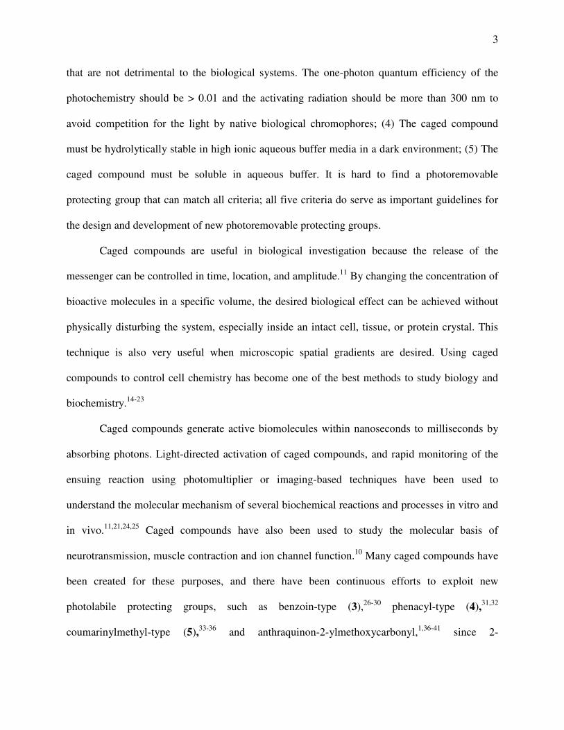
that are not detrimental to the biological systems. The one-photon quantum efficiency of the
photochemistry should be > 0.01 and the activating radiation should be more than 300 nm to
avoid competition for the light by native biological chromophores; (4) The caged compound
must be hydrolytically stable in high ionic aqueous buffer media in a dark environment; (5) The
caged compound must be soluble in aqueous buffer. It is hard to find a photoremovable
protecting group that can match all criteria; all five criteria do serve as important guidelines for
the design and development of new photoremovable protecting groups.
Caged compounds are useful in biological investigation because the release of the
messenger can be controlled in time, location, and amplitude.11 By changing the concentration of
bioactive molecules in a specific volume, the desired biological effect can be achieved without
physically disturbing the system, especially inside an intact cell, tissue, or protein crystal. This
technique is also very useful when microscopic spatial gradients are desired. Using caged
compounds to control cell chemistry has become one of the best methods to study biology and
biochemistry.14-23
Caged compounds generate active biomolecules within nanoseconds to milliseconds by
absorbing photons. Light-directed activation of caged compounds, and rapid monitoring of the
ensuing reaction using photomultiplier or imaging-based techniques have been used to
understand the molecular mechanism of several biochemical reactions and processes in vitro and
in vivo.11,21,24,25 Caged compounds have also been used to study the molecular basis of
neurotransmission, muscle contraction and ion channel function.10 Many caged compounds have
been created for these purposes, and there have been continuous efforts to exploit new
photolabile protecting groups, such as benzoin-type (3),26-30 phenacyl-type (4),31,32
coumarinylmethyl-type (5),33-36 and anthraquinon-2-ylmethoxycarbonyl,1,36-41 since 2-
4
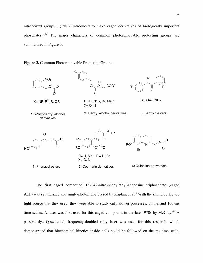
nitrobenzyl groups (1) were introduced to make caged derivatives of biologically important
phosphates.1,37 The major characters of common photoremovable protecting groups are
summarized in Figure 3.
Figure 3. Common Photoremovable Protecting Groups
HO
O R'O
O O
R'
RO O
O
O
XR''
NROBr
OR
O
4: Phenacyl esters
R= H, Me R'= H, BrX= O, N
5: Coumarin derivatives 6: Quinoline derivatives
NO2
O X
O
R
OHX COO-
OO
X
R'
X= NR1R2, R, OR
1:o-Nitrobenzyl alcoholderivatives
R= H, NO2, Br, MeOX= O, N
2: Benzyl alcohol derivatives
X= OAc, NR2
3: Benzoin esters
R
The first caged compound, P3-1-(2-nitro)phenylethyl-adenosine triphosphate (caged
ATP) was synthesized and single-photon photolyzed by Kaplan, et al.1 With the shuttered Hg arc
light source that they used, they were able to study only slower processes, on 1-s and 100-ms
time scales. A laser was first used for this caged compound in the late 1970s by McCray.42 A
passive dye Q-switched, frequency-doubled ruby laser was used for this research, which
demonstrated that biochemical kinetics inside cells could be followed on the ms-time scale.

5
Although they could focus a laser beam through a microscope objective to a very small area in
two-dimensions, on the order of a few (µm)2, for one-photon photolysis of caged compounds in
or near biological tissues, a serious problem arises because photodamage of the biological
material occurs. This damage is due to considerable absorbance by proteins and nucleic acids at
short wavelengths, so multiphoton excitation provides a good method to reduce the damage.
Infrared (IR) light and multiphoton excitation have been used as a less damaging method
for the photolysis of caged compounds (Figure 4). Multiphoton excitation (usually two-photon
excitation) provides an excellent way to photolyze the caged compounds with high spatial
resolution in living cells. Because the non-linear (quadratic) two-photon absorption is confined
to the focus of the laser beam, the light-induced uncaging processes are localized in this small
volume. For a two-photon photolysis experiment, the successful wavelength used in one-photon
photolysis experiment is approximately doubled. At very high intensities, a normal UV-
chromophore can be excited by two IR photons. Also, because cells and tissues are relatively
transparent to IR light, there is much less photodamage, light absorption and scattering, with
much deeper penetration in this photolysis processes. Molecular two-photon excitation (2PE)
was first predicted by Maria Göppert-Mayer in 1931.43 Two-photon excitation requires high peak
intensities, typically 1020 to 1030 photons/(cm2· s) for the observation of two-photon absorption.
Two-photon excitation for biological imaging and two-photon uncaging of biomolecules was
first demonstrated in 1990 by Denk, Strickler and Webb.44 They suggested that it would be better
to use lower energy IR photons than UV photons. In one-photon photolysis, UV photons that are
not involved in uncaging can photo-oxidize proteins, leading to damage, while the excess IR
photons that are not involved in uncaging can only be absorbed into vibrational states of proteins,
leading to at most a temperature increase of the sample. The highest two-photon absorption

6
probability is in the region of highest peak power density and the peak power is low outside this
region. The two-photon photolysis is highly localized into a small volume and the photodamage
is minimized. The most commonly used laser is the argon ion-pumped, titanium-sapphire solid-
state pulsed laser that gives femtosecond laser outputs at wavelengths above 700 nm. It is easier
for non-laser specialists to use this type of laser to apply two-photon photolysis to the
compounds that have UV-absorption above 350 nm.
Figure 4. The Three-dimensional Spatial Selectivity of Single vs. Two-photon Excitation
The two-photon uncaging action cross-section, δu, is a measure of the sensitivity of a
chromophore to two-photon photolysis. It is the product of the two-photon absorbance cross-
section, δa, and the uncaging quantum yield, Qu2. To be useful in biological systems, δu should
exceed 0.1 GM (Goeppert-Mayer, 10-50 cm4 s/ photon).35 To measure the two-photon uncaging
cross-sections, δu, Furuta and Tsien35 illustrated a method of using fluorescein as a reference

7
compound whose two-photon fluorescence quantum yield (Qf2 = 0.9 mol/ein) and absorbance
cross-section (δaF = 30 GM at 740 nm)45 are known. A value of δu is calculated from the
following equation:
s
FaFfpu CtF
CQN
��= )(
2δφδ
Where Np is the number of product molecules formed per unit time (molecules/s,
determined by HPLC analysis); φ is the collection efficiency of the detector used to measure the
fluorescence of fluorescein emitted at a right angle to the beam and passed though a 535/45 nm
bandpass filter; CF is the concentration of fluorescein (mol/L); <F(t)> is the time averaged
fluorescent photon flux (photons/s) collected by the detector; and Cs is the initial concentration
of caged substrate (mol/L).35
The design and synthesis of two-photon sensitive caged biomolecules that can be used
inside living cells is challenging for organic chemists. Only the photoremovable protecting
groups that possess sufficiently large two-photon uncaging action cross-sections, δu, have
potential for biological applications.35 As an added advantage, chromophores with sensitivity to
multiphoton excitation tend to be highly sensitive to single-photon excitation. That means if the
caged compound has a large two-photon uncaging action cross-section, it also has high one-
photon uncaging quantum efficiency. Although o-nitrobenzyl-type groups have been most
widely used as photoremovable protecting groups, recently, coumarin derivatives were reported
as more two-photon sensitive caged compounds.35,36,46-49 For example, 6-bromo-7-
hydroxycoumarin-4-ylmethyl has been used as a multiphoton-sensitive protecting group for

8
neurotransmitters, DNA and RNA, diols, and an inhibitor of nitric oxide synthase. MNI-
glutamate has been reported to release glutamate upon two-photon excitation in sufficient
quantities to be useful for investigating the function of glutamate receptors. The calcium cage
azid-1 can effectively release calcium under two-photon excitation.50 A potentially biologically
useful multiphoton-sensitive protection group is 8-bromo-7-hydroxyquinoline.51
Caged compounds have been used to study the fast kinetics of signal transduction, such
as with neurotransmitters, but they have not been widely used to regulate slower processes such
as gene expression or protein synthesis. Furthermore, the use of multiphoton excitation to
mediate the release of biological effectors has not found much widespread application, despite its
advantages over single photon processes. Further expansion of the prospects of applications of
caged compounds will enable less invasive mapping of local responses to the messengers
involved in signal transduction, gene expression, and protein synthesis. This technology has the
potential to become a powerful drug delivery method that can be used as a research tool and for
therapy.

9
PART 2. LIGHT-SWITCHABLE GENE EXPRESSION
Understanding physiological processes is a major requirement for modern scientists to
develop gene therapy. How to regulate the expression of transferred exogenous genes within the
human body is a serious problem. By specially mutating the genetic code, we can observe the
microscopic details of cellular functions and learn how the genes work. Several regulatable
transgene systems that have been created for eukaryotes, and this technique has yielded a
tremendous amount of information about physiological function. Among these, using small
molecules to control gene expression in complex biological systems is a powerful tool to study
gene function. They can control gene expression in time and they are more practical for real gene
therapy to regulate gene expression than using heat shock or heavy metals.52,53
In order to improve temporal and spatial resolution and provide better control of absolute
expression levels, light-switchable gene expression systems have been reported. For example,
phytochrome controls gene expression by reversibly interconverting between its inactive and
active forms.54 A light-switch gene promoter system developed by Sae Shimizu-Sato55 can be
rapidly and reversibly induced by a flash of light. Their achievement is based on the two forms
of holoprotein reversibly interconverted by exposure to IR light.56 It is hard to find general
application of this technique. Ando57 reported the photolysis of caged RNA/DNA to regulate
gene expression in zebrafish embryos. The first example of using a caged small molecule to
control gene expression was reported by John Koh, et al58 in 2000. This regulation was achieved
by using hormone analogues whose agonist properties are blocked by a photoremovable
protecting group. Another light-activatable ecdysone-inducible gene expression system has been
created in Lawrence’s lab.59

10
Because multiphoton photolysis can provide high localization, less damage to tissue,
minimized scattering, and deeper penetration to regulation of gene expression, multiphoton
excitation of caged compounds could be an invaluable tool for these purposes. We can use
multiphoton sensitive photoremovable protecting groups to cage biological active molecules and
release them by photolysis. Our plan is to create a light activated gene expression system for
zebrafish based on the use of caged regulators of these processes. Photochemical control of gene
expression in a complex biological system, such as zebrafish, would be a significant method for
investigating gene functions involved in a multitude of physiological processes: signal
transduction, cell-to-cell communication, neuronal signaling, cell cycle regulation, development,
motility, and many others. A system that could be mediated by a multiphoton excitation process
(high localization, less damage to tissue, minimized scattering, deeper penetration) would have
the additional advantage of facilitating control at the single cell level deep inside tissue, which
will be especially useful for studying vertebrate development. Zebrafish has been widely used to
study vertebrate development because it has two major advantages: (1) Zebrafish have
transparent bodies, which make it possible to monitor their tissues and neurons in vivo; (2)
Zebrafish are small and they are easy to maintain and reproduce them in the research laboratory.
In order to prevent constitutive gene expression, a distinct regulator gene can be
cotransferred to govern the expression of the target gene. In 1994, Wang et al.60 developed a
regulatory system, which can be used in both animal and human gene transfer studies. This
system can be switched on/off in response to a small chemical compound, such as mifepristone
(RU486). Because the physiological properties of mifepristone have been well studied, creating a
caged mifepristone to regulate a light-switchable gene expression system with multiphoton
excitation is practical. Mifepristone can regulate the gene expression system by an

11
autoregulatory feedback loop. At lower concentrations, mifepristone acts as an agonist on the
progesterone receptor.
Wang’s system has been commercialized by Invitrogen under GeneSwitchTM. A
photochemically activated system, works as shown in Figure 5. The GeneSwitchTM system
includes pGene/V5-His, pSwitch, mifepristone, and a control expression plasmid containing the
lacZ gene and pGene/V5-His/lacZ. In the absence of mifepristone, the translation of the GAL4-
DBD/hPR-LBD/p65-AD fusion gene by the pSwitch regulatory vector is controlled by the
minimal thymidine kinase (TK) promoter. This kind of expression only provides inactive protein
(GeneSwitchTM) in the nucleus. In this system, mifepristone binds to the receptor at the
nanomolar level (Kd ~ 3 nM), and causes a conformation change in the hPR-LBD. Mifepristone
can activate expression of the gene of interest by dimerizing and binding GeneSwitchTM protein
(GAL4-DBD/hPR-LBD/p65-AD) to GALUAS. The ligand-bound protein can regulate the
expression of its own gene to synthesize GeneSwitchTM protein by activation of an
autoregulatory feedback loop, therefore only a small amount of uncaged compound (~ 0.1 nM) is
necessary to activate gene expression.60 Modified with a 42-amino acid deletion in the
progesterone receptor-ligand binding domain (hPR-LBD), the system is not activated by
endogenous progesterone. We expect the photolabile-protecting group Bhc-diol can deactivate
mifepristone, and Bhc-diol-mifepristone can diffuse into cells. When the cells are exposed to a
flash of light, the physiological activity of mifepristone can be reactivated.
The control of gene expression in complex biological systems would be invaluable for
studying physiological processes. Because of the advantages afforded by multiphoton excitation,
further development of applications for the use of photoremovable protecting groups capable of
releasing biomolecules through multi-photon processes will fuel efforts to answer questions

12
about the temporal and spatial relevance of signaling by physiological messengers. It will impact
many areas of biology and medicine, including, but no limited to, developmental biology,
neuroscience, pharmacology, molecular biology, and medical diagnostics. Moreover, the
development of a light activated gene expression system should be particularly useful for
studying development and gene function in zebrafish.
Figure 5. Photoactivated Gene Expression Based on GeneSwitchTM

13
CHAPTER 2
RESULTS AND DISCUSSIONS
PART 1. DEVELOPMENT OF A NEW PHOTOREMOVABLE PROTECTING GROUP FOR
ALDEHYDES AND KETONES
The coumarin-based caged compounds have received wide attention due to their ability to
mediate biological activities in animal cells with light. Although the 4-coumarinylmethyl group
has been used as a fluorescent tag for biological molecules,33 this highly fluorescent group had
not been used as a caging group until Toshiaki Furuta et al. illustrated a method for the synthesis
of esters of diethyl phosphate and cAMP.61 4-(7-methoxycoumarinyl)methyl (7, MCM),40,49 4-
(7-hydroxycoumarinyl)methyl (8, HCM),34,62 4-(7-acetoxycoumarinyl)methyl (9, ACM),34,62 4-
(6,7-dimethoxycoumarinyl)methyl (10, DMCM),46 4-(6,7-biscarboxymethoxycoumarinyl)methyl
(11, BCMCM),36,63 4-(7-dimethylaminocoumarinyl)methyl (12, DMACM),36,63-65 4-(7-
diethylaminocoumarinyl)methyl (13, DEACM),36,63-65 4-(7-carboxymethoxycoumarinyl)methyl
(14, CMCM),66 and 4-(6-bromo-7-hydroxycoumarinyl)methyl (15, Bhc)35,57,67-71 have been
successfully used to “cage” carboxylates, amines, diols, phosphates, and alcohols and phenols.71
Furthermore, Bhc (15) caged compounds can be photolyzed under two-photon excitation with a
large absorbance cross-section35,57,67-70 (Figure 6).
Although many photoremovable protecting groups are known, such groups for ketones
and aldehydes are limited, especially under physiological conditions.72-76 This is surprising
because carbonyl groups are one of the most common functional groups in organic compounds

14
and biological effectors, especially drugs. Synthetically useful photoremovable protecting groups
for carbonyl groups such as N,N-dimethylhydrazones77 require the generation of singlet oxygen,
while others require a triplet sensitizer, as in the case of dithioacetals.72,78 Both methods cannot
be used in biological systems. o-Nitrophenylethylene glycol derivatives (16)79 and (18)73,80
release carbonyl compounds upon exposure to 350 nm light in organic solvents (Scheme 1).
Similarly, polymer-supported o-nitrophenylethylene glycols74 offer photoremovable protection to
aldehydes, releasing them both in benzene and in a stream of air after exposure to a visible-light
mercury lamp for 7 h. The photolysis is not efficient, so it is impractical to apply this
photoremovable protecting group to biological tissues. These protecting groups require
significant synthetic adaptation for physiological use, and they would still suffer from very poor
sensitivity to multiphoton excitation.
Figure 6. Coumarin Based Photoremovable Protecting Groups and Bhc with Biological Utility.
O
Br
O
X
O
O R
O
O NH
OR
O O
OR
O
O
R'
R
P OROOR'
O
O
R'
R''
X'
O
O O
OPh
15 : Bhc-X
X =
7 6-MCM : R' = OCH3, R'' = H 7-MCM : R' = H, R'' = OCH3 8 HCM : R' = H, R'' = OH 9 ACM : R' = H, R'' = CH3CO10 DMCM : R' = R'' = OCH311 BCMCM : R' = R'' = OCH2CO2H12 DMACM : R' = H, R'' = N(CH3)213 DEACM : R' = H, R'' = N(CH2CH3)214 CMCM : R' = H, R'' = OCH2CO2H

15
Scheme 1. o-Nitrophenylethylene Glycol as Photoremovable Protecting Groups for Aldehydes
and Ketones
NO2
O
O R
R'
NO2
NO2
O
O R
R'
NO
O
OHR
R'O
R
R'O
protecting group
remnant
+
+
16 17
(a) hν (350 nm), benzene, 6 h, 31-90%; (b) hν (350 nm), CH3CN or Et2O, 1-2 h, 69-97%
a
b
18
Using Bhc as a basis, we set out to prepare a good multiphoton-sensitive photoremovable
protecting group that can release biologically active ketones or aldehydes in living cells, tissues,
and animals. The general idea that acetals and ketals of 6-bromo-4-(1,2-dihydroxyethyl)-7-
hydroxycoumarin (19, Bhc-diol-acetals/ketals) would be capable of releasing aldehydes and
ketones upon single- or two- photon photolysis under simulated physiological conditions such as
KMOPS buffer (100 mM KCl and 10 mM MOPS titrated to pH 7.2 with KOH) is outlined in
Scheme 2. By studying the (7-methoxycoumarin-4-yl)methyl-caged phosphates, carboxylates,
and sulfonates, Bendig et al. proposed a photochemical SN1 reaction (solvent-assisted
photoheterolysis) mechanism for the coumarin-type photoremovable protecting groups.40 Upon
photolysis of Bhc under physiological conditions, zwitterion 22, a possible intermediate
suggested by Bendig et al., is generated from the diradical 21 by rapid intramolecular single

16
electron transfer. Bulk solvent (H2O or -OH) trapping of the resulting cation followed by
dissociation to Bhc-diol (20) and the carbonyl compound is competitive with recombination to
regenerate the stating ketal.
Scheme 2. Photolysis of Bhc-diol-Protected Aldehydes and Ketones
O
Br
HO O
O
OR
R'
O
Br
HO O
OH
HO
R
R'O
O
Br
HO O
O
OR
R'
O
Br
HO O
OR
R'H2O
hνKMOPSpH 7.2
Osingle electron transfer
19 20
21 22
hν
We have devised a method to accomplish this using Bhc-diol (20), which we have
synthesized in five steps from commercially available materials (Scheme 3).
4-(6-Bromo-7-hydroxycoumarinyl)methyl chloride (25, Bhc-Cl) was prepared in 90%
yield by a Pechman condensation of 4-bromoresorcinol (23) with ethyl-4-chloroacetoacetate (24)
in concentrated sulfuric acid35. The phenolic hydroxy group of Bhc-Cl was protected as the
acetate by using acetic anhydride and pyridine, followed by formation of the triphenyl
phosphonium salt 27 in acetonitrile at 85 oC with triphenylphosphine overnight. Olefination81 of

17
the 6-bromo-7-hydroxycoumarin triphenyl phosphonium salt was accomplished by a Wittig
reaction with aqueous formaldehyde, using sodium carbonate as the base. The reaction
proceeded with loss of the acetate protection group at the same time to reveal Bhc-vinyl (28).
Osmium teteroxide dihydroxylation of the 6-bromo-7-hydroxy-4-vinylcoumarin (28) provided
Bhc-diol (20).
Scheme 3. Synthesis of Bhc-diol
OH
Br
HO O
Br
AcO
Cl
OOEt
O OCl
O
Br
AcO
P+Ph3Cl-
O
O
Br
HO
Cl
O
O
Br
HO
HO OH
OO
Br
HO O
20: Bhc-diol
25: BhcCl
+
Reagents and Conditions:(a) H2SO4, 6d, 90%; (b)pyridine, acetic anhydride,85%; (c) Ph3P, CH3CN, 82%; (d) CH2O, H2O, Na2CO3, 72%; (e) H2O, OsO4, acetone, N-methylmorpholine N-oxide, 80%
ba
c d e
23 24 26
27 28
Acetalization with benzaldehyde and piperonal and ketalization with acetophone and
cyclohexanone provided four caged compounds with which to test the effectiveness of
photochemical release of ketones from Bhc-diol. The Bhc-diol-protected aldehydes and ketones
were synthesized as shown in Scheme 4.
Bhc-diol was refluxed in toluene with benzaldehyde (29a), piperonal (29b),
acetophenone (29c), or cyclohexanone (29d) in the presence of pyridinium p-toluene-sulfonate
(PPTS); solid magnesium sulfate was added to remove water. This afforded Bhc-diol-acetals and
Bhc-diol-ketals as a mixture of diastereomers (in the case of 30a-c) in modest yields.

18
Scheme 4. Synthesis of Bhc-diol-protected Aldehydes and Ketones
O
Br
HO
HO OH
O
(CH2)5
O
O
CHO
O
O O
Br
HO
OO
R'R
O
O
CHO
O
20: Bhc-diol
a
(a) 29a-d, pyridinium p-toluenesulfonate, MgSO4 (anhydrous), toluene,
BuOH, reflux, 110oC, 8h, 57%, 28%, 22%, 39% for 30a-d, respectively.
30a : R = H, R' = Ph
30b: R = H, R' =
30c: R = Me, R' = Ph
30d: R/R'=
29a 29b
29c 29d
30a-d
Each of the Bhc-diol-protected compounds was tested in vitro for their optical properties
(UV spectrum, extinction coefficient). Stability and its resistance to spontaneous hydrolysis in
the dark under simulated physiological condition can be measured readily by HPLC, looking for
disappearance of starting Bhc-diol-protected aldehydes and ketones and appearance of the caging
group remnants. A small amount (2-5%) of decomposition of the conjugate was observed after
seven days in pH 7.2 KMOPS buffer (Figure 7).

19
Figure 7. Time Courses of Spontaneous Hydrolysis of the Bhc-diol-protected Aldehydes and
Ketones in the Dark under Simulated Physiological Condition
Irradiation of Bhc-diol-protected aldehydes and ketones with 365 nm light in pH 7.2
KMOPS buffer released the free aldehydes and ketones (Figure 8). The photochemical
experiments were carried out by using a UV lamp and taking aliquots for HPLC analysis at
periodic intervals. Comparing the time courses for these reactions reveals that Bhc-diol-
benzaldehyde and Bhc-diol-piperonal were photolyzed slightly more efficiently than the
acetophenone and cyclohexanone derivatives. All of these four Bhc-diol-protected acetals/ketals
can be fully deprotected by photolysis in two minutes. From these data, single-photon uncaging

20
quantum efficiencies, Qu1, were determined as previously described, Qu1 = (I·σσσσ·t90%)-1.82,83 The
UV intensity of the lamp I was measured by using potassium ferrioxalate actinometry83 in the
same setup. Release of piperonal or acetophenone was monitored at 313 and 240 nm,
respectively, and the progress curves plotted as an exponential rise to max. Concentrations were
determined using an external standard.
Figure 8. The Time Courses of One-photon Photolysis of Bhc-diol-protected Aldehydes and
Ketones

21
One-photon quantum efficiencies of Bhc-diol-protected aldehydes and ketones are
summarized in Table 1 along with selected absorption data. The quantum efficiencies for single-
photon photolysis of Bhc-diol-protected benzaldehyde, piperonal, acetophenone, and
cyclohexanone are similar to those of other Bhc-protected compounds.35,51,57,68,69
Table 1. Photochemical Properties of Bhc-ketals or acetals
Bhc-ketals or acetals
�max (nm)
�365 (M-1 cm-1)
�370 (M-1 cm-1)
Qu1 (mol/ein)
�u (GM)*
(740nm)
Bhc-diol-benzaldehyde
370 18000 18500 0.057 0.90
Bhc-diol-piperonal 370 19500 19800 0.035 0.60
Bhc-diol-cyclohexanone
370 12600 12800 0.032 0.51
Bhc-diol-acetophenone
370 18000 18400 0.030 1.23
*GM=10-50cm4s mol-1
To be useful in biological systems, �u > 0.1 GM
The extent of carbonyl compound release as measured by HPLC was half of what was
expected. To explore the possibility of secondary photochemical degradation, KMOPS-buffered
solutions of piperonal and acetophenone were each exposed to 365 nm light for one min, but
neither compound showed any signifant decomposition. Irradiation of a 1:1 mixture of Bhc-diol
Calculation of One-Photon Quantum Efficiency
Qu1 = (I·σ·t90%)-1
I - lamp intensity (ein×cm2/s) (determined by potassium ferrioxalate actinometry) ε - molar extinction coefficient (cm-1 M-1) σ - decadic extinction coefficient (103 times ε, cm2/mol) t90% - irradiation time in second for 90% conversion to product

22
and piperonal or acetophenone in KMOPS buffer also showed no change in concentration of the
carbonyl compounds. Degradation of the carbonyl compounds after uncaging appears to not be
the result of a direct or a Bhc-diol-mediated photochemical process.
The two-photon uncaging cross-section, δu, of Bhc-diol-protected aldehydes and ketones
was measured as an external standard as previously described.51 The experimental setup is shown
in Figure 9. Measurements were carried out in microcuvettes (10 × 1 × 1 mm illuminated
dimensions) with an effective filling volume of 20 �L using light from a fs-pulsed, mode-locked
Ti:Sapphire laser focused on the center of the cuvette chamber with a 25 mm focal length lens
optimized for IR lasers. The two-photon uncaging cross-section (δu) was estimated by
referencing to fluorescein, a compound with a known two-photon fluorescence quantum yield
(Qf2 = 0.9 mol/ein) and absorbance cross-section (�aF = 30 GM at 740 nm).47 The progress of the
uncaging reaction was measured by HPLC and graphed as a function of time (Figure 10).
Figure 9: Apparatus for the Measurement of Two-photon Uncaging Cross-sections

23
Figure 10. The Time Courses of Two-photon Photolysis of Bhc-diol-protected Aldehydes and
Ketones
Calculation of Two-Photon Uncaging Action Cross-Section �u
s
FaFfpu CtF
CQN��
= )(2δφδ
Np-number of product molecules formed per unit time CF-the concentration of fluorescein �-the collection efficiency of the detector �aF-the fluorescein absorbance cross-section Qf2-the two-photon fluorescence quantum yield of fluorescein <F(t)>-the time averaged fluorescent photon flux Cs-the initial concentration of caged substrate

24
The values of δ (Table 1) determined for Bhc-diol-acetals/ketals are similar to the values
obtained for other Bhc-protected compounds. Any discrepancies are probably a result of
differences in laser power and the optical setups employed. The longer time constants (min), as
compared to the single-photon kinetics (s), are due to the small volume of the sample that is
actually irradiated (~1 fL) relative to the bulk solution (20 L). Sufficient quantities of the Bhc-
diol-acetals/ketals for HPLC analysis must diffuse into the laser’s focal volume, where uncaging
efficiency is high, undergo photolysis, and then diffuse back out into the bulk solution.
The results indicate that Bhc-diol can be used as a photolabile protecting group to cage
biologically active messengers containing carbonyl functionality. Since Bhc has been used in
biological systems, this represents the first example of a photolabile protecting group capable of
releasing aldehydes and ketones by single- and two-photon excitation under physiological
conditions.

25
PART 2. LIGHT-SWITCHABLE GENE EXPRESSION SYSTEM
After we achieved the above accomplishment, we began to synthesize the desired Bhc-
diol-protected biological messengers, such as mifepristone, that have a carbonyl group in their
structures (Figure 11).
Important gene messengers such as the progesterone receptor antagonist, (31,
mifepristone, also known as RU486) (synthetic steroid), induce gene expression in mammalian
cells.60 Because mifepristone’s carbonyl group is important for its action on the progesterone
receptor, Bhc-diol can be used as a photoremovable-protecting group for releasing mifepristone
inside living cells, tissues, and animals. As the physiological properties of mifepristone are well
known, it is ideal to induce mammalian cells with Bhc-diol caged mifepristone.
Figure 11. Mifepristone and Bhc-diol-mifepristone
O
NOH
31: Mifepristone (RU486)
NOH
O
O
O
Br
HO O
32: Bhc-diol-mifepristone
The previous ketal functionalization is affected with Bhc-diol in an acid medium.
Because mifepristone is a relatively complicated molecule with several functional groups, we
have expected that there would be some problems to prepare the Bhc-diol caged mifepristone
(32).

26
The standard method of making ketals from ketones and 1,2-diols is by refluxing the
substrates with pyridinium p-toluenesulfonate in dry toluene and anhydrous magnesium sulfate
to remove the water generated. A trace quantity of Bhc-diol-mifepristone derivative has been
synthesized in our lab, having carried out the reaction with Bhc-diol and mifepristone. The mass
spectrum and NMR data indicate that we have produced the caged mifepristone as a mixture of
diastereomers in very low yield with migration of conjugated doubled bonds.
After making numerous efforts to modify the reaction condition, such as changing the
solvent from toluene to benzene, using p-toluenesulfonic acid instead of pyridinium p-
toluenesulfonate, and using molecular sieves instead of MgSO4, there was no improvement.
Other more efficient methods to make ketals of �, �-unsaturated ketones were examined.
Several methods were tried, such as microwave ketalization and TMSOTf catalysis, and TMSCl
catalyzed ketalzation.84 Under TMSCl condition, THF was used as the solvent to make Bhc-diol
dissolve in the reaction mixture. This reaction gave Bhc-diol-mifepristone derivative 33 in good
yield (Scheme 5).
HPLC analysis indicated that the product of the ketalization of Bhc-diol and mifepristone
with TMSCl is a mixture of four similar compounds that have different retention time on HPLC.
From LC-Mass data, they have the same molecular weight, 712/714, which is the desired
molecular weight of the product. UV spectral analysis also gave very similar data that has two
major absorptions, at 288 nm and 326 nm.

27
Scheme 5. Synthesis of Bhc-diol-mifepristone
O
Br
HO
HOOH
OO
NOH
NOH
∗
O
Br
HO O
+
(a) 5 equiv. TMSCl, THF, 80%
a
20 31 33
O
∗O
Because two new stereocenters have been generated with no control in this reaction, four
diastereomers were produced. The Bhc-diol-mifepristone was mixed in KMOPS buffer and
photolyzed under a 365 nm UV lamp, but there was no change in concentration even after 10
min. One of the possibilities that might cause a photolysis problem is the low solubility of the
Bhc-diol-mifepristone in aqueous solution. Several different conditions were tried: HEPES
buffer (pH = 7.4) was used instead of KMOPS buffer; a large amount of an organic solvent, such
as acetonitrile, methanol, or DMSO was used to increase its solubility in aqueous buffer. The
best condition was when 200 �L of DMSO were added to dissolve the Bhc-diol-mifepristone in
3 mL KMOPS buffer. The photolysis under UV light (365 nm) still took very long under these
conditions (Figure 12). It is impractical for biological systems. The other possibility for the
photolysis problem is that Bhc excitation might be quenched by the aniline moiety of
mifepristone. Mifepristone analogs, also capable of inducing expression, might alleviate this
problem.

28
Figure 12. The Time Courses of One-photon Photolysis of Bhc-diol-mifepristone
A modification is to make the trimethylammonium salt of Bhc-diol-mefipristone and to
test its photochemistry. We will make trimethylammonium salts of Bhc-diol-mifepristone (34)
by using CH3I in diethyl ether with Bhc-diol-mifepristone (33) (Scheme 6). The 1H NMR and
mass spectrum showed that the product obtained under this condition was the
trimethylammonium salt of Bhc-diol-mefipristone, 34. Further characterization will be done to
confirm its formation. The ammonium salt might increase the solubility of the caged compounds
and change the electronic distribution of the compound in a way that will facilitate the photolysis
of the messenger.
Analogs of mifepristone might work for the same purpose in the gene expression system.
One alternative (35, desmethylmifepristone) has been synthesized in our lab and coupled to
BhcOH (Figure 13) but surprisingly, the carbamate caged compound (36, Bhc-

29
desmethylmifepristone) cannot mask the messenger’s physiological activity. Another possible
alternative is hydroxymifepristone 3685 (Scheme 7). The hydroxymifepristone will be also
coupled with BhcOH as a carbonate (37) (Scheme 8). The hydroxymifepristone can also be
modified by amination. As a precursor of carbamate 41, intermediate 39 has been made by a
Mitsunobu reaction by using diisopropyl azodicarboxylate (DIAD), phthalimide, and
triphenylphosphine (Scheme 9).
Scheme 6. Formation of Trimethyl Ammonium Salts of Bhc-diol-mifepristone
(a) CH3I, (CH3CH2)2O
a
33
NOH
O
O
O
Br
HO O
34
NOH
O
O
O
Br
HO O
I
Figure 13. Desmethylmifepristone and Bhc-desmethylmifepristone
O
NOH
O
Br
HO
O
O
O
O
HNOH
35 : Desmethylmifepristone 36 : Bhc-Desmethylmifepristone

30
Scheme 7. Synthesis of (6α- and 6β-)hydroxymifepristone
O
N
OH
O
N
OH
OH
(a) SeO2, dioxane, 39%.
a
31 37
Scheme 8. Synthesis of Bhc-mifepristone carbonate
N
O
OH
OH
O
Br
HO
OH
O
N
O
O
OH
O
Br
HO
O
O
O
+
37 38

31
Scheme 9. Synthesis of Bhc-mifepristone carbamate.
O
OHN
OH
O
OHN
NH2
O
OHN
NHO
Br
HO O
OH
O
Br
HO O
O
O
37 40
41
O
OHN
NOO
39
a
(a) DIAD, phthalimide, THF, Ph3P.
The synthesis and application of a new photoremovable protecting group Bhc-diol has
been described in this thesis. The presented data and results indicated Bhc-diol possesses
sufficient one-photon quantum efficiency and two-photon uncaging cross-section for this
purpose, and it is comparable to other known coumarin derivatives. The successful use of caged
32
compounds in cellular studies requires solubility in aqueous solutions and hydrolytic stability in
the absence of light. Our Bhc-diol-caged aldehydes and ketones have good solubility in water
and they are hydrolytically stable in the dark. The data we have collected shows that Bhc-diol
has the ability to be used as a photoremovable protecting group to cage aldehydes and ketones
and the promise of Bhc-diol as a good photo-mediator of drug delivery. Upon photolysis, the
Bhc-diol caged aldehydes and ketones can release free carbonyl compounds under simulated
physiological conditions. Although there were some problems to photolyze Bhc-diol-
mifepristone efficiently, Bhc-diol still has the potential to cage other biologically active
messengers containing carbonyl functional groups. After synthesizing the desired Bhc-diol-
mifepristone (32, without conjugated double bonds migration), solving the problem involved in
photolysis of Bhc-diol-mifepristone, or developing Bhc caged mifepristone analogs, a practical
light-switchable gene expression system could be created. Since Bhc has been widely used in
biological systems, Bhc-diol is the first example of a photoremovable protecting group capable
of releasing aldehydes and ketones by single- and two-photon excitation under physiological
conditions. Further investigation into the mechanism of photolysis and utility in biological
systems is underway.

33
CHAPTER 3
EXPERIMENTAL SECTION
All reagents and solvents were purchased from commercial sources and used without
further purification with the following exceptions. Toluene was dried by passing it through
activated alumina under nitrogen pressure (Solv-Tek, Berryville, VA). Pyridine and acetonitrile
were refluxed with calcium hydride under nitrogen, and then distilled. 1H NMR and 13C NMR
spectra were recorded on a Varian Mercury Plus 400 MHz spectrometer using tetramethylsilane
(TMS) as an internal standard. Chemical shift data for the proton resonances were reported in
parts per million (δ) relative to internal standard TMS (δ 0.00). Chromatographic solvent
proportions are expressed on a volume: volume basis. FTIR spectra were recorded on a Brucker
Vector 22 spectrophotometer. UV spectra were recorded on a Cary 300 Bio UV-Visible
spectrophotometer (Varian). HPLC analysis (analytical and preparative) was performed on a
Varian ProStar HPLC system with an autosampler and diode array detector using Microsorb C-
18 reverse phase columns. Mass spectrometry was performed on a Sciex API-1 Plus quadrupole
mass spectrometer with an electrospray ionization source. KMOPS buffer consisted of 100 mM
KCl and 10 mM MOPS titrated to pH 7.2 with KOH. Thin layer and column chromatography
were performed on precoated silica gel 60 F254 plates (EM Science) and 230-400 mesh silica gel
60 (EM Science), respectively. Melting points were determined on a Mel-Temp (Laboratory
Devices, Inc.), and are uncorrected.

34
6-BROMO-4-CHLOROMETHYL-7-ACETYLOXYCOUMARIN (26).
OHO
Br
O
Cl
OAcO
Br
O
Cl
acetic anhydride
85%
pyridine
Under a nitrogen atmosphere, 6-bromo-4-chloromethyl-7-hydroxycoumarin (780 mg,
2.69 mmol) and acetic anhydride (0.64 ml, 6.75 mmol) were stirred in anhydrous pyridine (3 ml)
for 4 h. The pyridine was removed under vacuum, and the resulting crude residue was purified
by flash chromatography through silica gel (hexane/ethyl acetate 1:1) to afford a white solid (760
mg, 85%, mp 180 oC dec). 1H NMR (400 MHz, CDCl3) δ 7.91 (1H, s), 7.21 (1H, s), 6.58 (1H, s),
4.63 (2H, s), 2.41 (3H, s); 13C NMR (100 MHz, CDCl3) 168.06, 159.45, 153.68, 150.99, 148.31,
128.61, 116.96, 113.30, 112.40, 41.19, 21.03; FTIR (neat) 1732, 1599, 1396, 1217, 1181, 1137,
1022, 906, 862, 728, 658, 629 cm-1.
PHOSPHONIUM SALT (27).
OAcO
Br
O
Cl
OAcO
Br
O
PPh3 ClPh3P, CH3CN,
82%
Under a nitrogen atmosphere, a mixture of 6-bromo-4-chloromethyl-7-
acetyloxycoumarin (650 mg, 1.96 mmol) and triphenyl phosphine (1.028 g, 3.92 mmol) in
acetonitrile (5 ml) was heated to 85 oC. After 30 min, the suspended mixture dissolved. Heating

35
was continued for 18 h, during which period the white phosphonium salt precipitated. The
mixture was cooled, filtered and the filtrand was washed several times with boiling benzene to
yield a white solid (948 mg, 82%), which was carried to the next step without further
purification.

36

37

38
6-BROMO-7-HYDROXY-4-VINYLCOUMARIN(BHC-VINYL) (28).
OAcO
Br
O
PPh3 Cl
OAcO
Br
O
CH2O, H2O
72%
Na2CO3,
Phosphonium salt (310 mg, 0.522 mmol) was taken up in a 37% formaldehyde solution
(4 ml, aqueous). The mixture was stirred for 15 min, and then an aqueous solution of 15%
Na2CO3 (0.5 ml) was added intermittently by a syringe. Each subsequent addition was made after
the orange-yellow color of the phosphorone formed had disappeared. When the addition of the
base was complete, the mixture was stirred at room temperature for 2 h, and then extracted three
times with chloroform. The combined chloroform extracts were dried over anhydrous sodium
sulfate and evaporated. The crude product was purified by flash chromatography through silica
gel (hexane/ethyl acetate 6:4) to afford a white solid (100 mg, 72%, mp 210-230 oC dec).
1HNMR (400 MHz, (CD3)2CO) δ 8.00 (1H, s), 7.21 (1H, dd, J = 17.2, 11.6 Hz), 6.94 (1H, s),
6.35 (1H, s), 6.14 (1H, dd, J = 17.2, 1.2 Hz), 5.77 (1H, dd, J = 11.2, 0.8 Hz); 13C NMR (100
MHz, (CD3)2CO) 160.08, 157.31, 154.88, 150.18, 130.47, 129.19, 123.31, 112.80, 108.38,
106.08, 103.87; FTIR (neat) 3392, 1700, 1607, 1412, 1365, 1310, 1268, 1223, 1155, 954 cm-1;
MS m/z 267 (MH+, 81Br), 265 (MH+, 79Br).

39

40

41
6-BROMO-4-(1,2-DIHYDROXYETHYL)-7-HYDROXYCOUMARIN(BHC-DIOL) (20).
OAcO
Br
O OAcO
Br
O
HOOH
H2O, OsO4, Acetone
80%
N-methylmorpholineN-oxide
Bhc-vinyl (200 mg, 0.749 mmol) and one small piece of osmium tetroxide (which was
not weighed because of its toxicity and the difficulty in accurately weighing it, due to rapid
sublimation) were added to a solution of 4-methylmorpholine N-oxide monohydrate (101 mg,
0.747 mmol) in water (4 ml) and acetone (2 ml). The resulting solution was vigorously stirred at
room temperature for 18 h, during which time the mixture turned a light brown color. The
reaction was neutralized to pH 7 with 3 N sulfuric acid. The acetone was evaporated, and the
resulting aqueous solution was extracted five times by ethyl acetate. The combined organic
extracts were evaporated, and the resulting crude product was purified by flash chromatography
through silica gel (hexane/ethyl acetate 2:8) to afford a white solid (180 mg, 80%, mp 200-220
oC dec). 1HNMR (400 MHz, (CD3)2CO) δ 8.05 (1H, s), 6.92 (1H, s), 6.41 (1H, s), 5.13 (1H, dd, J
= 4.4, 4.4 Hz), 3.88 (1H, dd, J = 12.0, 4.4 Hz), 3.71 (1H, dd, J = 11.2, 5.6 Hz); 13C NMR (100
MHz, (CD3)2CO) 160.78, 157.55, 156.15, 155.57, 129.94, 113.09, 111.09, 106.56, 104.44, 71.42,
67.03; FTIR (neat) 3332, 1726, 1604, 1437, 1158, 1120, 882, 773, 695, 540 cm-1; MS m/z 303
(MH+, 81Br), 301 (MH+, 79Br).

42

43

44
GENERAL PROCEDURE FOR THE SYNTHESIS OF 6-BROMO-7-
HYDROXYCOUMARIN ACETALS AND KETALS.
Under a nitrogen atmosphere, aldehyde or a ketone (2 equiv.) in 10 µl of 1-butanol was
added to a mixture of Bhc-diol (1 equiv.), pyridinium p-toluene sulfonate (PPTS, 1 equiv.), and
anhydrous MgSO4 (100 mg) in anhydrous toluene (2 ml). The reaction mixture was stirred at 110
oC for 8 h, and then filtered, washing the solid filtrand with chloroform. The filtrate was
evaporated, and the resulting crude product was purified by flash chromatography through silica
gel (ethyl acetate/hexanes 1:1) to give the Bhc-protected aldehyde or ketone. With the exception
of Bhc-diol-cyclohexanone, all Bhc-diol protected aldehydes and ketones were isolated as a
mixture of diastereomers.
BHC-DIOL-BENZALDEHYDE (30a).
O
Br
HO O
HOOH
CHO
O O
Br
HO
OO
+
pyridinium p-toluenesulfonate
MgSO4 (anhydrous)
toluene, reflux, 110oC, 8h57%
Bhc-diol (15 mg, 0.05 mmol), PPTS (13 mg, 0.05 mmol), benzaldehyde (10 µl, 0.10
mmol); yield = 11 mg (57%, mp 106-180 oC dec). 1HNMR (400 MHz, CDCl3) δ 7.50 (6H, m),
[7.07 (s), 7.06 (s) (1H)], [6.67 (s), 6.55 (s) (1H)], [6.14 (s), 6.02 (s) (1H)], 5.40 (1H, m), [4.73
(dd, J = 7.6, 7.6 Hz), 4.57 (dd, J = 8.4, 8.4 Hz) (1H)], [4.09 (dd, J = 8.4, 5.6 Hz), 3.86 (dd, J =

45
7.2, 7.2 Hz) (1H)]; 13C NMR (100 MHz, CDCl3) (160.44, 160.33), (155.37, 155.27), (154.91,
154.85), (152.14, 151.85), 136.57, 135.58, (129.96, 129.76), (128.68, 128.62), 126.67, (126.54,
126.44), (112.24, 112.15), 110.70, 109.60, 106.68, 105.18, (104.74, 104.67), (73.80, 73.30),
(70.62, 70.23); FTIR (neat) 3407, 2922, 1698, 1602, 1396, 1308, 1271, 1222, 1153, 1095, 1021,
873, 756, 699 cm-1; MS m/z 391 (MH+, 81Br), 389 (MH+, 79Br).

46

47

48
BHC-DIOL-PIPERONAL (30b).
O
Br
HO O
HOOH
O
O
CHO
O O
Br
HO
OO
OO
+ pyridinium p-toluenesulfonate
MgSO4 (anhydrous)
toluene, reflux, 110oC, 8h28%
Bhc-diol (15 mg, 0.05 mmol), PPTS (13 mg, 0.05 mmol), piperonal (15 mg, 0.10 mmol);
yield = 6 mg (28%, mp 176-195 oC dec). 1HNMR (400 MHz, CDCl3) δ [7.58 (s), 7.50 (s) (1H)],
7.06 (3H, m), [6.87 (s), 6.85 (s) (1H)], [6.64 (s), 6.54 (s) (1H)], 6.02 (2H, m), [6.02 (s), 5.92
(1H)], [5.40 (dd, J = 6.8, 6.8 Hz), 5.35 (dd, J = 6.8, 5.6 Hz) (1H)], [4.74 (dd, J = 7.6, 7.6 Hz),
4.54 (dd, J = 8.0, 8.0 Hz) (1H)], [4.07 (dd, J = 7.6, 5.2 Hz), 3.82 (dd, J = 7.6, 7.6 Hz) (1H)]; 13C
NMR (100 MHz, CDCl3) (160.38, 160.27), (155.34, 155.25), (154.96, 154.90), (152.10, 151.77),
(148.97, 148.84), 148.01, (130.44, 129.46), (126.70, 126.55), (120.96, 120.76), (112.28, 112.20),
(110.74, 109.61), (108.29, 108.22), 106.92, (106.72, 106.69), (105.05, 104.65), (104.76, 104.70),
(101.40, 101.30), (73.76, 73.28), (70.62, 70.12); FTIR (neat) 3423, 2960, 2923, 1722, 1605,
1400, 1260, 1094, 1029, 869, 801 cm-1; MS m/z 435 (MH+, 81Br), 433 (MH+, 79Br).

49

50

51
BHC-DIOL-ACETOPHENONE (30c).
O
Br
HO O
HOOH
+
O O
Br
HO
OO
CH3
22%
pyridinium p-toluenesulfonate
MgSO4 (anhydrous)
toluene, reflux, 110oC, 8h
O
Bhc-diol (20 mg, 0.07 mmol), PPTS (33 mg, 0.14 mmol), acetophenone (40 µL, 0.35
mmol); yield = 6 mg (22%, mp 170-220 oC dec). 1HNMR (400 MHz, CDCl3) δ 7.51 (6H, m),
[7.05 (s), 7.01 (s) (1H)], [6.64 (s), 6.40 (s) (1H)], [5.40 (dd, J = 7.2, 7.2 Hz), 5.07 (dd, J = 7.6,
5.2 Hz) (1H)], [4.65 (dd, J = 8.8, 6.8 Hz), 4.24 (dd, J = 8.0, 8.0 Hz) (1H)], [3.89 (dd, J = 8.0, 5.2
Hz), 3.71 (dd, J = 8.0, 8.0 Hz) (1H)], [1.83 (s), 1.78 (s) (3H)]; 13C NMR (100 MHz, CDCl3)
160.59, 155.40, (154.81, 154.63), 152.56, 141.98, (128.60, 128.53), (128.47, 128.34), 126.66,
125.27, 124.70, 112.15, 111.11, 110.20, 106.63, (104.61, 104.51), 74.41, 72.94, (69.86, 69.33),
(28.16, 27.74); FTIR (neat) 3416, 2922, 1697, 1602, 1395, 1308, 1224, 1155, 1096, 874, 760,
699 cm-1; MS m/z 405 (MH+, 81Br), 403 (MH+, 79Br).

52

53

54
BHC-DIOL-CYCLOHEXANONE (30d).
O
Br
HO O
HOOH
+
O
O O
Br
HO
OO
39%
pyridinium p-toluenesulfonate
MgSO4 (anhydrous)
toluene, reflux, 110oC, 8h
Bhc-diol (15 mg, 0.05 mmol), PPTS (13 mg, 0.05 mmol), cyclohexanone (10 µL, 0.10
mmol); yield = 7.5 mg (39%, mp 180-230 oC dec). 1HNMR (4500 MHz, CDCl3) δ 7.54 (1H, s),
7.04 (1H, s), 6.58 (1H, s), 5.26 (1H, dd, J = 8.0, 8.0 Hz), 4.53 (1H, dd, J = 8.0, 8.0 Hz), 3.77 (1H,
dd, J = 8.0, 8.0 Hz), 1.70 (10H, m); 13C NMR (100 MHz, CDCl3) 160.66, 155.26, 154.72,
152.69, 126.63, 112.38, 111.56, 109.89, 106.62, 104.60, 72.94, 69.09, 35.67, 34.81, 25.01, 23.89,
23.79; FTIR (neat) 3386, 2935, 2862, 1707, 1604, 1395, 1272, 1231, 1155, 1107, 1036, 875, 700
cm-1; MS m/z 383 (MH+, 81Br), 381 (MH+, 79Br).

55

56

57
BHC-DIOL-MIFEPRISTONE (33).
O
Br
HO
HOOH
OO
NOH
TMSCl
THF
NOH
O
O
O
Br
HO O
+
80%
Mifepristone (50 mg, 0.12 mmol) is added to a solution of Bhc-diol (90 mg, 0.30 mmol)
and dry THF (2 ml) under a nitrogen atmosphere. To the mixture is added chlorotrimethylsilane
(75 µl, 0.58 mmol) and the mixture is stirred at reflux for 48 h. A saturated aqueous solution of
sodium bicarbonate (5 ml) is added and the mixture extracted three times with ether, washed
with brine, and the combined ether extracts were dried with anhydrous sodium sulfate and
evaporated. The crude product was purified by flash chromatography on silica gel (hexane/ethyl
acetate 4:6) to afford a yellow solid (68mg, 80%). 1HNMR (400 MHz, CDCl3) δ 7.51 (1H, m),
7.10 (2H, m), 7.02 (1H, m), 6.68 (2H, m), [6.57 (s), 6.55 (s), 6.49 (s), 6.41 (s) (1H)], 5.22 (1H,
m), 4.49 (1H, m), 3.70 (1H, m), 2.95 (6H, m), 1.79 (3H, s), 0.97 (3H, s); FTIR (neat) 2919, 2357,
1718, 1604, 1516, 1395, 1351, 1308, 1268, 1215, 1150, 1113, 1091, 1062, 1012, 945, 876, 854,
818, 745, 665 cm-1; MS m/z 714 (MH+, 81Br), 712 (MH+, 79Br).

58

59

60
(6αααα- AND 6ββββ-) HYDROXYMIFEPRISTONE (37).
O
NOH
SeO2
O
NOH
OH
dioxane
39%
(11β,17β)-11-(4-Dimethylamino-phenyl)-17-hydroxyl-17-(1-propynyl)-estra-4,9-dien-3-
one (mifepristone, RU486) (107 mg, 0.243 mmol) was dissolved in 50 ml dry dioxane in a 100
mL three-neck flask equipped with a reflux condenser. After addition of SeO2 (33 mg, 0.303
mmol) the mixture was stirred at 80 oC under a N2 atmosphere. The slightly yellow colored
reaction mixture became red. The reaction was stopped after 20 h by addition of 30 mL 5%
aqueous KOH solution. Thereafter, the solution was extracted four times with ethyl acetate, and
the combined organic layers were washed with water to neutral pH, dried over sodium sulfate,
and evaporated. A mixture of products was obtained as a yellowish oil, which was further
purified by column chromatography on silica gel (hexane/ethyl acetate 4:6) to afford a yellow
solid (43 mg, 39%). 1HNMR (400 MHz, CDCl3) δ 7.06 (2H, d), 6.66 (2H, d), 5.91 (1H, s), 4.50
(1H, s), [4.35 (s), 4.34 (s), (1H)], 2.91 (6H, s), 1.89 (3H, s), 0.56 (3H, s); MS m/z 446 (MH+).
[Spectra data match the published literature values.85]

61
DETERMINATION OF THE QUANTUM EFFICIENCY FOR SINGLE PHOTON
EXCITATION.
To start a photolysis experiment, the UV absorption spectrum of the compound to be
photolyzed is recorded. Based on the spectrum, an appropriate light source (similar wavelength
as absorption peak) and vessel are selected. Most molecules have a rather broad absorption
spectrum, often with several different absorption maxima. Usually, it is more efficient to select a
light source with maximum emission matching one absorption maximum, and it is usually
advantageous to select the maximum at the longest possible wavelength. Since the energy of the
photons decreases as the wavelength increases, the severity of the side reactions may be
decreased with longer wavelength light. For two-photon photolysis experiment, successful
wavelength used in one-photon photolysis experiment was doubled.
The quantum yield for any process is the fraction of the photons used in the process, that
is, the number of moles of compound undergoing the process divided by the number of moles of
photons absorbed (1 mol of photons = 1 einstein). Therefore, the sum of the quantum yields for
all the processes in which the photons have participated is equal to 1.
The energy of the light beam can be measured with a calibrated thermopile. Because it is
extremely difficult to perform this type of absolute measurement routinely, indirect methods are
usually preferred. They use as reference a reaction for which the quantum yield has been
previous been measured. By determining the extent of product formation for the known reaction
under conditions absolutely identical to those used for the unknown conversion, it is possible to
determine how many quanta of light have been used. The chemical yield of the product
formation in the unknown reaction is measured, and divided by the number of quanta to arrive at
the yield of product for each quantum of light absorbed, the quantum yield. The reference

62
photochemical reaction mentioned above uses a starting material that is called an actinometer. In
order to use an actinometer properly one must be certain that the quantum yield of the reference
reaction and the reaction of interest are measured at the exact same wavelength(s). In this regard,
the best actinometer is probably ferric oxalate.83 Upon irradiation, the ferric ion is converted into
Fe2+, along with oxalate oxidation. The extent of conversion is determined by adding 1,10-
phenanthroline complexes. The quantum yield has been carefully measured by Parker and
Hatchard and found to vary little over a wide range of wavelengths.
UV absorption spectra of KMOPS-buffered solutions (3 mL) of the substrates (100 �M)
in quartz cuvettes (21-Q-10, Starna, Atascadero, CA) were measured. Molar extinction
coefficients were calculated. Then the test photolysis experiments were carried out by
monitoring the UV absorption at 30 s, 1min, 2min, 3min, 4 min and 5min. The rough time
needed for photolysis was estimated. Another 3 mL of KMOPS-buffered Bhc-diol caged
aldehyde and ketone solutions were irradiated with 365-nm UV light from a mercury lamp
(Spectroline SB-100P; Spectronics Corporation, Westbury, NY). The spectral output of the lamp
is a distribution across the UV-A wavelengths (310-400 nm) with an intense band at 365 nm. A
20 �l aliquot of the solution was removed for analysis by HPLC at the point 0 s, 5 s, 10 s, 15 s,
20 s, 30 s, 40 s, 50 s, 60 s, 90 s, 120 s, and 240 s using an external standard method to determine
concentrations. The compound was eluted with an isocratic mixture of acetonitrile and water
containing 0.1% trifluoroacetic acid (flow rate of 1 mL/min). The exact HPLC conditions are
shown in Table 2. The progress curves were plotted as simple decaying exponentials. Quantum
efficiencies (Qu1) were calculated according to a published method,35 using Qu1 =(I⋅�⋅t90%)-1,
where I is the irradiation intensity in ein�cm-2�s-1, � is the decadic extinction coefficient (103
times �, molar extinction coefficient) in cm2�mol-1, and t90% is the irradiation time in seconds for

63
90% conversion to product. The UV intensity of the lamp I was measured by using potassium
ferrioxalate actinometry83 in the same setup. 3 mL of 6 mM potassium ferrioxalate solution were
irradiated for 20 s and mixed properly. 2 mL of this solution was mixed with 3 mL aqueous
buffer (600 mL1 M NaOAc and 360 mL 1N H2SO4 diluted to 1 L with water), 3 mL of 0.1%
1,10-phenanthroline solution (10 mg in 100 mL water), and 1 mL of 2 M KF solution and the
mixture was diluted with water to 25 mL in a volumetric flask. A blank solution was prepared
without irradiation in the same way. These solutions were allowed to stand for 30 min in the
dark. The UV absorption of these solutions was measured at 510 nm and the UV lamp intensity I
was calculated by the following equation.
I =V3∆D510
103ε510V2φFe2+ t
V3 is the volume of dilution (25 mL); V2 is the volume of irradiated solution taken for
analysis; ∆D510 is the absorption of the absorption of the solution; ε510 is the molar extinction
coefficient of actinometry; φFe2+ is the quantum yields for production of ferrous ions from
potassium ferrioxalate at about 365 nm (1.21); and t is the time of irradiation. Release of
piperonal or acetophenone was monitored at 313 and 240 nm, respectively, and the progress
curves plotted as an exponential rise to max. Concentrations were determined using an external
standard by making a calibration curve for each compound detected.

64
Table 2. HPLC Conditions of Single- and Two-photon Photolysis
Bhc-diol protected
aldehydes and ketones
Solvent conditions for HPLC (volume percentage)
Compounds were monitored
Detection wavelengths (nm)
Retention time (min)
Bhc-diol-benzaldehyde
Acetonitrile 65%; H2O (0.1% TFA) 35%
Bhc-diol-benzaldehyde 325 5.533
Bhc-diol-piperonal 329 5.944 Bhc-diol-piperonal Acetonitrile 60%; H2O (0.1% TFA) 40%
Piperonal 313 4.234
Bhc-diol-cyclohexanone
Acetonitrile 68%; H2O (0.1% TFA) 32%
Bhc-diol-cyclohexanone 328 5.910
Bhc-diol-acetophenone 328 3.832 Bhc-diol-acetophenone Acetonitrile 82%; H2O (0.1% TFA) 18%
Acetophenone 240 3.454
DETERMINATION OF THE DARK HYDROLYSIS RATE.
Substrates were dissolved in a KMOPS-buffered solution and stored in the dark at room
temperature. HPLC analysis was carried out periodically as described for single photon
photolysis.
CONTROL PHOTLOLYSIS EXPERIMENTS.
Piperonal (97 µM) and acetophenone (104 µM) were each dissolved in KMOPS buffer. A
small amount of MeOH was used as a co-solvent to effect dissolution. The solutions were

65
exposed to the 365-nm mercury lamp for 1 min, and any change in concentration was monitored
by HPLC as in the quantum efficiency determination. KMOPS-buffered solutions of Bhc-diol
(100 µM) and carbonyl compound (97 µM, 104 µM) were photolyzed as before, and any
changes in concentration of Bhc-diol, piperonal, or acetophenone were monitored by HPLC.
MEASUREMENT OF THE TWO-PHOTON UNCAGING CROSS-SECTION.
Measurements were carried out in microcuvettes (10×1×1 mm illuminated dimensions)
with an effective filling volume of 20 �l (26.10F-Q-10, Starna, Atascadero, CA) using light from
a fs-pulsed and mode-locked Ti:Sapphire laser (Mira 900 pumped by a Verdi, Coherent, Santa
Clara, CA) focused on the center of the cuvette chamber with a 25 mm focal length lens
optimized for IR lasers (06LXP003/076, Melles-Griot, Irvine, CA). The two-photon uncaging
cross-section (�u2) was estimated by referencing to fluorescein, a compound with a known two-
photon fluorescence quantum yield (Qf2 = 0.9 mol/ein) and absorbance cross-section (�aF = 30
GM at 740 nm).47 A fluorescein solution was prepared: 10 µL of 1 M NaOH in 10 mL of water,
and 1 mg of fluorescein. Then 300 µL of this solution was dissolved in 3 mL of water. Its
concentration was measured by UV spectrophotometry (ε = 88000 cm-1 M-1). The fluorescein
solution (20 µL) was put into the microcuvette and placed in the laser path. The fluorescence
intensity was measured by a radiometer, both at the beginning and the end of the experiment. A
series of 20-�l aliquots of the solution, containing the caged compound, were placed in the
cuvette chamber and irradiated three times at each exposure time: 0, 5, 10, 20, 30, and 40 min.
Each aliquot was analyzed for the concentration of the caged compounds. The same HPLC
conditions were used as in the one-photon photolysis experiment. The progress curves were
plotted as simple decaying exponentials. Release of piperonal or acetophenone was monitored at

66
313 and 240 nm, respectively, and the progress curves plotted as an exponential rise to max.
Concentrations were determined using an external standard by making a calibration curve for
each compound detected.
The two-photon uncaging cross-sections were calculated according to the following
equation:
s
FaFfpu CtF
CQN��
= )(2δφδ
Where Np is the number of product molecules formed per unit time (molecules/s,
determined by HPLC analysis as in the single photon analysis); φ is the collection efficiency of
the detector (SED033 on an IL-1700, International Light, Newburyport, MA) used to measure
the fluorescence of fluorescein emitted at a right angle to the beam and passed though a 535/45
nm bandpass filter (Chroma Technologies, Brattleboro, VT); CF is the concentration of
fluorescein (mol/L); <F(t)> is the time averaged fluorescent photon flux (photons/s) collected by
the detector; and Cs is the initial concentration of caged substrate (mol/L).35
The collection efficiency of the detector φ was calculated by the following equation:
224 nRAy
πφ =
A: area of detector (0.38 cm2) y: fraction of integrated emission spectrum transmitted by interference filter (0.465) R: distance from the center of the cuvette to the detector (2.25 cm) n: refractive index of water (1.333)

67
The number of product molecules formed per unit time Np was calculated by the
following equation:
The time averaged fluorescent photon flux (photons/s) collected by the detector <F(t)>
was calculated by the following equation:
tAVHCAVC
N ssssp
''−=
Cs: concentration of substrate Vs: volume of substrate A’: 6.022 × 10²³ molecules/mole H: fraction of substrate remaining t: time (s)
rhcFA
tFλ>=< )(
F: fluorescence reading (A) A: area of detector (0.38 cm2) λ: wavelength (535 × 10-9 m) r: spectral response of detector (0.09385 at 535 nm) h: Plank’s constant (6.63 × 10⁻³⁴ J ⋅ s) c: speed of light (3.00 × 10⁸ m/s)

68
REFFERENCES
(1) Kaplan, J. H.; Forbush, B., III “Rapid Photorelease of Adenosine 5′-Triphosphate from a
Protected Analogue: Utilization by the Na:K Pump of Human Red Blood Cell Ghosts.”
Biochemistry 1978, 17, 1929-1935.
(2) Pillai, V. N. R. “Photoremovable Protecting Groups in Organic Synthesis.” Synthesis
1980, 1-26.
(3) Binkley, R. W.; Flechtner, T. W. “Photoremovable Protecting Groups.” In Synthetic
Organic Photochemistry; Horspool, W. M., Ed. Plenum: New York, 1984; pp 375-423.
(4) Dorman, G.; Prestwich, G. D. “Using Photolabile Ligands in Drug Discovery and
Development.” Trends Biotechnol. 2000, 18, 64-77.
(5) Kotzyba-Hibert, F.; Kapfer, I.; Goeldner, M. “Recent Trends in Photoaffinity Labeling.”
Angew. Chem., Int. Ed. 1995, 34, 1296-1312.
(6) Fedan, J. S.; Hogaboom, G. K.; O'Donnell, J. P. “Photoaffinity Labels as
Pharmacological Tools.” Biochem. Pharmacol. 1984, 33, 1167-1180.
(7) Hatanaka, Y.; Nakayama, H.; Kanaoka, Y. “Diazirine-based Photoaffinity Labeling:
Chemical Approach to Biological Interfaces.” Rev. Heteroatom. Chem. 1996, 14, 213-
243.
(8) Brunner, J. “New Photolabeling and Crosslinking Methods.” Annu. Rev. Biochem. 1993,
62, 483-514.

69
(9) Prestwich, G. D.; Dorman, G.; Elliott, J. T.; Marecak, D. M.; Chaudhary, A.
“Benzophenone Photoprobes for Phosphoinositides, Peptides and Drugs.” Photochem.
Photobiol. 1997, 65, 222-234.
(10) Caged Compounds; Marriott, G., Ed. Academic Press: New York, 1998; Vol. 291.
(11) Adams, S. R.; Tsien, R. Y. “Controlling Cell Chemistry with Caged Compounds.” Annu.
Rev. Physiol. 1993, 55, 755-784.
(12) McOmie, J. F. W. “Recent Developments with Protecting Groups.” Chem. Ind. (London)
1979, 603-609.
(13) Greene, T. W. Protective Groups in Organic Synthesis; John Wiley and Sons: New York,
1981.
(14) Corrie, J. E. T.; Katayama, Y.; Reid, G. P.; Anson, M.; Trentham, D. R. “The
development and application of photosensitive caged compounds to aid time-resolved
structure determination of macromolecules.” Philos. Trans. R. Soc. London, Ser. A 1992,
340, 233-244.
(15) Gurney, A. M.; Lester, H. A. “Light-Flash Physiology with Synthetic Photosensitive
Compounds.” Physiol. Rev. 1987, 67, 583-617.
(16) Gurney, A. M. “Photolabile Calcium Buffers to Selectively Activate Calcium-dependent
Processes.” Cell. Neurobiol. 1991, 153-177.
(17) Homsher, E.; Millar, N. C. “Caged Compounds and Striated Muscle Contraction.” Annu.
Rev. Physiol. 1990, 52, 875-896.
(18) Kaplan, J. H. “Photochemical Manipulation of Divalent Cation Levels.” Annu. Rev.
Physiol. 1990, 52, 897-914.

70
(19) Kaplan, J. H.; Somlyo, A. P. “Flash Photolysis of Caged Compounds: New Tools for
Cellular Physiology.” Trends Neurosci. 1989, 12, 54-59.
(20) Lester, H. A.; Nerbonne, J. M. “Physiological and Pharmacological Manipulations with
Light Flashes.” Annu. Rev. Biophys. Bioeng. 1982, 11, 151-175.
(21) McCray, J. A.; Trentham, D. R. “Properties and Uses of Photoreactive Caged
Compounds.” Annu. Rev. Biophys. Chem. 1989, 18, 239-270.
(22) Nerbonne, J. M. “Design and Application of Photolabile Intracellular Probes.” In Optical
Methods in Cell Physiology; De Weer, P. and Salzberg, B. M., Eds.; Wiley-Interscience:
New York, 1986; pp 417-445.
(23) Somlyo, A. P.; Somlyo, A. V. “Flash Photolysis Studies of Excitation-contraction
Coupling, Regulation, and Contraction in Smooth Muscle.” Annu. Rev. Physiol. 1990, 52,
857-874.
(24) Nerbonne, J. M. “Caged Compounds: Tools for Illuminating Neuronal Responses and
Connections.” Curr. Opin. Neurobiol. 1996, 6, 379-386.
(25) Corrie, J. E. T.; Trentham, D. R. “Caged Nucleotides and Neurotransmitters.” In
Biological Applications of Photochemical Switches; Morrison, H., Ed. John Wiley &
Sons: New York, 1993; pp 243-305.
(26) Sheehan, J. C.; Wilson, R. M. “Photolysis of Desyl Compounds. A New Photolytic
Cyclization.” J. Am. Chem. Soc. 1964, 86, 5277-5281.
(27) Givens, R. S.; Athey, P. S.; Kueper, W., III; Matuszewski, B.; Xue, J.-y. “Photochemistry
of α-Keto Phosphate Esters: Photorelease of a Caged cAMP.” J. Am. Chem. Soc. 1992,
114, 8708-8710.

71
(28) Papageorgiou, G.; Corrie, J. E. T. “Synthesis and Properties of Carbamoyl Derivatives of
Photolabile benzoins.” Tetrahedron 1997, 53, 3917-3932.
(29) Pirrung, M. C.; Fallon, L.; McGall, G. “Proofing of Photolithographic DNA Synthesis
with 3',5'-Dimethoxybenzoinyloxycarbonyl-Protected Deoxynucleoside
Phosphoramidites.” J. Org. Chem. 1998, 63, 241-246.
(30) Hansen, K. C.; Rock, R. S.; Larsen, R. W.; Chan, S. I. “A Method for Photoinitiating
Protein Folding in a Nondenaturing Environment.” J. Am. Chem. Soc. 2000, 122, 11567-
11568.
(31) Sheehan, J. C.; Umezawa, K. “Phenacyl Photosensitive Blocking Groups.” J. Org. Chem.
1973, 38, 3771-3774.
(32) Conrad, P. G., II; Givens, R. S.; Weber, J. F. W.; Kandler, K. “New Phototriggers:
Extending the p-Hydroxyphenacyl π-π* Absorption Range.” Org. Lett. 2000, 2, 1545-
1547.
(33) Givens, R. S.; Matuszewski, B. “Photochemistry of Phosphate Esters: an Efficient
Method for the Generation of Electrophiles.” J. Am. Chem. Soc. 1984, 106, 6860-6861.
(34) Furuta, T.; Iwamura, M. “New Caged Groups: 7-Substituted Coumarinylmethyl
Phosphate Esters.” In Methods Enzymol.1998; Vol. 291; pp 50-63.
(35) Furuta, T.; Wang, S. S. H.; Dantzker, J. L.; Dore, T. M.; Bybee, W. J.; Callaway, E. M.;
Denk, W.; Tsien, R. Y. “Brominated 7-hydroxycoumarin-4-ylmethyls: photolabile
protecting groups with biologically useful cross-sections for two photon photolysis.”
Proc. Natl. Acad. Sci. U.S.A. 1999, 96, 1193-1200.

72
(36) Hagen, V.; Bendig, J.; Frings, S.; Eckardt, T.; Helm, S.; Reuter, D.; Kaupp, U. B.
“Highly Efficient and Ultrafast Phototriggers for cAMP and cGMP by Using Long-
Wavelength UV/Vis-Activation.” Angew. Chem., Int. Ed. Engl. 2001, 40, 1046-1048.
(37) Engels, J.; Schlager, E.-J. “Synthesis, Structure, and Reactivity of Adenosine Cyclic
3′,5′-Phosphate Benzyl Triesters.” J. Med. Chem. 1977, 20, 907-911.
(38) Papageorgiou, G.; Ogden, D. C.; Barth, A.; Corrie, J. E. T. “Photorelease of Carboxylic
Acids from 1-Acyl-7-nitroindolines in Aqueous Solution: Rapid and Efficient
Photorelease of L-Glutamate.” J. Am. Chem. Soc. 1999, 121, 6503-6504.
(39) Givens, R. S.; Jung, A.; Park, C.-H.; Weber, J.; Bartlett, W. “New Photoactivated
Protecting Groups. 7. p-Hydroxyphenacyl: A Phototrigger for Excitatory Amino Acids
and Peptides.” J. Am. Chem. Soc. 1997, 119, 8369-8370.
(40) Schade, B.; Hagen, V.; Schmidt, R.; Herbich, R.; Krause, E.; Eckardt, T.; Bendig, J.
“Deactivation Behavior and Excited State Properties of (Coumarin-4-yl)methyl
Derivatives. 1. Photocleavage of (7-Methoxycoumarin-4-yl)methyl-Caged Acids with
Fluorescence Enhancement.” J. Org. Chem. 1999, 64, 9109-9117.
(41) Furuta, T.; Hirayama, Y.; Iwamura, M. “Anthraquinon-2-ylmethoxycarbonyl (Aqmoc): A
New Photochemically Removable Protecting Group for Alcohols.” Org. Lett. 2001, 3,
1809-1812.
(42) McCray, J. A.; Herbette, L.; Kihara, T.; Trentham, D. R. “A New Approach to Time-
resolved Studies of ATP-requiring Biological Systems: Laser Flash Photolysis of Caged
ATP.” Proc. Natl. Acad. Sci. U.S.A, 1980, 77, 7237-7241.
(43) Göppert-Mayer, M. “Über Elementarakte mit zwei Quantensprüngen.” Ann. Phys.
(Berlin) 1931, 9, 273-294.

73
(44) Denk, W.; Strickler, J. H.; Webb, W. W. “Two Photon Laser Scanning Fluorescence
Microscopy.” Science 1990, 248, 73-76.
(45) Xu, C.; Webb, W. W. “Measurement of Two-Photon Excitation Cross Sections of
Molecular Fluorophores with Data from 690 to 1050 nm.” J. Opt. Soc. Am. B 1996, 13,
481-491.
(46) Eckardt, T.; Hagen, V.; Schade, B.; Schmidt, R.; Schweitzer, C.; Bendig, J. “Deactivation
Behavior and Excited-State Properties of (Coumarin-4-yl)methyl Derivatives. 2.
Photocleavage of Selected (Coumarin-4-yl)methyl-Caged Adenosine Cyclic 3 ,5 -
Monophosphates with Fluorescence Enhancement.” J. Org. Chem. 2002, 67, 703-710.
(47) Hagen, V.; Bendig, J.; Frings, S.; Weisner, B.; Schade, B.; Helm, S.; Lorenz, D.; Kaupp,
U. B. “Synthesis Photochemistry and Application of (7-Methoxycoumarin-4-yl)methyl-
caged 8-bromoadenosine cyclic 3',5'-monophosphate and 8-Bromoguanosine cyclic 3',5'-
monophosphate Photolyzed in the Nanosecond Time Region.” J. Photochem. Photobiol.,
B 1999, 53, 91-102.
(48) Sarker, A. M.; Kaneko, Y.; Neckers, D. C. “Photochemistry and Photophysics of Novel
Photoinitiators: N,N,N-tributyl-N-(4-methylene-7-methoxycoumarin) ammonium
borates.” J. Photochem. Photobiol. A. Chem. 1998, 117, 67-74.
(49) Furuta, T.; Torigai, H.; Sugimoto, M.; Iwamura, M. “Photochemical Properties of New
Photolabile cAMP Derivatives in a Physiological Saline Solution.” J. Org. Chem. 1995,
60, 3953-3956.
(50) Brown, E. B.; Shear, J. B.; Adams, S. R.; Tsien, R. Y.; Webb, W. W. “Photolysis of
Caged Calcium in Femtoliter Volumes Using Two-Photon Excitation.” Biophys. J. 1999,
76, 489-499.

74
(51) Fedoryak, O. D.; Dore, T. M. “Brominated Hydroxyquinoline as a Photolabile Protecting
Group with Sensitivity to Multiphoton Excitation.” Org. Lett. 2002, 4, 3419-3422.
(52) Yarranton, G. T. “Inducible Vectors for Expression in Mammalian Cells.” Curr. Opin.
Biotech. 1992, 3, 506-511.
(53) Clackson, T. “Controlling Mammalian Gene Expression with Small Molecules.” Curr.
Opin. Chem. Biol. 1997, 1, 210-218.
(54) Quail, P. H. “Phytochrome: A Light-activated Molecular Switch that Regulates Plant
Gene Expression.” Annu. Rev. Genet. 1991, 25, 389-409.
(55) Shimizu-Sato, S.; Huq, E.; Tepperman, J. M.; Quail, P. H. “A Light-Switchable Gene
Promoter System.” Nature Biotechnol. 2002, 20, 1041-1044.
(56) Quail, P. H. “Phytochrome Photosensory Signalling Networks.” Nature rev. Mol. Cell.
Biol. 2002, 3, 85-93.
(57) Ando, H.; Furuta, T.; Tsien, R. Y.; Okamoto, H. “Photo-mediated gene activation using
caged RNA/DNA in zebrafish embryos.” Nat. Genet. 2001, 28, 317-325.
(58) Cruz, F. G.; Koh, J. T.; Link, K. H. “Light-Activated Gene Expression.” J. Am. Chem.
Soc. 2000, 122, 8777-8778.
(59) Lin, W.; Albanese, C.; Pestell, R. G.; Lawrence, D. S. “Spatially Discrete, Light-Driven
Protein Expression.” Chem. Biol. 2002, 9, 1347-1353.
(60) Wang, Y.; O'Malley, B. W., Jr.; Tsai, S. Y.; O'Malley, B. W. “A Regulatory System for
Use in Gene Transfer.” Proc. Natl. Acad. Sci. U.S.A. 1994, 91, 8180-8184.
(61) Furuta, T.; Torigai, H.; Osawa, T.; Iwamura, M. “Direct Esterification of Phosphates with
Various Halides and its Application to Synthesis of cAMP Alkyl triesters.” J. Chem. Soc.,
Perkin Trans. 1993, 1, 3139-3142.

75
(62) Furuta, T.; Momotake, A.; Sugimoto, M.; Hatayama, M.; Torigai, H.; Iwamura, M.
“Acyloxycoumarinylmethyl-Caged cAMP, the Photolabile and Membrane-Permeable
Derivative of cAMP That Effectively Stimulates Pigment-Dispersion Response of
Melanophores.” Biochem. Biophys. Res. Commun. 1996, 228, 193-198.
(63) Hagen, V.; Frings, S.; Bendig, J.; Lorenz, D.; Wiesner, B.; Kaupp, U. B. “Fluorescence
Spectroscopic Quantification of the Release of Cyclic Nucleotides from Photocleavable
[Bis(carboxymethoxy)coumarin-4-yl]methyl esters inside Cells.” Angew. Chem., Int. Ed.
2002, 41, 3625-3628.
(64) Geissler, D.; Kresse, W.; Wiesner, B.; Bendig, J.; Kettenmann, H.; Hagen, V. “DMACM-
Caged Adenosine Nucleotides: Ultrafast Phototriggers for ATP, ADP, and AMP
Activated by Long-Wavelength Irradiation.” ChemBioChem. 2003, 4, 162-170.
(65) Hagen, V.; Frings, S.; Wiesner, B.; Helm, S.; Kaupp, U. B.; Bendig, J. “[7-
(Dialkylamino)coumarin-4-yl]methyl-Caged Compounds as Ultrafast and Effective
Long-Wavelength Phototriggers of 8-Bromo-substituted Cyclic Nucleotides.”
ChemBioChem. 2003, 4, 434-442.
(66) Takaoka, K.; Tatsu, Y.; Yumoto, N.; Nakajima, T.; Shimamoto, K. “Synthesis and
Photoreactivity of Caged Blockers for Glutamate Transporters.” Bioorg. Med. Chem.
Lett. 2003, 13, 965-970.
(67) Tsien, R. Y.; Furuta, T. Preparation of Halogenated Coumarins, Quinoline-2-ones,
Xanthenes, Thioxanthenes, Selenoxanthenes, and Anthracenes as Photolabile Protecting
Groups with Increased Photosensitivities. 2000, Patent WO/00/31588,

76
(68) Montgomery, H. J.; Perdicakis, B.; Fishlock, D.; Lajoie, G. A.; Jervis, E.; Guillemette, J.
G. “Photo-Control of Nitric Oxide Synthase Activity Using a Caged Isoform Specific
Inhibitor.” Bioorg. Med. Chem. 2002, 10, 1919-1927.
(69) Lin, W.; Lawrence, D. S. “A Strategy for the Construction of Caged Diols Using a
Photolabile Protecting Group.” J. Org. Chem. 2002, 67, 2723-2726.
(70) Lu, M.; Fedoryak, O. D.; Moister, B. R.; Dore, T. M. “Bhc-diol as a Photolabile
Protecting Group for Aldehydes and Ketones.” Org. Lett. 2003, 5, 2119-2122.
(71) Suzuki, A. Z.; Watanabe, T.; Kawamoto, M.; Nishiyama, K.; Yamashita, H.; Ishii, M.;
Iwamura, M.; Furuta, T. “Coumarin-4-ylmethoxycarbonyls as Phototriggers for Alcohols
and Phenols.” Org. Lett. 2003, 5, 4867-4870.
(72) McHale, W. A.; Kutateladze, A. G. “An Efficient Photo-SET-Induced Cleavage of
Dithiane-Carbonyl Adducts and Its Relevance to the Development of Photoremovable
Protecting Groups for Ketones and Aldehydes.” J. Org. Chem. 1998, 63, 9924-9931.
(73) Gravel, D.; Hebert, J.; Thoraval, D. “o-Nitrophenylethylene Glycol as a Photoremovable
Protective Group for Aldehydes and Ketones: Syntheses, Scope, and Limitations.” Can.
J. Chem. 1983, 61, 400-410.
(74) Aurell, M. J.; Boix, C.; Ceita, M. L.; Llopis, C.; Tortajada, A.; Mestres, R. “Polymer-
Supported o-Nitrophenylethylene Glycols for Photoremovable Protection of Aldehydes.”
J. Chem. Res., Synop. 1995, 452-453.
(75) Ceita, L.; Mestres, R.; Tortajada, A. “o-Nitroaryldioxolanes for Protection of
Pheromones. Study of the Photorelease of Carbonyl Compounds.” Boletin de la Sociedad
Quimica del Peru 1998, 64, 55-61.

77
(76) Ceita, L.; Maiti, A. K.; Mestres, R.; Tortajada, A. “o-Nitroaryldioxolanes for Protection
of Pheromones. Study of the Photodelivery of Carbonyl Compounds.” J. Chem. Res.,
Synop 2001, 403-404.
(77) Friedrich, E.; Lutz, W.; Eichenauer, H.; Enders, D. “Mild Cleavage of N,N-
dimethylhydrazones to Carbonyl Compounds with Singlet Oxygen.” Synthesis 1977, 893-
894.
(78) Takahashi, T. T.; Nakamura, C. Y.; Satoh, J. Y. “Novel Dethioacetalization by
Photolysis.” J. Chem. Soc., Chem. Commun. 1977, 680.
(79) Hébert, J.; Gravel, D. “o-Nitrophenylethylene Glycol: A Photosensitive Protecting Group
for Aldehydes and Ketones.” Can. J. Chem. 1974, 52, 187-189.
(80) Blanc, A.; Bochet, C. G. “Bis(o-nitrophenyl)ethanediol: A Practical Photolabile
Protecting Group for Ketones and Aldehydes.” J. Org. Chem. 2003, 68, 1138-1141.
(81) Findlay, J. A.; Mebe, P.; Stern, M. D.; Givner, M. L. “Total Synthesis of 3,17β-
Dihydroxy-6-oxaestra1,3,5(10),7-tetraen and Related Miroestrol Analogues.” Can. J.
Chem. 1980, 58, 1427-1434.
(82) Adams, S. R.; Kao, J. P. Y.; Grynkiewicz, G.; Minta, A.; Tsien, R. Y. “Biologically
Useful Chelators that Release Ca2+ upon Illumination.” J. Am. Chem. Soc. 1988, 110,
3212-3220.
(83) Hatchard, C. G.; Parker, C. A. “A New Sensitive Chemical Actinometer II. Potassium
Ferrioxalate as a Standard Chemical Actinometer.” Proc. R. Acad. London, Ser. A 1956,
235, 518-536.
(84) Chan, T. H.; Brook, M. A.; Chaly, T. “A Simple Procedure for the Acetalization of
Carbonyl Compounds.” Synthesis 1983, 203-205.

78
(85) Strommer, R.; Strauss, W.; Emmert, H.; Sailer, R.; Steiner, R.; Reisinger, E.; Haslinger,
E.; Schramm, H. W. “Synthesis and Biological Activity of Oxidation Products of the
Antiprogesterone Mifepristone. ” Monatsh. Chem. 2001, 132, 387-392.