Embed Size (px)
Citation preview
Synthesis and Biological Evaluation of 1-Amino-2-PhosphonomethylcyclopropanecarboxylicAcids, New Group III Metabotropic Glutamate Receptor Agonists
Pauline Sibille,†,¶ Sebastien Lopez,‡ Isabelle Brabet,| Ornella Valenti,⊥,# Nadia Oueslati,| Florence Gaven,| Cyril Goudet,|
Hugues-Olivier Bertrand,X Jacques Neyton,@ Michael J. Marino,⊥,§ Marianne Amalric,‡ Jean-Philippe Pin,| andFrancine C. Acher*,†
Laboratoire de Chimie et de Biochimie Pharmacologiques et Toxicologiques, CNRS UMR-8601, UniVersity Paris Descartes, 45 rue des SaintsPeres, 75270 Paris Cedex 06, France, Laboratoire de Neurobiologie de la Cognition, CNRS UMR 6155, Aix-Marseille UniVersite, Marseille,France, Institut de Ge´nomique Fonctionnelle, UniVersitede Montpellier, CNRS, UMR5203, 141 Rue de la Cardonille, and INSERM, U661,F-34094 Montpellier cedex 5, France, Department of Molecular Neurology, Merck Research Laboratories, West Point, PennsylVania, Accelrys,Parc-Club Orsay UniVersite, 20 rue J. Rostand, 91898 Orsay cedex, France, and Laboratoire de Neurobiologie, UMR 8544-CNRS,Ecole Normale Supe´rieure, 46 rue d’Ulm, 75005 Paris, France
ReceiVed March 8, 2007
Stereoisomers of 1-amino-2-phosphonomethylcyclopropanecarboxylic acid (APCPr), conformationallyrestricted analogues ofL-AP4 (2-amino-4-phosphonobutyric acid), have been prepared and evaluated atrecombinant group III metabotropic glutamate receptors. They activate these receptors over a broad rangeof potencies. The most potent isomer (1S,2R)-APCPr displays a similar pharmacological profile as that ofL-AP4 (EC50 0.72, 1.95,>500, 0.34µM at mGlu4, 6, 7, 8 receptors, respectively, and no effect at group I/IImGluRs). It was characterized on native receptors located in the basal ganglia (BG) where it induced arobust and reversible inhibition of synaptic transmission. It was testedin ViVo in haloperidol-induced catalepsy,a model of Parkinsonian akinesia, by direct infusion in the globus pallidus of the BG. At a dose of 0.5nmol/µL, catalepsy was significantly antagonized. This study reveals that (1S,2R)-APCPr is a potent groupIII mGluR agonist and confirms that these receptors may be considered as a therapeutic target in theParkinson’s disease.
Introduction
The acidic amino acidL-glutamic acid is the major excitatoryneurotransmitter in the mammalian central nervous system(CNS). This excitatory amino acid activates both ionotropic andmetabotropic receptors.1 The ionotropic receptors are directlyresponsible for the fast depolarization of postsynaptic cellswhereas the metabotropic receptors regulate the activity of ionchannels or enzymes producing second messengers via GTP-binding proteins. So far, eight mGlu receptor subtypes have beencloned and classified into three groups, according to sequencesimilarity, transduction mechanism, and agonist pharmacology.2
Group I includes mGlu1 and mGlu5 receptors, which arepositively coupled to phosphatidyl inositol hydrolysis whereasgroup II (mGlu2, mGlu3) and group III receptors (mGlu4,mGlu6, mGlu7, and mGlu8) are both negatively coupled toadenylyl cyclase but are endowed with different pharmacologyand neuronal localization.
With the discovery of potent and selective ligands, mGlureceptors have become valuable therapeutic targets.3-7 However,while several subtype-selective ligands for group I and IIreceptors have been discovered, only few are known for group
III. Yet recent evidence point out an important role for groupIII mGlu receptors for the treatment of psychiatric disorderssuch as anxiety and depression,8 pain9,10and neurodegenerativedisorders, such as Parkinson’s disease (PDa).5,11,12The role ofgroup III mGlu receptors13,14 and especially mGlu415,16 in thenormalization of the abnormal neurotransmission in the basalganglia in animal models of PD has recently been emphasizedsuggesting that selective agonists of this receptor class couldprovide symptomatic benefits for Parkinsonian patients.
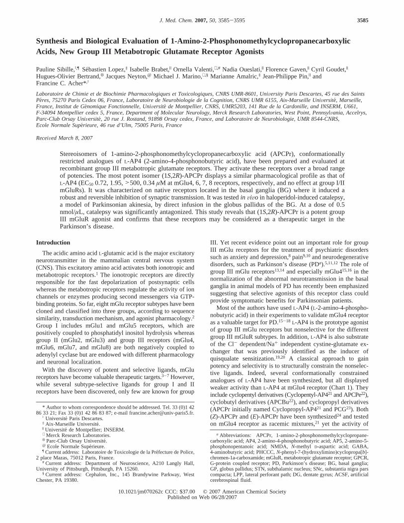
Most of the authors have usedL-AP4 (L-2-amino-4-phospho-nobutyric acid) in their experiments to validate mGlu4 receptoras a valuable target for PD.15-18 L-AP4 is the prototype agonistof group III mGlu receptors but nonselective for the differentgroup III mGluR subtypes. In addition,L-AP4 is also substrateof the Cl- dependent/Na+ independent cystine-glutamate ex-changer that was previously identified as the inducer ofquisqualate sensitization.19,20 A classical approach to gainpotency and selectivity is to structurally constrain the nonselec-tive ligands. Indeed, several conformationally constrainedanalogues ofL-AP4 have been synthesized, but all displayedweaker activity thanL-AP4 at mGlu4 receptor (Chart 1). Theyinclude cyclopentyl derivatives (Cyclopentyl-AP421 and APCPe22),cyclobutyl derivatives (APCBu22), and cyclopropyl derivatives(APCPr initially named Cyclopropyl-AP421 and PCG23). Both(Z)-APCPr and (E)-APCPr have been synthesized24 and testedon mGlu4 receptor as racemic mixtures,21 yet the activity of
* Author to whom correspondence should be addressed. Tel. 33 (0)1 4286 33 21; Fax 33 (0)1 42 86 83 87; e-mail [email protected].
† UniversiteParis Descartes.‡ Aix-Marseille Universite.| Universitede Montpellier; INSERM.⊥ Merck Research Laboratories.X Parc-Club Orsay Universite´.@ Ecole Normale Supe´rieure.¶ Current address: Laboratoire de Toxicologie de la Pre´fecture de Police,
2 place Mazas, 75012 Paris, France.# Current address: Department of Neuroscience, A210 Langly Hall,
University of Pittsburgh, Pittsburgh, PA 15260.§ Current address: Cephalon, Inc., 145 Brandywine Parkway, West
Chester, PA 19380.
a Abbreviations: APCPr, 1-amino-2-phosphonomethylcyclopropane-carboxylic acid; AP4, 2-amino-4-phosphonobutyric acid; AP5, 2-amino-5-phosphonopentanoic acid; NMDA,N-methyl D-aspartic acid; GABA,4-aminobutyric acid; PHCCC,N-phenyl-7-(hydroxylimino)cyclopropa[b]-chromen-1a-carboxamide; mGluR, metabotropic glutamate receptor; GPCR,G-protein coupled receptor; PD, Parkinson’s disease; BG, basal ganglia;GP, globus pallidus; STN, subthalamic nucleus; SNc, substantia nigra parscompacta; LPP, lateral perforant path; DG, dentate gyrus; ACSF, artificialcerebrospinal fluid.
3585J. Med. Chem.2007,50, 3585-3595
10.1021/jm070262c CCC: $37.00 © 2007 American Chemical SocietyPublished on Web 06/28/2007
the pure enantiomers may differ from that of the racemates. Inthis study, we report on the synthesis of the four stereoisomersof 1-amino-2-phosphonomethylcyclopropanecarboxylic acidAPCPr (1 and2, Chart 2) and their functional activity on eachgroup III mGlu receptor expressed in HEK 293 cells. The mostpotent stereoisomer was further characterizedin Vitro in brainslices andin ViVo in a rat haloperidol-induced catalepsy modelof Parkinsonian akinesia.
Results
Chemistry. tert-Butyl (1S,2R)-1-(tert-butoxycarbonylamino)-2-(hydroxymethyl)cyclopropane-1-carboxylate3 and ethyl(1R,2R)-1-(tert-butoxycarbonylamino)-2-(hydroxymethyl)cyclo-
propane-1-carboxylate4 were prepared from (R)-(+)-benzylg-lycerol according to literature references.25-28 Those alcoholswere then submitted to bromination using CBr4 and polymer-bound PPh3 in CH2Cl2 in the presence of triethylamine (Scheme1A). The next step in the synthesis of APCPr, the introductionof the phosphonate functionality, was difficult to achieve.Bromide5 was reacted with different phosphorus reactants underArbuzov29 or Michaelis-Becker conditions (P(OMe)3, P(OiPr)3,HP(O)(OEt)2/NaH/toluene, HP(O)(OPh)2/DBU/acetonitrile) butgave mostly the cyclopropane-cleavage product7, along withthe phosphonate8 and bicyclic product9 in minor quantities(Scheme 1B). Replacing the bromide by a chloride or an iodidedid not solve the problem, but decreasing the electrodonorcapacity of the nitrogen of substrate5 by changing the amineprotective group (Boc) to a more electron-withdrawing group(trifluoroacetyl) afforded the phosphonate12 with a significantdecrease of cyclopropane cleavage and no other secondaryproduct besides the alkene derivative. Indeed the intermediatecarbocation resulting from the cyclopropane ring opening is lessprone to be formed because, as the nitrogen free doublet isattracted by the trifluoroacetyl group, it is prevented fromforming the iminium ion and stabilizing the carbocation (Scheme1C). The protected APCPr12and the alkene13were separatedby flash chromatography (Scheme 2). Acid hydrolysis allowedcomplete deprotection of12 to give (-)-(1S,2R)-1-amino-2-phosphonomethylcyclopropanecarboxylic acid (1S,2R)-APCPr(-)-1 which was purified by ionic chromatography. (-)-(1R,2R)-1-Amino-2-phosphonomethylcyclopropanecarboxylic acid(-)-2 and enantiomers(+)-1 and (+)-2 were obtained in anidentical manner (Scheme 2).
Pharmacological Activities of APCPrs at mGlu Receptors.The effects of the four stereoisomers of APCPr (Chart 2) wereexamined on all group III mGlu receptors (mGlu4, mGlu6,mGlu7, and mGlu8). These receptors were transiently expressedin HEK 293 cells as previously described,30 and the total inositolphosphate production resulting from the receptor activation wasdetermined. Since group III mGlu receptors are not normallycoupled to PLC but rather inhibit adenylyl cyclase, this couplingwas made possible by coexpressing this receptor with thechimeric G-protein alpha subunit GRqi.30,31 We previouslyreported that this assay gave more accurate results than the
Chart 1. Cyclic Analogues ofL-AP4:a Structures and EC50
Values at the mGlu4 Receptor
a Abbreviations: AP4, 2-amino-4-phosphonobutyric acid; SOP, serine-O-phosphate; cyclopentyl-AP4, 1-amino-3-phosphonocyclopentanecarboxy-lic acid; APCPe, 1-amino-2-phosphonomethylcyclopentanecarboxylic acid;APCBu, 1-amino-2-phosphonomethylcyclobutanecarboxylic acid; APCPr,1-amino-2-phosphonomethylcyclopropanecarboxylic acid.b This study.c Reference 21.d Reference 22.e Reference 23.
Chart 2. The Four Stereoisomers of APCPr:1 and2 are theZandE Diastereoisomers, Respectively21
Scheme 1.Synthesis of Bromides5 and6. Arbuzov and SideReactionsa
a Reagents and conditions: (a) polymer-bound PPh3, CBr4, NEt3, CH2Cl2;(b) P(OMe)3, reflux.
3586 Journal of Medicinal Chemistry, 2007, Vol. 50, No. 15 Sibille et al.
classical measurement of the inhibition of the forskolin-activatedadenylyl cyclase activity and that the pharmacology of thesereceptors was not altered.30
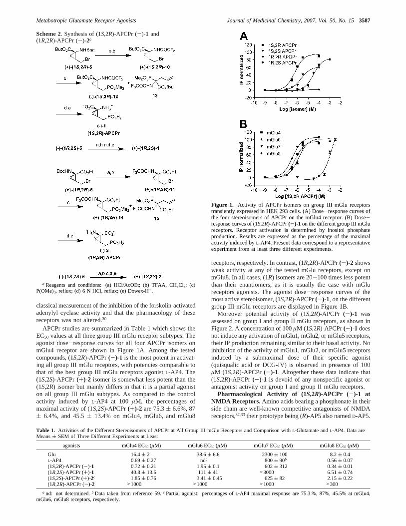
APCPr studies are summarized in Table 1 which shows theEC50 values at all three group III mGlu receptor subtypes. Theagonist dose-response curves for all four APCPr isomers onmGlu4 receptor are shown in Figure 1A. Among the testedcompounds, (1S,2R)-APCPr(-)-1 is the most potent in activat-ing all group III mGlu receptors, with potencies comparable tothat of the best group III mGlu receptors agonistL-AP4. The(1S,2S)-APCPr(+)-2 isomer is somewhat less potent than the(1S,2R) isomer but mainly differs in that it is a partial agoniston all group III mGlu subtypes. As compared to the controlactivity induced byL-AP4 at 100 µM, the percentages ofmaximal activity of (1S,2S)-APCPr(+)-2 are 75.3( 6.6%, 87( 6.4%, and 45.5( 13.4% on mGlu4, mGlu6, and mGlu8
receptors, respectively. In contrast, (1R,2R)-APCPr(-)-2 showsweak activity at any of the tested mGlu receptors, except onmGlu8. In all cases, (1R) isomers are 20-100 times less potentthan their enantiomers, as it is usually the case with mGlureceptors agonists. The agonist dose-response curves of themost active stereoisomer, (1S,2R)-APCPr(-)-1, on the differentgroup III mGlu receptors are displayed in Figure 1B.
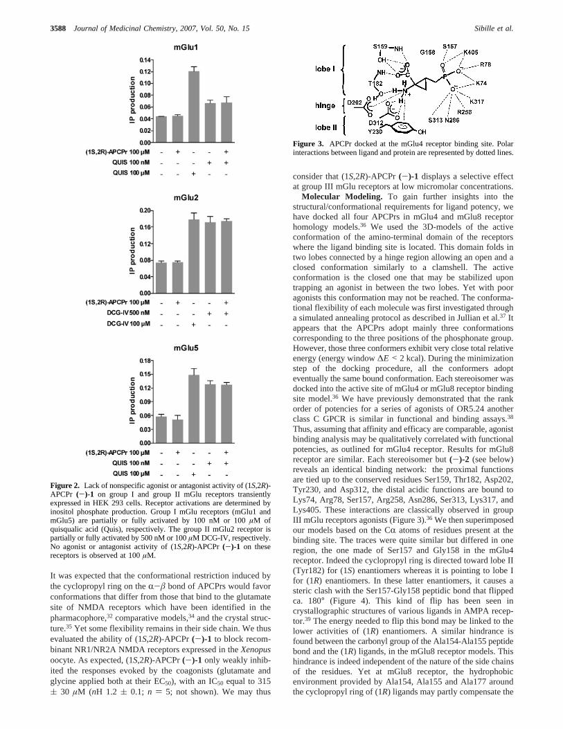
Moreover potential activity of (1S,2R)-APCPr (-)-1 wasassessed on group I and group II mGlu receptors, as shown inFigure 2. A concentration of 100µM (1S,2R)-APCPr(-)-1 doesnot induce any activation of mGlu1, mGlu2, or mGlu5 receptors,their IP production remaining similar to their basal activity. Noinhibition of the activity of mGlu1, mGlu2, or mGlu5 receptorsinduced by a submaximal dose of their specific agonist(quisqualic acid or DCG-IV) is observed in presence of 100µM (1S,2R)-APCPr (-)-1. Altogether these data indicate that(1S,2R)-APCPr (-)-1 is devoid of any nonspecific agonist orantagonist activity on group I and group II mGlu receptors.
Pharmacological Activity of (1S,2R)-APCPr (-)-1 atNMDA Receptors.Amino acids bearing a phosphonate in theirside chain are well-known competitive antagonists of NMDAreceptors,32,33their prototype being (R)-AP5 also namedD-AP5.
Scheme 2. Synthesis of (1S,2R)-APCPr (-)-1 and(1R,2R)-APCPr (-)-2a
a Reagents and conditions: (a) HCl/AcOEt; (b) TFAA, CH2Cl2; (c)P(OMe)3, reflux; (d) 6 N HCl, reflux; (e) Dowex-H+.
Table 1. Activities of the Different Stereoisomers of APCPr at All Group III mGlu Receptors and Comparison withL-Glutamate andL-AP4. Data areMeans( SEM of Three Different Experiments at Least
agonists mGlu4 EC50 (µM) mGlu6 EC50 (µM) mGlu7 EC50 (µM) mGlu8 EC50 (µM)
Glu 16.4( 2 38.6( 6.6 2300( 100 8.2( 0.4L-AP4 0.69( 0.27 nda 800( 90b 0.56( 0.07(1S,2R)-APCPr (-)-1 0.72( 0.21 1.95( 0.1 602( 312 0.34( 0.01(1R,2S)-APCPr (+)-1 40.8( 13.6 111( 41 >3000 6.51( 0.74(1S,2S)-APCPr (+)-2c 1.85( 0.76 3.41( 0.45 625( 82 2.15( 0.22(1R,2R)-APCPr (-)-2 >1000 >1000 >1000 >300
a nd: not determined.b Data taken from reference 59.c Partial agonist: percentages ofL-AP4 maximal response are 75.3.%, 87%, 45.5% at mGlu4,mGlu6, mGlu8 receptors, respectively.
Figure 1. Activity of APCPr isomers on group III mGlu receptorstransiently expressed in HEK 293 cells. (A) Dose-response curves ofthe four stereoisomers of APCPr on the mGlu4 receptor. (B) Dose-response curves of (1S,2R)-APCPr(-)-1 on the different group III mGlureceptors. Receptor activation is determined by inositol phosphateproduction. Results are expressed as the percentage of the maximalactivity induced byL-AP4. Present data correspond to a representativeexperiment from at least three different experiments.
Metabotropic Glutamate Receptor Agonists Journal of Medicinal Chemistry, 2007, Vol. 50, No. 153587
It was expected that the conformational restriction induced bythe cyclopropyl ring on theR-â bond of APCPrs would favorconformations that differ from those that bind to the glutamatesite of NMDA receptors which have been identified in thepharmacophore,32 comparative models,34 and the crystal struc-ture.35 Yet some flexibility remains in their side chain. We thusevaluated the ability of (1S,2R)-APCPr(-)-1 to block recom-binant NR1/NR2A NMDA receptors expressed in theXenopusoocyte. As expected, (1S,2R)-APCPr(-)-1 only weakly inhib-ited the responses evoked by the coagonists (glutamate andglycine applied both at their EC50), with an IC50 equal to 315( 30 µM (nH 1.2 ( 0.1; n ) 5; not shown). We may thus
consider that (1S,2R)-APCPr (-)-1 displays a selective effectat group III mGlu receptors at low micromolar concentrations.
Molecular Modeling. To gain further insights into thestructural/conformational requirements for ligand potency, wehave docked all four APCPrs in mGlu4 and mGlu8 receptorhomology models.36 We used the 3D-models of the activeconformation of the amino-terminal domain of the receptorswhere the ligand binding site is located. This domain folds intwo lobes connected by a hinge region allowing an open and aclosed conformation similarly to a clamshell. The activeconformation is the closed one that may be stabilized upontrapping an agonist in between the two lobes. Yet with pooragonists this conformation may not be reached. The conforma-tional flexibility of each molecule was first investigated througha simulated annealing protocol as described in Jullian et al.37 Itappears that the APCPrs adopt mainly three conformationscorresponding to the three positions of the phosphonate group.However, those three conformers exhibit very close total relativeenergy (energy window∆E < 2 kcal). During the minimizationstep of the docking procedure, all the conformers adopteventually the same bound conformation. Each stereoisomer wasdocked into the active site of mGlu4 or mGlu8 receptor bindingsite model.36 We have previously demonstrated that the rankorder of potencies for a series of agonists of OR5.24 anotherclass C GPCR is similar in functional and binding assays.38
Thus, assuming that affinity and efficacy are comparable, agonistbinding analysis may be qualitatively correlated with functionalpotencies, as outlined for mGlu4 receptor. Results for mGlu8receptor are similar. Each stereoisomer but(-)-2 (see below)reveals an identical binding network: the proximal functionsare tied up to the conserved residues Ser159, Thr182, Asp202,Tyr230, and Asp312, the distal acidic functions are bound toLys74, Arg78, Ser157, Arg258, Asn286, Ser313, Lys317, andLys405. These interactions are classically observed in groupIII mGlu receptors agonists (Figure 3).36 We then superimposedour models based on the CR atoms of residues present at thebinding site. The traces were quite similar but differed in oneregion, the one made of Ser157 and Gly158 in the mGlu4receptor. Indeed the cyclopropyl ring is directed toward lobe II(Tyr182) for (1S) enantiomers whereas it is pointing to lobe Ifor (1R) enantiomers. In these latter enantiomers, it causes asteric clash with the Ser157-Gly158 peptidic bond that flippedca. 180° (Figure 4). This kind of flip has been seen incrystallographic structures of various ligands in AMPA recep-tor.39 The energy needed to flip this bond may be linked to thelower activities of (1R) enantiomers. A similar hindrance isfound between the carbonyl group of the Ala154-Ala155 peptidebond and the (1R) ligands, in the mGlu8 receptor models. Thishindrance is indeed independent of the nature of the side chainsof the residues. Yet at mGlu8 receptor, the hydrophobicenvironment provided by Ala154, Ala155 and Ala177 aroundthe cyclopropyl ring of (1R) ligands may partly compensate the
Figure 2. Lack of nonspecific agonist or antagonist activity of (1S,2R)-APCPr (-)-1 on group I and group II mGlu receptors transientlyexpressed in HEK 293 cells. Receptor activations are determined byinositol phosphate production. Group I mGlu receptors (mGlu1 andmGlu5) are partially or fully activated by 100 nM or 100µM ofquisqualic acid (Quis), respectively. The group II mGlu2 receptor ispartially or fully activated by 500 nM or 100µM DCG-IV, respectively.No agonist or antagonist activity of (1S,2R)-APCPr (-)-1 on thesereceptors is observed at 100µM.
Figure 3. APCPr docked at the mGlu4 receptor binding site. Polarinteractions between ligand and protein are represented by dotted lines.
3588 Journal of Medicinal Chemistry, 2007, Vol. 50, No. 15 Sibille et al.
steric destabilization. It may thus explain a significanly lowerEC50 at mGlu8 receptor than at mGlu4 receptor with theseagonists (Table 1). Based on mGlu receptor pharmacophoremodels the region of the 1R cyclopropyl ring is stericallyrestricted,40,41 this is in concordance with what we report here.It should also be noted that the active conformation of thebinding domain of the receptor that is modeled in this study,may not be relevant for weak agonists such as (1R,2R)-APCPr(-)-2 or partial agonists as (1S,2S)-APCPr(+)-2. Activities ofthe (1S) enantiomers are close to each other and indeed the tracesare quite similar even in the Ser157-Gly158 region (Figure 5).Nevertheless, the methylphosphonate protons of the less potent(1S,2S)-APCPr(+)-2 are directed to the same “forbidden zone”described above. This situation may be responsible for the partialagonism of this stereoisomer. A steric hindrance with the twohomologous residues of mGlu8 receptor has been reported tobe the cause of the partial agonism of FP429 at mGlu8 subtype.42
In that previous study, FP429 was a full agonist at mGlu4receptor and a partial agonist at mGlu8 receptor, we demon-
strated that the side chains of Ala154-Ala155 were responsiblefor that property and suggested that hindrance with those sidechains induced either a partial closing of the bilobate domainor a weaker stabilization of that domain resulting in partialagonism. In the present case, hindrance would occur both withSer157 and/or Gly158 of the mGlu4 receptor and with Ala154and/or Ala155 of the mGlu8 receptor, resulting also in anonoptimal closed conformation of the two subtype bindingdomains. This effect being analogous with mGlu4 and mGlu8receptors, mutagenesis experiments may not help to investigatethe cause of partial agonism.
Electrophysiology.As (1S,2R)-APCPr(-)-1 was shown tostrongly activate recombinant group III mGlu receptors, we nextchose to investigate the effect of this compound on nativereceptors. Previous studies have shown that group III mGlureceptors are presynaptically localized at glutamatergic andGABAergic nerve terminals and that activation of these recep-tors produces a marked reduction of Glu or GABA transmission.Therefore, (1S,2R)-APCPr(-)-1 was testedin Vitro for its abilityto inhibit the excitatory transmission in the basal ganglia (BG)at the synapse between the subthalamic nucleus and thesubstantia nigra pars compacta (STN-SNc synapse) and at thehippocampal lateral perforant path-dentate gyrus synapse (LPP-DG). Group III modulation of transmission at the subthalamo-nigral synapse has been previously demonstrated to be solely
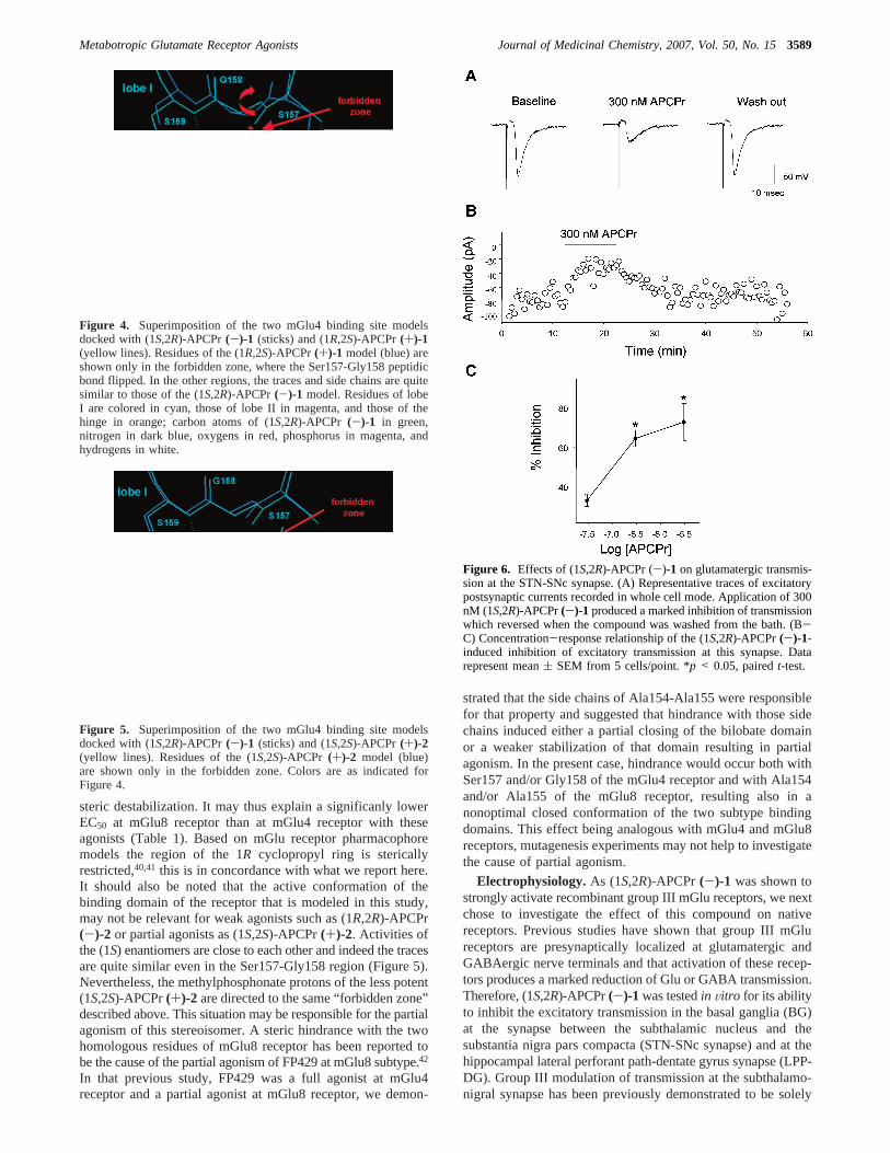
Figure 4. Superimposition of the two mGlu4 binding site modelsdocked with (1S,2R)-APCPr(-)-1 (sticks) and (1R,2S)-APCPr(+)-1(yellow lines). Residues of the (1R,2S)-APCPr(+)-1 model (blue) areshown only in the forbidden zone, where the Ser157-Gly158 peptidicbond flipped. In the other regions, the traces and side chains are quitesimilar to those of the (1S,2R)-APCPr(-)-1 model. Residues of lobeI are colored in cyan, those of lobe II in magenta, and those of thehinge in orange; carbon atoms of (1S,2R)-APCPr (-)-1 in green,nitrogen in dark blue, oxygens in red, phosphorus in magenta, andhydrogens in white.
Figure 5. Superimposition of the two mGlu4 binding site modelsdocked with (1S,2R)-APCPr (-)-1 (sticks) and (1S,2S)-APCPr (+)-2(yellow lines). Residues of the (1S,2S)-APCPr (+)-2 model (blue)are shown only in the forbidden zone. Colors are as indicated forFigure 4.
Figure 6. Effects of (1S,2R)-APCPr (-)-1 on glutamatergic transmis-sion at the STN-SNc synapse. (A) Representative traces of excitatorypostsynaptic currents recorded in whole cell mode. Application of 300nM (1S,2R)-APCPr(-)-1 produced a marked inhibition of transmissionwhich reversed when the compound was washed from the bath. (B-C) Concentration-response relationship of the (1S,2R)-APCPr(-)-1-induced inhibition of excitatory transmission at this synapse. Datarepresent mean( SEM from 5 cells/point. *p < 0.05, pairedt-test.
Metabotropic Glutamate Receptor Agonists Journal of Medicinal Chemistry, 2007, Vol. 50, No. 153589
meditated by mGlu4 receptors43 while in the hippocampus, onlythe mGlu8 receptors are involved.44
Consistent with the observations in recombinant systems, wefound that bath application of (1S,2R)-APCPr(-)-1 induced arobust and reversible inhibition of synaptic transmission at boththe STN-SNc synapse (Figure 6) and the LPP-DG synapse(Figure 7). The effects where dose dependent and at bothsynapses reached significance at the 300 nM dose. These datasuggest that (1S,2R)-APCPr(-)-1 activates native mGlu recep-tors in a manner consistent with our recombinant findings.
In Vi Wo Effect of (1S,2R)-APCPr (-)-1: AntiparkinsonianEffect When Injected into the CNS. A growing number ofanimal studies suggest that the globus pallidus (GP) plays akey role in the pathophysiology of PD.45 Increased GABAneurotransmission at the striato-pallidal synapse has beenthought to underlie the alteration of GP firing activity, whichis the hallmark of the pathophysiological dysfunction observedin PD. In particular, it was recently shown that activation ofthe mGlu4 receptor with the positive allosteric modulatorPHCCC orL-AP4 decreases GABAergic transmission at thestriato-pallidal synapse and reverses the akinesia produced bydopamine depletion in the reserpine model of PD.12,15 This isconsistent with a presynaptic inhibition of GABA transmissionin the GP. We therefore tested the effects of (1S,2R)-APCPr(-)-1 in haloperidol-induced catalepsy, a classical model ofParkinsonian akinesia, by directly infusing the compound intothe GP. Systemic injection of the dopamine D1/D2 receptorantagonist, haloperidol (1 mg/kg i.p.), produced a profound
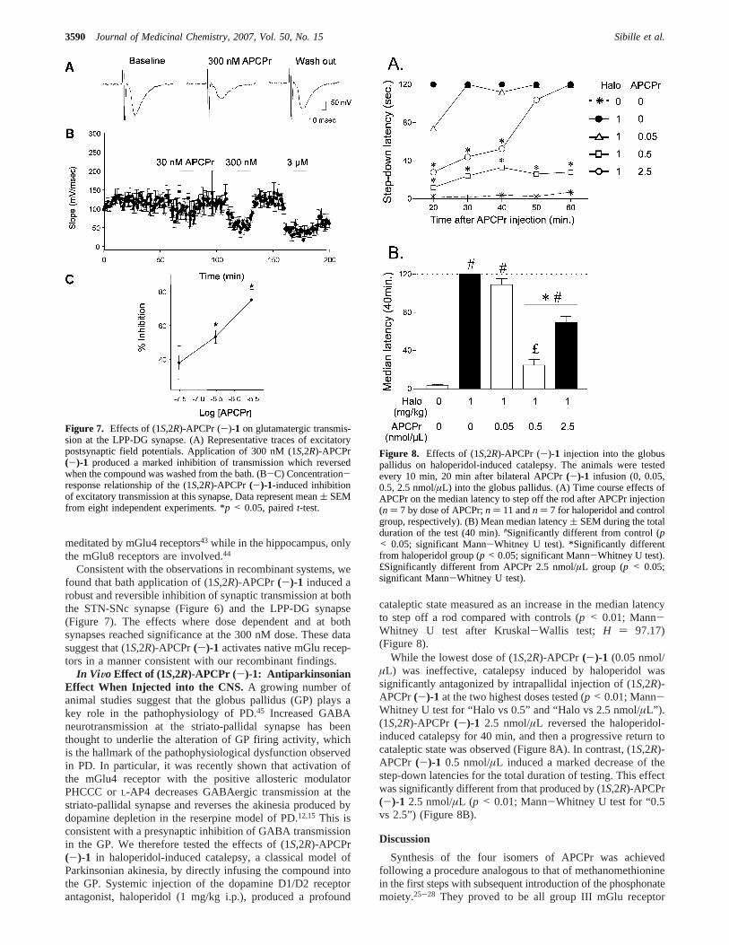
cataleptic state measured as an increase in the median latencyto step off a rod compared with controls (p < 0.01; Mann-Whitney U test after Kruskal-Wallis test; H ) 97.17)(Figure 8).
While the lowest dose of (1S,2R)-APCPr(-)-1 (0.05 nmol/µL) was ineffective, catalepsy induced by haloperidol wassignificantly antagonized by intrapallidal injection of (1S,2R)-APCPr(-)-1 at the two highest doses tested (p < 0.01; Mann-Whitney U test for “Halo vs 0.5” and “Halo vs 2.5 nmol/µL”).(1S,2R)-APCPr (-)-1 2.5 nmol/µL reversed the haloperidol-induced catalepsy for 40 min, and then a progressive return tocataleptic state was observed (Figure 8A). In contrast, (1S,2R)-APCPr (-)-1 0.5 nmol/µL induced a marked decrease of thestep-down latencies for the total duration of testing. This effectwas significantly different from that produced by (1S,2R)-APCPr(-)-1 2.5 nmol/µL (p < 0.01; Mann-Whitney U test for “0.5vs 2.5”) (Figure 8B).
Discussion
Synthesis of the four isomers of APCPr was achievedfollowing a procedure analogous to that of methanomethioninein the first steps with subsequent introduction of the phosphonatemoiety.25-28 They proved to be all group III mGlu receptor
Figure 7. Effects of (1S,2R)-APCPr (-)-1 on glutamatergic transmis-sion at the LPP-DG synapse. (A) Representative traces of excitatorypostsynaptic field potentials. Application of 300 nM (1S,2R)-APCPr(-)-1 produced a marked inhibition of transmission which reversedwhen the compound was washed from the bath. (B-C) Concentration-response relationship of the (1S,2R)-APCPr (-)-1-induced inhibitionof excitatory transmission at this synapse, Data represent mean( SEMfrom eight independent experiments. *p < 0.05, pairedt-test.
Figure 8. Effects of (1S,2R)-APCPr (-)-1 injection into the globuspallidus on haloperidol-induced catalepsy. The animals were testedevery 10 min, 20 min after bilateral APCPr(-)-1 infusion (0, 0.05,0.5, 2.5 nmol/µL) into the globus pallidus. (A) Time course effects ofAPCPr on the median latency to step off the rod after APCPr injection(n ) 7 by dose of APCPr;n ) 11 andn ) 7 for haloperidol and controlgroup, respectively). (B) Mean median latency( SEM during the totalduration of the test (40 min).#Significantly different from control (p< 0.05; significant Mann-Whitney U test). *Significantly differentfrom haloperidol group (p < 0.05; significant Mann-Whitney U test).£Significantly different from APCPr 2.5 nmol/µL group (p < 0.05;significant Mann-Whitney U test).
3590 Journal of Medicinal Chemistry, 2007, Vol. 50, No. 15 Sibille et al.
agonists, the (1S) stereoisomers being more potent than the (1R)stereoisomers. (Z)-APCPr, which was previously described asan mGlu4 receptor agonist, is composed of (1S,2R)- and (1R,2S)-APCPr whereas (E)-APCPr from the same study is the racemicmixture of (1R,2R)- and (1S,2S)-APCPr.21 The EC50 values ofthese racemates cannot be compared to those of the enantio-merically pure compounds because they were not tested in thesame system. Nevertheless, they are concordant as the EC50sof the most potent isomers of each racemate (1S,2R)-APCPrfor (Z)-APCPr and (1S,2S)-APCPr for (E)-APCPr, and the EC50sof racemates are found in the same range, respectively.
The present syntheses were performed in order to characterizethe most potent enantiomer of the APCPr series and to improvethe reported activity and selectivity. Unfortunately, selectivitywas not found among the four isomers, although the beststereoisomer (1S,2R)-APCPr (-)-1 is as potent asL-AP4. So,the additional cyclopropyl ring of conformationally restrainedAPCPr does not improve affinity, as the entropic gain is likelycompensated by a loss of enthalpy; neither induces a betterselectivity between group III mGlu receptors. However, (1S,2R)-APCPr (-)-1 may be more selective with respect to othersystems whereL-AP4 plays a role46 such as the cystine-glutamate exchanger previously identified as the quisqualateeffect.19,47,48 The large range of EC50s covered by the fourAPCPr stereoisomers may be interpreted on the basis ofhomology models of the binding site in its active conformation(Figures 4 and 5). As we observed previously, contacts betweenagonists and lobe I are tight, particularly with the S157-G158residues of the mGlu4 receptor (and homologous residues ofother subtypes); consequently, steric hindrance in that regioncauses a large affinity decrease as determined for (1R,2S)-(+)-1and (1R,2R)-APCPr (-)-2 or partial agonism as with (1S,2S)-APCPr(+)-2. A similar interpretation may explain the inactivityof APCPe and APCBu22 (Chart 1).
As expected, (1S,2R)-APCPr(-)-1 is inactive at group I andgroup II mGlu receptors and is only a weak inhibitor of NMDAreceptors. It may thus be considered as inactive at these receptorsat µM concentrations and may be used in further biological as-says. Indeed it was shown to inhibit the excitatory transmissionat the STN-SNc and the LPP-DG synapses. These results suggestthat (1S,2R)-APCPr (-)-1 activates native mGlu4 and mGlu8receptors in a manner consistent with the recombinant data.Finally the effect of direct infusion of (1S,2R)-APCPr (-)-1into the GP was tested in an animal model of PD. The GP isknown to be critically involved in motor control, and excessivestriatal inhibition of the GP is believed to play a key role in themotor deficits observed in Parkinson’s disease. Activation ofthe mGlu4 receptor decreases striatal inhibition of the GPand has previously been shown to exhibit antiparkinsonian ef-fects.12,15,49-51 Consistent with its potency at mGlu4 receptor,(1S,2R)-APCPr(-)-1 was able to reverse haloperidol-inducedakinesia. The finding of a somewhat biphasic effect in thesestudies may be explained by the activation of the mGlu7 receptorexpected at higher doses. In fact, the inhibition of striato-pallidaltransmission byL-AP4 exhibits an unusual biphasic concentra-tion-response relationship that was interpreted to suggest anactivation of mGlu7 receptor at concentrations above 30µm.15
The fact that the mGlu7 receptor is abundantly expressed inthe GP52 and does not yet play a significant role in themodulation of striato-pallidal transmission suggests that thisreceptor may be involved in modulating collateral inhibition atpallido-pallidal synapses. Therefore, activation of mGlu7 recep-tor in the GP may increase coherent activity across the nucleusand produce a proparkinsonian effect.45
Conclusion
The four stereoisomers of APCPrs have been synthesized andevaluated on cloned mGlu receptors. They all activate groupIII receptors over a large range of potency. The (1S,2R) isomeris the most active and shows similar potency asL-AP4. It wastested on brain slices and in an animal model of akinesia, adisabling symptom in Parkinson’s disease. (1S,2R)-APCPr(-)-1was able to reverse the cataleptic effect. This result confirmsthat group III mGlu receptors may be considered as a therapeutictarget for symptomatic treatment in the Parkinson’s disease.
Experimental Section
Chemistry. General Procedures.All chemicals and solventswere of the best quality available from commercial suppliers andused without further purification.1H (250.13 MHz),13C (62.9 MHz),and 31P (101.25 MHz) NMR spectra were recorded on an ARX250 Bruker spectrometer. Chemical shifts (δ, ppm) are given withreference to residual1H or 13C of deuterated solvents (CDCl3 7.24,77.00; CD3OD 3.30, 49.0; D2O 4.80) or external reference (H3PO4
95%). TLC was performed on Merck 60F254 precoated silica gelplates. Products were visualized by 2% (w/v) ninhydrin in ethanoland TDM reagent.53 Column chromatography was performed witha Biotage FLASH40i chromatography module (prepacked cartridgesystem). Optical rotations were measured at the sodium D line (589nm) or at 546 nm at 20°C, with a Perkin-Elmer 241 polarimeterusing a 1 dmpath length cell. High-pressure liquid chromatography(HPLC) was performed at 0°C with an Altex Chromatem 380pump, a Rheodyne 7125 valve (100µL loop), a Pye-Unicam LC-UV detector set at 210 nm, and a Shimadzu CR-3A integrator, usinga Crownpak CR(+) (150 mm * 4 mm i.d.) column equilibratedwith pH 1.0 HClO4 at a 0.1 mL/min flow rate.
(+)-tert-Butyl (1S,2R)-1-(tert-Butoxycarbonylamino)-2-(bro-momethyl)cyclopropane-1-carboxylate 5.To a solution of3 (2.2g, 7.6 mmol, 1 equiv) in CH2Cl2 (180 mL) under argon at 0°Cwere added CBr4 (5.3 g, 15.9 mmol, 2 equiv) and NEt3 (1.1 mL,7.9 mmol, 1 equiv) followed by polymer-bound PPh3 (5.0 g, 15.0mmol, 2 equiv) in small portions. After being stirred at 25°C for5 h, the reaction mixture was filtered through a short plug of celite.After evaporation of the filtrate, the crude product was purified byflash chromatography. Elution with CH2Cl2 afforded5 (1.5 g, 4.3mmol, 56.4%) as a white solid: TLC (silica gel, CH2Cl2/EtOAc,8:2, ninhydrin visualization)Rf 0.78;1H NMR (CDCl3) δ 5.17 (brs, 1H), 3.52 (m, 1H), 3.41 (m, 1H), 2.15 (m, 1H), 1.69 (m, 1H),1.44 (s, 9H), 1.43 (s, 9H), 1.08 (m, 1H);13C NMR (CDCl3) δ 170.8,156.3, 81.8, 80.2, 41.2, 32.5, 29.2, 28.2, 27.9, 24.2; [R]20D 2.4(c 1, CHCl3).
(+)-Ethyl (1R,2R)-1-(tert-Butoxycarbonylamino)-2-(bromom-ethyl)cyclopropane-1-carboxylate 6.The (1R,2R) bromide6 wassynthesized from4 in an identical manner to the (1S,2R) bromide5 described above: TLC (silica gel, CH2Cl2/EtOAc, 9:1, ninhydrinvisualization)Rf 0.64; 1H NMR (CDCl3) δ 5.20 (br s, 1H), 4.19(m, 2H), 3.72 (dd,J ) 6.8, 10.4 Hz, 1H), 3.54 (t,J ) 10.0 Hz,1H), 1.95 (m, 1H), 1.70 (dd,J ) 6.0, 12.8 Hz, 1H), 1.52 (m, 1H),1.45 (s, 9H), 1.30 (t,J ) 7.2 Hz, 3H);13C NMR (CDCl3) δ 170.6,155.7, 80.2, 61.8, 41.0, 32.9, 30.6, 28.3, 25.5, 14.2; [R]20D 53.1(c 1, CHCl3).
tert-Butyl 1-[N-(tert-Butoxycarbonyl)amino]-1-[(dimethoxy-phosphoryl)methyl]-but-3-enecarboxylate 7.To 5 (64 mg, 0.18mmol, 1 equiv) under Ar was added P(OMe)3 (25 µL, 0.21 mmol,1.2 equiv). The resulting mixture was heated under reflux for 4 h.The solution was cooled and evaporated. The crude mixture wasextracted with EtOAc (200 mL), and the organic phase was washedwith H2O (100 mL), saturated NaHCO3 solution (2× 100 mL),and brine (3× 100 mL), dried over MgSO4, and evaporated. Theresidue was purified by flash chromatography. Elution withincreasing amounts of EtOAc (10-100%) in CH2Cl2 afforded7(21 mg, 0.06 mmol, 30.8%) as a colorless oil: TLC (silica gel,EtOAc, TDM visualization)Rf 0.31;1H NMR (CDCl3) δ 5.61 (m,2H), 5.12 (m, 2H), 3.79 (t,J ) 10.4 Hz, 6H), 3.37 (m, 1H), 2.92
Metabotropic Glutamate Receptor Agonists Journal of Medicinal Chemistry, 2007, Vol. 50, No. 153591
(m, 1H), 1.47 (s, 9H), 1.41 (s, 9H);13C NMR (CDCl3) δ 167.4,153.5, 131.6 (d,J ) 11.3 Hz), 119.6, 83.6, 79.6, 63.9 (d,J ) 147.7Hz), 54.3 (d,J ) 7.0 Hz), 53.8 (d,J ) 7.0 Hz), 35.2, 28.2, 27.7;31P NMR (CDCl3) δ 22.2.
tert-Butyl (1S,2R)-1-[N-(tert-Butoxycarbonyl)amino]-2-[(dimethoxyphosphoryl)methyl]cyclopropanecarboxylate 8. 8(4mg, 0.01 mmol, 5.8%) was obtained as a colorless oil from theprevious reaction: TLC (silica gel, EtOAc, TDM visualization)Rf
0.28; 1H NMR (CDCl3) δ 5.86 (br s, 1H), 3.78 (d,J ) 6.4 Hz,3H), 3.74 (d,J ) 6.4 Hz, 3H), 2.24 (m, 1H), 1.85 (m, 1H), 1.73(m, 1H), 1.43 (s, 9H), 1.41 (s, 9H), 1.29 (m, 1H), 0.88 (m, 1H);13C NMR (CDCl3) δ 171.9, 156.6, 81.2, 79.4, 51.9 (d,J ) 6.5Hz), 51.5 (d,J ) 6.5 Hz), 38.2, 29.7, 28.3, 27.9, 24.4 (d,J )151.2 Hz), 21.1;31P NMR (CDCl3) δ 28.4.
tert-Butyl (1S,6R)-3-Oxo-4-oxa-2-azabicyclo[4.1.0]heptane-1-carboxylate 9. 9 (9 mg, 0.04 mmol, 23.5%) was obtained fromthe previous reaction as colorless crystals: TLC (silica gel, CH2-Cl2/EtOAc 80:20, TDM visualization)Rf 0.32; 1H NMR (CDCl3)δ 5.87 (s, 1H), 4.53 (m, 1H), 4.14 (m, 1H), 2.11 (m, 1H), 1.59 (m,1H), 1.46 (s, 9H), 1.34 (m, 1H);13C NMR (CDCl3) δ 168.6, 152.9,83.3, 66.5, 38.7, 28.0, 21.7, 18.9.
(+)-tert-Butyl (1S,2R)-1-[N-(Trifluoroacetyl)amino]-2-(bro-momethyl)cyclopropane-1-carboxylate 10.To 5 (1.5 g, 4.3 mmol,1 equiv) was added a solution of 2 M HCl/AcOEt (130 mL). Afterthe mixture was stirred at 25°C for 2h, the HCl/AcOEt wasevaporated and the residue was dried under vacuum. The crudeamine hydrochloride was used without further purification: TLC(silica gel, CH2Cl2/CH3OH/NH4OH, 85:17:2.5, ninhydrin visualiza-tion) Rf 0.65;1H NMR (CD3OD) δ 4.87 (br s, 1H), 3.78 (dd,J )7.8, 11.1 Hz, 1H), 3.61 (m, 1H), 2.32 (m, 1H), 1.81 (dd,J ) 6.5,9.6 Hz, 1H), 1.52 (s, 9H), 1.39 (dd,J ) 6.9, 14.1 Hz, 1H);13CNMR (CD3OD) δ 170.3, 87.5, 43.8, 31.0, 30.9, 29.8, 22.8.
The amine hydrochloride was suspended in CH2Cl2 (130 mL)under argon. Trifluoroacetic anhydride TFAA (2.3 mL, 16.3 mmol,4 equiv) was added dropwise. The mixture was stirred for 1 h.After evaporation,10 was obtained as a pale yellow oil (1.41 g,4.1 mmol, 95.3%): TLC (silica gel, CH2Cl2/EtOAc, 8:2, ninhydrinvisualization)Rf 0.75; 1H NMR (CDCl3) δ 6.98 (br s, 1H), 3.58(dd,J ) 6.8, 10.8 Hz, 1H), 3.58 (t,J ) 9.6 Hz, 1H), 2.29 (m, 1H),1.93 (dd,J ) 5.9, 9.2 Hz, 1H), 1.42 (s, 9H), 1.23 (t,J ) 6.6 Hz,1H); 13C NMR (CDCl3) δ 168.3, 158.8 (q,J ) 37.4 Hz), 115.5 (q,J ) 288.5 Hz), 83.3, 39.9, 31.3, 28.7, 27.8, 23.9.
(+)-Ethyl (1R,2R)-1-[N-(Trifluoroacetyl)amino]-2-(bromom-ethyl)cyclopropane-1-carboxylate 11.The (1R,2R) bromide11was synthesized from6 in an identical manner to the (1S,2R)bromide10 described above.
Amine hydrochloride: TLC (silica gel, CH2Cl2/CH3OH/NH4-OH, 85:17:2.5, ninhydrin visualization)Rf 0.75; 1H NMR (CD3-OD) δ 4.36 (q,J ) 7.2 Hz, 2H), 3.88 (dd,J ) 6.0, 10.8 Hz, 1H),3.54 (t,J ) 10.4 Hz, 1H), 2.31 (m, 1H), 1.80 (m, 2H), 1.37 (t,J) 7.2 Hz, 3H);13C NMR (CD3OD) δ 170.0, 66.1, 42.8, 32.6, 31.5,24.2, 16.4.
11: TLC (silica gel, CH2Cl2/EtOAc, 8:2, ninhydrin visualization)Rf 0.73;1H NMR (CDCl3) δ 8.01 (br s, 1H), 4.24 (q,J ) 7.2 Hz,2H), 3.75 (dd,J ) 6.8, 10.4 Hz, 1H), 3.54 (t,J ) 10.0 Hz, 1H),2.08 (m, 1H), 1.89 (dd,J ) 6.4, 7.6 Hz, 1H), 1.67 (dd,J ) 6.4,9.2 Hz, 1H), 1.27 (t,J ) 7.2 Hz, 3H);13C NMR δ 173.9, 158.6(q, J ) 37.9 Hz), 115.2 (q,J ) 287.3 Hz), 62.8, 39.8, 32.4, 29.3,24.8, 13.7.
(-)-tert-Butyl (1S,2R)-1-[N-(Trifluoroacetyl)amino]-2-[(dimethoxyphosphoryl))methyl]cyclopropane-1-carboxylate 12.To 10 (1.41 g, 4.1 mmol, 1 equiv) under argon was added P(OMe)3
(2.0 mL, 16.9 mmol, 4 equiv). The resulting mixture was heatedunder reflux for 8 h. The solution was cooled and evaporated. Thecrude mixture was extracted with EtOAc (200 mL), and the organicphase was washed with H2O (100 mL), saturated NaHCO3 solution(2 × 100 mL), and brine (3× 100 mL), dried over MgSO4, andevaporated. The residue was purified by flash-chromatography.Elution with increasing amounts of EtOAc (10-100%) in CH2Cl2afforded12 (782 mg, 2.1 mmol, 50.9%): TLC (silica gel, EtOAc,TDM visualization)Rf 0.35; 1H NMR (CDCl3) δ 8.83 (br s, 1H),
3.76 (d,J ) 6.5 Hz, 3H), 3.72 (d,J ) 6.5 Hz, 3H), 2.30 (ddd,J )4.4, 15.6, 19.2 Hz, 1H), 1.88 (m, 1H), 1.77 (m, 1H), 1.51 (m, 1H),1.31 (s, 9H), 1.01 (m, 1H);13C NMR (CDCl3) δ 168.9, 158.6 (q,J ) 36.3 Hz), 115.6 (q,J ) 288.3 Hz), 81.9, 52.7 (d,J ) 6.5 Hz),52.4 (d,J ) 6.5 Hz), 37.0 (d,J ) 8.9 Hz), 27.3, 24.2 (d,J ) 142.0Hz), 20.0 (d,J ) 12.1 Hz), 19.3 (d,J ) 4.2 Hz);31P NMR (CDCl3)δ 33.0; [R]20D -51.6 (c 1, CH3OH).
tert-Butyl 1-[N-(Trifluoroacetyl)amino]-1-(dimethoxyphos-phoryl)-3-butenecarboxylate 13.This compound was isolated asthe major secondary product of the previous Arbuzov reaction(13.1% yield). TLC (silica gel, EtOAc, TDM visualization)Rf 0.39;1H NMR (CDCl3) δ 7.31 (br s, 1H), 5.44 (m, 1H), 5.15 (d,J ) 5.6Hz, 1H), 5.09 (s, 1H), 3.81 (d,J ) 11.0 Hz, 3H), 3.74 (d,J ) 11.0Hz, 3H), 3.49 (m, 1H), 2.97 (m, 1H), 1.47 (s, 9H);13C NMR(CDCl3) δ 166.3, 155.4 (q,J ) 37.0 Hz), 129.7 (d,J ) 13.1 Hz),121.9, 115.4 (q,J ) 288.7 Hz), 85.3, 64.6 (d,J ) 147.8 Hz), 54.8(d, J ) 7.0 Hz), 53.8 (d,J ) 7.0 Hz), 34.0, 27.6;31P NMR (CDCl3)δ 19.4.
(+)-Ethyl (1R,2R)-1-[N-(Trifluoroacetyl)amino]-2-[(dimethox-yphosphoryl))methyl]cyclopropane-1-carboxylate 14.The (1R,2R)phosphonate14was synthesized from11 in an identical manner tothe (1S,2R) phosphonate12 described above. The recovery waslower with a 17.5% yield: TLC (silica gel, EtOAc, TDM visualiza-tion) Rf 0.16; 1H NMR (CDCl3) δ 8.19 (br s, 1H), 4.17 (m, 2H),3.72 (d,J ) 4.8 Hz, 3H), 3.68 (d,J ) 4.8 Hz, 3H), 2.05 (ddd,J )0.8, 7.2, 18.4 Hz, 2H), 1.82 (m, 1H), 1.66 (dd,J ) 5.8, 8.0 Hz,1H), 1.47 (dd,J ) 5.8, 9.2 Hz, 1H), 1.22 (t,J ) 7.2 Hz, 3H);13CNMR (CDCl3) δ 169.2, 158.1 (q,J ) 37.4 Hz), 115.7 (q,J )287.8 Hz), 61.8, 52.4 (d,J ) 6.3 Hz), 52.3 (d,J ) 6.3 Hz), 37.2(d, J ) 10.5 Hz), 23.2 (d,J ) 3.2 Hz), 22.5, 21.8 (d,J ) 104.9Hz), 13.9;31P NMR (CDCl3) δ 32.6; [R]20D 8.9 (c 1, CHCl3).
Ethyl 1-[N-(Trifluoroacetyl)amino]-1-(dimethoxyphosphoryl)-3-butenecarboxylate 15.This compound is the major secondaryproduct of the previous Arbuzov reaction (27.1% yield). TLC (silicagel, EtOAc, TDM visualization)Rf 0.17;1H NMR (CDCl3) δ 7.30(br s, 1H), 5.49 (m, 1H), 5.19 (d,J ) 4.4 Hz, 1H), 5.13 (s, 1H),4.35 (q,J ) 7.2 Hz, 2H), 3.86 (d,J ) 10.8 Hz, 3H), 3.79 (d,J )11.2 Hz, 3H), 3.55 (c,J ) 7.2 Hz, 1H), 3.04 (c,J ) 7.2 Hz, 1H),1.33 (t,J ) 7.2 Hz, 3H);13C NMR (CDCl3) δ 167.5, 155.5 (q,J) 35.8 Hz), 129.7 (d,J ) 12.6 Hz), 121.0, 115.3 (q,J ) 288.3Hz), 64.3 (d,J ) 14.7 Hz), 63.2, 55.0 (d,J ) 6.8 Hz), 54.0 (d,J) 7.4 Hz), 34.1, 13.9;31P NMR (CDCl3) δ 19.0.
(-)-(1S,2R)-1-Amino-2-phosphonomethylcyclopropane-carboxylic Acid 1. Compound12 (440 mg, 1.17 mmol) wasrefluxed in 6 N HCl (10 mL) for 22 h. The solvent was evaporated,and the remaining yellow oil was diluted in 700 mL H2O (pH 3),applied to an AG50-X4 cation-exchange column. The column waseluted with H2O. Evaporation of the combined ninhydrin positivefractions containing the desired material gave a white solid (219mg, 1.12 mmol, 96.0%): HPLCtR 15.9 min; 1H NMR (D2O) δ1.82-2.10 (br m, 3H), 1.55-1.69 (m, 2H);13C NMR (D2O) δ174.7, 41.3 (d,J ) 11.0 Hz), 28.0 (d,J ) 133.6 Hz), 24.7 (d,J )3.7 Hz), 21.3 (d,J ) 8.4 Hz);31P NMR (D2O) δ 22.0; [R]20D -51.5(c 1, H2O); Anal. (C5H10NO5P·2/3H2O) C, N. H: calcd; 5.52; found,5.25.
(-)-(1R,2R)-1-Amino-2-phosphonomethylcyclopropane-carboxylic Acid 2. The (1R,2R) phosphonate2 was synthesizedfrom 14 in an identical manner to the (1S,2R) phosphonic acid1described above. HPLCtR 18.1 min;1H NMR (D2O) δ 1.72-2.09(br m, 4H), 1.34 (t,J ) 7.0 Hz, 1H);13C NMR (D2O) δ 175.9,40.6, 28.5 (d,J ) 133.1 Hz), 23.3 (d,J ) 3.6 Hz), 22.3 (d,J ) 7.0Hz); 31P NMR (D2O) δ 23.5; [R]20
546 -9.8 (c 1, H2O); Anal. (C5H10-NO5P·2/3H2O) C, N. H: calcd, 5.52; found, 5.11.
The enantiomers were prepared in an identical fashion.(-)-tert-Butyl (1R,2S)-1-(tert-butoxycarbonylamino)-2-(bro-
momethyl)cyclopropane-1-carboxylate ((-)-5): [R]20D -2.8(c 1, CHCl3). (-)-Ethyl (1S,2S)-1-(tert-butoxycarbonylamino)-2-(bromomethyl)cyclopropane-1-carboxylate ((-)-6): [R]20D -46.5(c 1, CHCl3). (+)-tert-Butyl (1R,2S)-1-[N-(trifluoroacetyl)amino]-2-[(dimethoxyphosphoryl))methyl]cyclopropane-1-carboxylate((+)-12): [R]20D 54.0° (c 1, CH3OH). (-)-Ethyl (1S,2S)-1-[N-
3592 Journal of Medicinal Chemistry, 2007, Vol. 50, No. 15 Sibille et al.
(trifluoroacetyl)amino]-2-[(dimethoxyphosphoryl))methyl]cy-clopropane-1-carboxylate ((-)-14): [R]20D -7.8 (c 1, CHCl3). (+)-(1R,2S)-1-Amino-2-phosphonomethylcyclopropanecarboxylic acid((+)-1): [R]20D 52.3 (c 1, H2O). (+)-(1S,2S)-1-amino-2-phospho-nomethylcyclopropanecarboxylic acid ((+)-2): HPLC tR 19.9 min;[R]20
546 8.2 (c 1, H2O).Molecular Modeling. Conformational Analyses.All molecules
were protonated on theR-amino group and deprotonated on theR-carboxylic and distal phosphonic groups. They were subjectedto a conformational search using Discover 3.00 (Insight II Accelrys,San Diego, CA). The flexibility of each molecule was investigatedthrough a simulated annealing protocol using the CVFF forcefield(Accelrys) and setting the dielectric constant to 80, as alreadydescribed.37,40 After initial minimization, the temperature of thesystem was raised up to 900 K for 1 ps and cooled down to 300 Kduring 5 ps. The resulting conformation was minimized again, usinga combination of steepest-descent (until derivative less than 5 kcal/mol) and conjugated-gradient (derivative less than 0.05 kcal/mol)methods. This procedure was repeated 100 times for each molecule,and each final minimized conformation was archived. In order toclassify them into families, the 100 conformations were comparedthrough a clustering analysis based on two distances,d1 theR-amine/phosphonate distance andd2 theR-carboxylate/phospho-nate distance.
Docking. Each stereoisomer was docked into the active site ofmGlu4 or mGlu8 receptor ATD (homology modeling model36,54).The ligands were first manually positioned by superimposing theR-carboxylate, the amine, and theγ-acidic group on those ofglutamate. The obtained protein-ligand complex was thereforesubjected to energy minimization (steepest-descent convergence,1 kcal. mol-1. Å-1; conjugate-gradient convergence, 0.1 kcal. mol-1.Å-1). This was performed using the Discover 3.00 calculationengine with the CFF force field (Insight II, Accelrys). Nonbondcutoff method and dielectric constant were set to cell-multipodeand distance dependent (ε ) 1R), respectively. In order to obtainoptimal proximal bindings between ligand and protein,36 threedistances between theR-carboxylic and amino groups of the ligandand three residues of the proximal pocket (S159, A180, T182) wereconstrained using a quadratic potential. During the minimizationprocedure, the force constant was gradually decreased from 1000to 100 kcal mol-1.Å-2. Additionally, in order to compensate forsome forcefield discrepancies and to maintain the correct geometryof the cyclopropyl ring bearing charged substituents on theR-carbon, distances and dihedral angles of this rigid part of theligand were tethered using a decreasing force contant from 1000to 500 kcal mol-1.Å-2.
Pharmacology. Culture and Transfection of HEK 293 Cells.HEK 293 cells were cultured in Dulbecco’s modified Eagle’smedium (DMEM, Gibco BRL) supplemented with 10% fetal calfserum and transfected by electroporation as previously described.30
Electroporation was carried out in a total volume of 300µL with10 µg of carrier DNA, plasmid DNA containing mGlu4a, mGlu6,mGlu7, or mGlu8a receptor (5µg), and 10 million cells. To allowthose receptors to activate PLC, an effect easier to measure thanthe inhibition of cAMP production, these receptors were coex-pressed with the chimeric G-protein Gqi9 as previously described.30
We previously reported that the pharmacological profiles of thesereceptors were identical to that characterized by measuring theinhibition of cAMP formation.
Determination of Inositol Phosphates (IP) Accumulation.Determination of IP accumulation in transfected cells was per-formed as previously described after labeling the cells overnightwith [3H]myoinositol (23.4 Ci/mmol, NEN, France).30 The stimula-tion was conducted for 30 min in a medium containing 10 mMLiCl and the agonist at the indicated concentration. The basal IPformation was determined after 30 min incubation in the presenceof 10 mM LiCl and the Glu-degrading enzyme glutamate pyruvatetransaminase (1 unit/mL) and 2 mM pyruvate to avoid the possibleaction of Glu released from the cells. Results are expressed as theamount of IP produced over the radioactivity present in themembranes. The dose-response curves were fitted using the
equationy ) (ymax -ymin)/[1 + (x/EC50)n] + ymin and the kaleida-graph program.
Pharmacological Assay on Recombinant NMDA Receptors.Recombinant NR1/NR2A NMDA receptors were expressed inXenopus laeVis oocytes after nuclear coinjection of the correspond-ing pcDNA3-based expression plasmids containing either the ratNR1-1a or the rat NR2A insert. Oocytes were prepared, injected,voltage-clamped, and superfused as described previously.55 Theexternal solution contained (in mM) 100 NaCl, 2.5 KCl, 0.3 BaCl2,5 HEPES, and 10 Tricine. The pH was adjusted to 7.3 with NaOH.Tricine was used to remove traces of contaminant Zn, which actsas a very potent allosteric inhibitor of NR1/NR2A NMDA receptors(see ref 55). NMDA currents were induced by simultaneousapplication ofL-glutamate and glycine and recorded at-60 mVand at room temperature (20-24 °C). Their inhibition by (1S,2R)-APCPr (-)-1 was measured by adding various amounts of thecompound during a response to glutamate and glycine, both appliedat their EC50 (5 µM for glutamate and 2µM for glycine,respectively). Inhibition dose-response curves were fitted with thefollowing Hill equation: IAPCPr/Icontrol ) 1 - 1/(1 + (IC50/[APCPr])nH), where nH is the Hill coefficient and IC50 is theconcentration of APCPr producing 50% current inhibition.
Electrophysiology.All patch clamp experiments were performedon slices from 15 to 20-d-old Sprague Dawley rats (Taconic,Germantown, NY). Animals were killed by decapitation, and brainswere rapidly removed and submerged in an ice-cold solutioncontaining (in mM): choline chloride 126, KCl 2.5, NaH2PO4 1.2,MgCl2 1.3, MgSO4 8, glucose 10, and NaHCO3 26, equilibratedwith 95% O2/5% CO2.43 The brain was glued to the chuck of avibrating blade microtome (Leica Microsystems, Nussloch GmbH),and parasagittal slices (250µm thick) were obtained. Slices wereimmediately transferred to a 500 mL holding chamber containingartificial cerebrospinal fluid (in mM): NaCl 124, KCl 2.5, MgSO4
1.3, NaH2PO4 1.0, CaCl2 2, glucose 20, and NaHCO3 26,equilibrated with 95% O2/5% CO2. In all experiments 5µMglutathione, 500µM pyruvate, and 250µM kynurenic acid wereincluded in the choline chloride buffer and in the holding chamberACSF. Whole-cell patch-clamp recordings were obtained asdescribed previously.15 During recording, slices were maintainedfully submerged on the stage of a 1 mL brain slice chamber at 32°C and perfused continuously with equilibrated ACSF (2-3 mL/min). Neurons were visualized using a differential interferencecontrast microscope and an infrared video system. Patch electrodeswere pulled from borosilicate glass on a two-stage puller and hadresistances in the range of 3-7 MΩ when filled with the followinginternal solution: (in mM): 140 cesium methane sulfonate, 16HEPES, 10 NaCl, 2 EGTA, 2 MgATP, 0.2 mM Na3-GTP.Excitatory postsynaptic currents (EPSCs) were evoked by electri-cally stimulation, in the presence of blockers of GABAA (25µMbicuculline methobromide) and GABAB (100 nM CPG 55845)receptors. Bipolar tungsten stimulation electrodes were placedlocally in the SNc∼100 µm rostral to the recording site. EPSCswere evoked from a holding potential of-70 mV by single pulsesthat ranged from 5 to 25 V, 200-400µs, delivered once every 30s. All field recording experiments were performed on hippocampalslices from adult (100-160 g) male Sprague Dawley rats (Taconic,Germantown, NY). Slices were obtained using previously describedmethods.44,56,57For hippocampal field recordings a patch electrodefilled with ACSF was placed in the dendritic region of the dentategyrus. Field excitatory postsynaptic potentials (fEPSPs) wereisolated and characterized as previously described.56,57Compoundswere applied to the bath using a three-way stopcock and werealways applied for 10-20 min in order to achieve a plateauconcentration.
Behavior Assays. Animals.Male Wistar rats (Charles River,L’Arbresle, France), were housed in groups of two per cage andmaintained in temperature-controlled conditions with a 12 h light/dark cycle (7.00 A.M. to 7.00 P.M., lights off). Water and foodwere providedad libitum. All procedures were conducted in accor-dance with the requirements of the French “Ministe`re de l’Agricultureet de la Peˆche” Decret no. 87-848, October 19, 1987, and to the
Metabotropic Glutamate Receptor Agonists Journal of Medicinal Chemistry, 2007, Vol. 50, No. 153593
European Communities council directive of November 24, 1986(86/609/EEC).
Catalepsy Measurement.Male Wistar rats (n ) 39, 280-300g, Charles River, France) were used for this experiment. At periodof 85 min after haloperidol injection (1 mg/kg intraperitonealinjection i.p.), APCPr(-)-1 (0, 0.05, 0.5, and 2.5 nmol/µL; n )11, 7, 7, and 7, respectively) was infused bilaterally into the globuspallidus, and the animals were tested in the horizontal bar testimmediately afterward. Each animal was gently placed with itsforepaws on a metal rod suspended 9 cm above the floor, and thetime elapsing before it climbed down from the bar was recordedin seconds (with a cutoff time of 120 s) each 10 min for the 60-min testing. In addition, a control group (n ) 7) receiving bothvehicle of haloperidol and APCPr(-)-1 solutions was used in thisexperiment.
Surgery. The animals were anesthetized by systemic injectionof xylazine (15 mg/kg) and ketamine (100 mg/kg) and placed in astereotaxic instrument (David Kopf, Tujunga, CA) with the incisorbar positioned-3.0 mm under the interaural line for surgicalprocedures based on the stereotaxic coordinates.58 All the animalswere implanted with 10 mm bilateral stainless steel guide cannulae(23 gauge) positioned 3 mm above the injection site in the globuspallidus at the following coordinates: (AP)-0.92 mm, (L)(3.0mm, and (DV)-4 mm from bregma. The guide cannulae werethen anchored to the skull with four stainless steel screws and dentalciment. Stainless steel wire inlet cannulae (10 mm) were placedinside to prevent occlusion.
Injection Procedure. The bilateral intrapallidal injections wereperformed with stainless steel injector needles (13 mm, 30 gauge)inserted inside the implanted guide cannulae and fitted so that theyprotruded 3 mm below, within the globus pallidus. The injectorswere connected via a polyethylene catheter (Tygon, i.d. 0.25 mm)to Hamilton microsyringes (10µL) fitted to a micropump (CMA/100, Stockholm, Sweden). The flow delivered by the pump wasset at 0.166µL/min for a volume of 0.5µL/side. At the end ofinjection, injector needles were left in place for an additional 3min to allow the diffusion of the solution. Immediately afterward,step-down latencies were recorded.
Drugs. The mixed D1/D2 dopaminergic receptor antagonisthaloperidol (Haldol injectable solution 5 mg/mL; Janssen, Boulogne,France) was dissolved in physiological 0.9% saline solution andinjected systemically at a dose of 1 mg/kg. The selective group IIImetabotropic glutamate receptors agonist APCPr(-)-1 was freshlydissolved in distilled water. The APCPr(-)-1 solution was adjustedto a pH of 6.5-7.5 with 0.1 N NaOH.
Data and Statistical Analysis.Catalepsy data were analyzedby using a multiple Kruskal-Wallis “H” test. The median latencywas calculated for each dose and for each 10 min period. Individualcomparisons were performed using the nonparametric Mann-Whitney U test.
Acknowledgment. This work was supported by grants fromthe Fondation de France (Parkinson committee), RETINA-France, the French National Research Agency (ANR-05-NEUR-021-01), the French Ministry of Education and Research, andthe Centre National de la Recherche Scientifique.
Supporting Information Available: Chemical schemes for thepreparation oftert-butyl (1S,2R)-1-(tert-butoxycarbonylamino)-2-(hydroxymethyl)cyclopropane-1-carboxylate3 and ethyl (1R,2R)-1-(tert-butoxycarbonylamino)-2-(hydroxymethyl)cyclopropane-1-carboxylate4. Table of ratios of8 over7 derivatives obtained withvarious leaving groups or protecting groups of5. Combustionanalysis data of1 and2. 1H NMR, 13C NMR, and31P NMR spectraof 1. Superimposition of mGlu8 receptor binding site docked with(-)-1 and (+)-1, and with (-)-1 and (+)-2. This material isavailable free of charge via the Internet at http://pubs.acs.org
References(1) Ozawa, S.; Kamiya, H.; Tsuzuki, K. Glutamate receptors in the
mammalian central nervous system.Prog. Neurobiol.1998, 54, 581-618.
(2) Pin, J.-P.; Acher, F. The metabotropic glutamate receptors: structure,activation mechanism and pharmacology.Curr. Drug Targets-CNSNeur. Dis.2002, 1, 297-317.
(3) Spooren, W. P. M.; Gasparini, F.; Salt, T. E.; Kuhn, R. Novelallosteric antagonists shed light on mGlu5 receptors and CNSdisorders.Trends Pharmacol. Sci.2001, 22, 331-337.
(4) Niswender, C. M.; Jones, C. K.; Conn, P. J. New therapeutic frontiersfor metabotropic glutamate receptors.Curr. Top. Med. Chem.2005,5, 847-857.
(5) Conn, P. J.; Battaglia, G.; Marino, M. J.; Nicoletti, F. Metabotropicglutamate receptors in the basal ganglia motor circuit.Nature ReV.Neurosci.2005, 6, 787-798.
(6) Marino, M. J.; Hess, J. F.; Liverton, N. Targeting the metabotropicglutamate receptor mGluR4 for the treatment of diseases of the centralnervous system.Curr. Top. Med. Chem.2005, 5, 885-895.
(7) Marino, M. J.; Conn, P. J. Glutamate-based therapeutic approaches:allosteric modulators of metabotropic glutamate receptors.Curr. Opin.Pharmacol.2006, 6, 98-102.
(8) Swanson, C. J.; Bures, M.; Johnson, M. P.; Linden, A. M.; Monn, J.A.; Schoepp, D. D. Metabotropic glutamate receptors as novel targetsfor anxiety and stress disorders.Nat. ReV. Drug DiscoVery 2005, 4,131-144.
(9) Neugebauer, V. Metabotropic glutamate receptors-important modu-lators of nociception and pain behavior.Pain 2002, 98, 1-8.
(10) Li, W.; Neugebauer, V. Differential changes of group II and groupIII mGluR function in central amygdala neurons in a model of arthriticpain.J. Neurophysiol.2006, 96, 1803-1815.
(11) Kearney, J. A. F.; Albin, R. L. mGluRs: a target for pharmacotherapyin Parkinson disease.Exp. Neurol.2003, 184, S30-S36.
(12) Marino, M. J.; Williams, D. L. J.; O’Brien, J. A.; Valenti, O.;McDonald, T. P.; Clements, M. K.; Wang, R.; DiLella, A. G.; Hess,J. F.; Kinney, G. G.; Conn, P. J. Allosteric modulation of group IIImetabotropic glutamate receptor 4: a potential approach to Parkin-son’s disease treatment.Proc. Natl. Acad. Sci. U.S.A.2003, 100,13668-13673.
(13) Marino, M. J.; Valenti, O.; Conn, P. J. Glutamate receptors andParkinson’s disease.Drugs Aging2003, 20, 377-397.
(14) Rouse, S. T.; Marino, M. J.; Bradley, S. R.; Awad, H.; Wittmann,M.; Conn, P. J. Distribution and roles of metabotropic glutamatereceptors in the basal ganglia motor circuit: implications for treatmentof Parkinson’s disease and related disorders.Pharmacol. Ther.2000,88, 427-435.
(15) Valenti, O.; Marino, M. J.; Wittmann, M.; Lis, E.; DiLella, A. G.;Kinney, G. G.; Conn, P. J. Group III Metabotropic GlutamateReceptor-Mediated Modulation of the Striatopallidal Synapse.J.Neurosci.2003, 23, 7218-7226.
(16) Matsui, T.; Kita, H. Activation of group III metabotropic glutamatereceptors presynaptically reduces both gabaergic and glutamatergictransmission in the rat globus pallidus.Neuroscience2003, 122, 727-737.
(17) Awad-Granko, H.; Conn, P. J. Activation of groups I or IIImetabotropic glutamate receptors inhibits excitatory transmission inthe rat subthalamic nucleus.Neuropharmacology2001, 41, 32-41.
(18) Zhang, C.; Albin, R. L. Increased response to intrastriatal L(1)-2-amino-4-phosphonobutyrate (L-AP4) in unilateral 6-hydroxydopam-ine-lesioned Rats.Exp. Neurol.2000, 165, 278-284.
(19) Chase, L. A.; Roon, R. J.; Wellman, L.; Beitz, A. J.; Koerner, J. F.L-quisqualic acid transport into hippocampal neurons by a cystinesensitive carrier is required for the induction of quisqualate sensitiza-tion. Neuroscience2001, 106, 287-301.
(20) Vestergaard, H. T.; Vogensen, S. B.; Madsen, U.; Ebert, B. Analoguesof homoibotenic acid show potent and selective activity followingsensitisation by quisqualic acid.Eur. J. Pharmacol.2004, 488, 101-109.
(21) Johansen, P. A.; Chase, L. A.; Sinor, A. D.; Koerner, J. F.; Johnson,R. L.; Robinson, M. B. Type 4a metabotropic glutamate receptor:identification of new potent agonists and differenciation from theL-(+)-2-amino-4-phosphonobutanoic acid-sensitive receptor in thelateral perforant pathway in rats.Mol. Pharmacol.1995, 48, 140-149.
(22) Johnson, R. L.; Rao, K. S. 2,3-Ethylene- and 2,3-trimethylene-bridgedanalogues of the group III metabotropic glutamate receptor ligand2-amino-4-phosphonobutanoic acid.Bioorg. Med. Chem. Lett.2005,15, 57-60.
(23) Amori, L.; Serpi, M.; Marinozzi, M.; Costantino, G.; Diaz, M. G.;Hermit, M. B.; Thomsen, C.; Pellicciari, R. Synthesis and preliminarybiological evaluation of (2S,1′R,2′S)- and (2S,1′S,2′R)-2-(2′-phospho-nocyclopropyl)glycines, two novel conformationally constrainedl-AP4 analogues.Bioorg. Med. Chem. Lett.2006, 16, 196-199.
3594 Journal of Medicinal Chemistry, 2007, Vol. 50, No. 15 Sibille et al.
(24) Kroona, H. B.; Peterson, N. L.; Koerner, J. F.; Johnson, R. L.Synthesis of the 2-amino-4-phosphonobutanoic acid analogues (E)-and (Z)-2-amino-2,3-methano-4-phosphonobutanoic acid and theirevaluation as inhibitors of hippocampal excitatory neurotransmission.J. Med. Chem.1991, 34, 1692-1699.
(25) Burgess, K.; Ho, K.-K.; Ke, C.-Y. Synthesis of a valuable cyclopropylchiron for preparation of 2,3-methanoamino acids.J. Org. Chem.1993, 58, 3767-3768.
(26) Burgess, K.; Ho, K.-K.; Moye-Sherman, D. Asymmetric synthesisof 2,3-methanoamino acids.Synlett1994, 575-583.
(27) Burgess, K.; Lim, D. Y. Asymmetric syntheses of the stereoisomersof protected 2,3-methanoglutamine.Tetrahedron Lett.1995, 36,7815-7818.
(28) Burgess, K.; Ke, C.-Y. Large scale syntheses of N-protected 2,3-methanomethionine strereoisomers.Synthesis1996, 1463-1467.
(29) Bhattacharya, A. K.; Thyagarayan, G. The Michaelis-Arbuzovrearrangement.Chem. ReV. 1981, 81, 415-430.
(30) Gomeza, J.; Mary, S.; Brabet, I.; Parmentier, M.-L.; Restituito, S.;Bockaert, J.; Pin, J.-P. Coupling of mGluR2 and mGluR4 to GR15,GR16 and chimeric GRq/i proteins: characterization of new antago-nists.Mol. Pharmacol.1996, 50, 923-930.
(31) Conklin, B. R.; Farfel, Z.; Lustig, K. D.; Julius, D.; Bourne, H. R.Substitution of three amino acids switches receptor specificity of GqRto that of GiR. Nature1993, 363, 274-276.
(32) Ortwine, D. F.; Malone, T. C.; Bigge, C. F.; Drummond, J. T.;Humblet, C.; Johnson, G.; Pinter, G. W. Generation of N-methyl-D-aspartate agonist and competitive antagonist pharmacophoremodels. Design and synthesis of phosphonoalkyl-substituted tetrahy-droisoquinolines as novel antagonists.J. Med. Chem.1992, 35, 1345-1370.
(33) Whitten, J. P.; Harrison, B. L.; Weintraub, H. J.; McDonald, I. A.Modeling of competitive phosphono amino acid NMDA receptorantagonists.J. Med. Chem.1992, 35, 1509-1514.
(34) Laube, B.; Schemm, R.; Betz, H. Molecular determinants of liganddiscrimination in the glutamate-binding pocket of the NMDAreceptor.Neuropharmacology2004, 47, 994-1007.
(35) Furukawa, H.; Singh, S. K.; Mancusso, R.; Gouaux, E. Subunitarrangement and function in NMDA receptors.Nature 2005, 438,185-192.
(36) Bertrand, H.-O.; Bessis, A.-S.; Pin, J.-P.; Acher, F. C. Common andselective molecular determinants involved in metabotropic glutamatereceptor agonist activity.J. Med. Chem.2002, 45, 3171-3183.
(37) Jullian, N.; Brabet, I.; Pin, J.-P.; Acher, F. C. Agonist selectivity ofmGluR1 and mGluR2 metabotropic receptors: a different environ-ment but similar recognition of an extended glutamate conformation.J. Med. Chem.1999, 42, 1546-1555.
(38) Luu, P.; Acher, F.; Bertrand, H.-O.; Fan, J.; Ngai, J. Moleculardeterminants of ligand selectivity in a vertebrate odorant receptor.J. Neurosci.2004, 24, 10128-10137.
(39) Hogner, A.; Kastrup, J. S.; Jin, R.; Liljefors, T.; Mayer, M. L.;Egebjerg, J.; Larsen, I. K.; Gouaux, E. Structural basis for AMPAreceptor avtivation and ligand selectivity: crystal structures of fiveagonist complexex with the GluR2 ligand-binding core.J. Mol. Biol.2002, 322, 93-109.
(40) Bessis, A.-S.; Jullian, N.; Coudert, E.; Pin, J.-P.; Acher, F. C.Extended glutamate activates metabotropic receptor types 1, 2 and4: selective features at mGluR4 binding site.Neuropharmacology1999, 38, 1543-1551.
(41) Pin, J.-P.; De Colle, C.; Bessis, A.-S.; Acher, F. New perspective inthe development of selective metabotropic glutamate receptor ligands.Eur. J. Pharmacol.1999, 375, 277-294.
(42) Frauli, M.; Hubert, N.; Schann, S.; Triballeau, N.; Bertrand, H.-O.;Acher, F. C.; Neuville, P.; Pin, J.-P.; Pre´zeau, L. Amino-pyrrolidinetricarboxylic acids (APTCs) give new insight into group III metabo-tropic glutamate receptor activation mechanism.Mol. Pharmacol.2007, 71, 704-712.
(43) Valenti, O.; Mannaioni, G.; Seabrook, G. R.; Conn, P. J.; Marino,M. J. Group III metabotropic glutamate-receptor-mediated modulationof excitatory transmission in rodent substantia nigra pars compactadopamine neurons.J. Pharmacol. Exp. Ther.2005, 313, 1296-1304.
(44) Cai, Z.; Saugstad, J. A.; Sorensen, S. D.; Ciombor, K. J.; Zhang, C.;Schaffhauser, H.; Hubalek, F.; Pohl, J.; Duvoisin, R. M.; Conn, P. J.Cyclic AMP-dependent protein kinase phosphorylates group IIImetabotropic glutamate receptors and inhibits their function aspresynaptic receptors.J. Neurochem.2001, 78, 756-766.
(45) Bergman, H.; Feingold, A.; Nini, A.; Raz, A.; Slovin, H.; Abeles,M.; Vaadia, E. Physiological aspects of information processing inthe basal ganglia of normal and parkinsonian primates.TrendsNeurosci.1998, 21, 32-38.
(46) Natale, N. R.; Magnusson, K. R.; Nelson, J. K. Can selective ligandsfor glutamate binding proteins be rationally designed?Curr. Top.Med. Chem.2006, 6, 823-847.
(47) Robinson, M. B.; Whittemore, E. R.; Marks, R. L.; Koerner, J. F.Exposure of hippocampal slices to quisqualate sensitizes synapticresponses to phosphonate-containing analogues of glutamate.BrainRes.1986, 381, 187-190.
(48) Harris, E. W.; Stevens, D. R.; Cotman, C. W. Hippocampal cellsprimed with quisqualate are depolarized by AP4 and AP6, ligandsfor a putative glutamate uptake site.Brain Res.1987, 418, 361-365.
(49) MacInnes, N.; Messenger, M. J.; Duty, S. Activation of group IIImetabotropic glutamate receptors in selected regions of the basalganglia alleviates akinesia in the reserpine-treated rat.Br. J. Phar-macol.2004, 141, 15-22.
(50) Lopez, S.; Turle-Lorenzo, N.; Amalric, M. Intrapallidal injectionsof group III metabotropic glutamate receptors agonist reduce akineticdeficits in parkinsonian rats.BehaV. Pharmacol.2005, 16, B13.
(51) Ossowska, K.; Konieczny, J.; Wardas, J.; Pietraszek, M.; Kuter, K;,Wolfarth, S.; Pilc, A. An influence of ligands of metabotropicglutamate receptor subtypes on parkinsonian-like symptoms andthe striatopallidal pathway in rats.Amino Acids2007, 32, 179-188.
(52) Kosinski, C. M. S. R. B.; Conn, P. J.; Levey, A. I.; Landwehrmeyer,G. B.; Penney, J. B. J.; Young, A. B.; Standaert, D. G. Localizationof metabotropic glutamate receptor 7 mRNA and mGluR7a proteinin the rat basal ganglia.J. Comp. Neurol.1999, 415, 266-284.
(53) Von Arx, E.; Faupel, M.; Brugger, M. Das 4, 4′-Tetramethyldiamino-diphenylmethan Reagens (TDM). Eine Modifikation der Chlor-o-Tolidin Farbereaktion fu¨r die Dunnschicht-chromatographie.J.Chromatogr.1976, 120, 224-228.
(54) Bessis, A.-S.; Bertrand, H.-O.; Galvez, T.; De Colle, C.; Pin, J.-P.;Acher, F. C. Three-dimensional model of the extracellular domainof the type 4a metabotropic glutamate receptor: new insights intothe activation process.Protein Sci.2000, 9, 2200-2209.
(55) Paoletti, P.; Ascher, P.; Neyton, J. High-affinity zinc inhibition ofNMDA NR1-NR2A receptors.J. Neurosci. 1997, 17, 5711-5725.
(56) Gereau, R. W.; Conn, P. J. Multiple presynaptic metabotropicglutamate receptors modulate excitatory and inhibitory synaptictransmission in hippocampal area CA1.J. Neurosci.1995, 15, 6879-6889.
(57) Macek, T. A.; Winder, D. G.; Gereau, R. W.; Ladd, C. O.; Conn, P.J. Differential involvement of group II and group III mGluRs asautoreceptors at lateral and medial perforant path synapses.J.Neurophysiol.1996, 76, 3798-3806.
(58) Paxinos, G. C. W.The Rat Brain in Stereotaxic Coordinates;Academic Press: Sydney, 2005.
(59) Corti, C.; Restituito, S.; Rimland, J. M.; Brabet, I.; Corso, M.; Pin,J.-P.; Ferraguti, F. Cloning and characterisation of alternative mRNAforms for the rat metabotropic glutamate receptors mGluR7 andmGluR8.Eur. J. Neurosci.1998, 10, 3629-3641.
JM070262C
Metabotropic Glutamate Receptor Agonists Journal of Medicinal Chemistry, 2007, Vol. 50, No. 153595


![RATIONAL DESIGN OF SUBTYPE- SELECTIVE ORTHOSTERIC … · RATIONAL DESIGN OF SUBTYPE-SELECTIVE ORTHOSTERIC AGONISTS FOR GROUP III METABOTROPIC GLUTAMATE RECEPTORS Acher F[1], Bertrand](https://img.pdfslide.net/doc/110x75/5fbd40265d3ee872e72f90a5/rational-design-of-subtype-selective-orthosteric-rational-design-of-subtype-selective.jpg)