Embed Size (px)
Citation preview
Synthesis and Characterization of Bionanoparticle Silica Composites and Mesoporous Silica with Large Pores
Zhongwei Niu1, Saswat Kabisatpathy1, Jinbo He2, L. Andrew Lee1, Jianhua Rong1, Lin Yang3, Godfrey Sikha4,
Branko N. Popov4, Todd S. Emrick2, Thomas P. Russell2, and Qian Wang1( )
1 Department of Chemistry and Biochemistry and Nanocenter, University of South Carolina, Columbia, SC 29208, USA 2 Department of Polymer Science and Engineering, University of Massachusetts, Amherst, MA 01003, USA3 Brookhaven National Laboratory, Upton, NY 11973, USA4 Department of Chemical Engineering, University of South Carolina, Columbia, SC 29208, USA
Received: 21 December 2008 /Revised: 15 March 2009 /Accepted: 24 March 2009
©Tsinghua University Press and Springer-Verlag 2009. This article is published with open access at Springerlink.com
00474Nano Res (2009) 2: 474 483DOI 10.1007/s12274-009-9043-6Research Article
ABSTRACTA sol–gel process has been developed to incorporate bionanoparticles, such as turnip yellow mosaic virus, cowpea mosaic virus, tobacco mosaic virus, and ferritin into silica, while maintaining the integrity and morphology of the particles. The structures of the resulting materials were characterized by transmission electron microscopy, small angle X-ray scattering, and N2 adsorption desorption analysis. The results show that the shape and surface morphology of the bionanoparticles are largely preserved after being embedded into silica. After removal of the bionanoparticles by calcination, mesoporous silica with monodisperse pores, having the shape and surface morphology of the bionanoparticles replicated inside the silica, was produced,. This study is expected to lead to both functional composite materials and mesoporous silica with structurally well-defi ned large pores.
KEYWORDSMesoporous silica, bionanoparticles, virus, ferritin, sol gel
Address correspondence to [email protected]
Introduction
Mesoporous silica has attracted increasing interest recently for applications including separations, catalysis, sensing, and biotechnology [1 4]. Many synthetic pathways have been reported for the preparation of porous silica using a variety of templates. In particular, syntheses of ordered mesoporous silica materials have been developed using liquid crystalline phases of ionic surfactants and amphiphilic block copolymers as templates
[1, 5 10]. Ordered mesoporous materials with pore size larger than 12 nm are in demand for use in both reactions and separations [4, 6, 11, 12]. On the other hand, nature employs protein assemblies, such as viruses and virus-like particles [13, 14], ferritins [15, 16], heat shock protein cages [17], and enzyme complexes [18 20] to form vital biosynthetic machineries. These protein shells, or bionanoparticles (BNPs), are highly organized nanoscale materials with robust chemical and physical properties while being amenable to modification using genetic
Nano Research
475Nano Res (2009) 2: 474 483
or chemical methods. A myriad of viruses and other BNPs have been genetically and chemically reprogrammed to function as drug/gene delivery vehicles [14, 21 23], vaccines [23 25], nanowires [26 28], cell-culturing scaffolds [29, 30], and composite materials [27, 31 35]. Most BNPs possess outstanding thermal and pH stabilities along with tremendous resistance to denaturation at high ionic concentrations. Such features, in combination with superb symmetry and size uniformity, make BNPs unique templates for the synthesis of mesoporous materials.
In the literatures, proteins, enzyme, and ferritins have been reported to be capable of encapsulation in a sol gel derived silica glass [36 39]. For example, Tartaj and coworkers showed that an aerosol-based process led to colloidal composites containing metallic nanomagnets with reconstituted apoferritins as nanocarriers [40], Mann and coworkers reported the use of the nematic phase of tobacco mosaic virus (TMV) assemblies as templates for mesostructured silica [41], and Royston et al. demonstrated that silica could be coated onto the surface of TMV under basic conditions [42]. Yet, it remains a challenge to develop a practical sol gel protocol using biological entities
as templates, while preserving the shape and surface features of the templates. In this work, a practical and versatile sol–gel process for incorporation of BNPs into silica, which maintains the integrity and morphology of the particles, is reported. The results showed that the shape of the bioparticles can be imprinted into mesoporous.
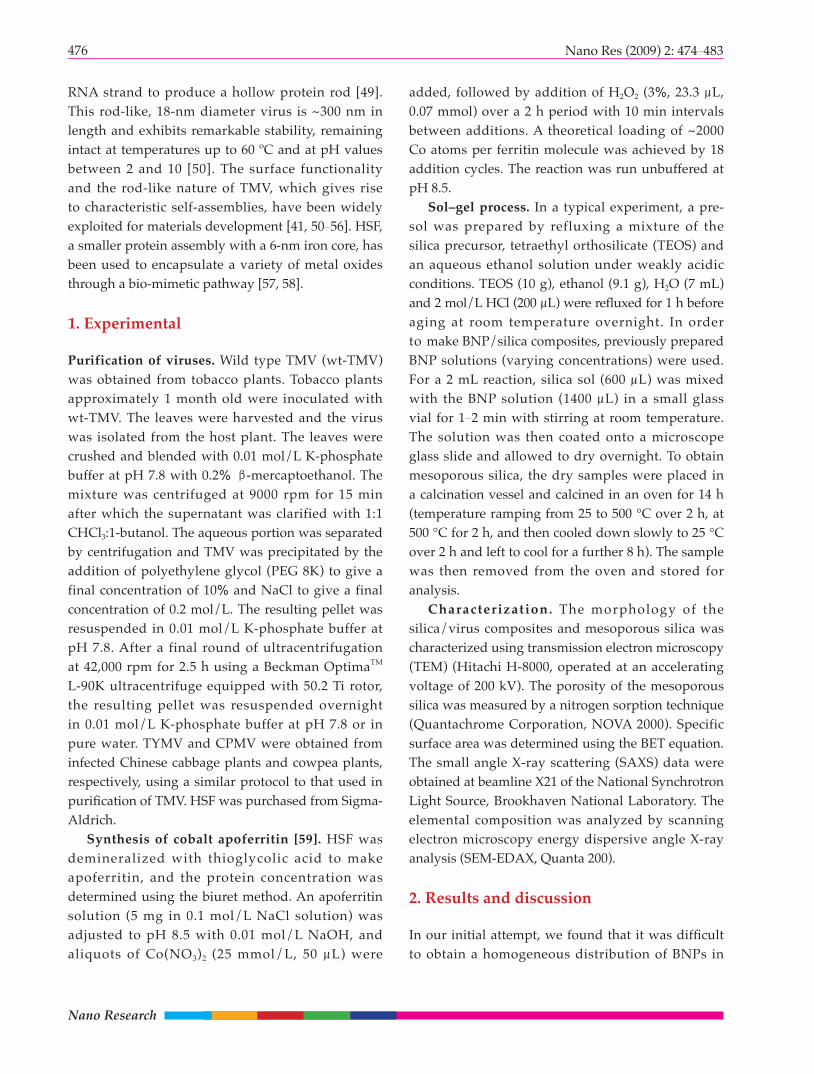
Four BNPs were employed in this study, namely turnip yellow mosaic virus (TYMV), cowpea mosaic virus (CPMV), TMV, and horse spleen ferritin (HSF), as shown in Fig. 1. TYMV is a non-enveloped plant virus composed of 180 identical protein subunits (20 kDa), assembled loosely in a T=3 icosahedral symmetry. The capsid consists of 32 knob-like structures corresponding to 20 hexamers and 12 pentamers of the coat protein (Figs. 1(d) and 1(e)) [43, 44]. CPMV is also a plant virus and can be obtained from infected cowpea plants. The physical, biological, and genetic properties have been well characterized over the past few years [45 48]. It is composed of 60 copies of two protein submits in an icosahedral symmetry. CPMV has a similar diameter to TYMV, i.e., ~30 nm; however, the surface features are quite different. TMV consists of 2130 identical protein subunits arranged in a helical motif around a single
Figure 1 X-ray crystal structure of (a) TYMV, (b) CPMV, (c) TMV, and (d) HSF. (e) and (f) show the structures and dimensions of the exterior view of the pentameric and hexameric units of TYMV which appear as knob-like protrusions. Structures were generated using PyMol (www.pymol.org) with coordinates from the RCSB Protein Data Bank (www.pdb.org)
(a) (b) (c) (d)
(e) (f)
Nano Research
476 Nano Res (2009) 2: 474 483
RNA strand to produce a hollow protein rod [49]. This rod-like, 18-nm diameter virus is ~300 nm in length and exhibits remarkable stability, remaining intact at temperatures up to 60 ºC and at pH values between 2 and 10 [50]. The surface functionality and the rod-like nature of TMV, which gives rise to characteristic self-assemblies, have been widely exploited for materials development [41, 50 56]. HSF, a smaller protein assembly with a 6-nm iron core, has been used to encapsulate a variety of metal oxides through a bio-mimetic pathway [57, 58].
1. Experimental
Purification of viruses. Wild type TMV (wt-TMV) was obtained from tobacco plants. Tobacco plants approximately 1 month old were inoculated with wt-TMV. The leaves were harvested and the virus was isolated from the host plant. The leaves were crushed and blended with 0.01 mol/L K-phosphate buffer at pH 7.8 with 0.2% β-mercaptoethanol. The mixture was centrifuged at 9000 rpm for 15 min after which the supernatant was clarified with 1:1 CHCl3:1-butanol. The aqueous portion was separated by centrifugation and TMV was precipitated by the addition of polyethylene glycol (PEG 8K) to give a final concentration of 10% and NaCl to give a final concentration of 0.2 mol/L. The resulting pellet was resuspended in 0.01 mol/L K-phosphate buffer at pH 7.8. After a final round of ultracentrifugation at 42,000 rpm for 2.5 h using a Beckman OptimaTM L-90K ultracentrifuge equipped with 50.2 Ti rotor, the resulting pellet was resuspended overnight in 0.01 mol/L K-phosphate buffer at pH 7.8 or in pure water. TYMV and CPMV were obtained from infected Chinese cabbage plants and cowpea plants, respectively, using a similar protocol to that used in purifi cation of TMV. HSF was purchased from Sigma-Aldrich.
Synthesis of cobalt apoferritin [59]. HSF was demineralized with thioglycolic acid to make apoferritin, and the protein concentration was determined using the biuret method. An apoferritin solution (5 mg in 0.1 mol/L NaCl solution) was adjusted to pH 8.5 with 0.01 mol/L NaOH, and aliquots of Co(NO3)2 (25 mmol/L, 50 μL) were
added, followed by addition of H2O2 (3%, 23.3 μL, 0.07 mmol) over a 2 h period with 10 min intervals between additions. A theoretical loading of ~2000 Co atoms per ferritin molecule was achieved by 18 addition cycles. The reaction was run unbuffered at pH 8.5.
Sol–gel process. In a typical experiment, a pre-sol was prepared by refluxing a mixture of the silica precursor, tetraethyl orthosilicate (TEOS) and an aqueous ethanol solution under weakly acidic conditions. TEOS (10 g), ethanol (9.1 g), H2O (7 mL) and 2 mol/L HCl (200 μL) were refl uxed for 1 h before aging at room temperature overnight. In order to make BNP/silica composites, previously prepared BNP solutions (varying concentrations) were used. For a 2 mL reaction, silica sol (600 μL) was mixed with the BNP solution (1400 μL) in a small glass vial for 1 2 min with stirring at room temperature. The solution was then coated onto a microscope glass slide and allowed to dry overnight. To obtain mesoporous silica, the dry samples were placed in a calcination vessel and calcined in an oven for 14 h (temperature ramping from 25 to 500 °C over 2 h, at 500 °C for 2 h, and then cooled down slowly to 25 °C over 2 h and left to cool for a further 8 h). The sample was then removed from the oven and stored for analysis.
Characterization. The morphology of the silica/virus composites and mesoporous silica was characterized using transmission electron microscopy (TEM) (Hitachi H-8000, operated at an accelerating voltage of 200 kV). The porosity of the mesoporous silica was measured by a nitrogen sorption technique (Quantachrome Corporation, NOVA 2000). Specific surface area was determined using the BET equation. The small angle X-ray scattering (SAXS) data were obtained at beamline X21 of the National Synchrotron Light Source, Brookhaven National Laboratory. The elemental composition was analyzed by scanning electron microscopy energy dispersive angle X-ray analysis (SEM-EDAX, Quanta 200).
2. Results and discussion
In our initial attempt, we found that it was diffi cult to obtain a homogeneous distribution of BNPs in
477Nano Res (2009) 2: 474 483
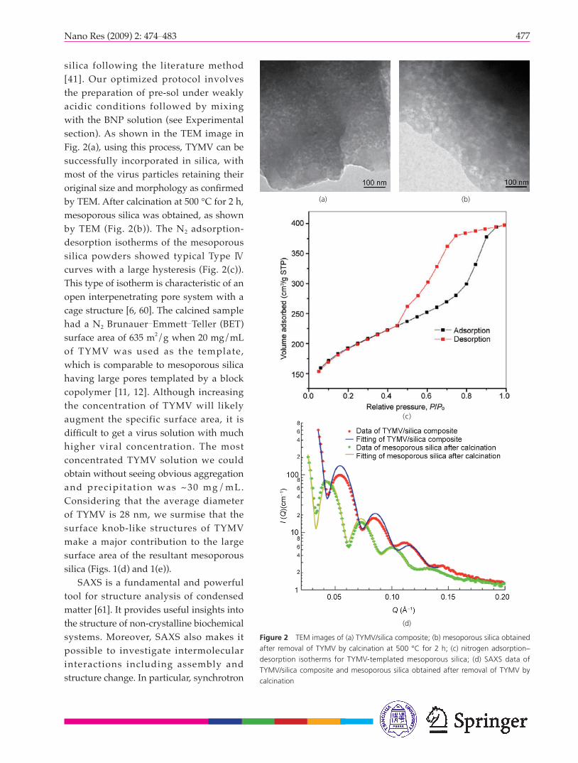
silica following the literature method [41]. Our optimized protocol involves the preparation of pre-sol under weakly acidic conditions followed by mixing with the BNP solution (see Experimental section). As shown in the TEM image in Fig. 2(a), using this process, TYMV can be successfully incorporated in silica, with most of the virus particles retaining their original size and morphology as confi rmed by TEM. After calcination at 500 °C for 2 h, mesoporous silica was obtained, as shown by TEM (Fig. 2(b)). The N2 adsorption-desorption isotherms of the mesoporous silica powders showed typical Type Ⅳ curves with a large hysteresis (Fig. 2(c)). This type of isotherm is characteristic of an open interpenetrating pore system with a cage structure [6, 60]. The calcined sample had a N2 Brunauer Emmett Teller (BET) surface area of 635 m2/g when 20 mg/mL of TYMV was used as the template, which is comparable to mesoporous silica having large pores templated by a block copolymer [11, 12]. Although increasing the concentration of TYMV will likely augment the specific surface area, it is diffi cult to get a virus solution with much higher viral concentration. The most concentrated TYMV solution we could obtain without seeing obvious aggregation and precipitat ion was ~30 mg/mL. Considering that the average diameter of TYMV is 28 nm, we surmise that the surface knob-like structures of TYMV make a major contribution to the large surface area of the resultant mesoporous silica (Figs. 1(d) and 1(e)).
SAXS is a fundamental and powerful tool for structure analysis of condensed matter [61]. It provides useful insights into the structure of non-crystalline biochemical systems. Moreover, SAXS also makes it possible to investigate intermolecular interactions including assembly and structure change. In particular, synchrotron
Figure 2 TEM images of (a) TYMV/silica composite; (b) mesoporous silica obtained after removal of TYMV by calcination at 500 °C for 2 h; (c) nitrogen adsorption–desorption isotherms for TYMV-templated mesoporous silica; (d) SAXS data of TYMV/silica composite and mesoporous silica obtained after removal of TYMV by calcination
(a) (b)
(c)
(d)
Nano Research
478 Nano Res (2009) 2: 474 483
SAXS is an effective method for the quantitative analysis of bionanoparticle structures [26, 34, 35]. To determine whether TYMV retains its original morphology, SAXS was performed on the TYMV/silica nanocomposite and the mesoporous silica obtained after removal of TYMV by calcination (Fig. 2(d)). For the TYMV/silica nanocomposite, model fi ts to the SAXS data gave an inner radius of 9.0 nm and a wall thickness of 2.5 nm, indicating that TYMV embedded in silica retained its spherical shape. However, the SAXS data of the imbedded TYMV were slightly different from that in solution. Using the Schultz polydisperse core and spherical shell model, TYMV was found to have an inner radius of 10.1 nm and a wall thickness of 3.5 nm, which is consistent with its X-ray crystallographic structure [44]. There is a large electron density difference between TYMV and silica, which produces a high intensity in the SAXS, enabling analysis of the interactions between viral particles and silica matrix. Assuming the protein skeleton was not deformed after the sol gel process, the data suggested that the silica was not only formed on top of the exterior surface of TYMV, but also penetrated deeply into the coat proteins of the virus. In addition, the SAXS data showed that the pores generated after calcination were monodisperse with a diameter of 20.6 nm. This further confirms that BNPs can be used as an effective template for the generation of mesoporous silica with a controlled pore shape and size.
Another spherical bionanoparticle, CPMV was
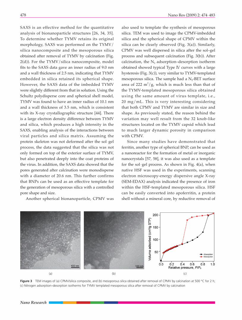
also used to template the synthesis of mesoporous silica. TEM was used to image the CPMV-imbedded silica and the spherical shape of CPMV within the silica can be clearly observed (Fig. 3(a)). Similarly, CPMV was well dispersed in silica after the sol–gel process and subsequent calcination (Fig. 3(b)). After calcination, the N2 adsorption–desorption isotherm obtained showed typical Type Ⅳ curves with a large hysteresis (Fig. 3(c)), very similar to TYMV-templated mesoporous silica. The sample had a N2-BET surface area of 222 m2/g, which is much less than that of the TYMV-templated mesoporous silica obtained us ing the same amount of virus template, i.e., 20 mg/mL. This is very interesting considering that both CPMV and TYMV are similar in size and shape. As previously stated, the reason behind the variation may well result from the 32 knob-like structures located on the TYMV capsid which lead to much larger dynamic porosity in comparison with CPMV.
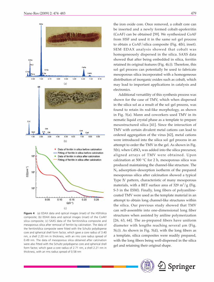
Since many studies have demonstrated that ferritin, another type of spherical BNP, can be used as a nanoreactor for the formation of metal or inorganic nanocrystals [57, 58], it was also used as a template for the sol gel process. As shown in Fig. 4(a), when native HSF was used in the experiments, scanning electron microscopy-energy dispersive angle X-ray (SEM-EDAX) analysis indicated the presence of iron within the HSF-templated mesoporous silica. HSF can be easily converted into apoferritin, a protein shell without a mineral core, by reductive removal of
Figure 3 TEM images of (a) CPMV/silica composite, and (b) mesoporous silica obtained after removal of CPMV by calcination at 500 °C for 2 h; (c) Nitrogen adsorption–desorption isotherms for TYMV templated mesoporous silica after removal of CPMV by calcination
(a) (b) (c)
479Nano Res (2009) 2: 474 483
(b)
Figure 4 (a) EDAX data and optical images (inset) of the HSF/silica composite; (b) EDAX data and optical images (inset) of the CoAF/silica composite; (c) SAXS data of the ferritin/silica composite and mesoporous silica after removal of ferritin by calcination. The data of the ferritin/silica composite were fi tted with the Schultz polydisperse core and spherical shell form factor, which gave a core radius of 3.40 nm, a shell 2.20 nm in thickness, with an rms core radius spread of 0.49 nm. The data of mesoporous silica obtained after calcination were also fi tted with the Schultz polydisperse core and spherical shell form factor, which gave a core radius of 2.71 nm, a shell 2.21 nm in thickness, with an rms radius spread of 0.58 nm
(c)
the iron oxide core. Once removed, a cobalt core can be inserted and a newly formed cobalt-apoferritin (CoAF) can be obtained [59]. We synthesized CoAF from HSF and used it in the same sol gel process to obtain a CoAF/silica composite (Fig. 4(b), inset). SEM EDAX analysis showed that cobalt was homogeneously dispersed in the silica. SAXS data showed that after being embedded in silica, ferritin retained its original features (Fig. 4(c)). Therefore, this sol gel process can potentially be used to fabricate mesoporous silica incorporated with a homogeneous distribution of inorganic oxides such as cobalt, which may lead to important applications in catalysis and electronics.
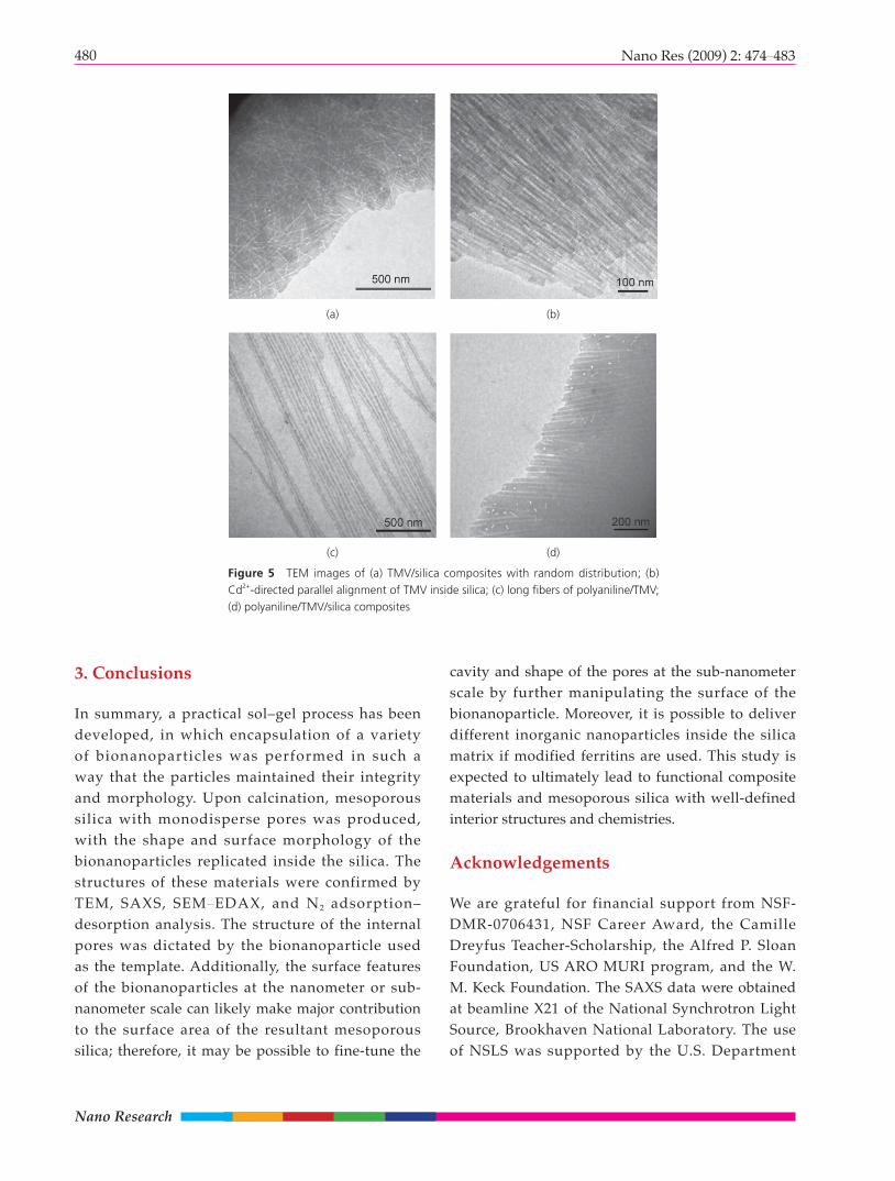
Additional versatility of this synthesis process was shown for the case of TMV, which when dispersed in the silica sol as a result of the sol gel process, was found to retain its rod-like morphology, as shown in Fig. 5(a). Mann and coworkers used TMV in its nematic liquid crystal phase as a template to prepare mesostructured silica [41]. Since the interaction of TMV with certain divalent metal cations can lead to ordered aggregation of the virus [62], metal cations were introduced into the silica sol gel process in an attempt to order the TMV in the gel. As shown in Fig. 5(b), when CdSO4 was added into the silica precursor, aligned arrays of TMV were obtained. Upon calcination at 500 °C for 2 h, mesoporous silica was produced maintaining the channel-like structure. The N2 adsorption–desorption isotherm of the prepared mesoporous silica after calcination showed a typical Type Ⅳ pattern, characteristic of many mesoporous materials, with a BET surface area of 329 m2/g (Fig. S-3 in the ESM). Finally, long fibers of polyaniline-coated TMV were used as the template material in an attempt to obtain long channel-like structures within the silica. Our previous study showed that TMV can self-assemble into one-dimensional long fiber structures when assisted by aniline polymerization [26, 63, 64]. The as-prepared fibers have uniform diameter with lengths reaching several μm (Fig. 5(c)). As shown in Fig. 5(d), with the long fibers as a template, silica composites were readily prepared, with the long fi bers being well-dispersed in the silica gel and retaining their original shape.
(a)
Nano Research
480 Nano Res (2009) 2: 474 483
Figure 5 TEM images of (a) TMV/silica composites with random distribution; (b) Cd2+-directed parallel alignment of TMV inside silica; (c) long fi bers of polyaniline/TMV; (d) polyaniline/TMV/silica composites
3. Conclusions
In summary, a practical sol–gel process has been developed, in which encapsulation of a variety of bionanoparticles was performed in such a way that the particles maintained their integrity and morphology. Upon calcination, mesoporous silica with monodisperse pores was produced, with the shape and surface morphology of the bionanoparticles replicated inside the silica. The structures of these materials were confirmed by TEM, SAXS, SEM EDAX, and N2 adsorption–desorption analysis. The structure of the internal pores was dictated by the bionanoparticle used as the template. Additionally, the surface features of the bionanoparticles at the nanometer or sub-nanometer scale can likely make major contribution to the surface area of the resultant mesoporous silica; therefore, it may be possible to fine-tune the
cavity and shape of the pores at the sub-nanometer scale by further manipulating the surface of the bionanoparticle. Moreover, it is possible to deliver different inorganic nanoparticles inside the silica matrix if modified ferritins are used. This study is expected to ultimately lead to functional composite materials and mesoporous silica with well-defined interior structures and chemistries.
Acknowledgements
We are grateful for financial support from NSF-DMR-0706431, NSF Career Award, the Camille Dreyfus Teacher-Scholarship, the Alfred P. Sloan Foundation, US ARO MURI program, and the W. M. Keck Foundation. The SAXS data were obtained at beamline X21 of the National Synchrotron Light Source, Brookhaven National Laboratory. The use of NSLS was supported by the U.S. Department
(a) (b)
(c) (d)
481Nano Res (2009) 2: 474 483
of Energy, Office of Science, Office of Basic Energy Sciences, under Contract No. DE-AC02-98CH10886.
Electronic Supplementary Material: Nitrogen adsorption–desorption isotherms for TYMV- and TMV-templated mesoporous silica, and the TEM image of ferritin-templated silica composite materials are available in the online version of this article at http://dx.doi.org/10.1007/s12274-009-9043-6 and are accessible free of charge.
References
[1] Ying, J. Y.; Mehnert, C. P.; Wong, M. S. Synthesis and
applications of supramolecular-templated mesoporous
materials. Angew. Chem. Int. Ed. 1999, 38, 56 77.
[2] Davis, M. E. Ordered porous materials for emerging
applications. Nature 2002, 417, 813 821.
[3] Yiu, H. H. P.; Wright, P. A. Enzymes supported on ordered
mesoporous solids: A special case of an inorganic
organic hybrid. J. Mater. Chem. 2005, 15, 3690 3700.
[4] Hartmann, M. Ordered mesoporous materials for
bioadsorption and biocatalysis. Chem. Mater. 2005, 17,
4577 4593.
[5] Lu, Y. F.; Yang, Y.; Sellinger, A.; Lu, M. C.; Huang, J.
M.; Fan, H. Y.; Haddad, R.; Lopez, G.; Burns, A. R.;
Sasaki, D. Y.; Shelnutt, J.; Brinker, C. J. Self-assembly of
mesoscopically ordered chromatic polydiacetylene/silica
nanocomposites. Nature 2001, 410, 913 917.
[6] Yang, P. D.; Zhao, D. Y.; Chmelka, B. F.; Stucky, G. D.
Triblock-copolymer-directed syntheses of large-pore
mesoporous silica fi bers. Chem. Mater. 1998, 10, 2033
2036.
[7] Mal, N. K.; Fujiwara, M.; Tanaka, Y. Photocontrolled
reversible release of guest molecules from coumarin-
modified mesoporous silica. Nature 2003, 421, 350
353.
[8] El-Safty, S. A. Review on the key controls of designer
copolymer-silica mesophase monoliths (HOM-type)
with large particle morphology, ordered geometry and
uniform pore dimension. J. Porous Mater. 2008, 15, 369
387.
[9] Sanchez, C.; Boissiere, C.; Grosso, D.; Laberty, C.;
Nicole, L. Design, synthesis, and properties of inorganic
and hybrid thin films having periodically organized
nanoporosity. Chem. Mater. 2008, 20, 682 737.
[10] Wan, Y.; Shi, Y. F.; Zhao, D. Y. Supramolecular
aggregates as templates: Ordered mesoporous polymers
and carbons. Chem. Mater. 2008, 20, 932 945.
[11] Matos, J. R.; Kruk, M.; Mercuri, L. P.; Jaroniec, M.; Zhao,
L.; Kamiyama, T.; Terasaki, O.; Pinnavaia, T. J.; Liu, Y.
Ordered mesoporous silica with large cage-like pores:
Structural identifi cation and pore connectivity design by
controlling the synthesis temperature and time. J. Am.
Chem. Soc. 2003, 125, 821 829.
[12] Fan, J.; Yu, C. Z.; Lei, J.; Zhang, Q.; Li, T. C.; Tu, B.;
Zhou, W. Z.; Zhao, D. Y. Low-temperature strategy to
synthesize highly ordered mesoporous silicas with very
large pores. J. Am. Chem. Soc. 2005, 127, 10794
10795.
[13] Douglas, T.; Young, M. Viruses: Making friends with old
foes. Science 2006, 312, 873 875.
[14] Lee, L. A.; Wang, Q. Adaptations of nanoscale viruses
and other protein cages for medical applications.
Nanomedicine 2006, 2, 137 149.
[15] Kramer, R. M.; Li, C.; Carter, D. C.; Stone, M. O.; Naik, R.
R. Engineered protein cages for nanomaterial synthesis. J.
Am. Chem. Soc. 2004, 126, 13282 13286.
[16] M e l d r u m , F. C . ; H e y w o o d , B . R . ; M a n n , S .
Magnetoferritin: In vitro synthesis of a novel magnetic
protein. Science 1992, 257, 522 523.
[17] Flenniken, M. L.; Willits, D. A.; Brumfield, S.; Young,
M. J.; Douglas, T. The small heat shock protein cage
from methanococcus jannaschii is a versatile nanoscale
platform for genetic and chemical modification. Nano
Lett. 2003, 3, 1573 1576.
[18] Seebeck, F. P.; Woycechowsky, K. J.; Zhuang, W.; Rabe,
J. P.; Hilvert, D. A simple tagging system for protein
encapsulation. J. Am. Chem. Soc. 2006, 128, 4516
4517.
[19] Domingo, G. J.; Orru, S.; Perham, R. N. Multiple display
of peptides and proteins on a macromolecular scaffold
derived from a multienzyme complex. J. Mol. Biol. 2001,
305, 259 267.
[20] Paavola, C. D.; Chan, S. L.; Li, Y.; Mazzarella, K. M.;
McMillan, R. A.; Trent, J. D. A versatile platform for
nanotechnology based on circular permutation of a
chaperonin protein. Nanotechnology 2006, 17, 1171
1176.
[21] Campos, S. K.; Barry, M. A. Current advances and future
challenges in adenoviral vector biology and targeting.
Curr. Gene Ther. 2007, 7, 189 204.
Nano Research
482 Nano Res (2009) 2: 474 483
[22] Manchester, M.; Singh, P. Virus-based nanoparticles
(VNPs): Platform technologies for diagnostic imaging.
Adv. Drug Deliv. Rev. 2006, 58, 1505 1522.
[23] Ramqvist, T.; Andreasson, K.; Dalanis, T. Vaccination,
immune and gene therapy based on virus-like particles
against viral infections and cancer. Expert Opin. Biol.
Ther. 2007, 7, 997 1007.
[24] Canizares, M. C.; Nicholson, L.; Lomonossoff, G. P.
Use of viral vectors for vaccine production in plants.
Immunol. Cell Biol. 2005, 83, 263 270.
[25] Streatfield, S. J. Oral hepatitis B vaccine candidates
produced and delivered in plant material. Immunol. Cell
Biol. 2005, 83, 257 262.
[26] Niu, Z.; Bruckman, M.; Kotakadi, V. S.; He, J.; Emrick,
T.; Russel l , T. P.; Yang, L.; Wang, Q. Study and
characterization of tobacco mosaic virus head-to-tail
assembly assisted by aniline polymerization. Chem.
Commun. 2006, 3019 3021.
[27] Mao, C.; Solis, D. J.; Reiss, B. D.; Kottmann, S. T.;
Sweeney, R. Y.; Hayhurst, A.; Georgiou, G.; Iverson,
B.; Belcher, A. M. Virus-based toolkit for the directed
synthesis of magnetic and semiconducting nanowires.
Science 2004, 303, 213 217.
[28] Niu, Z.; Bruckman, M.; Harp, B.; Mello, C. M.; Wang, Q.
Bacteriophage M13 as scaffold for preparing conductive
polymeric composite fibers. Nano Res. 2008, 1, 235
241.
[29] Rong, J. H.; Lee, L. A.; Li, K.; Harp, B.; Mello, C. M.; Niu,
Z. W.; Wang, Q. Oriented cell growth on self-assembled
bacteriophage M13 thin films. Chem. Commun. 2008,
5185 5187.
[30] Kaur, G.; Valarmathi, M. T.; Potts, J. D.; Wang, Q. The
promotion of osteoblastic differentiation of rat bone
marrow stromal cells by a polyvalent plant mosaic virus.
Biomaterials 2008, 29, 4074 4081.
[31] Li, T.; Niu, Z. W.; Emrick, T.; Russell, T. R.; Wang, Q.
Core/shell biocomposites from the hierarchical assembly
of bionanoparticles and polymer. Small 2008, 4, 1624
1629.
[32] Lee, L. A.; Niu, Z.; Wang, Q. Viruses and virus-like protein
assemblies Chemically programmable nanoscale
building blocks. Nano Res. accepted.
[33] Rong, J.; Oberbeck, F.; Wang, X.; Li, X.; Oxsher, J.; Niu,
Z.; Wang, Q. Tobacco mosaic virus templated synthesis
of one dimensional inorganic/polymer hybrid fibres. J.
Mater. Chem. 2009, 19, 2841 2845.
[34] Lin, Y.; Boker, A.; He, J.; Sill, K.; Xiang, H.; Abetz, C.; Li,
X.; Wang, J.; Emrick, T.; Long, S.; Wang, Q.; Balazs, A.;
Russell, T. P. Self-directed self-assembly of nanoparticle/
copolymer mixtures. Nature 2005, 434, 55 59.
[35] Russell, J. T.; Lin, Y.; Böker, A.; Long, S.; Carl, P.; Zettl,
H.; He, J.; Sill, K.; Tangiraia, R.; Emrick, T.; Littrell, K.;
Thiyagarajan, P.; Cookson, D.; Fery, A.; Wang, Q.; Russell,
T. P. Self-assembly and cross-linking of bionanoparticles
at liquid liquid interfaces. Angew. Chem. Int. Ed. 2005,
44, 2420 2426.
[36] Avnir, D.; Coradin, T.; Lev, O.; Livage, J. Recent bio-
applications of sol gel materials. J. Mater. Chem. 2006,
16, 1013 1030.
[37] Ferrer, M. L.; del Monte, F.; Levy, D. A novel and simple
alcohol-free sol gel route for encapsulation of labile
proteins. Chem. Mater. 2002, 14, 3619 3621.
[38] Gill, I.; Ballesteros, A. Encapsulation of biologicals
within silicate, siloxane, and hybrid sol-gel polymers: An
effi cient and generic approach. J. Am. Chem. Soc. 1998,
120, 8587 8598.
[39] Lan, E. H.; Dunn, B.; Valentine, J. S.; Zink, J. I .
Encapsulation of the ferritin protein in sol gel derived
silica glasses. J. Sol-Gel Sci. Techn. 1996, 7, 109 116.
[40] Tartaj, P.; Gonzalez-Carreno, T.; Ferrer, M. L.; Serna, C. J.
Metallic nanomagnets randomly dispersed in spherical
colloids: Toward a universal route for the preparation of
colloidal composites containing nanoparticles. Angew.
Chem. Int. Ed. 2004, 43, 6304 6307.
[41] Fowler, C. E.; Shenton, W.; Stubbs, G.; Mann, S. Tobacco
mosaic virus liquid crystals as templates for the interior
design of silica mesophases and nanoparticles. Adv.
Mater. 2001, 13, 1266 1269.
[42] Royston, E.; Lee, S. Y.; Culver, J. N.; Harris, M. T.
Characterization of silica-coated tobacco mosaic virus. J.
Coll. Int. Sci. 2006, 298, 706 712.
[43] Klug, A.; Finch, J. T.; Franklin, R. E. Structure of turnip
yellow mosaic virus. Nature 1957, 179, 683 684.
[44] Canady, M. A.; Larson, S. B.; Day, J.; McPherson, A.
Crystal structure of turnip yellow mosaic virus. Nat.
Struct. Biol. 1996, 3, 771 781.
[45] Wang, Q.; Raja, K. S.; Janda, K. D.; Lin, T. W.; Finn, M.
G. Blue fluorescent antibodies as reporters of steric
accessibility in virus conjugates. Bioconjugate Chem.
2003, 14, 38 43.
[46] Wang, Q.; Lin, T. W.; Johnson, J. E.; Finn, M. G. Natural
supramolecular building blocks: Cysteine-added mutants
483Nano Res (2009) 2: 474 483
of cowpea mosaic virus. Chem. Biol. 2002, 9, 813 819.
[47] Wang, Q.; Kaltgrad, E.; Lin, T. W.; Johnson, J. E.; Finn,
M. G. Natural supramolecular building blocks: Wild-type
cowpea mosaic virus. Chem. Biol. 2002, 9, 805 811.
[48] Wang, Q.; Chan, T. R.; Hilgraf, R.; Fokin, V. V.; Sharpless,
K. B.; Finn, M. G. Bioconjugation by copper(I)-catalyzed
azide-alkyne [3+2] cycloaddition. J. Am. Chem. Soc.
2003, 125, 3192 3193.
[49] Klug, A. The tobacco mosaic virus particle: Structure and
assembly. Philos. Trans. R. Soc. B 1999, 354, 531 535.
[50] Shenton, W.; Douglas, T.; Young, M.; Stubbs, G.;
Mann, S. Inorganic organic nanotube composites from
template mineralization of tobacco mosaic virus. Adv.
Mater. 1999, 11, 253 256.
[51] Fonoberov, V. A.; Balandin, A. A. Phonon confinement
effects in hybrid virus- inorganic nanotubes for
nanoelectronic applications. Nano Lett. 2005, 5, 1920
1923.
[52] Knez, M.; Sumser, M.; Bittner, A. M.; Wege, C.; Jeske,
H.; Martin, T. P.; Kern, K. Spatially selective nucleation of
metal clusters on the tobacco mosaic virus. Adv. Funct.
Mater. 2004, 14, 116 124.
[53] Yi, H.; Nisar, S.; Lee, S. Y.; Powers, M. A.; Bentley, W. E.;
Payne, G. F.; Ghodssi, R.; Rubloff, G. W.; Harris, M. T.;
Culver, J. N. Patterned assembly of genetically modifi ed
viral nanotemplates via nucleic acid hybridization. Nano
Lett. 2005, 5, 1931 1936.
[54] Yi, H.; Rubloff, G. W.; Culver, J. N. TMV microarrays:
Hybridization-based assembly of DNA-programmed viral
nanotemplates. Langmuir 2007, 23, 2663 2667.
[55] Tan, W. S.; Lewis, C. L.; Horelik, N. E.; Pregibon, D.
C.; Doyle, P. S.; Yi, H. Hierarchical assembly of viral
nanotemplates with encoded microparticles via nucleic
acid hybridization. Langmuir 2008, 24, 12483 12488.
[56] Balci, S.; Leinberger, D. M.; Knez, M.; Bittner, A. M.;
Boes, F.; Kadri, A.; Wege, C.; Jeske, H.; Kern, K. Printing
and aligning mesoscale patterns of tobacco mosaic virus
on surfaces. Adv. Mater. 2008, 20, 2195 2200.
[57] Wong, K. K. W.; Douglas, T.; Gider, S.; Awschalom, D.
D.; Mann, S. Biomimetic synthesis and characterization
of magnetic proteins (magnetoferritin). Chem. Mater.
1998, 10, 279 285.
[58] Douglas, T.; Dickson, D. P. E.; Betteridge, S.; Charnock,
J.; Garner, C. D.; Mann, S. Synthesis and structure of
an iron(Ⅲ) sulfide-ferritin bioinorganic nanocomposite.
Science 1995, 269, 54 57.
[59] Stark, V.; Douglas, T. Nanophase cobalt oxyhydroxide
mineral synthesized within the protein cage of ferritin.
Inorg. Chem. 1999, 39, 1828 1830.
[60] Kuang, D. B.; Brezesinski, T.; Smarsly, B. Hierarchical
porous silica materials with a trimodal pore system using
surfactant templates. J. Am. Chem. Soc. 2004, 126,
10534 10535.
[61] Svergun, D. I.; Koch, M. H. J. Small-angle scattering
studies of biological macromolecules in solution. Rep.
Prog. Phys. 2003, 66, 1735 1782.
[62] Nedoluzhko, A.; Douglas, T. Ordered association of
tobacco mosaic virus in the presence of divalent metal
ions. J. Inorg. Biochem. 2001, 84, 233 240.
[63] Niu, Z. W.; Bruckman, M. A.; Li, S. Q.; Lee, L. A.;
Lee, B.; Pingali, S. V.; Thiyagarajan, P.; Wang, Q.
Assembly of tobacco mosaic virus into fibrous and
macroscopic bundled arrays mediated by surface aniline
polymerization. Langmuir 2007, 23, 6719 6724.
[64] Niu, Z.; Liu, J.; Lee, L. A.; Bruckman, M. A.; Zhao, D.;
Koley, G.; Wang, Q. Biological templated synthesis of
water-soluble conductive polymeric nanowires. Nano
Lett. 2007, 7, 3729 3733.