Embed Size (px)
Citation preview

pubs.acs.org/IC Published on Web 11/10/2009 r 2009 American Chemical Society
11032 Inorg. Chem. 2009, 48, 11032–11037
DOI: 10.1021/ic901205c
Synthesis and Optical Properties of Guanosine 50-Monophosphate-MediatedCdS Nanostructures: An Analysis of their Structure, Morphology, and Electronic
Properties
Anil Kumar*,†, ‡ and Vinit Kumar†
†Department of Chemistry, and ‡Centre of Nanotechnology, Indian Institute of Technology Roorkee,Roorkee - 247667, India
Received June 23, 2009
The present manuscript reports the synthesis, characterization, and analysis of electronic properties of water-solubleguanosine 50-monophosphate (GMP)-mediated CdS quantum dots (Q-dots). The morphology, size, and sizedistribution of these particles have been analyzed by transmission electron microscopy. These particles display theonset of absorption at 2.7 eV and emission at 2.2 eV. In comparison to other monophosphates of RNA (AMP, UMP,and CMP), GMP-mediated CdS exhibits enhanced electronic properties. The participation of different functionalgroups of GMP in the stabilization of CdS nanoparticles has been analyzed by FTIR and 1H and 31P NMRspectroscopic techniques. Two types of binding sites involving phosphorus centers are indicated by IR and 31P NMRstudies. The conversion of CdS Q-dots to nanorods has been monitored by using electron microscopy, steady-stateoptical and fluorescence measurements, and a fluorescence lifetime system coupled with anisotropy accessories. Theobserved change in the morphology and electronic behavior of GMP- and RNA-mediated CdS nanostructures isdiscussed on the basis of their structural difference.
1. Introduction
A large number of biomolecules like proteins, lipids,nucleic acids, antibodies, antigens, and enzymes havingnanoscale dimensions constitute the fundamental buildingblocks of natural systems.1,2 In these systems, several biolo-gical molecules exist as supramolecular aggregates consistingof complex microstructures and are being utilized to developbiological machines in the nanoscale domain for applicationsin ultrasmall electronics and devices.3-8 Among differentbiomolecules, functionalized biopolymers like DNA9 andRNA10,11 have lately been employed extensively for templat-ing of nanomaterials. These consist of long-range nano-scale order, which could be tailored to fabricate artificial
nanostructures with varied properties. Integration of the func-tionalized biomolecules to inorganic nanomaterials may gener-ate newmaterialswith enhanced optical, fluorescence,magnetic,and electronic properties and increased photostability.4,12,13 Inrecent years, there has been enormous interest in making nano-hybrids of quantized semiconductor and biomolecules,14-16 inefforts to replace the fluorescent organic dyes employed asbiomarkers,17 since semiconductor quantumdots (Q-dots) haveseveral added advantages over organic dyes in regard to theirtunable optical and fluorescing properties, increased solubility,and enhanced chemical andphotostability.Anunderstandingofthe interaction/binding of semiconductor quantum dots withbiomolecules through its various functional sites may provide akey to optimizing and controlling the growth needed for thedesign of new nanostructures.In previous studies, nucleotide triphosphates have been
shown to be the effective ligands for the stabilization of
*To whom correspondence should be addressed. Tel.: þ91-1332-285799.Fax: þ91-1332-273560. E-mail: [email protected].
(1) Prasad, P. N. Nanophotonics; John Wiley & Sons: New Jersey, 2004;Chapter 12, pp 337-353.
(2) Willner, I.; Willner, B.; Katz, E. Bioelechem 2007, 70, 2–11.(3) Mann, S. Angew. Chem., Int. Ed. 2008, 47, 5306–5320.(4) Niemeyer, C. M. Angew. Chem., Int. Ed. 2001, 40, 4128–4158.(5) Gracia-Garibay, M. A. Nat. Mater. 2008, 7, 431–432.(6) Sotiropoulou, S.; Sierra-Sastre, Y.; Mark, S. S.; Batt, C. A. Chem.
Mater. 2008, 20, 821–834.(7) Sanchez, C.; Arribart, H.; Guille, M. M. G.Nat. Mater. 2005, 4, 277–
287.(8) Johnston, A. P. R.; Zelikin, A. N.; Caruso, F. Adv. Mater. 2007, 19,
3727–3730.(9) Storhoff, J. J.; Mirkin, C. A. Chem. Rev. 1999, 99, 1849–1862.(10) Feldheim, D. L.; Eaton, B. E. ACS Nano 2007, 1, 154–159.(11) Kumar, A.; Kumar, V. J. Phys. Chem. C 2008, 112, 3633–3640.
(12) Ma, N.; Sargent, E. H.; Kelley, S. O. J. Mater. Chem. 2008, 18, 954–964.
(13) Whaley, S. R.; English, D. S.; Hu, E. L.; Barbara, P. F.; Belcher, A.M. Nature. 2000, 405, 665–668.
(14) Dooley, C. J.; Rouge, J.;Ma,N.; Invernale,M.;Kelly, S. O. J.Mater.Chem. 2007, 17, 1687–1691.
(15) Berti, L.; Burley, G. A. Nat. Nanotechnol. 2008, 3, 81-87 andreferences therein.
(16) Green, M.; Symeth-Boyle, D.; Harries, J.; Taylor, R. Chem. Com-mun. 2005, 4830–4832.
(17) Stephens, D. J.; Allan, V. J. Science 2003, 300, 82–86.
Article Inorganic Chemistry, Vol. 48, No. 23, 2009 11033
fluorescing CdS nanoparticles;14,16 it is the monophosphate-(s) of nucleotides, which constitute different components ofRNA and DNA. In the present work, we have examinednucleotide monophosphates, namely, guanosine 50-mono-phosphate (GMP), adenosine 50-monophosphate (AMP),cytidine 50-monophosphate (CMP), and uridine 50-mono-phosphate (UMP), as templates for the synthesis of CdSnanoparticles and assessed the contribution of each of thecomponents toward electronic properties. We did not comeacross any extensive studies on these systems in the literature.Among the investigated components, GMP (Figure 1)stabilizes CdS nanostructures effectively and exhibits a moreprominent excitonic band, relatively higher fluorescencequantum efficiency, and a longer emission lifetime. Agingof these nanoparticles transforms them to nanorods. Adifference in the morphology and electronic behavior ofGMP- and RNA-mediated CdS nanostructures is alsodiscussed.
2. Experimental Section
Materials. Cadmium perchlorate, guanosine 50-monopho-sphate, adenosine 50-monophosphate, cytidine 50-monopho-sphate, uridine 50-monophosphate (Sigma); FeS, perchloricacid, and NaOH (Merck) were of analytical grade and wereused without any further purification. Nitrogen (99.9% Sigma)was used for deoxygenating the solution during synthesis. Allexperiments were performed in an aqueous medium.
Equipment.UV-visible absorption and steady-state emissionspectra were recorded on a Shimadzu UV-2100S spectrophoto-meter and Shimadzu RF-5301-PC spectrofluorophotometer,respectively. Electron microscopy and selected area electrondiffraction patterns were performed on a digital FEI-TECNAItransmission electron microscope operating at 200 KeV. Thesize of different nanostructures was determined from theirtransmission electron microscopy (TEM) micrographs usingdigital micrograph software. Sample(s) for TEM analysis wereprepared by applying a drop of the colloidal solution to acarbon-coated copper grid. The excess solution was then re-moved after about 2 min with a tissue paper. X-ray diffractionpatterns were obtained on a Philips DW1140/90 X-ray diffracto-meter using the Cu KR line (1.5418 A�) of the X-ray source. The1H and 31P NMR spectra were obtained on a Bruker Avance 500spectrometer working at 500MHz. For 31PNMR studies, a 10%solution of H3PO4 was used as an external reference. All NMRdatawere collected in deuterated aqueous solutions. IR spectra inthe mid-IR range were recorded on a Thermo Nicolet NexusFTIR spectrophotometer. The time-resolved fluorescence andfluorescence anisotropy decay measurements were made on aHoriba Jobin Yvon-IBH single-photon counter using thetime domain method. NanoLEDs and LDs were used as excita-tion sources. An iterative reconvolution technique using a multi-exponential fitting program was used for the analysis of decaycurves.
Synthesis of Nanoparticles.GMP-, AMP-, CMP-, and UMP-stabilized CdS nanopatricles were synthesized by adding variedconcentrations of these substrates (0.001-0.015 g/100 mL) andCd2þ (2� 10-4 mol dm-3) at pH 9.2 followed by the injection ofSH- at room temperature (∼ 20 �C). In all cases, the reactionmixture(s) were purged with nitrogen under continuous stirringfor about 15 min prior to the injection of SH-. Among these,CMP forms a complex with Cd2þ and precipitates out, even atits lowest concentration, that is, 0.001 g/100 mL. Thus, it couldnot becomepossible to produceCMP-templatedCdS.However,UMP-stabilized CdS particles could be produced by employinga relatively much higher concentration of nucleotide (0.025 g/100 mL) and by the slow addition of Cd2þ (2� 10-4 mol dm-3)to the nucleotide solution at 0 �C.All of the samples were purged
with nitrogen under continuous stirring for 15 min prior to theinjection of SH- prepared by the acid decomposition of FeS. Anincrease in the concentration of these solutes blue-shifts both theonset and the exciton absorption. Electronic properties ofGMP- and AMP-mediated CdS were optimized by varying theexperimental conditions of pH (9.2-10.2) and the concentrationof the nucleotide (0.001-0.015 g/100 mL). The best conditionsunder which the strong excitonic absorption and emissionwere observed correspond to 0.015 g/100 mL of the nucleotide,Cd2þ =2� 10-4 mol dm-3, and HS- = 1� 10-4 mol dm-3 atpH 9.2. Both GMP and AMP produced highly water-solubleCdS nanoparticles.
3. Results and Discussion
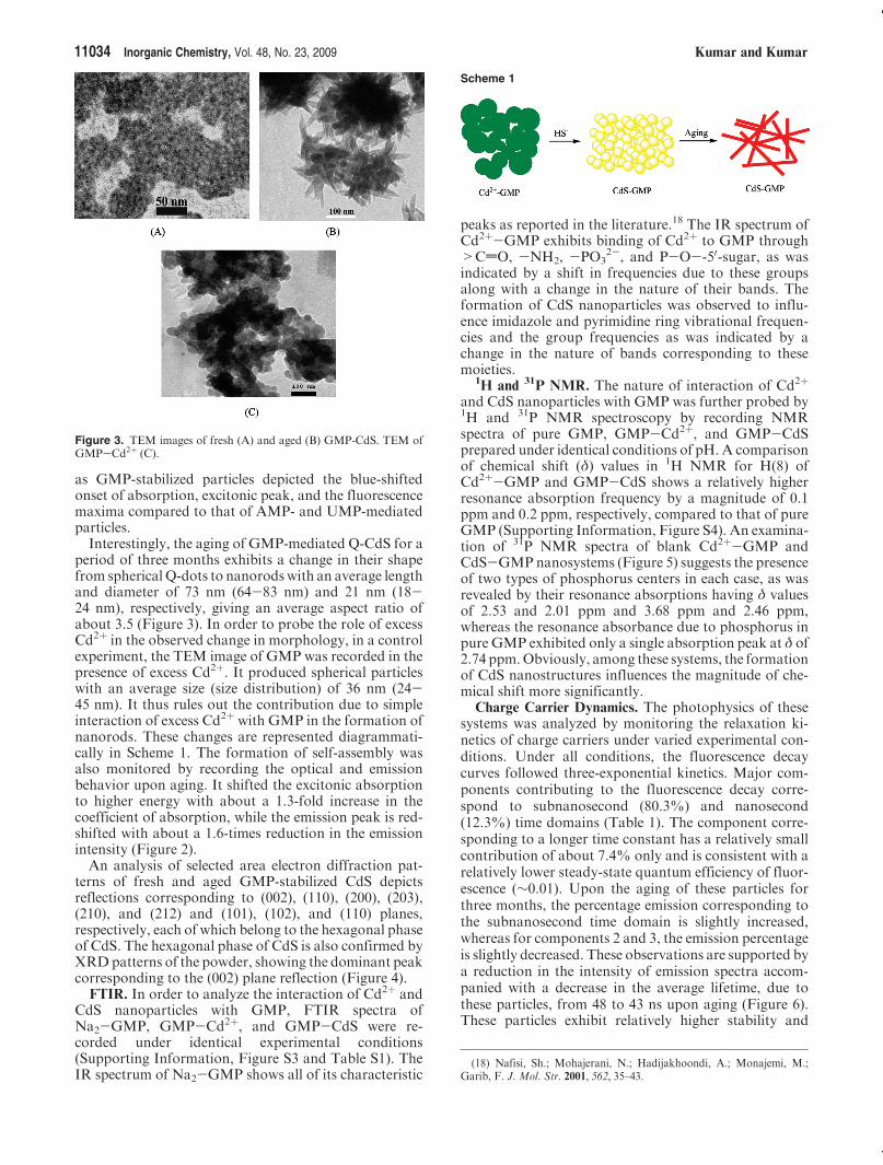
Results. Optical Properties. For a typical case ofGMP, the effect of variation of its concentration (0.001g/100 mL to 0.015 g/100 mL) on the optical and emissionbehavior of CdS is shown in Figure S1 (SupportingInformation). An increase in the concentration of GMPblue-shifts the onset of absorption and emission maximafrom 2.5 to 2.7 eV and 2.0 to 2.2 eV, respectively. Acomparison of electronic and emission data of GMP-,AMP-, and UMP-mediated particles demonstrates that,for the latter two systems, the excitonic absorption andemission maxima are slightly red-shifted. The emissionband becomes broad, and intensity is reduced to about40% in each case (Figure 2).
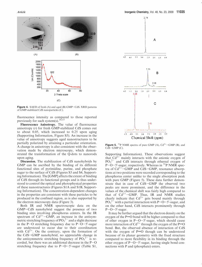
TEM and SAED. Electron micrographs of GMP-,AMP-, and UMP-mediated CdS particles consisted ofaggregates of a large number of spherical particles havinga size (size distribution) of 5 nm (2-8 nm), 7.5 nm (3-12 nm), and 10 nm (8-15 nm), respectively (Figure 3;Supporting Information, Figure S2). The difference intheir size is consistent with their electronic spectral data,
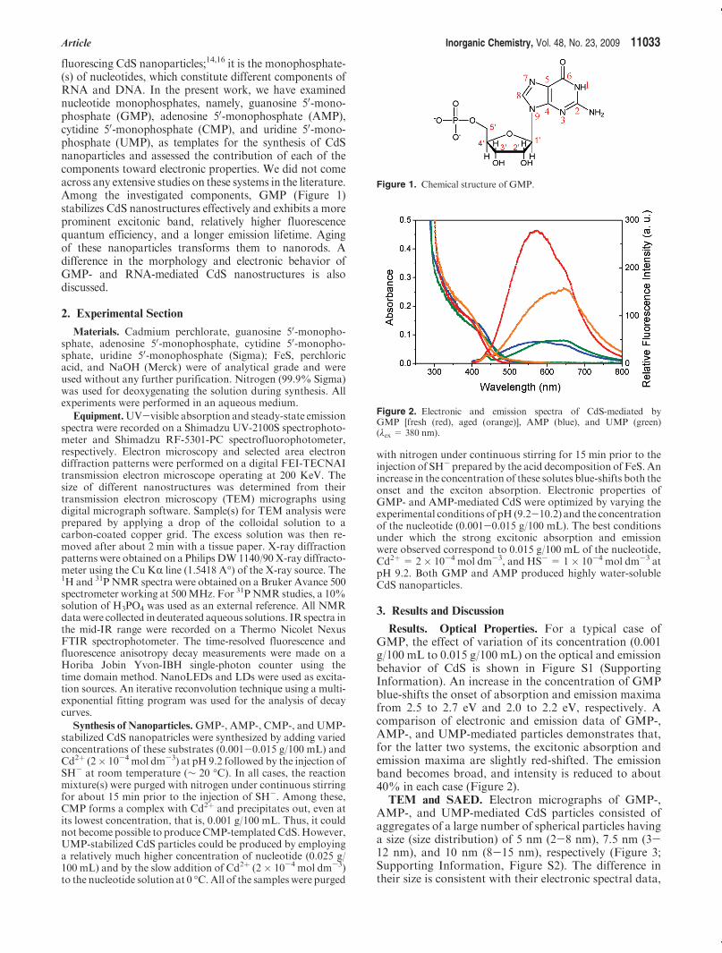
Figure 1. Chemical structure of GMP.
Figure 2. Electronic and emission spectra of CdS-mediated byGMP [fresh (red), aged (orange)], AMP (blue), and UMP (green)(λex = 380 nm).
11034 Inorganic Chemistry, Vol. 48, No. 23, 2009 Kumar and Kumar
as GMP-stabilized particles depicted the blue-shiftedonset of absorption, excitonic peak, and the fluorescencemaxima compared to that of AMP- and UMP-mediatedparticles.Interestingly, the aging of GMP-mediated Q-CdS for a
period of three months exhibits a change in their shapefrom spherical Q-dots to nanorodswith an average lengthand diameter of 73 nm (64-83 nm) and 21 nm (18-24 nm), respectively, giving an average aspect ratio ofabout 3.5 (Figure 3). In order to probe the role of excessCd2þ in the observed change in morphology, in a controlexperiment, the TEM image of GMP was recorded in thepresence of excess Cd2þ. It produced spherical particleswith an average size (size distribution) of 36 nm (24-45 nm). It thus rules out the contribution due to simpleinteraction of excess Cd2þ with GMP in the formation ofnanorods. These changes are represented diagrammati-cally in Scheme 1. The formation of self-assembly wasalso monitored by recording the optical and emissionbehavior upon aging. It shifted the excitonic absorptionto higher energy with about a 1.3-fold increase in thecoefficient of absorption, while the emission peak is red-shifted with about a 1.6-times reduction in the emissionintensity (Figure 2).An analysis of selected area electron diffraction pat-
terns of fresh and aged GMP-stabilized CdS depictsreflections corresponding to (002), (110), (200), (203),(210), and (212) and (101), (102), and (110) planes,respectively, each of which belong to the hexagonal phaseof CdS. The hexagonal phase of CdS is also confirmed byXRDpatterns of the powder, showing the dominant peakcorresponding to the (002) plane reflection (Figure 4).
FTIR. In order to analyze the interaction of Cd2þ andCdS nanoparticles with GMP, FTIR spectra ofNa2-GMP, GMP-Cd2þ, and GMP-CdS were re-corded under identical experimental conditions(Supporting Information, Figure S3 and Table S1). TheIR spectrum of Na2-GMP shows all of its characteristic
peaks as reported in the literature.18 The IR spectrum ofCd2þ-GMP exhibits binding of Cd2þ to GMP through>CdO, -NH2, -PO3
2-, and P-O--50-sugar, as wasindicated by a shift in frequencies due to these groupsalong with a change in the nature of their bands. Theformation of CdS nanoparticles was observed to influ-ence imidazole and pyrimidine ring vibrational frequen-cies and the group frequencies as was indicated by achange in the nature of bands corresponding to thesemoieties.
1H and 31P NMR. The nature of interaction of Cd2þ
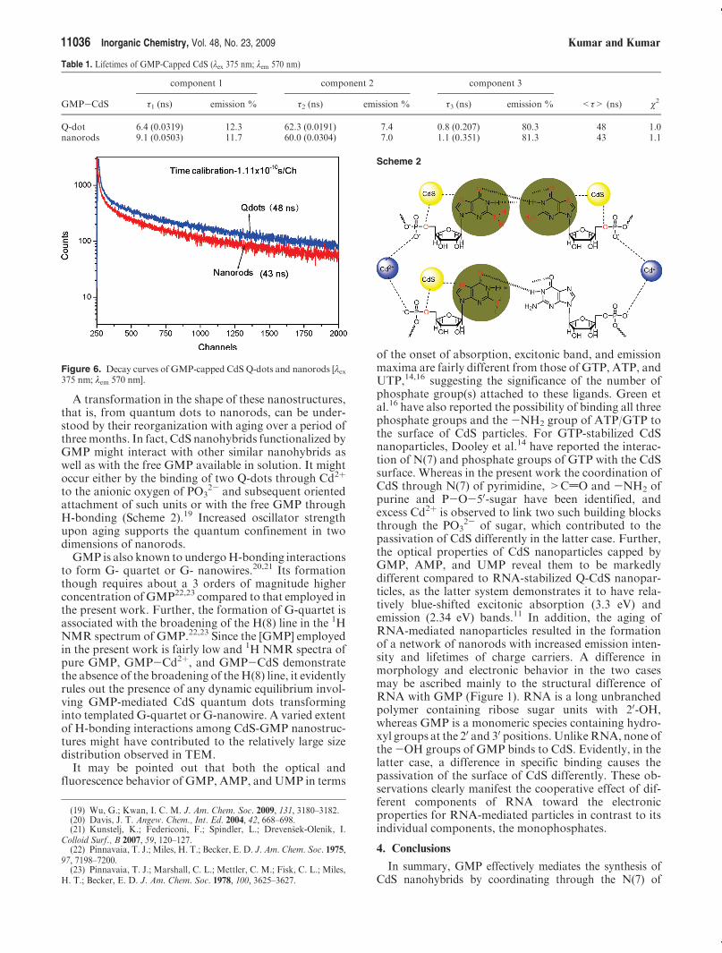
and CdS nanoparticles with GMP was further probed by1H and 31P NMR spectroscopy by recording NMRspectra of pure GMP, GMP-Cd2þ, and GMP-CdSprepared under identical conditions of pH. A comparisonof chemical shift (δ) values in 1H NMR for H(8) ofCd2þ-GMP and GMP-CdS shows a relatively higherresonance absorption frequency by a magnitude of 0.1ppm and 0.2 ppm, respectively, compared to that of pureGMP (Supporting Information, Figure S4). An examina-tion of 31P NMR spectra of blank Cd2þ-GMP andCdS-GMPnanosystems (Figure 5) suggests the presenceof two types of phosphorus centers in each case, as wasrevealed by their resonance absorptions having δ valuesof 2.53 and 2.01 ppm and 3.68 ppm and 2.46 ppm,whereas the resonance absorbance due to phosphorus inpure GMP exhibited only a single absorption peak at δ of2.74 ppm.Obviously, among these systems, the formationof CdS nanostructures influences the magnitude of che-mical shift more significantly.
Charge Carrier Dynamics. The photophysics of thesesystems was analyzed by monitoring the relaxation ki-netics of charge carriers under varied experimental con-ditions. Under all conditions, the fluorescence decaycurves followed three-exponential kinetics. Major com-ponents contributing to the fluorescence decay corre-spond to subnanosecond (80.3%) and nanosecond(12.3%) time domains (Table 1). The component corre-sponding to a longer time constant has a relatively smallcontribution of about 7.4% only and is consistent with arelatively lower steady-state quantum efficiency of fluor-escence (∼0.01). Upon the aging of these particles forthree months, the percentage emission corresponding tothe subnanosecond time domain is slightly increased,whereas for components 2 and 3, the emission percentageis slightly decreased. These observations are supported bya reduction in the intensity of emission spectra accom-panied with a decrease in the average lifetime, due tothese particles, from 48 to 43 ns upon aging (Figure 6).These particles exhibit relatively higher stability and
Figure 3. TEM images of fresh (A) and aged (B) GMP-CdS. TEM ofGMP-Cd2þ (C).
Scheme 1
(18) Nafisi, Sh.; Mohajerani, N.; Hadijakhoondi, A.; Monajemi, M.;Garib, F. J. Mol. Str. 2001, 562, 35–43.
Article Inorganic Chemistry, Vol. 48, No. 23, 2009 11035
fluorescence intensity as compared to those reportedpreviously for such system(s).14,15
Fluorescence Anisotropy. The value of fluorescenceanisotropy (r) for fresh GMP-stabilized CdS comes outto about 0.05, which increased to 0.25 upon aging(Supporting Information, Figure S5). An increase in thevalue of anisotropy suggests aged nanostructures to bepartially polarized by attaining a particular orientation.A change in anisotropy is also consistent with the obser-vation made by electron microscopy, which demon-strated the transformation of the Q-dots to nanorodsupon aging.
Discussion. The stabilization of CdS nanohybrids byGMP can be ascribed by the binding of its differentfunctional sites of pyrimidine, purine, and phosphatesugar to the surface of CdS (Figures S3 and S4, Support-ing Information). The [GMP] affects the extent of bindingof CdS through its functional groups and is thus under-stood to control the optical and photophysical propertiesof these nanostructures (Figures S1A and S1B, Support-ing Information). The concentration-dependent changesin the properties are consistent with these particles to beproduced in the confined region, as is also supported bythe electron microscopy data (Figure 3).Both IR and NMR spectroscopic data on the
GMP-CdS nanohybrid suggest the presence of twobinding sites involving phosphorus centers. In the IRspectrum of Cd2þ-GMP, an increase in the antisym-metric stretching frequency of PO3
2- and a slight decreasein the P-O stretching frequency due to P-O-50-sugarare understood to occur due to their coordinationwith Cd2þ. On the contrary, upon the formation ofthe CdS-GMP nanohybrid, no appreciable change inthe antisymmetric stretching frequency of PO3
2- is re-corded, but there was an additional decrease in the P-Ostretching frequency due to P-O-50-sugar (Table S1,
Supporting Information). These observations suggestthat Cd2þ mainly interacts with the anionic oxygen ofPO3
2- and CdS interacts through ethereal oxygen ofP-O-50-sugar, respectively. Whereas in 31P NMR spec-tra of Cd2þ-GMP and CdS-GMP, resonance absorp-tions at two positions were recorded corresponding to thephosphorus center unlike to the single absorption peakwith pure GMP (Figure 5). These data further demon-strate that in case of CdS-GMP the observed twopeaks are more prominent, and the difference in thevalues of the chemical shift was fairly high compared tothat of Cd2þ-GMP. Thus, IR and NMR studiesclearly indicate that Cd2þ gets bound mainly throughPO3
2-with a partial interaction with P-O-50-sugar, andon the other hand, CdS interacts prominently throughP-O-50-sugar.Itmay be further argued that the electron density on the
oxygen of the PdO bond will be higher compared to thatof ether oxygen in P-O-50-sugar, which should causemore interaction of Cd2þ through the oxygen of the PdObond. But, the observed absence of interaction of CdSwith the oxygen of PdO though can be understoodbecause of its planar geometry with the fixed structurecompared to more flexibility in its binding through theether oxygen of P-O-50-sugar, having single bond con-nections with P and (phosphate) esters.
Figure 4. SAED of fresh (A) and aged (B) GMP-CdS. XRD patternsof GMP-stabilized CdS nanoparticles (C).
Figure 5. 31P NMR spectra of pure GMP (A), Cd2þ-GMP (B), andCdS-GMP (C).
11036 Inorganic Chemistry, Vol. 48, No. 23, 2009 Kumar and Kumar
A transformation in the shape of these nanostructures,that is, from quantum dots to nanorods, can be under-stood by their reorganization with aging over a period ofthreemonths. In fact, CdS nanohybrids functionalized byGMP might interact with other similar nanohybrids aswell as with the free GMP available in solution. It mightoccur either by the binding of two Q-dots through Cd2þ
to the anionic oxygen of PO32- and subsequent oriented
attachment of such units or with the free GMP throughH-bonding (Scheme 2).19 Increased oscillator strengthupon aging supports the quantum confinement in twodimensions of nanorods.GMP is also known to undergoH-bonding interactions
to form G- quartet or G- nanowires.20,21 Its formationthough requires about a 3 orders of magnitude higherconcentration ofGMP22,23 compared to that employed inthe present work. Further, the formation of G-quartet isassociated with the broadening of the H(8) line in the 1HNMR spectrum of GMP.22,23 Since the [GMP] employedin the present work is fairly low and 1H NMR spectra ofpure GMP, GMP-Cd2þ, and GMP-CdS demonstratethe absence of the broadening of theH(8) line, it evidentlyrules out the presence of any dynamic equilibrium invol-ving GMP-mediated CdS quantum dots transforminginto templated G-quartet or G-nanowire. A varied extentof H-bonding interactions among CdS-GMP nanostruc-tures might have contributed to the relatively large sizedistribution observed in TEM.It may be pointed out that both the optical and
fluorescence behavior of GMP, AMP, andUMP in terms
of the onset of absorption, excitonic band, and emissionmaxima are fairly different from those of GTP, ATP, andUTP,14,16 suggesting the significance of the number ofphosphate group(s) attached to these ligands. Green etal.16 have also reported the possibility of binding all threephosphate groups and the -NH2 group of ATP/GTP tothe surface of CdS particles. For GTP-stabilized CdSnanoparticles, Dooley et al.14 have reported the interac-tion of N(7) and phosphate groups of GTP with the CdSsurface. Whereas in the present work the coordination ofCdS through N(7) of pyrimidine, >CdO and -NH2 ofpurine and P-O-50-sugar have been identified, andexcess Cd2þ is observed to link two such building blocksthrough the PO3
2- of sugar, which contributed to thepassivation of CdS differently in the latter case. Further,the optical properties of CdS nanoparticles capped byGMP, AMP, and UMP reveal them to be markedlydifferent compared to RNA-stabilized Q-CdS nanopar-ticles, as the latter system demonstrates it to have rela-tively blue-shifted excitonic absorption (3.3 eV) andemission (2.34 eV) bands.11 In addition, the aging ofRNA-mediated nanoparticles resulted in the formationof a network of nanorods with increased emission inten-sity and lifetimes of charge carriers. A difference inmorphology and electronic behavior in the two casesmay be ascribed mainly to the structural difference ofRNA with GMP (Figure 1). RNA is a long unbranchedpolymer containing ribose sugar units with 20-OH,whereas GMP is a monomeric species containing hydro-xyl groups at the 20 and 30 positions. UnlikeRNA, none ofthe-OH groups of GMP binds to CdS. Evidently, in thelatter case, a difference in specific binding causes thepassivation of the surface of CdS differently. These ob-servations clearly manifest the cooperative effect of dif-ferent components of RNA toward the electronicproperties for RNA-mediated particles in contrast to itsindividual components, the monophosphates.
4. Conclusions
In summary, GMP effectively mediates the synthesis ofCdS nanohybrids by coordinating through the N(7) of
Table 1. Lifetimes of GMP-Capped CdS (λex 375 nm; λem 570 nm)
component 1 component 2 component 3
GMP-CdS τ1 (ns) emission % τ2 (ns) emission % τ3 (ns) emission % <τ> (ns) χ2
Q-dot 6.4 (0.0319) 12.3 62.3 (0.0191) 7.4 0.8 (0.207) 80.3 48 1.0nanorods 9.1 (0.0503) 11.7 60.0 (0.0304) 7.0 1.1 (0.351) 81.3 43 1.1
Figure 6. Decay curves of GMP-capped CdS Q-dots and nanorods [λex375 nm; λem 570 nm].
Scheme 2
(19) Wu, G.; Kwan, I. C. M. J. Am. Chem. Soc. 2009, 131, 3180–3182.(20) Davis, J. T. Angew. Chem., Int. Ed. 2004, 42, 668–698.(21) Kunstelj, K.; Federiconi, F.; Spindler, L.; Dreven�sek-Olenik, I.
Colloid Surf., B 2007, 59, 120–127.(22) Pinnavaia, T. J.; Miles, H. T.; Becker, E. D. J. Am. Chem. Soc. 1975,
97, 7198–7200.(23) Pinnavaia, T. J.; Marshall, C. L.; Mettler, C. M.; Fisk, C. L.; Miles,
H. T.; Becker, E. D. J. Am. Chem. Soc. 1978, 100, 3625–3627.

Article Inorganic Chemistry, Vol. 48, No. 23, 2009 11037
pyrimidine: the >CdO and -NH2 of purine and P-O-50-sugar. Transformation of theirmorphology from sphericalQ-dots to nanorods upon aging is understood to propagateby the binding of Cd2þ through PO3
2- of twoCdS-GMPQ-dots followed by oriented attachment of various such unitsthrough H-bonding. Selective binding of nanoparticles todifferent sites of biomolecule(s) thus provides a means tocontrol the morphology and optical properties of nanohy-brids. Synthesis of such nanohybrids provides a method tofabricate biomolecule(s) templated nanostructures with tun-able optical and electronic properties.
Acknowledgment. The financial support of CSIR,New Delhi is gratefully acknowledged for undertakingthis work. V.K. acknowledges CSIR, New Delhi for theaward of SRF. Thanks are also due to the Head, IIC,IITR, Roorkee for providing us the facilities of NMR,TEM, FESEM, XRD, and single photon counter.
Supporting Information Available: Electronic and emissionspectra; TEM, 1H NMR, FTIR spectra and spectral data;fluorescence anisotropy. Thismaterial is available free of chargevia the Internet at http://pubs.acs.org.


![Structure of guanosine-3[prime],5[prime]-cytidine](https://img.pdfslide.net/doc/110x75/6185f12859d7806a1a3467d8/structure-of-guanosine-3prime5prime-cytidine-.jpg)


