Embed Size (px)
Citation preview
DOI: 10.1002/chem.200500876
Synthesis of Unsymmetrical Tweezer Receptor Libraries and Identification ofReceptors for Lys-d-Ala-d-Ala in Aqueous Solution
Jon Shepherd, Tom Gale, Kim B. Jensen, and Jeremy D. Kilburn*[a]
Introduction
The selective binding of peptides with synthetic receptorsremains a major challenge, particularly, if binding is to besuccessful in competitive aqueous media,[1] which is essentialif such systems are to be of use in the physiological milieu.In recent years, a variety of receptors, featuring one or morepeptidic arms attached to a rigid template, have proved tobe effective and sequence selec-tive for peptides, despite the in-herent flexibility of many ofthese receptor systems.[2-4] Inaddition, the use of a combina-torial, split-and-mix approachto randomize the amino acidcontent in the peptidic arms hasprovided a powerful method forthe construction of libraries ofpotential peptide receptors,[5]
and this approach has beenused successfully for the identi-fication of sequence selectivereceptors for a variety of pep-tides, in both nonpolar organ-ic[3] and aqueous solvents.[2,4] The incorporation of a specific
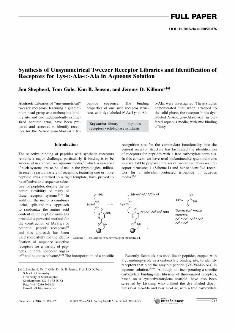
recognition site for the carboxylate functionality into thegeneral receptor structure has facilitated the identificationof receptors for peptides with a free carboxylate terminus.In this context, we have used bis(aminoalkyl)guanindiniumsas a scaffold to prepare libraries of two-armed “tweezer” re-ceptor structures 1 (Scheme 1) and hence identified recep-tors for a side-chain-protected tripeptide in aqueousmedia.[2e]
Recently, Schmuck has used linear peptides, capped witha guanidinopyrrole as a carboxylate binding site, to identifyreceptors that bind the amyloid peptide (Val-Val-Ile-Ala) inaqueous solution.[2c,d,6] Although not incorporating a specificcarboxylate binding site, libraries of three-armed receptors,based on a cyclotriveratrylene scaffold, have also beenscreened by Liskamp who utilized the dye-labeled dipep-tides d-Ala-d-Ala and d-Ala-d-Lac, with a free carboxylate
Abstract: Libraries of “unsymmetrical”tweezer receptors, featuring a guanidi-nium head group as a carboxylate bind-ing site and two independently synthe-sized peptidic arms, have been pre-pared and screened to identify recep-tors for the N-Ac-Lys-d-Ala-d-Ala tri-
peptide sequence. The bindingproperties of one such receptor struc-ture, with dye-labeled N-Ac-Lys-d-Ala-
d-Ala, were investigated. These studiesdemonstrated that when attached tothe solid-phase, the receptor binds dye-labeled N-Ac-Lys-d-Ala-d-Ala, in buf-fered aqueous media, with mm bindingaffinity.
Keywords: library · peptides ·receptors · solid-phase synthesis
[a] J. Shepherd, Dr. T. Gale, Dr. K. B. Jensen, Prof. J. D. KilburnSchool of ChemistryUniversity of SouthamptonSouthampton, SO17 1BJ (UK)Fax: (+44)2380-596-805E-mail : [email protected]
Scheme 1. Two-armed tweezer receptor structures 1.
Chem. Eur. J. 2006, 12, 713 – 720 C 2006 Wiley-VCH Verlag GmbH&Co. KGaA, Weinheim 713
FULL PAPER
terminus, as substrates in phosphate buffer.[2a,b] These devel-opments demonstrate the potential of such receptor systemsto provide potent and sequence selective peptide receptorsfor use as novel therapeutics or biosensors.
In our own work with two-armed “tweezer” receptors,based around a bis(aminoalkyl)guanindinium scaffold, the li-braries prepared to date have limited diversity; this is be-cause both arms of the receptor are synthesized simultane-ously on the solid support, which leads to “symmetrical”tweezer receptors containing two arms with an identicalamino acid sequence.[2e] To increase the diversity of struc-tures that can be prepared by using this combinatorial ap-proach, we have now developed routes to libraries of analo-gous “unsymmetrical” tweezer receptors in which the twoarms are synthesized independently. These libraries havebeen screened to identify receptor structures that are ableto bind to the bacterial cell wall precursor peptide N-Ac-Lys-d-Ala-d-Ala.[7] One such receptor structure was resyn-thesized and used to determine the association constantwith N-Ac-Lys-d-Ala-d-Ala. In free solution, although aUV binding study provides evidence for an association be-tween the receptor and the tripeptide, the data does notallow for the determination of a simple 1:1 (receptor:sub-strate) binding constant. When attached to the solid support,however, the receptor binds to the tripeptide in an aqueousbuffered solution with a mm association constant. Herein wedescribe these studies in detail.
Results and Discussion
Our approach to tweezer receptor libraries has relied onEdman degradation to identify the amino acid componentsin selected “hit” beads from screening experiments. Whilethis proved to be relatively straightforward for the “symmet-rical” receptor structures reported previously,[2e] the con-struction of “unsymmetrical” structures requires a morecomplicated procedure and careful use of orthogonal pro-tecting groups. We have used two strategies for the construc-tion of such libraries, with both cases utilizing the orthogo-nally protected guanidinium derivative 6, prepared in fivesteps from the previously described[8] thiourea 2, as thestarting point.
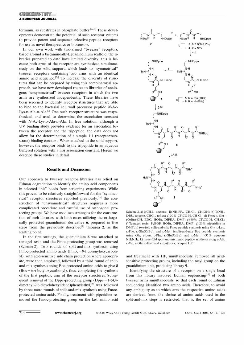
In the first strategy, the guanidinium 6 was attached totentagel resin and the Fmoc-protecting group was removed(Scheme 2). Two rounds of split-and-mix synthesis usingFmoc-protected amino acids (Fmoc=9-fluorenyloxycarbon-yl), with acid-sensitive side chain protection where appropri-ate, were then employed, followed by a third round of split-and-mix synthesis using Boc-protected amino acids to give 8(Boc= tert-butyloxycarbonyl), thus, completing the synthesisof the first peptidic arm of the receptor structures. Subse-quent removal of the Dppe-protecting group (Dppe=1-(4,4-dimethyl-2,6-dicyclohexylidene)phenylethyl)[9] was followedby three more rounds of split-and-mix synthesis using Fmoc-protected amino acids. Finally, treatment with piperidine re-moved the Fmoc-protecting group on the last amino acid
and treatment with HF, simultaneously, removed all acid-sensitive protecting groups, including the tosyl group on theguanidinium unit, producing library 9.
Identifying the structure of a receptor on a single beadfrom this library involved Edman sequencing[10] of bothtweezer arms simultaneously, so that each round of Edmansequencing identified two amino acids. Therefore, to avoidany ambiguity as to which arm the respective amino acidsare derived from, the choice of amino acids used in thesplit-and-mix steps is restricted, that is, the set of amino
Scheme 2. a) i) CH3I, acetone; ii) NH4PF6, CH2Cl2, CH3OH; b) TsNH2,DBU, toluene, CHCl3, reflux; c) 30% CF3CO2H, CH2Cl2; d) Fmoc-l-Glu-(OtBu)-OH, EDC, HOBt, DIPEA, DMF; e) 60% CF3CO2H, CH2Cl2;f) Tentagel resin, PyBOP, HOBt, DIPEA, DMF; g) 20% piperidine inDMF; h) two-fold split-and-mix Fmoc peptide synthesis using Gly, l-Leu,l-Phe, l-Glu(OtBu), and l-Met; i) split-and-mix Boc peptide synthesisusing Gly, l-Leu, l-Phe, l-Glu(OtBu), and l-Met; j) 35% aqueousNH2NH2; k) three-fold split-and-mix Fmoc peptide synthesis using l-Ala,l-Val, l-Gln, l-Hist, and l-Lys(Boc); l) liquid HF.
www.chemeurj.org C 2006 Wiley-VCH Verlag GmbH&Co. KGaA, Weinheim Chem. Eur. J. 2006, 12, 713 – 720714
acids used at the first position of the first arm (AA1) mustbe different from those incorporated at the first position ofthe second arm (AA4). The same restriction applies to AA2
versus AA5 and AA3 versus AA6.Following this approach, a library containing 15625 mem-
bers was prepared by using the amino acids Gly, l-Leu, l-Phe, l-Glu(OtBu), and l-Pro for positions AA1–AA3 andthe amino acids l-Ala, l-Val, l-Gln, l-Ser(OtBu), and l-Lys-(Boc) for positions AA4–AA6. Edman sequencing on ran-domly selected beads from the library confirmed that thestructure of the receptor on each bead could be unambigu-ously determined for each case.
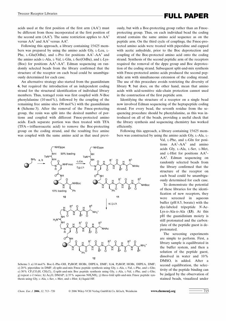
An alternative strategy also started from the guanidinium6, but required the introduction of an independent codingstrand for the structural identification of individual librarymembers. Thus, tentagel resin was first coupled with N-Bocphenylalanine (10 mol%), followed by the coupling of theremaining free amine sites (90 mol%) with the guanidinium6 (Scheme 3). After the removal of the Fmoc-protectinggroup, the resin was split into the desired number of por-tions and coupled with different Fmoc-protected aminoacids. Each separate portion was then treated with TFA(TFA= trifluoroacetic acid) to remove the Boc-protectinggroup on the coding strand, and the resulting free aminewas coupled with the same amino acid as that used previ-
ously, but with a Boc-protecting group rather than an Fmoc-protecting group. Thus, on each individual bead the codingstrand contains the same amino acid sequence as on thepeptide arm. On the third cycle of couplings, the Fmoc-pro-tected amino acids were treated with piperidine and cappedwith acetic anhydride, prior to the Boc deprotection andcoupling of the Boc-protected amino acid onto the codingstrand. Synthesis of the second peptidic arm of the receptorsrequired the removal of the dppe group and Boc deprotec-tion of the coding strand. Subsequent split-and-mix synthesiswith Fmoc-protected amino acids produced the second pep-tidic arm with simultaneous extension of the coding strand.The use of this procedure avoids restricting the diversity oflibrary 9, but does, on the other hand, mean that aminoacids with acid-sensitive side-chain protection cannot usedin the construction of the first peptidic arm.
Identifying the structure of a receptor on a single beadnow involved Edman sequencing of the heptapeptide codingstrand. For every bead, the seventh residue from the se-quencing procedure should be phenylalanine, as this was in-troduced on all of the beads, providing a useful check thatthe library synthesis and sequencing chemistry has workedefficiently.
Following this approach, a library containing 15625 mem-bers was constructed by using the amino acids Gly, l-Ala, l-
Val, l-Phe, and l-Gln for posi-tions AA1–AA3 and aminoacids Gly, l-Ala, l-Ser, l-Met,and l-Hist for positions AA4–AA6. Edman sequencing onrandomly selected beads fromthe library confirmed that thestructure of the receptor oneach bead could be unambigu-ously determined for each case.
To demonstrate the potentialof these libraries for the identi-fication of new receptors, theywere screened in aqueousbuffer (pH 8.5, borate) with thedye-labeled tripeptide N-Ac-Lys-d-Ala-d-Ala (13). At thispH the guanidinium moiety isstill protonated and the carbox-ylate of the peptidic guest is de-protonated.
The screening experimentsare simple to perform. First, alibrary sample is equilibrated inthe buffer system, and then asolution of the peptide guest,dissolved in water and 10%DMSO, is added. After asecond equilibration, the selec-tivity of the peptide binding canbe judged by the observation ofstained beads, visualized under
Scheme 3. a) 10 mol% Boc-L-Phe-OH, PyBOP, HOBt, DIPEA, DMF; b) 6, PyBOP, HOBt, DIPEA, DMF;c) 20% piperidine in DMF; d) split-and-mix Fmoc peptide synthesis using Gly, l-Ala, l-Val, l-Phe, and l-Gln;e) 30% CF3CO2H, CH2Cl2; f) split-and-mix Boc peptide synthesis using Gly, l-Ala, l-Val, l-Phe, and l-Gln;g) repeat c–f twice; h) Ac2O, DMAP; i) 35% aqueous NH2NH2; j) three-fold split-and-mix Fmoc peptide syn-thesis using Gly, l-Ala, l-Ser, l-Met, and l-Hist; k) liquid HF.
Chem. Eur. J. 2006, 12, 713 – 720 C 2006 Wiley-VCH Verlag GmbH&Co. KGaA, Weinheim www.chemeurj.org 715
FULL PAPERTweezer Receptor Libraries
a microscope. Additional aliquots of the peptide guest canbe added to increase the peptide concentration, and thus,provide optimal selectivity, as adjudged by the number ofhighly stained beads against a background of lightly or un-stained beads.
By using this strategy, both tweezer receptor libraries 9and 12 were screened with dye-labeled 13. For library 9, theselectivity was disappointing and there were a large numberof beads stained, with little discrimination between thedegree of staining. By using library 12, however, the selec-tivity after equilibration (48 h, at a peptide concentration of30 mm) was high, showing <2% of the highly red-coloredbeads. As a control experiment, the library was also incubat-ed with the disperse red dye 14 (30 mm), but selective bind-ing of the dye was not observed. Ten of the most intensivelystained beads from the screening experiment of 13 with li-brary 12 were selected and sequenced by Edman degrada-tion[10] (Table 1).
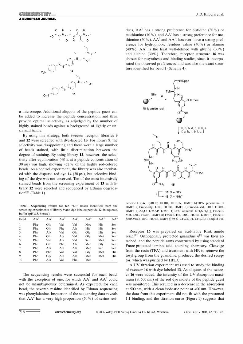
The sequencing results were successful for each bead,with the exception of one, for which AA5 and AA6 couldnot be unambiguously determined. As expected, for eachbead, the seventh residue identified by Edman sequencingwas phenylalanine. Inspection of the sequencing data revealsthat AA6 has a very high proportion (70%) of serine resi-
dues, AA5 has a strong preference for histidine (30%) ormethionine (40%), and AA4 has a strong preference for me-thionine (50%). AA3 and AA2, however, have a strong pref-erence for hydrophobic residues valine (40%) or alanine(40%). AA1 is the least well-defined with glycine (30%)and alanine (30%). Therefore, receptor structure 16 waschosen for resynthesis and binding studies, since it incorpo-rated the observed preferences, and was also the exact struc-ture identified for bead 1 (Scheme 4).
Receptor 16 was prepared on acid-labile Rink amideresin.[11] Orthogonally protected guanidine 6[8] was then at-tached, and the peptide arms constructed by using standardFmoc-protected amino acid coupling chemistry. Cleavagefrom the resin (TFA) and treatment with HF, to remove thetosyl group from the guanidine, produced the desired recep-tor, which was purified by HPLC.
A UV titration experiment was used to study the bindingof tweezer 16 with dye-labeled 13. As aliquots of the tweez-er 16 were added, the intensity of the UV absorption maxi-mum (at 500 nm) of the red dye moiety of the peptide guestwas monitored. This resulted in a decrease in the absorptionat 500 nm, with a clean isobestic point at 400 nm. However,the data from this experiment did not fit with the presumed1:1 binding, and the titration curve (Figure 1) suggests that
Table 1. Sequencing results for ten “hit” beads identified from thescreening experiments of library 9 and dye-labeled peptide 12, in aqueousbuffer (pH 8.5, borate).
Bead AA0 AA1 AA2 AA3 AA4 AA5 AA6
1 Phe Gly Val Val Met His Ser2 Phe Gly Phe Ala His His Ser3 Phe Ala Val Gln Gly His Ser4 Phe Gln Ala Val Gly Met Ser5 Phe Val Ala Val Ser Met Ser6 Phe Gln Phe Ala Met Gly Ser7 Phe Ala Ala Ala Met Ser Ser8 Phe Phe Val Val Gly Met His9 Phe Gly Ala Ala Met Met His10 Phe Ala Val Phe Met – –
Scheme 4. a) 6, PyBOP, HOBt, DIPEA, DMF; b) 20% piperidine inDMF; c) Fmoc-Gly, DIC, HOBt, DMF; d) Fmoc-l-Val, DIC, HOBt,DMF; e) Ac2O, DMAP, DMF; f) 35% aqueous NH2NH2; g) Fmoc-l-Met, DIC, HOBt, DMF; h) Fmoc-l-His, DIC, HOBt, DMF; i) Fmoc-l-Ser(OtBu), DIC, HOBt, DMF; j) 95% CF3CO2H, CH2Cl2; k) liquid HF.
www.chemeurj.org C 2006 Wiley-VCH Verlag GmbH&Co. KGaA, Weinheim Chem. Eur. J. 2006, 12, 713 – 720716
J. D. Kilburn et al.
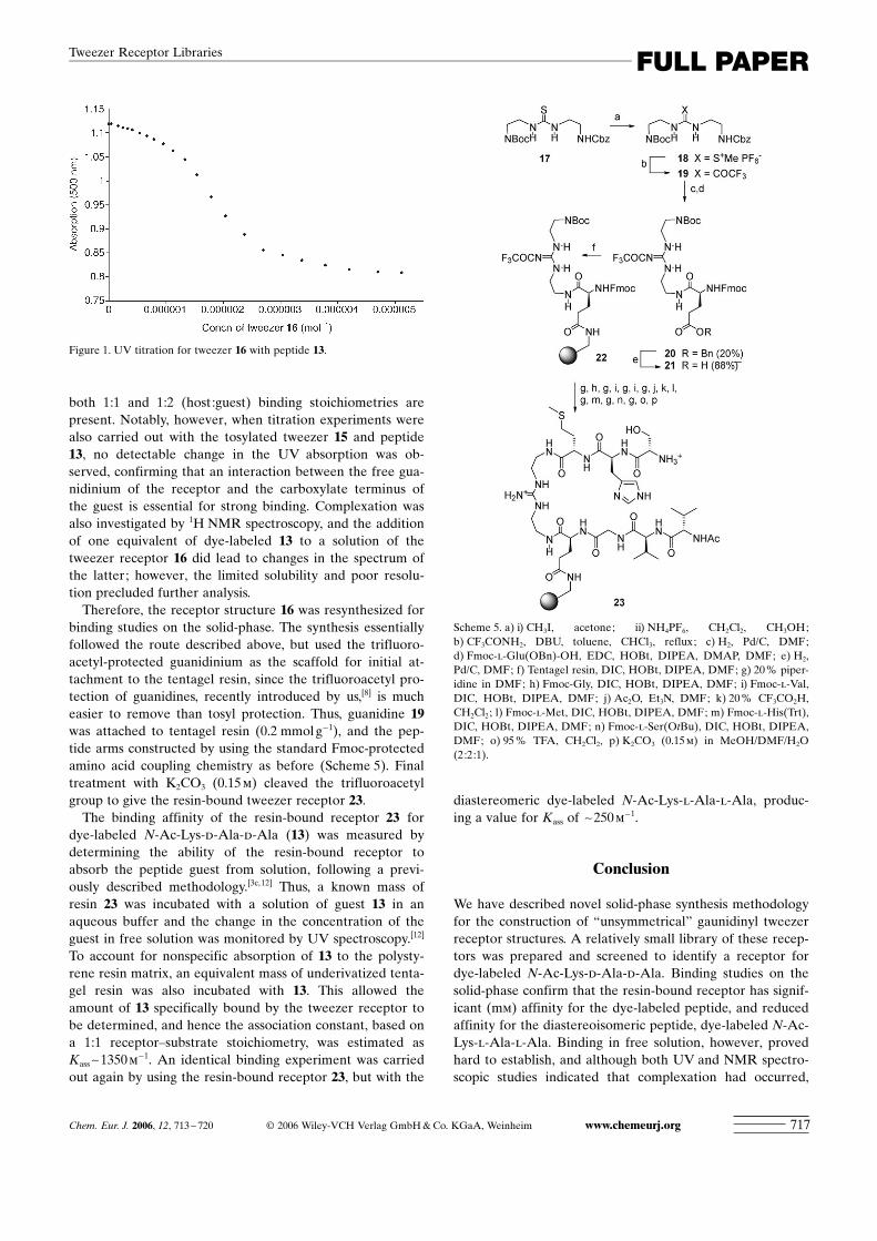
both 1:1 and 1:2 (host:guest) binding stoichiometries arepresent. Notably, however, when titration experiments werealso carried out with the tosylated tweezer 15 and peptide13, no detectable change in the UV absorption was ob-served, confirming that an interaction between the free gua-nidinium of the receptor and the carboxylate terminus ofthe guest is essential for strong binding. Complexation wasalso investigated by 1H NMR spectroscopy, and the additionof one equivalent of dye-labeled 13 to a solution of thetweezer receptor 16 did lead to changes in the spectrum ofthe latter; however, the limited solubility and poor resolu-tion precluded further analysis.
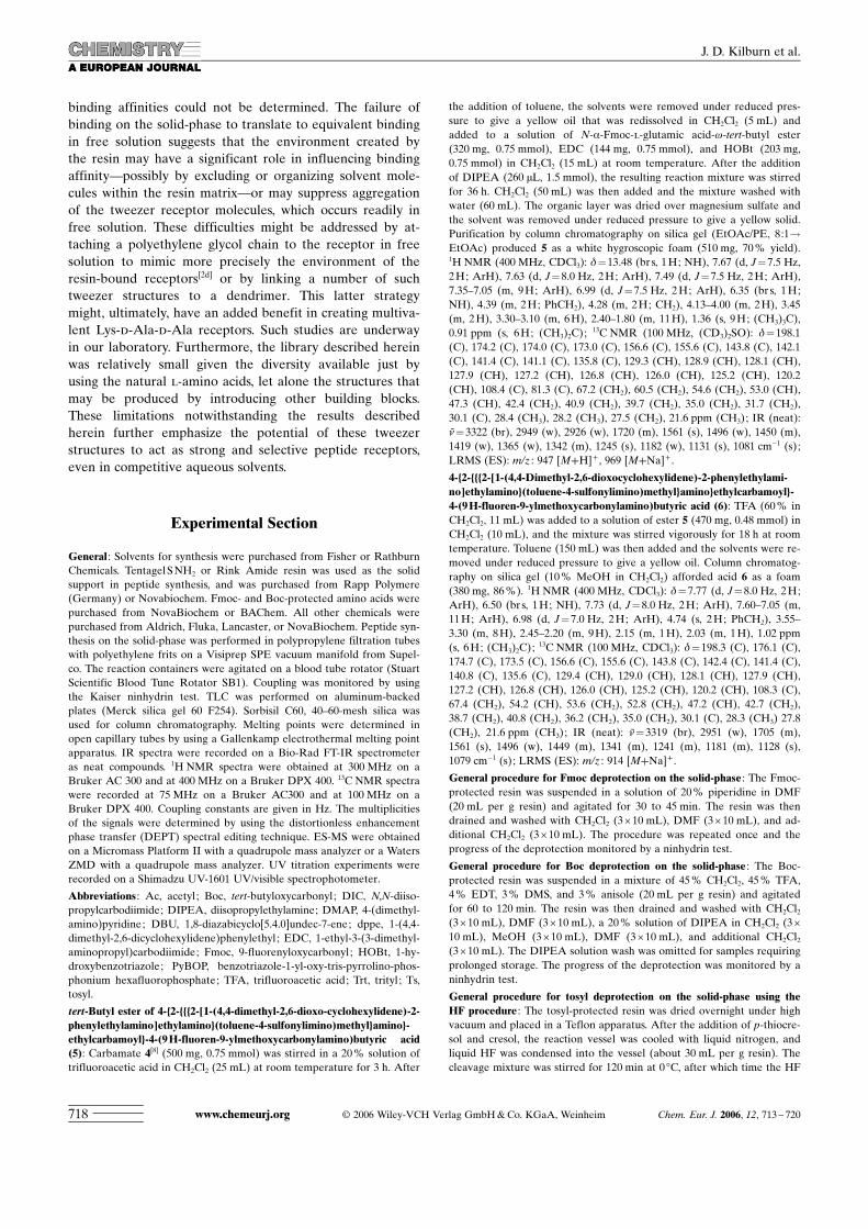
Therefore, the receptor structure 16 was resynthesized forbinding studies on the solid-phase. The synthesis essentiallyfollowed the route described above, but used the trifluoro-acetyl-protected guanidinium as the scaffold for initial at-tachment to the tentagel resin, since the trifluoroacetyl pro-tection of guanidines, recently introduced by us,[8] is mucheasier to remove than tosyl protection. Thus, guanidine 19was attached to tentagel resin (0.2 mmolg�1), and the pep-tide arms constructed by using the standard Fmoc-protectedamino acid coupling chemistry as before (Scheme 5). Finaltreatment with K2CO3 (0.15m) cleaved the trifluoroacetylgroup to give the resin-bound tweezer receptor 23.
The binding affinity of the resin-bound receptor 23 fordye-labeled N-Ac-Lys-d-Ala-d-Ala (13) was measured bydetermining the ability of the resin-bound receptor toabsorb the peptide guest from solution, following a previ-ously described methodology.[3c,12] Thus, a known mass ofresin 23 was incubated with a solution of guest 13 in anaqueous buffer and the change in the concentration of theguest in free solution was monitored by UV spectroscopy.[12]
To account for nonspecific absorption of 13 to the polysty-rene resin matrix, an equivalent mass of underivatized tenta-gel resin was also incubated with 13. This allowed theamount of 13 specifically bound by the tweezer receptor tobe determined, and hence the association constant, based ona 1:1 receptor–substrate stoichiometry, was estimated asKass~1350m�1. An identical binding experiment was carriedout again by using the resin-bound receptor 23, but with the
diastereomeric dye-labeled N-Ac-Lys-l-Ala-l-Ala, produc-ing a value for Kass of ~250m�1.
Conclusion
We have described novel solid-phase synthesis methodologyfor the construction of “unsymmetrical” gaunidinyl tweezerreceptor structures. A relatively small library of these recep-tors was prepared and screened to identify a receptor fordye-labeled N-Ac-Lys-d-Ala-d-Ala. Binding studies on thesolid-phase confirm that the resin-bound receptor has signif-icant (mm) affinity for the dye-labeled peptide, and reducedaffinity for the diastereoisomeric peptide, dye-labeled N-Ac-Lys-l-Ala-l-Ala. Binding in free solution, however, provedhard to establish, and although both UV and NMR spectro-scopic studies indicated that complexation had occurred,
Figure 1. UV titration for tweezer 16 with peptide 13.
Scheme 5. a) i) CH3I, acetone; ii) NH4PF6, CH2Cl2, CH3OH;b) CF3CONH2, DBU, toluene, CHCl3, reflux; c) H2, Pd/C, DMF;d) Fmoc-l-Glu(OBn)-OH, EDC, HOBt, DIPEA, DMAP, DMF; e) H2,Pd/C, DMF; f) Tentagel resin, DIC, HOBt, DIPEA, DMF; g) 20% piper-idine in DMF; h) Fmoc-Gly, DIC, HOBt, DIPEA, DMF; i) Fmoc-l-Val,DIC, HOBt, DIPEA, DMF; j) Ac2O, Et3N, DMF; k) 20% CF3CO2H,CH2Cl2; l) Fmoc-l-Met, DIC, HOBt, DIPEA, DMF; m) Fmoc-l-His(Trt),DIC, HOBt, DIPEA, DMF; n) Fmoc-l-Ser(OtBu), DIC, HOBt, DIPEA,DMF; o) 95% TFA, CH2Cl2, p) K2CO3 (0.15m) in MeOH/DMF/H2O(2:2:1).
Chem. Eur. J. 2006, 12, 713 – 720 C 2006 Wiley-VCH Verlag GmbH&Co. KGaA, Weinheim www.chemeurj.org 717
FULL PAPERTweezer Receptor Libraries
binding affinities could not be determined. The failure ofbinding on the solid-phase to translate to equivalent bindingin free solution suggests that the environment created bythe resin may have a significant role in influencing bindingaffinity—possibly by excluding or organizing solvent mole-cules within the resin matrix—or may suppress aggregationof the tweezer receptor molecules, which occurs readily infree solution. These difficulties might be addressed by at-taching a polyethylene glycol chain to the receptor in freesolution to mimic more precisely the environment of theresin-bound receptors[2d] or by linking a number of suchtweezer structures to a dendrimer. This latter strategymight, ultimately, have an added benefit in creating multiva-lent Lys-d-Ala-d-Ala receptors. Such studies are underwayin our laboratory. Furthermore, the library described hereinwas relatively small given the diversity available just byusing the natural l-amino acids, let alone the structures thatmay be produced by introducing other building blocks.These limitations notwithstanding the results describedherein further emphasize the potential of these tweezerstructures to act as strong and selective peptide receptors,even in competitive aqueous solvents.
Experimental Section
General : Solvents for synthesis were purchased from Fisher or RathburnChemicals. TentagelSNH2 or Rink Amide resin was used as the solidsupport in peptide synthesis, and was purchased from Rapp Polymere(Germany) or Novabiochem. Fmoc- and Boc-protected amino acids werepurchased from NovaBiochem or BAChem. All other chemicals werepurchased from Aldrich, Fluka, Lancaster, or NovaBiochem. Peptide syn-thesis on the solid-phase was performed in polypropylene filtration tubeswith polyethylene frits on a Visiprep SPE vacuum manifold from Supel-co. The reaction containers were agitated on a blood tube rotator (StuartScientific Blood Tune Rotator SB1). Coupling was monitored by usingthe Kaiser ninhydrin test. TLC was performed on aluminum-backedplates (Merck silica gel 60 F254). Sorbisil C60, 40–60-mesh silica wasused for column chromatography. Melting points were determined inopen capillary tubes by using a Gallenkamp electrothermal melting pointapparatus. IR spectra were recorded on a Bio-Rad FT-IR spectrometeras neat compounds. 1H NMR spectra were obtained at 300 MHz on aBruker AC 300 and at 400 MHz on a Bruker DPX 400. 13C NMR spectrawere recorded at 75 MHz on a Bruker AC300 and at 100 MHz on aBruker DPX 400. Coupling constants are given in Hz. The multiplicitiesof the signals were determined by using the distortionless enhancementphase transfer (DEPT) spectral editing technique. ES-MS were obtainedon a Micromass Platform II with a quadrupole mass analyzer or a WatersZMD with a quadrupole mass analyzer. UV titration experiments wererecorded on a Shimadzu UV-1601 UV/visible spectrophotometer.
Abbreviations : Ac, acetyl; Boc, tert-butyloxycarbonyl; DIC, N,N-diiso-propylcarbodiimide; DIPEA, diisopropylethylamine; DMAP, 4-(dimethyl-amino)pyridine; DBU, 1,8-diazabicyclo[5.4.0]undec-7-ene; dppe, 1-(4,4-dimethyl-2,6-dicyclohexylidene)phenylethyl; EDC, 1-ethyl-3-(3-dimethyl-aminopropyl)carbodiimide; Fmoc, 9-fluorenyloxycarbonyl; HOBt, 1-hy-droxybenzotriazole; PyBOP, benzotriazole-1-yl-oxy-tris-pyrrolino-phos-phonium hexafluorophosphate; TFA, trifluoroacetic acid; Trt, trityl ; Ts,tosyl.
tert-Butyl ester of 4-{2-{{{2-[1-(4,4-dimethyl-2,6-dioxo-cyclohexylidene)-2-phenylethylamino]ethylamino}(toluene-4-sulfonylimino)methyl}amino}-ethylcarbamoyl}-4-(9H-fluoren-9-ylmethoxycarbonylamino)butyric acid(5): Carbamate 4[8] (500 mg, 0.75 mmol) was stirred in a 20% solution oftrifluoroacetic acid in CH2Cl2 (25 mL) at room temperature for 3 h. After
the addition of toluene, the solvents were removed under reduced pres-sure to give a yellow oil that was redissolved in CH2Cl2 (5 mL) andadded to a solution of N-a-Fmoc-l-glutamic acid-w-tert-butyl ester(320 mg, 0.75 mmol), EDC (144 mg, 0.75 mmol), and HOBt (203 mg,0.75 mmol) in CH2Cl2 (15 mL) at room temperature. After the additionof DIPEA (260 mL, 1.5 mmol), the resulting reaction mixture was stirredfor 36 h. CH2Cl2 (50 mL) was then added and the mixture washed withwater (60 mL). The organic layer was dried over magnesium sulfate andthe solvent was removed under reduced pressure to give a yellow solid.Purification by column chromatography on silica gel (EtOAc/PE, 8:1!EtOAc) produced 5 as a white hygroscopic foam (510 mg, 70% yield).1H NMR (400 MHz, CDCl3): d=13.48 (br s, 1H; NH), 7.67 (d, J=7.5 Hz,2H; ArH), 7.63 (d, J=8.0 Hz, 2H; ArH), 7.49 (d, J=7.5 Hz, 2H; ArH),7.35–7.05 (m, 9H; ArH), 6.99 (d, J=7.5 Hz, 2H; ArH), 6.35 (br s, 1H;NH), 4.39 (m, 2H; PhCH2), 4.28 (m, 2H; CH2), 4.13–4.00 (m, 2H), 3.45(m, 2H), 3.30–3.10 (m, 6H), 2.40–1.80 (m, 11H), 1.36 (s, 9H; (CH3)3C),0.91 ppm (s, 6H; (CH3)2C);
13C NMR (100 MHz, (CD3)2SO): d=198.1(C), 174.2 (C), 174.0 (C), 173.0 (C), 156.6 (C), 155.6 (C), 143.8 (C), 142.1(C), 141.4 (C), 141.1 (C), 135.8 (C), 129.3 (CH), 128.9 (CH), 128.1 (CH),127.9 (CH), 127.2 (CH), 126.8 (CH), 126.0 (CH), 125.2 (CH), 120.2(CH), 108.4 (C), 81.3 (C), 67.2 (CH2), 60.5 (CH2), 54.6 (CH2), 53.0 (CH),47.3 (CH), 42.4 (CH2), 40.9 (CH2), 39.7 (CH2), 35.0 (CH2), 31.7 (CH2),30.1 (C), 28.4 (CH3), 28.2 (CH3), 27.5 (CH2), 21.6 ppm (CH3); IR (neat):n=3322 (br), 2949 (w), 2926 (w), 1720 (m), 1561 (s), 1496 (w), 1450 (m),1419 (w), 1365 (w), 1342 (m), 1245 (s), 1182 (w), 1131 (s), 1081 cm�1 (s);LRMS (ES): m/z : 947 [M+H]+ , 969 [M+Na]+ .
4-{2-{{{2-[1-(4,4-Dimethyl-2,6-dioxocyclohexylidene)-2-phenylethylami-no]ethylamino}(toluene-4-sulfonylimino)methyl}amino}ethylcarbamoyl}-4-(9H-fluoren-9-ylmethoxycarbonylamino)butyric acid (6): TFA (60% inCH2Cl2, 11 mL) was added to a solution of ester 5 (470 mg, 0.48 mmol) inCH2Cl2 (10 mL), and the mixture was stirred vigorously for 18 h at roomtemperature. Toluene (150 mL) was then added and the solvents were re-moved under reduced pressure to give a yellow oil. Column chromatog-raphy on silica gel (10% MeOH in CH2Cl2) afforded acid 6 as a foam(380 mg, 86%). 1H NMR (400 MHz, CDCl3): d=7.77 (d, J=8.0 Hz, 2H;ArH), 6.50 (br s, 1H; NH), 7.73 (d, J=8.0 Hz, 2H; ArH), 7.60–7.05 (m,11H; ArH), 6.98 (d, J=7.0 Hz, 2H; ArH), 4.74 (s, 2H; PhCH2), 3.55–3.30 (m, 8H), 2.45–2.20 (m, 9H), 2.15 (m, 1H), 2.03 (m, 1H), 1.02 ppm(s, 6H; (CH3)2C);
13C NMR (100 MHz, CDCl3): d=198.3 (C), 176.1 (C),174.7 (C), 173.5 (C), 156.6 (C), 155.6 (C), 143.8 (C), 142.4 (C), 141.4 (C),140.8 (C), 135.6 (C), 129.4 (CH), 129.0 (CH), 128.1 (CH), 127.9 (CH),127.2 (CH), 126.8 (CH), 126.0 (CH), 125.2 (CH), 120.2 (CH), 108.3 (C),67.4 (CH2), 54.2 (CH), 53.6 (CH2), 52.8 (CH2), 47.2 (CH), 42.7 (CH2),38.7 (CH2), 40.8 (CH2), 36.2 (CH2), 35.0 (CH2), 30.1 (C), 28.3 (CH3) 27.8(CH2), 21.6 ppm (CH3); IR (neat): n=3319 (br), 2951 (w), 1705 (m),1561 (s), 1496 (w), 1449 (m), 1341 (m), 1241 (m), 1181 (m), 1128 (s),1079 cm�1 (s); LRMS (ES): m/z : 914 [M+Na]+ .
General procedure for Fmoc deprotection on the solid-phase : The Fmoc-protected resin was suspended in a solution of 20% piperidine in DMF(20 mL per g resin) and agitated for 30 to 45 min. The resin was thendrained and washed with CH2Cl2 (3O10 mL), DMF (3O10 mL), and ad-ditional CH2Cl2 (3O10 mL). The procedure was repeated once and theprogress of the deprotection monitored by a ninhydrin test.
General procedure for Boc deprotection on the solid-phase : The Boc-protected resin was suspended in a mixture of 45% CH2Cl2, 45% TFA,4% EDT, 3% DMS, and 3% anisole (20 mL per g resin) and agitatedfor 60 to 120 min. The resin was then drained and washed with CH2Cl2(3O10 mL), DMF (3O10 mL), a 20% solution of DIPEA in CH2Cl2 (3O10 mL), MeOH (3O10 mL), DMF (3O10 mL), and additional CH2Cl2(3O10 mL). The DIPEA solution wash was omitted for samples requiringprolonged storage. The progress of the deprotection was monitored by aninhydrin test.
General procedure for tosyl deprotection on the solid-phase using theHF procedure : The tosyl-protected resin was dried overnight under highvacuum and placed in a Teflon apparatus. After the addition of p-thiocre-sol and cresol, the reaction vessel was cooled with liquid nitrogen, andliquid HF was condensed into the vessel (about 30 mL per g resin). Thecleavage mixture was stirred for 120 min at 0 8C, after which time the HF
www.chemeurj.org C 2006 Wiley-VCH Verlag GmbH&Co. KGaA, Weinheim Chem. Eur. J. 2006, 12, 713 – 720718
J. D. Kilburn et al.
was evaporated under a stream of nitrogen. The resin was then washedwith diethyl ether (4O20 mL), 20% DIPEA in CH2Cl2 (3O20 mL),MeOH (3O20 mL), and CH2Cl2 (3O20 mL).
Receptor library 9 : TentaGelSNH2 resin (900 mg, 0.26 mmolg�1,0.23 mmol) was preswollen in CH2Cl2 and then drained. A solution ofacid 6 (314 mg, 0.35 mmol), PyBOP (182 mg, 0.35 mmol), HOBt (47 mg,0.35 mmol), and DIPEA (60 ml, 0.35 mmol) in DMF was added to theresin and the mixture agitated on a tube rotator for 18 h, before beingdrained and washed with CH2Cl2 (3O5 mL), DMF (3O5 mL), and addi-tional CH2Cl2 (3O5 mL). A ninhydrin test gave a negative result. Follow-ing Fmoc deprotection (see general procedure) the resin was divided intofive equal portions, and each portion was preswollen in DMF (2 mL). Asolution of Fmoc-protected amino acid (Gly, Leu, Phe, Glu(OtBu), orPro; 0.14 mmol), PyBOP (73 mg, 0.14 mmol), and HOBt (17 mg,0.14 mmol) in DMF (3 mL) was preactivated for a few minutes, and thenadded to each portion of the resin, followed by the addition of DIPEA(18 mg, 0.14 mmol). Each portion was agitated on a tube rotator for atleast 24 h at room temperature. After this time, the portions werewashed with CH2Cl2 (3O5 mL), DMF (3O5 mL), additional CH2Cl2 (3O5 mL), and then dried. The success of the coupling step was monitoredby a qualitative ninhydrin test. Each coupling cycle was repeated untilthe ninhydrin test showed that no free amino functions were present.After the successful coupling step, the resin was recombined and Fmoc-deprotected, as described in the general procedure, with monitoring by aqualitative ninhydrin test. This split-and-mix procedure was then repeat-ed with the same five amino acids (first, Fmoc-protected and then Boc-protected) to build up the first tripeptide side arm. For the next step inthe synthesis, the recombined resin was swollen in DMF (10 mL), treatedwith hydrazine hydrate (15 mL of a 35% aq solution), and agitated on atube rotator for 4 h, to remove the dppe-protecting group.[9] The resinwas then divided into five portions and each portion was preswollen inDMF (2 mL). A solution of Fmoc-protected amino acid (Ala, Val, Gln,Ser(OtBu), or Lys(Boc); 0.14 mmol), PyBOP (73 mg, 0.14 mmol), andHOBt (17 mg, 0.14 mmol) in DMF (3 mL) was preactivated for a few mi-nutes and then added to each portion of resin, followed by the additionof DIPEA (18 mg, 0.14 mmol). Each portion was agitated on a tube rota-tor for at least 24 h at room temperature. The portions were washed withCH2Cl2 (3O5 mL), DMF (3O5 mL), additional CH2Cl2 (3O5 mL), andthen dried. The success of the coupling step was monitored by a qualita-tive ninhydrin test. Each coupling cycle was repeated until the ninhydrintest showed that no free amino functions were present. After the success-ful coupling step, the resin was recombined and Fmoc-deprotected, as de-scribed in the general procedure, with monitoring by the qualitative nin-hydrin test. This split-and-mix procedure was then repeated twice morewith the same five Fmoc-protected amino acids to build up the secondtripeptide side arm. After the recombination of all of the portions of theresin, the protecting groups were removed by liquid HF treatment (seegeneral procedure) to give library 9.
Receptor library incorporating coding strand 12 : TentaGelSNH2 resin(900 mg, 0.26 mmolg�1, 0.23 mmol), was preswollen in CH2Cl2 and thendrained. A solution of Boc-Phe (6 mg, 23 mmol), PyBOP (12 mg,23 mmol), HOBt (31 mg, 23 mmol), and DIPEA (40 ml, 23 mmol) in DMF(3 mL) was added to the resin and the mixture agitated on a tube rotatorfor 18 h, before being drained and washed with CH2Cl2 (3O5 mL), DMF(3O5 mL), and additional CH2Cl2 (3O5 mL). A solution of acid 6(314 mg, 0.35 mmol), PyBOP (182 mg, 0.35 mmol), HOBt (47 mg,0.35 mmol), and DIPEA (60 mL, 0.35 mmol) in DMF was then added tothe resin and the mixture agitated on a tube rotator for a further 18 h,before being drained and washed with CH2Cl2 (3O5 mL), DMF (3O5 mL), and additional CH2Cl2 (3O5 mL). A ninhydrin test gave a nega-tive result. Following Fmoc deprotection (see general procedure), theresin was divided into five equal portions, and each portion was preswol-len in DMF (2 mL). A solution of Fmoc-protected amino acid (Gly, Ala,Phe, Val, or Leu; 0.14 mmol), PyBOP (73 mg, 0.14 mmol), and HOBt(17 mg, 0.14 mmol) in DMF (3 mL) was preactivated for a few minutesand then added to each portion of the resin, followed by the addition ofDIPEA (18 mg, 0.14 mmol). Each portion was agitated on a tube rotatorfor 24 h at room temperature. After this time, the portions were washedwith CH2Cl2 (3O5 mL), DMF (3O5 mL), additional CH2Cl2 (3O5 mL),
and then dried. The success of the coupling step was monitored by aqualitative ninhydrin test. Each coupling cycle was repeated until the nin-hydrin test showed that no free amino functions were present. Each por-tion was then Boc-deprotected to release the free amine of the codingstrand (see general procedure) and then coupled with a Boc-protectedamino acid (Gly, Ala, Phe, Val, and then Leu, corresponding with theFmoc-protected amino acids used in the previous coupling; 14 mmol) byusing PyBOP (7 mg, 14 mmol), HOBt (2 mg, 14 mmol), and DIPEA(2 mg, 14 mmol). Once complete, the portions were washed with CH2Cl2(3O5 mL), DMF (3O5 mL), additional CH2Cl2 (3O5 mL), and thendried. After the successful coupling step, the resin was recombined andFmoc-deprotected, as described in the general procedure, with monitor-ing by the qualitative ninhydrin test. This split-and-mix procedure wasthen repeated twice, with the same five amino acids, to build up the firsttripeptide side arm and the coding strand. For the next step in the syn-thesis, the recombined resin was swollen in DMF, Fmoc-deprotected (seegeneral procedure), treated with excess acetic anhydride and DMAP, andthen agitated on a tube rotator for 4 h to cap the first tripeptide arm. Theresin was then treated with hydrazine hydrate (15 mL of a 35% aq solu-tion) and agitated on a tube rotator for 4 h, to remove the dppe-protect-ing group.[9] Once complete, the resin was divided into five portions, pre-swollen in DMF (2 mL), and subjected to three rounds of split-and-mixsynthesis to build up the second tripeptide arm (Fmoc-protected aminoacids, Gly, Ala, Ser(OtBu), Hist(Trt), and then Met were used) and thecoding strand (with the corresponding Boc-protected amino acids), fol-lowing the exact same procedures described above for the synthesis ofthe first tripeptide arm and coding strand. Next, a solution of Fmoc-pro-tected amino acid (Ala, Val, Gln, Ser(OtBu), and then Lys(Boc);0.14 mmol), PyBOP (73 mg, 0.14 mmol), and HOBt (17 mg, 0.14 mmol)in DMF (3 mL) was preactivated for a few minutes and then added toeach portion of resin, followed by the addition of DIPEA (18 mg,0.14 mmol). Each portion was then agitated on a tube rotator for 24 h atroom temperature. After this time, the portions were washed withCH2Cl2 (3O5 mL), DMF (3O5 mL), additional CH2Cl2 (3O5 mL), andthen dried. The success of the coupling step was monitored by a qualita-tive ninhydrin test. As before, each coupling cycle was repeated until theninhydrin test showed that no free amino functions were present. Afterthe successful coupling step, the resin was recombined and Fmoc-depro-tected, as described in the general procedure, with monitoring by a quali-tative ninhydrin test. This split-and-mix procedure was then repeatedtwice more, with the same five Fmoc-protected amino acids, to build upthe second tripeptide side arm. After the recombination of all of the por-tions of the resin, the protecting groups were removed by liquid HFtreatment (see general procedure) to give library 12.
Screening experiments :
Screening of the tweezer receptor library 18 : A sample of library 12(15 mg) was equilibrated in borax buffer solution (300 mL) for 24 h. A so-lution of the tripeptide guest 13 (20 mM, 500 mL) in a 15% solution ofDMSO/borax buffer was then added to the library sample to give a guestconcentration of 12.5 mM. Equilibration was continued for 24 h. Analysisof the beads was carried out in flat-bottomed glass pots, under a Leica in-verted DML microscope (magnificationO40), and 10 highly red stainedbeads were selected and submitted for Edman sequencing.[10]
Benzyl ester of 4-{2-[N’-(2-tert-butoxycarbonylaminoethyl)-N’’-(2,2,2-tri-fluoroacetyl)guanidino]ethylcarbamoyl}-4-(9H-fluoren-9-ylmethoxycar-bonylamino)butyric acid (20): Carbamate 19[8] (224 mg, 0.47 mmol) wasdissolved in DMF (10 mL). Palladium on charcoal (10% by wt,10 mol%, 50 mg) was then added, and the mixture was stirred under ahydrogen atmosphere for 16 h. After this time, the mixture was filteredthrough Celite and the resulting filtrates were evaporated to give a paleyellow oil. N-a-Fmoc-l-glutamic acid-w-benzyl ester (238 mg,0.52 mmol), HOBt (127 mg, 0.94 mmol), DIPEA (410 ml, 2.36 mmol),DMAP (6 mg, 47 mmol), and EDC (99 mg, 0.52 mmol) were added to asolution of this oil in DMF (10 mL), and the resulting mixture was stirredfor 18 h. After this time, evaporation of all of the solvent and purificationof the residue by column chromatography (EtOAc/PE 1:1) produced 20as a white hygroscopic foam (73 mg, 20%). 1H NMR (300 MHz, CDCl3):d=9.65 (br s, 1H; NH), 7.76 (d, 2H, J=7.5 Hz; ArH), 7.57 (d, 2H, J=
Chem. Eur. J. 2006, 12, 713 – 720 C 2006 Wiley-VCH Verlag GmbH&Co. KGaA, Weinheim www.chemeurj.org 719
FULL PAPERTweezer Receptor Libraries
7.0 Hz; ArH), 7.42–7.16 (m, 9H; ArH), 7.16 (br s, 1H; NH), 7.01 (br s,1H; NH), 5.76 (br s, 1H; NH), 5.12 (s, 2H; CH2Ph), 4.45–4.30 (m, 2H),4.19 (m, 2H; CH2), 3.60–3.15 (m, 8H; CH2CH2), 2.49 (m, 2H;CH2COO), 2.13 (m, 1H), 2.01 (m, 1H), 1.40 ppm (s, 9H; (CH3)3C);13C NMR (75 MHz, CDCl3): d=173.3 (C), 173.2 (C), 162.7 (C), 157.6(C), 157.0 (C), 156.6 (C), 143.8 (C), 141.4 (C), 135.7 (C), 128.7 (CH),128.4 (CH), 128.3 (CH), 127.9 (CH), 127.2 (CH), 125.3 (CH), 120.1(CH), 116.7 (C), 80.6 (C), 67.3 (CH2), 66.8 (CH2), 54.8 (CH), 47.1 (CH2),41.9 (CH2), 41.0 (CH2), 39.6 (CH2), 38.9 (CH2), 30.4 (CH2), 28.4 ppm(CH3); IR (neat): n=1629 (m), 1522 (m), 1449 (m), 1242 (m), 1166 (m),1140 (m), 848 (w), 739 (m), 514 cm�1 (s); LRMS (ES): m/z : 783 [M+H]+ ,805 [M+Na]+ .
4-{2-[N’-(2-tert-Butoxycarbonylaminoethyl)-N’’-(2,2,2-trifluoroacetyl)gua-nidino]ethylcarbamoyl}-4-(9H-fluoren-9-ylmethoxycarbonylamino)buty-ric acid (21): Palladium on charcoal (10% by wt, 10 mol%, 8 mg) wasadded to a solution of ester 20 (55 mg, 70 mmol) in EtOH (5 mL). Themixture was stirred under a hydrogen atmosphere for 2 h and then fil-tered through Celite. Finally, the solvent was removed by evaporationunder reduced pressure to give 21 as an off-white hygroscopic foam(43 mg, 88%). 1H NMR (300 MHz, CDCl3): d=9.47 (br s, 1H; NH), 7.74(d, 2H, J=7.5 Hz; ArH), 7.55 (d, 2H, J=6.5 Hz; ArH), 7.44 (br s, 1H;NH), 7.42–7.22 (m, 4H; ArH), 6.18 (br s, 1H; NH), 5.88 (br s, 1H; NH),4.45–4.38 (m, 3H), 4.17 (m, 1H; CH), 3.70–2.95 (m, 8H; CH2CH2), 2.44(m, 2H; CH2COO), 2.18–1.82 (m, 2H), 1.40 ppm (s, 9H; (CH3)3C);13C NMR (75 MHz, CDCl3): d=180.1 (C), 176.2 (C), 173.7 (C), 171.7(C), 159.9 (C), 157.8 (C), 156.7 (C), 143.7 (C), 141.4 (C), 127.9 (CH),127.3 (CH), 125.3 (CH), 120.1 (CH), 80.7 (C), 67.5 (CH2), 55.7 (CH),54.6 (CH), 47.1 (CH2), 41.9 (CH2), 41.1 (CH2), 39.6 (CH2), 38.7 (CH2),30.0 (CH2), 28.4 ppm (CH3); IR (neat): n=2977 (br, w), 1628 (s), 1524(m), 1449 (m), 1243 (m), 1139 (s), 909 (m), 844 (m), 734 cm�1 (s); LRMS(ES): m/z : 693 [M+H]+ , 715 [M+Na]+ .
Resin-bound receptor 23 : TentaGelSNH2 resin (0.2 mmolg�1, 162.2 mg,32.4 mmol) was swollen in CH2Cl2 and drained. A solution of acid 21(34 mg, 48.7 mmol), DIC (15 ml, 97.3 mmol), HOBt (13 mg, 97.3 mmol),and DIPEA (17 ml, 97.3 mmol) in DMF (2 mL) was then added to theresin, and the mixture agitated on a tube rotator for 18 h. Any remainingamine residues were capped by treating the resin with an excess of aceticanhydride. The resin was then filtered and washed with CH2Cl2 (3O5 mL), DMF (3O5 mL), and additional CH2Cl2 (3O5 mL). A ninhydrintest was negative. There then followed three sequential cycles of cou-pling/Fmoc deprotection to add Fmoc-Gly, Fmoc-Val, and Fmoc-Val tothe resin, and to synthesize the first arm of the receptor. This was a-chieved by using Fmoc-protected amino acid (97 mmol), DIC (12 mg,97 mmol), HOBt (13 mg, 97 mmol), and DIPEA (17 ml, 97 mmol) in DMF(2 mL). Ninhydrin tests were used to monitor progress of each couplingreaction and the coupling reactions were repeated until complete. Afterdeprotection of the second Val residue, the chain was capped by thetreatment of the resin with acetic anhydride (9 ml, 97.3 mmol) andDIPEA (17 ml, 97.3 mmol) in DMF (2 mL). Boc deprotection (see generalprocedure) was followed by three sequential coupling/deprotection cyclesto add Fmoc-Met, Fmoc-His(Trt), and Fmoc-Ser(OtBu) to the resin alsoby using the Fmoc-protected amino acid (97 mmol), DIC (12 mg,97 mmol), HOBt (13 mg, 97 mmol), and DIPEA (17 ml, 97 mmol) in DMF(2 mL); this time to synthesize the second arm of the receptor. Ninhydrintests were used to monitor the progress of each coupling reaction. Trt de-protection was achieved by the treatment of the resin with 95% TFA/CH2Cl2 (2O1 h), followed by washing with CH2Cl2 (3O5 mL), 50%DIPEA/DMF (3O5 mL), DMF (3O5 mL), and additional CH2Cl2 (3O5 mL). Trifluoroacetyl deprotection was carried out by the treatment of
the resin with K2CO3 (0.15m) in MeOH/DMF/water (2:2:1, 2O3 h). Final-ly, the resin was washed as above, rinsed with diethyl ether, and dried invacuo.
Acknowledgements
We wish to thank EPSRC for supporting this work (GRL�98916/01) andthe BBSRC for a studentship (JS).
[1] For a recent review on peptide receptors see: a) M. W. Peczuh,A. D. Hamilton, Chem. Rev. 2000, 100, 2479–2493.
[2] a) C. Chamorro, R. M. J. Liskamp, Tetrahedron 2004, 60, 11145–11157; b) C. Chamorro, J.-W. Hofman, R. M. J. Liskamp, Tetrahe-dron 2004, 60, 8691–8699; c) C. Schmuck, M. Heil, Org. Biomol.Chem. 2003, 1, 633–636; d) C. Schmuck, M. Heil, ChemBioChem2003, 4, 1232–1238; e) K. B. Jensen, T. M. Braxmeier, M. Demarcus,J. D. Kilburn, Chem. Eur. J. 2002, 8, 1300–1309.
[3] a) H. Wennemers, M. C. Nold, M. Conza, K. J. Kulicke, M. Neubur-ger, Chem. Eur. J. 2003, 9, 442–448; b) H. Wennemers, M. Conza, J.Org. Chem. 2002, 67, 2696–2698; c) R. Arienzo, J. D. Kilburn, Tetra-hedron 2002, 58, 711–719; d) T. Braxmeier, M. Demarcus, T. Fes-smann, S. McAteer, J. D. Kilburn, Chem. Eur. J. 2001, 7, 1889–1898;e) J. D. Kilburn, T. Fessmann, Angew. Chem. 1999, 111, 2170–2174;Angew. Chem. Int. Ed. 1999, 38, 1993–1996.
[4] Other recent examples of sequence selective peptide receptors:a) C. Schmuck, L. Geiger, J. Am. Chem. Soc. 2004, 126, 8898–8899;b) S. Rensing, T. Schrader, Org. Lett. 2002, 4, 2161–2164; c) R. Xu,G. Greiveldinger, L. E. Marenus, A. Cooper, J. A. Ellman, J. Am.Chem. Soc. 1999, 121, 4898–4899; d) M. Sirish, H.-J. Schneider, J.Chem. Soc. Chem. Commun. 1999, 907–908; e) Md. A. Hossain, H.-J. Schneider, J. Am. Chem. Soc. 1998, 120, 11208–11209; f) R. Bre-slow, Z. Yang, R. Ching, G. Trojandt, F. Odobel, J. Am. Chem. Soc.1998, 120, 3536–3537.
[5] a) N. Srinivasan, J. D. Kilburn, Curr. Opin. Chem. Biol. 2004, 8, 305–310; b) B. Linton, A. D. Hamilton, Curr. Opin. Chem. Biol. 1999, 3,307–312.
[6] C. Schmuck, P. Frey, M. Heil, ChemBioChem 2005, 6, 628–631.[7] a) D. H. Williams, Nat. Prod. Rep. 1996, 13, 469–477; b) M. L.
Cohen, Science 1992, 257, 1050–1055; c) H. C. Neu, Science 1992,257, 1064–1073.
[8] S. Bartoli, K. B. Jensen, J. D. Kilburn, J. Org. Chem. 2003, 68, 9416–9422.
[9] S. R. Chhabra, B. Hothi, D. J. Evans, P. D. Wite, B. W. Bycroft, W. C.Chan, Tetrahedron Lett. 1998, 39, 1603–1606.
[10] P. Edman, G. Begg, Eur. J. Biochem. 1967, 1, 80–91; sequencing wasperformed by using an applied biosystems 477 A pulsed liquid-phasesequencer, coupled online to an applied biosystems 120 A phenyl-thiohydantoin (PTH) derivative analyzer.
[11] H. Rink, Tetrahedron Lett. 1987, 28, 3787–3790.[12] a) M. Conza, H. Wennemers, Chem. Commun. 2003, 866–867;
b) M. C. F. Monnee, A. J. Brouwer, L. M. Verbeek, A. M. A. van -Wageningen, R. M. J. Liskamp, Bioorg. Med. Chem. Lett. 2001, 11,1521–1525; c) Y. Cheng, T. Suenaga, W. C. Still, J. Am. Chem. Soc.1996, 118, 1813–1814.
Received: July 25, 2005Published online: October 14, 2005
www.chemeurj.org C 2006 Wiley-VCH Verlag GmbH&Co. KGaA, Weinheim Chem. Eur. J. 2006, 12, 713 – 720720
J. D. Kilburn et al.