Embed Size (px)
Citation preview

ARTICLE
Received 25 Jul 2014 | Accepted 24 Oct 2014 | Published 8 Dec 2014
Synthetic fossilization of soft biological tissuesand their shape-preserving transformation intosilica or electron-conductive replicasJason L. Townson1,2, Yu-Shen Lin1,2, Stanley S. Chou3, Yasmine H. Awad2, Eric N. Coker3,
C. Jeffrey Brinker3,4 & Bryan Kaehr3,4
Structural preservation of complex biological systems from the subcellular to whole organism
level in robust forms, enabling dissection and imaging while preserving 3D context,
represents an enduring grand challenge in biology. Here we report a simple immersion
method for structurally preserving intact organisms via conformal stabilization within silica.
This self-limiting process, which we refer to as silica bioreplication, occurs by condensation of
water-soluble silicic acid proximally to biomolecular interfaces throughout the organism.
Conformal nanoscopic silicification of all biomolecular features imparts structural rigidity
enabling the preservation of shape and nano-to-macroscale dimensional features upon drying
to form a biocomposite and further high temperature oxidative calcination to form silica
replicas or reductive pyrolysis to form electrically conductive carbon replicas of complete
organisms. The simplicity and generalizability of this approach should facilitate efforts in
biological preservation and analysis and could enable the development of new classes of
biomimetic composite materials.
DOI: 10.1038/ncomms6665 OPEN
1 Division of Molecular Medicine, Department of Internal Medicine, The University of New Mexico, Albuquerque, New Mexico 87131, USA. 2 Center for Micro-Engineered Materials, The University of New Mexico, Albuquerque, New Mexico 87131, USA. 3 Advanced Materials Laboratory, Sandia National Laboratories,Albuquerque, New Mexico 87185, USA. 4 Department of Chemical and Biological Engineering, The University of New Mexico, Albuquerque, New Mexico87131, USA. Correspondence and requests for materials should be addressed to B.K. (email: [email protected]).
NATURE COMMUNICATIONS | 5:5665 | DOI: 10.1038/ncomms6665 | www.nature.com/naturecommunications 1
& 2014 Macmillan Publishers Limited. All rights reserved.

The invention of electron microscopy (EM) in the early 20thcentury broadly impacted the physical and materialssciences, but proved particularly important for biological
research by enabling macromolecular complexes, ultrastructuresand other optically unresolvable features of cells and tissues to bestudied in context1,2. Throughout the technique’s history, EMinstrumentation has steadily improved allowing for increasinglybetter resolution and imaging in both scanning electron (SEM)and transmission electron microscopy (TEM; for example,aberration correction3), and providing procedures that enablein situ imaging of dynamic processes4 and hydrated samples5, aswell as other techniques for reconstructing three-dimensional(3D) images (for example, cryo-tomography6 and serialblock-face SEM7). Yet, despite consistent advancements ininstrumentation, techniques for the preparation of biologicalmaterials for EM remain largely unchanged since first put intopractice2. These often elaborate procedures designed to preservedelicate biological samples under the harsh conditions of EM(high-vacuum and energy flux) remain cumbersome to the non-specialist and thus limit their broader usage. For example,methods such as cryofixation (vitrification) currently come closestto capturing the most ‘accurate’, native state of a biomolecularstructure because of the rapid stabilization of the specimen withinvitreous ice through cold plunge, but, in particular for macro-scopic tissues or organisms, it requires specialized equipment,subsequent processing (for example, replication, fracture and/orcryo-focused-ion beam; cryo-FIB) and considerable expertise2.Freeze fracture, though an important development for elucidatingcellular ultrastructure, remains a highly specialized techniquelargely because of its overall complexity and multitude ofintricate steps8. Chemical fixation using aldehyde cross-linkersis a relatively simple approach, but dehydration via replacementof water with sequential, increasing concentrations of lowviscosity, high vapour pressure solvents (for example, ethanol,methanol, acetone) and subsequent drying necessary for imagingin vacuum can degrade sample architectures via de-wetting andother drying stresses, which can be significant for small features9.Alternatively, secondary and tertiary fixation is employed usingosmium tetroxide followed by uranyl acetate and subsequentdehydration using critical point or freeze drying5, but this routesubstantially increases both process complexity and use of toxicchemicals. Processing of specimens using embedding resinsresults in a moulded polymer block that enables thin sectioningfor cross-sectional observation in TEM, serial block-face SEM orfocused ion beam SEM (FIB/SEM)10, but this process destroys the3D structure as a whole. Finally, for observation using SEM,biological specimens generally require coating with metal orcarbon films to avoid charging. While these conductive coatingsimprove EM resolution, they have inherent granularity with grainsizes that can reach tens of nanometres as film thicknessincreases11; they can also alter topological features of softmaterials and can render only exposed surfaces conductive.Uniform deposition on complex 3D structures is challengingbecause of shadowing effects using evaporated or sputteredmetals.
To overcome limitations of existing methods of tissue/organism stabilization and imaging, the goal of this study wasthe development of a sample preparation procedure requiring fewsteps and minimal expertise or specialized equipment. Wepostulated a process that resulted in conformal structuralstabilization from subcellular to organism scales, avoidedembedding in polymer, and rendered an intrinsically conductivespecimen resistant to high intensity energy and long-termdegradation would provide new opportunities for biologicalanalysis (for example, internal imaging, elemental contrast) andestablish a new preparation method that complements the
substantial recent developments in EM instrumentation. As astarting point, we considered natural mineralization processesthat produce fossilized materials. Structural preservation ofbiological materials through fossilization requires an intricatealignment of optimum conditions that are achieved over longtime scales by complex geological processes. Even if theseare satisfied, preservation of soft tissue in natural fossils isextremely rare12. ‘Synthetic fossilization’ has been widelyexplored using, for example, wood, leaves, butterfly wings,pollen grains and diatoms as templates for material depositionand subsequent conversion13–18. However, these templates arealready mechanically stable, comprising stiff polysaccharides(wood, butterfly wings) or bioinorganic composites (diatoms).Thus in contrast to soft tissues, they are intrinsically resistant tostructural deformation upon drying and subsequent chemicalprocessing. The extension of ‘synthetic fossilization’ to softbiomaterials under shape-preserving conditions would providea new foundational approach for specimen preservation,create opportunities for conversion into more durable andEM-compatible materials, and serve as a facile approach tocreate new classes of biomimetic composite materials.
Here, we describe such an approach by which soft, biologicaltissues are replicated from the subcellular to the organismal scalein silica, a process we term silica bioreplication (SBR). We showshape and feature-preserving SBR of intact multicellular speci-mens (tissues derived from chicken embryos), inclusive of cells,extracellular matrices, tissues and organs as evidenced bygeneration of nearly identical inorganic (silica) replicas followingremoval of the biomolecular specimen template via hightemperature calcination (500–600 �C) under oxidizing conditions.In addition, following SBR, specimens can be subjected to hightemperature pyrolysis (800–1,000 �C) under reducing conditionsto convert the organic constituents into conductive carbon. Thisprocedure results in remarkable preservation of structure andenables whole, intact specimens to be imaged and sectioned adinfinitum and arbitrarily across the macro- to nanoscale withoutloss of resolution due to charging-induced specimen damage orimaging artifacts (as all internal and external features arecarbonized and equally conductive). Images of cellular structurecan be attained deep within tissue cavities of mechanicallysectioned or FIBed organs. Finally, the resistance to damage ofthese specimens under high accelerating voltage and beamcurrent provides excellent signal-to-noise ratios using back-scattered electron (BSE) detection allowing, for example, singleparticle chemical imaging of injected gold nanoparticles (AuNPs)in a fracture plane of a chicken embryo liver.
ResultsSilica bioreplication of chicken embryos. Recently, we observedthat silicification of cultured mammalian cells derived from arange of tissues preserves cellular structure from the nano (DNA,organelles and so on) to whole cell (micrometre) level19. Thisability to preserve intact cells with nanoscale fidelity laid thegroundwork to examine SBR of complex multicellular systems inwhich cells organize to form diverse tissue types with distinct 3Darchitectures. To explore these possibilities, we used chickenembryos (Gallus gallus domesticus), which have been commonlyused as exemplary in vivo models in the study of developmentalbiology, nanomedicine and other organism-scale processes20,21.As chicken embryos are primarily composed of soft tissue duringthe first 10 days of development and have well-formed internalorgans by day 17, their use over 3–17 days of development allowsus to demonstrate the efficacy of SBR for structural preservationof a wide spectrum of soft tissues and organs. First, embryos wereremoved from fertilized eggs at day 3 of development by cutting
ARTICLE NATURE COMMUNICATIONS | DOI: 10.1038/ncomms6665
2 NATURE COMMUNICATIONS | 5:5665 | DOI: 10.1038/ncomms6665 | www.nature.com/naturecommunications
& 2014 Macmillan Publishers Limited. All rights reserved.

the egg shell and removing the intact embryo and membranes21.Following sufficient development (0–14 days of incubation at37 �C and 465% relative humidity), embryos were euthanizedand dissected or fixed whole in 3.7% formaldehyde in phosphate-buffered saline (PBS) for a minimum of 24 h. After fixation,embryos or individual organs were rinsed in PBS and thenincubated for 7 days or more in acidic saline media (pH 3, 0.9%NaCl) containing silicic acid (Si(OH)4, 0.1 M) in a sealedcontainer at 37 �C. Under these isotonic conditions, Si(OH)4
self-condensation into bulk silica (SiO2), which would obscure all
structural detail, is minimized (formation of bulk gels would occuronly after approximately 3 weeks of aging); instead, as we haveobserved using individual proteins and matrices22, as well as singlecells19, condensation only occurs when catalysed by proximalbiomolecular components—first mediated via hydrogen-bondedinteractions with silica precursors—and subsequently catalysedamphoterically from the spectrum of acid and base moietiespresented at the biomolecular surface19. This enables the self-limiting formation of a nanoscopic (4 to 10-nm thick) silicareplica of all cellular-to-organism level features. Following
CalcinationFixation
550 °C, 6–12 h
Silica replicaCompositeTissue
Composite (hydrated) Composite (dehydrated) Replica
e
fg
i
j
k
h
Silicification(≥1 week)
Figure 1 | Silica bioreplication of a chicken embryo. (a) Schematic showing the silica bioreplication (SBR) process on an intact chicken embryo. The
corresponding SBR composite of a 9-day-old chicken embryo (before (b) and after (c) dehydration) and (d) after calcination at 500 �C to produce a
silica replica (scale bar (b–d), 5 mm). Embryo shows minimal shrinkage or shape change following dehydration (c) and calcination (d). (e–g) (Scale bars,
1 mm) Positions of the magnified images showing (h, scale bar, 10mm) surface dermal tissue, (i, scale bar, 20mm) subsurface cellular connective tissue,
(j, scale bar, 10 mm) subsurface cells of the ocular membrane, (k, scale bar, 5 mm) surface cells of the developing tongue. SEM images were acquired
following sputter coating of Au/Pd to a thickness of 10 nm.
NATURE COMMUNICATIONS | DOI: 10.1038/ncomms6665 ARTICLE
NATURE COMMUNICATIONS | 5:5665 | DOI: 10.1038/ncomms6665 | www.nature.com/naturecommunications 3
& 2014 Macmillan Publishers Limited. All rights reserved.

incubation in the silicic acid solution, embryos were washed inH2O (pH 3), incubated in 1:1 H2O/methanol (20 min) and 100%methanol (20 min) and air dried.
The SBR procedure applied to an intact chicken embryo isshown schematically in Fig. 1a and detailed in a flow chart inSupplementary Fig. 1. Figure 1b,c show optical images of theresultant biocomposite specimen, both hydrated (that is, aftersilicification but before solvent washing and air drying) anddehydrated (following solvent washing and air drying), of a9-day-old embryo.
Organism-scale high fidelity silica replicas. To assess the extentand fidelity of silica deposition upon the template, the organictemplate was removed via calcination at 500 �C producing aninorganic silica replica (Fig. 1d). Magnified SEM images of sur-face and subsurface tissues (Fig. 1e–g and corresponding mag-nifications Fig. 1h–k) detail SBR over the entirety of the organism.Overall the images in Fig. 1 reveal the high fidelity replicationafforded by SBR: over B6 orders of magnification from thesubcellular to organismal level and across diverse tissues types.
Following verification of exterior surface structural preserva-tion post silicification and calcination, we next examined theextent of silicification of internal organs and tissues. Figure 2ashows a calcined silica replica of a 4-day-old complete chickenembryo. We observed no substantial change in the overalldimensions of this embryo following high temperature treatment(500 �C) for 12 h, despite substantial weight loss (450%) due tovolatilization of the organic bulk biomolecular structure accom-panied by continued condensation of silica, as indicated bythermogravimetric analysis (TGA) of SBR chicken embryo tissueunder air (Supplementary Fig. 2). Indeed, in addition to thedetailed surface features including vertebrae, developing brain,eyes and skin folds preserved in the silica embryo replica, Fig. 2ashows SEM images of the calcined silica replica of an embryo inwhich the interior of the embryo, inclusive of a lobe of the liver,has been exposed by fracture of the specimen along the midline.
Closer examination at the fracture point (Fig. 2b) revealspreservation of complex structures from various tissue types,indicating silicification of tissues deep within the organism. Asapparent in Fig. 2a–c, successive magnification of the indicatedfracture point shows diverse cell types and extracellular matrix,including red blood cells and hepatocytes on the surface of thedeveloping liver. Figure 2d shows a single white blood cell replicanestled among red blood cells located deep within a blood vesselof a calcined and fractured liver. SEM of the surface of large bloodvessels on the silica heart replica (Fig. 2e–g) shows intact chickenred blood cell structures attached to replicated elastin andcollagenous fibres (B10–150 nm) and fibre bundles (B400–1,000 nm). Although these results might be anticipated on thebasis of our previous observation of individual cell replication19;the ready facile extension to a soft tissue—inclusive of all internaland external hierarchical structures—is remarkable given thefragility of unsupported tissues and organs in the absence ofhydration. Organism-scale shape preservation combined withhigh fidelity nanoscale resolution of all extracellular andsubcellular features within entire organs and throughoutcomplete organisms indicates that SBR is macroscopicallyextensive, providing structural stability to soft tissue, yetnanoscopically thin. This is attributed to self-limiting silicicacid condensation at all biomolecular interfaces catalysedamphoterically by proximal membrane-associated proteins,carbohydrates (or other components). Occlusion of the catalyticbiomolecular surface by silica naturally limits silica deposition too10 nm, as was observed previously using single cell templates19,and the resultant SBR composite appears virtually indistinct fromthe biological specimen. Remarkably, the thin but extensivenanoscopic silica layer stabilizes the organism-scale features ondrying and calcination to 500–600 �C despite substantial weightloss due to combustion of the organic template and furthersilica condensation. The stability of the SBR structure suggestedopportunities for further material transformations. Thus wewondered whether subjecting SBR tissues to pyrolysis under inertatmosphere would yield a dimensionally preserved conductive
Si
O
0.4 kev 2.0
22
1
1
Figure 2 | Structural preservation of deep tissue in silica replicas of chicken embryos. (a) Silica replica of a 4-day-old chicken embryo fractured
post silicification and subsequently calcined (scale bar, 1 mm). The inset shows an energy-dispersive X-ray spectrum (EDS) of the silica replica. Arrow in a
shows the exposed internal tissue magnified via SEM in b and further magnified in c, scale bar, 20mm. (d) Silica replica of a white blood cell (arrow)
nestled among SBR red blood cells within a blood vessel in chicken embryo liver (scale bar, 10mm). (e) Silica replica of heart from a 17-day-old chicken
embryo (scale bar, 2 mm). (f) SEM of the surface of a blood vessel (denoted by arrow in panel e) and further magnified in g showing red blood cell
replicas bound to presumable elastin and collagenous fibres with diameters spanning microns (1) to 10s of nanometres (2, indicates B60–80 nm fibres).
SEM images were acquired following sputter coating of Au/Pd to a thickness of 10 nm.
ARTICLE NATURE COMMUNICATIONS | DOI: 10.1038/ncomms6665
4 NATURE COMMUNICATIONS | 5:5665 | DOI: 10.1038/ncomms6665 | www.nature.com/naturecommunications
& 2014 Macmillan Publishers Limited. All rights reserved.
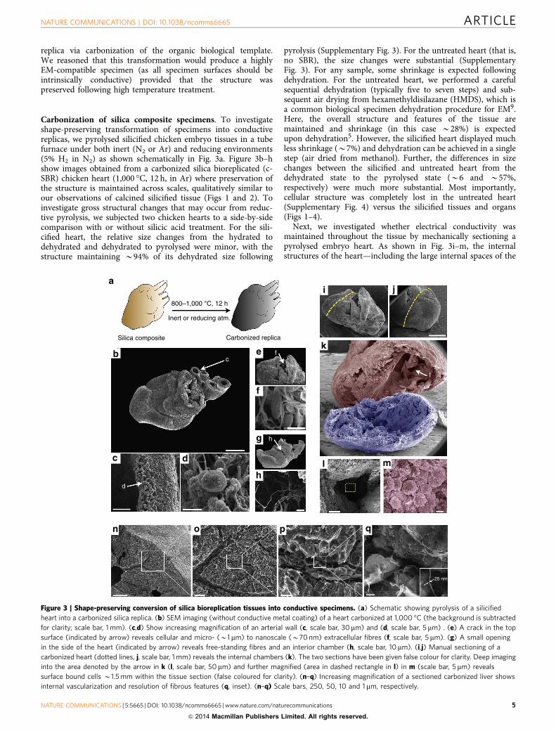
replica via carbonization of the organic biological template.We reasoned that this transformation would produce a highlyEM-compatible specimen (as all specimen surfaces should beintrinsically conductive) provided that the structure waspreserved following high temperature treatment.
Carbonization of silica composite specimens. To investigateshape-preserving transformation of specimens into conductivereplicas, we pyrolysed silicified chicken embryo tissues in a tubefurnace under both inert (N2 or Ar) and reducing environments(5% H2 in N2) as shown schematically in Fig. 3a. Figure 3b–hshow images obtained from a carbonized silica bioreplicated (c-SBR) chicken heart (1,000 �C, 12 h, in Ar) where preservation ofthe structure is maintained across scales, qualitatively similar toour observations of calcined silicified tissue (Figs 1 and 2). Toinvestigate gross structural changes that may occur from reduc-tive pyrolysis, we subjected two chicken hearts to a side-by-sidecomparison with or without silicic acid treatment. For the sili-cified heart, the relative size changes from the hydrated todehydrated and dehydrated to pyrolysed were minor, with thestructure maintaining B94% of its dehydrated size following
pyrolysis (Supplementary Fig. 3). For the untreated heart (that is,no SBR), the size changes were substantial (SupplementaryFig. 3). For any sample, some shrinkage is expected followingdehydration. For the untreated heart, we performed a carefulsequential dehydration (typically five to seven steps) and sub-sequent air drying from hexamethyldisilazane (HMDS), which isa common biological specimen dehydration procedure for EM9.Here, the overall structure and features of the tissue aremaintained and shrinkage (in this case B28%) is expectedupon dehydration5. However, the silicified heart displayed muchless shrinkage (B7%) and dehydration can be achieved in a singlestep (air dried from methanol). Further, the differences in sizechanges between the silicified and untreated heart from thedehydrated state to the pyrolysed state (B6 and B57%,respectively) were much more substantial. Most importantly,cellular structure was completely lost in the untreated heart(Supplementary Fig. 4) versus the silicified tissues and organs(Figs 1–4).
Next, we investigated whether electrical conductivity wasmaintained throughout the tissue by mechanically sectioning apyrolysed embryo heart. As shown in Fig. 3i–m, the internalstructures of the heart—including the large internal spaces of the
25 nm
h
f
I
c
d
800–1,000 °C, 12 h
Inert or reducing atm.
Carbonized replicaSilica composite
Figure 3 | Shape-preserving conversion of silica bioreplication tissues into conductive specimens. (a) Schematic showing pyrolysis of a silicified
heart into a carbonized silica replica. (b) SEM imaging (without conductive metal coating) of a heart carbonized at 1,000 �C (the background is subtracted
for clarity; scale bar, 1 mm). (c,d) Show increasing magnification of an arterial wall (c, scale bar, 30 mm) and (d, scale bar, 5 mm) . (e) A crack in the top
surface (indicated by arrow) reveals cellular and micro- (B1mm) to nanoscale (B70 nm) extracellular fibres (f, scale bar, 5mm). (g) A small opening
in the side of the heart (indicated by arrow) reveals free-standing fibres and an interior chamber (h, scale bar, 10mm). (i,j) Manual sectioning of a
carbonized heart (dotted lines, j, scale bar, 1 mm) reveals the internal chambers (k). The two sections have been given false colour for clarity. Deep imaging
into the area denoted by the arrow in k (l, scale bar, 50mm) and further magnified (area in dashed rectangle in l) in m (scale bar, 5 mm) reveals
surface bound cells B1.5 mm within the tissue section (false coloured for clarity). (n–q) Increasing magnification of a sectioned carbonized liver shows
internal vascularization and resolution of fibrous features (q, inset). (n–q) Scale bars, 250, 50, 10 and 1 mm, respectively.
NATURE COMMUNICATIONS | DOI: 10.1038/ncomms6665 ARTICLE
NATURE COMMUNICATIONS | 5:5665 | DOI: 10.1038/ncomms6665 | www.nature.com/naturecommunications 5
& 2014 Macmillan Publishers Limited. All rights reserved.

heart chambers—remained intact (that is, had not collapsed), andhigh resolution images of individual cells and surfaces could beacquired deep within the heart chamber (B1–2 mm). Impor-tantly, direct imaging of surfaces deep within tissues using EMpresents many challenges. Environmental SEM allows internalimaging of biological structures but is currently only amenable tovery thin samples of the order of single cells23. Otherwise, internalimaging generally requires serial sectioning of an embeddedspecimen followed by virtual reconstruction or, alternatively,careful dissection, preparation (fixation/dehydration) andmetallization of a specimen. With the latter, any furthersectioning would necessitate additional surface metallization.Here the intrinsically conductive internal surfaces combined withthe dynamic depth of field of an SEM enables imaging deepwithin internal cavities and allows biomolecular structures to bedirectly resolved within their 3D context and, if required,subsequent sectioning of the stabilized structure can beachieved manually, mechanically or by FIB without the needfor sputter coating or heavy metal staining. FIB/SEM may proveparticularly suitable for c-SBR specimens (Supplementary Fig. 5)as a means to shorten FIB processing time, which can take daysdue to the complexities of sample preparation, milling and imageprocessing10. To illustrate simple manual dissection of a c-SBRspecimen, Fig. 3n–q shows a sectioned liver with preservation offeatures down to B20–30 nm (Fig. 3q, inset). Here, the densespecimen appears to have been sectioned along intrinsic fractureplanes (for example, intercellular spaces) revealing a snapshot ofinternal surface topography, vascular hierarchy and cellular
organization that otherwise would be flattened usingmechanical methods of sectioning such as microtome or FIB.
Nanoparticle detection in the interior of an organ. The elec-trical conductivity of these specimens allows SEM interrogationusing high currents (10s of nA) and accelerating voltages (10–30 kV) that could otherwise damage even metal-coated samples(where metal coatings are typically B10–20 nm thick). This mayallow for chemical/elemental analysis using BSE imaging, whichrequires high current/kV for sufficient contrast. Consideringthe increasingly widespread interest in metal and other nano-particle materials for medicine24 and derivative studies25 (forexample, nanoparticle toxicology, biodistribution, tissue/particleinteractions), the ability to detect nanoparticles in deep tissue,particularly at low densities with single particle resolutionand within the intact 3D architecture of the tissue micro-environment, remains a challenge. Though BSE detection hasbeen occasionally applied to biological materials, examples haverequired specialized instrumentation (variable pressure/beamdeceleration26,27) or sample preparations (for example, singlecells grown on conductive substrates28) that are incompatiblewith normal tissue development.
Thus, we investigated whether SBR carbonization (c-SBR),combined with mechanical sectioning and BSE could detectintravenously injected nanoparticles within tissues. For thisexperiment, we synthesized 200 nm gold nanoparticles(AuNPs—stabilized with thiolated polyethylene glycol;
Interior
Interior
Exterior
(i)
(ii)(iii)
O
O
Si
Au
0.6 0.6
kev kev
1.5 1.52.4 2.4
Si
ee
fExterior
Figure 4 | Chemical fingerprinting of gold nanoparticles in the interior of a chicken liver. A mechanically fractured silica bioreplicated and carbonized
(c-SBR) chicken liver imaged using secondary electron (SE) (a, scale bar, 50mm) and backscattered electron detection (BSE) (b, scale bar, 50mm)
reveals gross morphology of the internal liver. The area within the dashed rectangle in a and b is expanded and imaged using SE (c, scale bar, 5 mm)
revealing a fenestrated sinusoid (i), space of disse (ii) and hepatocyte (iii). This same region is imaged using BSE to reveal single 200-nm diameter AuNPs
(d, scale bar, 5 mm; inset is a magnification of the centre bright spot). The spectrum in e was acquired from the centre particle (and representative
of spectra obtained from other points denoted as ‘e’) and the spectrum in f was acquired from the region ‘f’ denoted in d. (e,f) Show relative intensities
from single pixel acquisitions of 41,000 counts.
ARTICLE NATURE COMMUNICATIONS | DOI: 10.1038/ncomms6665
6 NATURE COMMUNICATIONS | 5:5665 | DOI: 10.1038/ncomms6665 | www.nature.com/naturecommunications
& 2014 Macmillan Publishers Limited. All rights reserved.

Supplementary Fig. 6) and injected them into a 16-day-oldchicken embryo. NPs were introduced by direct injection into avein of the chorioallantoic membrane and were allowed tocirculate for 1.5 h. Here, it was expected that AuNPs woulddeposit preferentially within the liver tissue soon after injectiondue to their relatively large size29. After harvesting and preparingthe liver (using c-SBR), the tissue was mechanically sectionedacross a large lobe to reveal the internal structure (Fig. 4).Figure 4a,b show lower magnification secondary electron (SE)and BSE images of the sectioned tissue that providecomplementary views; features such as microvilli andfenestrations are readily identified in SE mode while subsurfacestructures including cell nuclei are apparent using BSE detection.Focusing in and using a driving current of B20 nA at 10 kV, BSErevealed highly contrasted, individual AuNPs arrested on thewalls of the sinusoid endothelium (Fig. 4d and SupplementaryFig. 6). These particles could be chemically fingerprinted in situfrom the surrounding background using energy-dispersive X-rayspectroscopy as shown in Fig. 4e,f. BSE detection is essential asthe particles were indiscernible from the surrounding tissue usingsecondary electron (SE) detection (Fig. 4c). This indicates thatc-SBR procedures do not appear to detrimentally alter thephysical properties of AuNPs, and exemplifies the type ofproblem that is particularly well suited to be addressed usingthis approach. Depending on instrumentation, imagingconditions and sufficient Z-contrast with the carbonizedspecimen, particles that span the size ranges currentlyinvestigated for diagnostic and therapeutic applications (10sof nm to Bmicrons) should be detectable.
DiscussionThe simplicity of the technique, specimen stability, intrinsicconductivity post carbonization and level of resolution spanningsix orders of magnitude of magnification (that is, whole embryoto subcellular) distinguishes SBR from all previous bio-preserva-tion methods and should facilitate the examination of soft tissuesin their native 3D conformation that were previously difficult orimpossible to achieve. As an example, as BSEs are scarcer andemanate from a deeper interaction volume (B1–2 microns30) incomparison with SE, the sample subsurface could be resolvednon-destructively, revealing the architecture underlying a tissuesurface (Supplementary Fig. 7). Our ability to discover andimage nano-objects within a biological tissue/organism—findingessentially ‘a needle in a haystack’—while maintaining 3D contextis a new capability. While complete tissues and organs have beenpreserved, immunostained and optically imaged after stabilizationwithin hydrogels and refractive index matching31, further EMcharacterization of such hydrogel-stabilized samples requiredmultiple steps of solvent exchange, epoxy impregnation toprovide stability, staining to provide contrast and ultra-microtoming to achieve thin sections. Here stabilization ofcomplete organisms by ultra-thin conformal silica layers forms amechanically robust, refractory replica allowing transformation tocarbon, dissection and EM imaging ad infinitum at differentscales of magnification and with apparent generalizability to othersoft tissues derived from model organisms (for example, mouse;Supplementary Fig. 8). Although intracellular structures can beimaged (Supplementary Fig. 5), resolution of such featurescurrently does not approach methods that use sectioning (forexample, serial block-face); however, the size of samples that canbe used is only limited by the instrumentation implemented formaterial processing and imaging (for example, size of pyrolysisfurnace, chamber volume of SEM and so on). While detailedinterpretation of structures and structural accuracy with this newapproach require continuing efforts, our procedure to impart
shape-preserving, intrinsic conductivity across all specimenplanes could inform further design in instrumentation tooptimize the resolution of buried features, which may requirehigher energy fluxes and more sophisticated aberration correctionand deconvolution methods.
MethodsChicken embryo incubation and preparation. Ex ovo chicken embryo experi-ments were conducted under UNM protocol #10-100652-T-HSC, with all embryosused between day 3 and 17 (and as indicated in each experiment). All embryoswere handled and euthanized following approved UNM Institutional Animal Careand Use Committee (IACUC) procedures. Fertilized chicken eggs were obtainedfrom East Mountain Hatchery (Edgewood, NM) and placed in an automatedincubator (GQF 1500 professional, Savannah, GA) for 72–96 h, humidified (70%RH) and heated (37 �C). Following incubation, the egg shells were sterilized bybrief immersion in ethanol and physically cleaned with a paper towel. The eggshells were then scored using a rotary tool and cracked into a medium sterilizedweigh boat (VWR). Weigh boats were covered with a square plastic petri dish(VWR) and returned to the incubator until they were killed or time of injection.For particle injections, 0.1 ml of AuNPs (0.25 OD at l¼ 600 nm) were injected viaa pulled glass capillary needle into the vein of the chorioallantoic membrane andallowed to circulate for 90 min. Upon removal of embryos from the ex ovo egg,tissue was immersed in 3.7% paraformaldehyde in PBS for at least 24 h beforesilicification. For embryos at day 17 of development, individual organs were dis-sected from the chicken and fixed individually.
Silicification of specimens. Following fixation, tissues or whole embryos weresilicified by brief rinsing with PBS followed by subsequent immersion in silicifi-cation solution in a sealed container at 37 �C for 7–21 days. The silicificationsolution contains 0.1 M silicic acid derived from hydrolysis of tetramethyl ortho-silicate (TMOS) at pH 3 containing 0.154 M NaCl (0.9% saline solution). Forexample, to make a 100 ml solution, 0.1 ml of 1 N HCl is added to B98.5 ml of thesaline solution. Then, 1.5 ml of TMOS is added to this solution and stirred vig-orously (this can be accomplished by shaking in a sealed container) to hydrolysethe TMOS (it will appear dissolved upon hydrolysis) forming principally mono-silicic acid Si(OH)4. The approximate volume ratio of specimen to solution waskept at or below 1:20 as ratios exceeding 1:10 (specimen:silica solution; v/v) oftenwere observed to induce gelation of the solution (likely due to an increase insolution pH). No obvious difference in gross phenotype was apparent over thecourse of 3 weeks and gelation of solution (due to silica self-condensation)occurred only if the solution was not refreshed for over 3 weeks. Silica depositionupon specimens was apparent after a few days of immersion in the silicic acidsolution by a change in colour of the specimen from pink/brown to white.Following silicification, SBR tissues were rinsed in H2O (pH 3), 1:1 water/methanoland finally dried in air from 100% methanol.
Dehydration of non-silicified tissue. The non-silicified heart tissue shown inSupplementary Fig. 3 was fixed overnight in 3.7 volume% formaldehyde in PBSsolution, dehydrated by using 20-min sequential washes (33% ethanol (EtOH) inH2O; 50% EtOH; 66% EtOH; 2� 100% EtOH; 50% EtOH in HMDS; 100%HMDS) and allowed to dry in air for 16 h.
Calcination and pyrolysis of samples. Silicified samples were calcined by placingthem in a covered (but not air tight) pyrex dish and treating for 12–16 h in an oven(Fisher Scientific, Model #495A) at 500 �C under ambient atmospheric conditions.Ramp temperature was controlled at 1 �C per min, however, cooling rate wasuncontrolled.
For pyrolysis, silicified and non-silicified samples were placed uncovered in aceramic combustion boat (B20� 75 mm W� L alumina or porcelain) and heatedto either 800 or 1,000 �C in a quartz tube (25 mm OD; 20 mm ID) inserted in a tubefurnace (Lindberg/Blue Model #TF55035A) under constant gas flow (N2, Ar or 5%H2 in N2); the heating rate was 5 �C per min and final temperature was held for12 h. We found that an 800 �C holding temperature was sufficient for carbonizationof samples and higher temperatures were not required.
Scanning electron microscopy/energy-dispersive spectroscopy. SEM imageswere recorded using an FEI Quanta series SEM located at The University of NewMexico. This instrument was equipped with an energy-dispersive X-ray spectro-scopy from EDAX, which was used in single pixel mode for elemental identifica-tion. For SEM images shown in Fig. 1, samples were sputter-coated with Au/Pd. Inaddition, FIB milling shown in Supplementary Fig. 5 was performed on thisinstrument.
Synthesis of 200 nm gold nanoparticles. Gold nanoparticles (AuNPs) weresynthesized according to literature32. Briefly, 20 ml of HAuCl4 (0.1 mg ml� 1) wastitrated against trisodium citrate (10 mg ml� 1) at 90 �C. Two hundred nanometre
NATURE COMMUNICATIONS | DOI: 10.1038/ncomms6665 ARTICLE
NATURE COMMUNICATIONS | 5:5665 | DOI: 10.1038/ncomms6665 | www.nature.com/naturecommunications 7
& 2014 Macmillan Publishers Limited. All rights reserved.
particles are obtained with B0.35 ml of trisodium citrate. After heating, themixture was rapidly quenched in an ice bath, and the particles were washed withcentrifugation (500 r.c.f., 5 min). After resuspension in DI water, citrate cappingwas immediately exchanged by drop-wise addition of PEG-thiol in ethanol (MW5000, 2 mg ml� 1). The mixture was stirred for 24 h, and particles were purifiedagain by two cycles of centrifugation. The final product was dispersed in PBSatB0.25 OD (l¼ 600 nm).
References1. Bozzola, J. J. & Russell, L. D. Electron Microscopy: Principles and Techniques for
Biologists 2nd edn (Jones and Bartlett, 1999).2. Hayat, M. A. Principles and Techniques of Electron Microscopy: Biological
Applications (Cambridge Univ. Press, 2000).3. Dahmen, U. et al. Background, status and future of the transmission electron
aberration-corrected microscope project. Philos. Trans. R Soc. 367, 3795–3808(2009).
4. Browning, N. et al. Recent developments in dynamic transmission electronmicroscopy. Curr. Opin. Solid State Mater. Sci. 16, 23–30 (2012).
5. Kirk, S., Skepper, J. & Donald, A. Application of environmental scanningelectron microscopy to determine biological surface structure. J. Microsc. 233,205–224 (2009).
6. McIntosh, R., Nicastro, D. & Mastronarde, D. New views of cells in 3D: anintroduction to electron tomography. Trends Cell Biol. 15, 43–51 (2005).
7. Denk, W. & Horstmann, H. Serial block-face scanning electron microscopy toreconstruct three-dimensional tissue nanostructure. PLoS Biol. 2, 1900–1909(2004).
8. Severs, N. J. Freeze-fracture electron microscopy. Nat. Protoc. 2, 547–576(2007).
9. Bray, D. F., Bagu, J. & Koegler, P. Comparison of hexamethyldisilazane(HMDS), Peldri II, and critical-point drying methods for scanning electronmicroscopy of biological specimens. Microsc. Res. Tech. 26, 489–495 (1993).
10. Bushby, A. J. et al. Imaging three-dimensional tissue architectures by focusedion beam scanning electron microscopy. Nat. Protoc. 6, 845–858 (2011).
11. Kemmenoe, B. H. & Bullock, G. R. Structure-analysis of sputter-coated and ion-beam sputter-coated films: a comparative-study. J. Microsc. 132, 153–163 (1983).
12. Black, R. M. The Elements of Palaeontology (Cambridge Univ. Press, 1988).13. Miyako, E. et al. Self-assembled carbon nanotube honeycomb networks using a
butterfly wing template as a multifunctional nanobiohybrid. ACS Nano 7,8736–8742 (2013).
14. Goodwin, W. B., Gomez, I. J., Meredith, C. & Sandhage, K. H. Conversion ofpollen particles into three-dimensional ceramic replicas tailored for multimodaladhesion. Chem. Mater. 25, 4529–4536 (2013).
15. Zimmerman, A. B., Nelson, A. M. & Gillan, E. G. Titania and silica materialsderived from chemically dehydrated porous botanical templates. Chem. Mater.24, 4301–4310 (2012).
16. Van Opdenbosch, D., Fritz-Popovski, G., Paris, O. & Zollfrank, C. Silicareplication of the hierarchical structure of wood with nanometer precision.J. Mater. Res. 26, 1193–1202 (2011).
17. Paris, O., Burgert, I. & Fratzl, P. Biomimetics and biotemplating of naturalmaterials. MRS Bull. 35, 219–225 (2010).
18. Losic, D., Mitchell, J. G. & Voelcker, N. H. Diatomaceous lessons innanotechnology and advanced materials. Adv. Mater. 21, 2947–2958 (2009).
19. Kaehr, B. et al. Cellular complexity captured in durable silica biocomposites.Proc. Natl Acad. Sci. USA 109, 17336–17341 (2012).
20. Le Douarin, N. M. The avian embryo as a model to study the development of theneural crest: a long and still ongoing story. Mech. Dev. 121, 1089–1102 (2004).
21. Leong, H. S. et al. Intravital imaging of embryonic and tumor neovasculatureusing viral nanoparticles. Nat. Protoc. 5, 1406–1417 (2010).
22. Khripin, C. Y., Pristinski, D., Dunphy, D. R., Brinker, C. J. & Kaehr, B.Protein-directed assembly of arbitrary three-dimensional nanoporous silicaarchitectures. ACS Nano 5, 1401–1409 (2011).
23. de Jonge, N. & Ross, F. M. Electron microscopy of specimens in liquid. Nat.Nanotechnol. 6, 695–704 (2011).
24. Zhang, L. et al. Nanoparticles in medicine: therapeutic applications anddevelopments. Clin. Pharmacol. Ther. 83, 761–769 (2008).
25. Oberdorster, G., Stone, V. & Donaldson, K. Toxicology of nanoparticles: ahistorical perspective. Nanotoxicology 1, 2–25 (2007).
26. Ohta, K. et al. Beam deceleration for block-face scanning electron microscopyof embedded biological tissue. Micron 43, 612–620 (2012).
27. Ushiki, T. et al. Low-voltage backscattered electron imaging of non-coatedbiological samples in a low-vacuum environment using a variable-pressurescanning electron microscope with a YAG-detector. J. Electron Microsc. 47,351–354 (1998).
28. Pluk, H., Stokes, D., Lich, B., Wieringa, B. & Fransen, J. Advantages of indium–tin oxide-coated glass slides in correlative scanning electron microscopyapplications of uncoated cultured cells. J. Microsc. 233, 353–363 (2009).
29. Lipka, J. et al. Biodistribution of PEG-modified gold nanoparticles followingintratracheal instillation and intravenous injection. Biomaterials 31, 6574–6581(2010).
30. Brott, L. L. et al. Ultrafast holographic nanopatterning of biocatalyticallyformed silica. Nature 413, 291–293 (2001).
31. Chung, K. et al. Structural and molecular interrogation of intact biologicalsystems. Nature 497, 332–337 (2013).
32. Frens, G. Controlled nucleation for the regulation of the particle size inmonodisperse gold suspensions. Nature 241, 20–22 (1973).
AcknowledgementsThis work was supported by the U.S. Department of Energy, Office of Science, BasicEnergy Sciences, Materials Sciences and Engineering Division. J.L.T. acknowledgessupport from the Air Force Office of Scientific Research under grant #FA9550-14-1-0066.Y.-S.L. was supported by a fellowship from the New Mexico Cancer NanotechnologyTraining Center (R25CA153825). Y.H.A. acknowledges support from the NSF INSPIREprogram (CBET-1344298). Sandia National Laboratories is a multi-program laboratorymanaged and operated by Sandia Corporation, a wholly owned subsidiary of LockheedMartin Corporation, for the National Nuclear Security Administration of the U.S.Department of Energy under Contract No. DE-AC04-94AL85000.
Author contributionsJ.L.T. and B.K. conceived the study; J.L.T., C.J.B. and B.K. designed the research; J.L.T.,Y.-S.L., S.S.C., Y.H.A., E.N.C. and B.K. carried out the experiments; J.L.T., C.J.B. and B.K.analysed the data; J.L.T. and B.K. wrote the paper; all the authors commented on andedited the final version of the paper.
Additional informationSupplementary Information accompanies this paper at http://www.nature.com/naturecommunications
Competing financial interests: The authors declare no competing financial interests.
Reprints and permission information is available online at http://npg.nature.com/reprintsandpermissions/
How to cite this article: Townson, J. L. et al. Synthetic fossilization of soft biologicaltissues and their shape-preserving transformation into silica or electron-conductivereplicas. Nat. Commun. 5:5665 doi: 10.1038/ncomms6665 (2014).
This work is licensed under a Creative Commons Attribution 4.0International License. The images or other third party material in this
article are included in the article’s Creative Commons license, unless indicated otherwisein the credit line; if the material is not included under the Creative Commons license,users will need to obtain permission from the license holder to reproduce the material.To view a copy of this license, visit http://creativecommons.org/licenses/by/4.0/
ARTICLE NATURE COMMUNICATIONS | DOI: 10.1038/ncomms6665
8 NATURE COMMUNICATIONS | 5:5665 | DOI: 10.1038/ncomms6665 | www.nature.com/naturecommunications
& 2014 Macmillan Publishers Limited. All rights reserved.