Embed Size (px)
Citation preview
SYSMEX EDUCATIONAL ENHANCEMENT AND DEVELOPMENT | NOVEMBER 2016
SEED HAEMATOLOGY
The blast cell – a diagnostic heavyweight
Causes and cytological manifestations
Blast cells are described as precursor cells with the ability to
preserve themselves by dividing and to further differentiate.
Under pathological conditions, blast cells can be mobilised
from the bone marrow into the peripheral blood circulation.
In adults, this represents an alarming finding that can indicate
both reactive and malignant diseases such as leukaemia.
Therefore the detection of blast cells in the peripheral blood
is considered extremely important, and great responsibility
is placed on the investigating laboratory. As well as informa-
tion on the physiology, this article describes the possible
causes of the release of blast cells into the blood, the char-
acteristics by which they can be identified and how further
diagnosis is carried out.
Development, maturation and regulation
Haematopoietic precursor cells develop from the pluripotent
embryonic stem cells as a result of numerous development
stages. In the bone marrow, these cells are referred to as blast
cells (‘blastós’ is the Greek word for germ, bud, sprout or
shoot). For their further development, they are committed to
one specific line (erythropoiesis, granulopoiesis, monopoiesis,
thrombopoiesis and lymphopoiesis). Asymmetrical replication,
as shown in Fig. 1, allows blast cells to form both identical
daughter cells (replication) and to differentiate to form
mature blood cells.
A mature cell is developed after several differentiation
stages, involving gradual condensation of the nuclear chro-
matin. While blast cells have a homogeneous chromatin, the
nucleus shows chromatin clumping in the mature cells. The
nucleus-plasma relation also drops (see Fig. 2)
Fig. 1 Asymmetrical replication (example erythropoiesis)
Fig. 2 Schematic illustration: blast cell / mature cell
Blast cellNuclear chromatin fine, homogeneous Nucleus-plasma relation 70 – 95 %
Mature cellNuclear chromatin clumpedNucleus-plasma relation 30 – 50 %
Maturation
2SEED HAEMATOLOGY – The blast cell – a diagnostic heavyweight Sysmex Educational Enhancement and Development | November 2016
With leukaemia, blast cells can have a significantly changed
appearance. What all blast cells have in common is the
finely and evenly distributed, light nuclear chromatin.
Blast cells in the peripheral blood, causes and
cytological appearance
A shift of blast cells into the peripheral blood occurs physio-
logically only in neonates. The reason for this is the extra-
medullary haematopoiesis that still exists at the time of
birth. In adults, the presence of blast cells in the peripheral
blood is a serious finding. Essentially, it is important to dif-
ferentiate between a reactive and a leukaemic appearance
of blast cells. Examples are shown in Table 2. The extent of
the influx of blast cells can be used to differentiate between
a reactive and a malignant picture, as can the composition
of the other cell populations. Blood count values and clinical
data are also helpful.
Bone marrow barrier
Blast cells and other immature cells of haematopoiesis are
captured in the bone marrow due to their size and adhesion
properties, and do not enter the blood stream. This mecha-
nism is referred to as the ‘bone marrow barrier’. A disrup-
tion of this bone marrow barrier is associated with a leuco-
erythroblastic blood picture.
Physiological blast cells
These are medium to large cells (14 – 18 μm*) with the
specific characteristics of the nucleus and cytoplasm
described in Table 1. Table 2 Diagnostic reasons for the presence of blast cells in the peripheral blood
Reactive Malignant
Severe bacterial infections, sepsis
Acute leukaemias(AML, ALL, AUL)
Treatment with growth factors (G-CSF)
Myeloproliferative neoplasia (CML, PMF)
Regeneration afterchemotherapy
Myelodysplastic syndromes (RAEB 1 and 2)
Viral infections (mononucleosis) MDS/MPN overlap syndrome (CMML 1 and 2)
Bone marrow carcinosis Aggressive lymphoma of theB cell and T cell lines
Table 1 Diagnostic characteristics of physiological blast cells
Nucleus Cytoplasm
Shape: round/oval Narrow(constituting 5 – 30 % of the cell)
Nucleus-plasma relation: 70 – 95 %
Basophilic
Nucleoli: one to several (may not be visible)
Not granulated **
Finely distributed nucleic chromatin, no clumping
* Exception: megakaryoblast 150 μm ** Leukaemic blast cells may be granulated
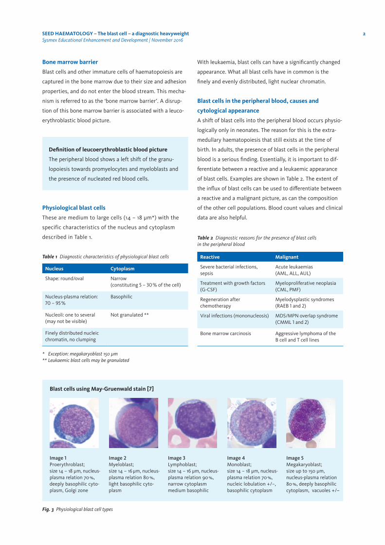
Fig. 3 Physiological blast cell types
Image 1 Proerythroblast; size 14 – 18 μm, nucleus-plasma relation 70 %, deeply basophilic cyto-plasm, Golgi zone
Image 3 Lymphoblast; size 14 – 16 μm, nucleus-plasma relation 90 %, narrow cytoplasm medium basophilic
Image 2 Myeloblast; size 14 – 16 μm, nucleus-plasma relation 80 %, light basophilic cyto-plasm
Image 4 Monoblast; size 14 – 18 μm, nucleus-plasma relation 70 %, nucleic lobulation +/–, basophilic cytoplasm
Image 5 Megakaryoblast; size up to 150 μm, nucleus-plasma relation 80 %, deeply basophilic cytoplasm, vacuoles +/–
Definition of leucoerythroblastic blood picture
The peripheral blood shows a left shift of the granu-
lopoiesis towards promyelocytes and myeloblasts and
the presence of nucleated red blood cells.
Blast cells using May-Gruenwald stain [7]
3SEED HAEMATOLOGY – The blast cell – a diagnostic heavyweight Sysmex Educational Enhancement and Development | November 2016
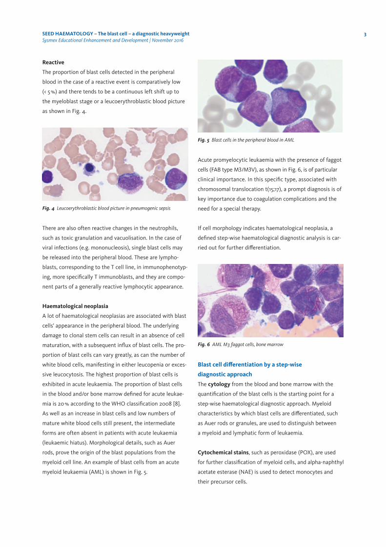
Reactive
The proportion of blast cells detected in the peripheral
blood in the case of a reactive event is comparatively low
(< 5 %) and there tends to be a continuous left shift up to
the myeloblast stage or a leucoerythroblastic blood picture
as shown in Fig. 4.
There are also often reactive changes in the neutrophils,
such as toxic granulation and vacuolisation. In the case of
viral infections (e.g. mononucleosis), single blast cells may
be released into the peripheral blood. These are lympho-
blasts, corresponding to the T cell line, in immunophenotyp-
ing, more specifically T immunoblasts, and they are compo-
nent parts of a generally reactive lymphocytic appearance.
Haematological neoplasia
A lot of haematological neoplasias are associated with blast
cells’ appearance in the peripheral blood. The underlying
damage to clonal stem cells can result in an absence of cell
maturation, with a subsequent influx of blast cells. The pro-
portion of blast cells can vary greatly, as can the number of
white blood cells, manifesting in either leucopenia or exces-
sive leucocytosis. The highest proportion of blast cells is
exhibited in acute leukaemia. The proportion of blast cells
in the blood and/or bone marrow defined for acute leukae-
mia is 20 % according to the WHO classification 2008 [8].
As well as an increase in blast cells and low numbers of
mature white blood cells still present, the intermediate
forms are often absent in patients with acute leukaemia
(leukaemic hiatus). Morphological details, such as Auer
rods, prove the origin of the blast populations from the
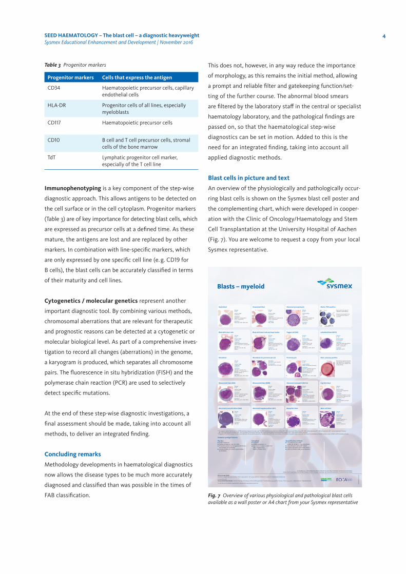
myeloid cell line. An example of blast cells from an acute
myeloid leukaemia (AML) is shown in Fig. 5.
Acute promyelocytic leukaemia with the presence of faggot
cells (FAB type M3/M3V), as shown in Fig. 6, is of particular
clinical importance. In this specific type, associated with
chromosomal translocation t(15;17), a prompt diagnosis is of
key importance due to coagulation complications and the
need for a special therapy.
If cell morphology indicates haematological neoplasia, a
defined step-wise haematological diagnostic analysis is car-
ried out for further differentiation.
Blast cell differentiation by a step-wise
diagnostic approach
The cytology from the blood and bone marrow with the
quantification of the blast cells is the starting point for a
step-wise haematological diagnostic approach. Myeloid
characteristics by which blast cells are differentiated, such
as Auer rods or granules, are used to distinguish between
a myeloid and lymphatic form of leukaemia.
Cytochemical stains, such as peroxidase (POX), are used
for further classification of myeloid cells, and alpha-naphthyl
acetate esterase (NAE) is used to detect monocytes and
their precursor cells.
Fig. 4 Leucoerythroblastic blood picture in pneumogenic sepsis
Fig. 5 Blast cells in the peripheral blood in AML
Fig. 6 AML M3 faggot cells, bone marrow
4SEED HAEMATOLOGY – The blast cell – a diagnostic heavyweight Sysmex Educational Enhancement and Development | November 2016
Immunophenotyping is a key component of the step-wise
diagnostic approach. This allows antigens to be detected on
the cell surface or in the cell cytoplasm. Progenitor markers
(Table 3) are of key importance for detecting blast cells, which
are expressed as precursor cells at a defined time. As these
mature, the antigens are lost and are replaced by other
markers. In combination with line-specific markers, which
are only expressed by one specific cell line (e. g. CD19 for
B cells), the blast cells can be accurately classified in terms
of their maturity and cell lines.
Cytogenetics / molecular genetics represent another
important diagnostic tool. By combining various methods,
chromosomal aberrations that are relevant for therapeutic
and prognostic reasons can be detected at a cytogenetic or
molecular biological level. As part of a comprehensive inves-
tigation to record all changes (aberrations) in the genome,
a karyogram is produced, which separates all chromosome
pairs. The fluorescence in situ hybridization (FISH) and the
polymerase chain reaction (PCR) are used to selectively
detect specific mutations.
At the end of these step-wise diagnostic investigations, a
final assessment should be made, taking into account all
methods, to deliver an integrated finding.
Concluding remarks
Methodology developments in haematological diagnostics
now allows the disease types to be much more accurately
diagnosed and classified than was possible in the times of
FAB classification.
This does not, however, in any way reduce the importance
of morphology, as this remains the initial method, allowing
a prompt and reliable filter and gatekeeping function/set-
ting of the further course. The abnormal blood smears
are filtered by the laboratory staff in the central or specialist
haematology laboratory, and the pathological findings are
passed on, so that the haematological step-wise
diagnostics can be set in motion. Added to this is the
need for an integrated finding, taking into account all
applied diagnostic methods.
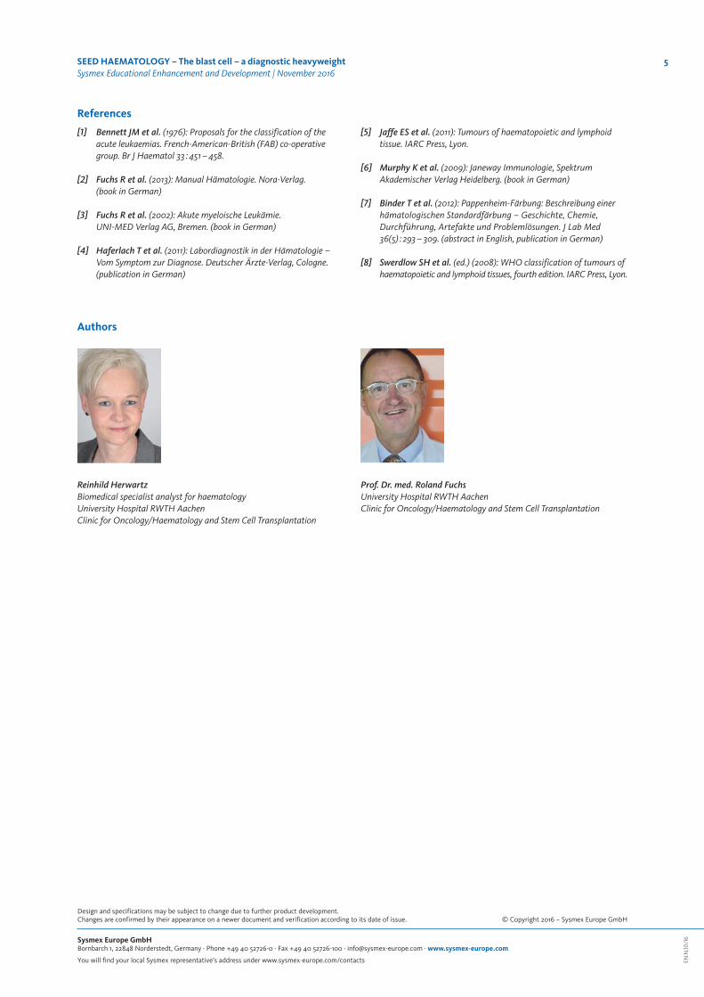
Blast cells in picture and text
An overview of the physiologically and pathologically occur-
ring blast cells is shown on the Sysmex blast cell poster and
the complementing chart, which were developed in cooper-
ation with the Clinic of Oncology/Haematology and Stem
Cell Transplantation at the University Hospital of Aachen
(Fig. 7). You are welcome to request a copy from your local
Sysmex representative.
Table 3 Progenitor markers
Progenitor markers Cells that express the antigen
CD34 Haematopoietic precursor cells, capillary endothelial cells
HLA-DR Progenitor cells of all lines, especially myeloblasts
CD117 Haematopoietic precursor cells
CD10 B cell and T cell precursor cells, stromal cells of the bone marrow
TdT Lymphatic progenitor cell marker, especially of the T cell line
Blasts – myeloid
Myeloblast
Size µm12–16
Nucleus shape round/oval
Cytoplasm basophilic, no granulation, (POX +, esterase Ø)
Incidence*physiologic, AML, MDS, CML
Abnormal promyelocyte
Size µm16–20
Nucleus shape oval
Cytoplasm dense, coarse, purple-red granulation, (POX ++, esterase Ø)
Incidence*AML-M3
Granulated blast
Size µm12–16
Nucleus shaperound/oval
Cytoplasm reddish granules, Golgi zone Ø, (POX +, esterase Ø)
Incidence*AML, RAEB
Blasts, POX-positive
Blasts with brown dye pre-cipitates of varied density.
Remark: All neutrophils from the promyelocyte stage on are POX +.
Blast with Auer rods
Size µm14–16
Nucleus shape round/oval
Cytoplasm needle-shaped red inclusions (Auer rods), (POX +, esterase Ø)
Incidence*AML-M1, -M2, -M3, -M6, RAEB-2
Faggot cell (M3)
Size µm14–18
Nucleus shape round/oval/lobulated
Cytoplasm bundles of Auer rods, (POX +, esterase Ø)
Incidence*AML-M3 and -M3V, t(15;17)
Blast with Auer rods and Auer bodies
Size µm14–16
Nucleus shape round/oval
Cytoplasm spherical red inclusions (Auer bodies), additionally Auer rods, (POX +, esterase Ø)
Incidence*AML-M1, -M2, -M6
Lobulated blast (M3V)
Size µm16–20
Nucleus shape (bi-)lobulated
Cytoplasm often fine reddish granulation, bundles of Auer rods +/–, (POX +, esterase Ø)
Incidence*AML-M3V, t(15;17)
Monoblast
Size µm14 –18
Nucleus shape round/oval,nucleoli +/–
Cytoplasm abundant cytoplasm, fine granules +/–, pseudopodia +/–, (POX Ø, esterase +)
Incidence*AML-M5A and -B, -M4, CMML
Promonocyte
Size µm14–18
Nucleus shape lobed, intermediate chromatin
Cytoplasm abundant cytoplasm, granula-tion +/–, often vacuoles, haemophagocytosis +/–, (POX Ø, esterase +)
Incidence*AML-M5A and -B, -M4, CMML
Monoblast (1), promonocyte (2)
Monoblast description: see to the left
Promonocyte description: see to the right
Blast, esterase positive
Blast with precipitates of brown dye; diagnostic for monoblastic AML only in case of diffuse and strong staining.
Monocytoid blast (M4)
Size µm16–18
Nucleus shape monocytoid
Cytoplasm abundant cytoplasm, granula-tion +/–, often vacuoles, (POX Ø, esterase +)
Incidence*AML-M5A and -B, -M4, CMML
Abnormal eosinophil (M4 Eo)
Size µm14–16
Nucleus shape round/ovalintermediate chromatin
Cytoplasm coarse, round, eosinophilic and blue-purple granules, (chloroacetate esterase +)
Incidence*AML-M4 Eo, inv(16), t(16;16)
Monocytoid blast (M5B)
Size µm16–18
Nucleus shape monocytoid
Cytoplasm abundant cytoplasm, granula-tion +/–, often vacuoles, (POX Ø, esterase +)
Incidence*AML-M5A and -B, -M4, CMML
Cup-like blast
Size µm14–16
Nucleus shape Nucleus with invaginated cytoplasm**
Cytoplasm Granulated cytoplasm. Crucial is the nucleus: fingerprint-like indentation; POX +
Incidence* AML with NPM1- and FLT3-mutations
Abnormal proerythroblast (M6)
Size µm14–16
Nucleus shape round/oval
Cytoplasm deep basophilic, flaky Golgi zone, (POX Ø, esterase Ø)
Incidence*AML-M6
Abnormal megakaryoblast (M7)
Size µm12–18
Nucleus shape round/oval
Cytoplasm undifferentiated blast, no granulation, cytoplasmic blebbing or pseudopodia, (POX Ø, esterase Ø)
Incidence*AML-M7
Mast cell blast
Size µm14–16
Nucleus shape often blurred
Cytoplasm basophilic granules +/–, (toluidine blue +)
Incidence*mast cell leukaemia
Basophilic blast
Size µm14 –16
Nucleus shape often blurred
Cytoplasm blue-purple granulation, often vacuoles, (toluidine blue +)
Incidence*acute basophilic leukaemia
ZE00
1199.EN.N.03/14
Common cytologic features
Nucleus ■ Shape: round/oval■ Nuclear-cytoplasmic ratio: 70–90%■ Chromatin: predominantly regularly distributed, not clumped, not condensed■ Varying numbers of nucleoli; may be hidden by chromatin
Cytoplasm ■ Basophilic■ Reddish granulation +/–■ Auer rods +/–; when +, evident for: AML, if blasts ≥20% RAEB-2, if blasts <20%
Quantifi cation of blasts■ <1% in PB and <5% in BM: in MDS: RA, RCMD +/– ring sideroblasts■ <5% in PB and 5–9% in BM: RAEB-1■ 5–19% in PB and 10–19% in BM: RAEB-2■ ≥20% in PB and/or BM: acute leukaemia
Sysmex Europe GmbHBornbarch 1, 22848 Norderstedt, Germany · Phone +49 40 52726-0 · Fax +49 40 52726-100 · [email protected] · www.sysmex-europe.com
© Copyright 2014 – Universitätsklinikum Aachen AÖR, Klinik für Onkologie, Hämatologie und StammzelltransplantationAuthors: Prof. Dr. med. Roland Fuchs, Reinhild Herwartz, Klinik für Onkologie, Hämatologie und Stammzelltransplantation, Universitätsklinikum Aachen
In cooperation with:
You will fi nd your local Sysmex representative’s address under www.sysmex-europe.com
Universitätsklinikum Aachen Klinik für Onkologie, Hämatologie und Stammzelltransplantation · Pauwelsstraße 30, 52074 Aachen, Germany · Phone +49 241 80-0 · [email protected] · www.ukaachen.de
2
1
Platelet phagocytosis
* For pragmatic reasons, the abbreviations of FAB classifi cation diagnoses have been used. The WHO classifi cation equivalents are as follows: M0 – AML with minimal diff erentiation; M1 – AML without maturation; M2 – AML with maturation; M3 – acute promyelocytic leukaemia; M4 – acute myelomonocytic leukaemia; M5 – acute monoblastic and monocytic leukaemia; M6 – acute erythroid leukaemia/proerythroblastic leukaemia; M7 – acute megakaryoblastic leukaemia.
** Invagination of the POX + cytoplasm into the nucleus. Defi nition of the cup-like blast population: indentation zone ≥25% of the nuclear surface, ≥10% of blasts show goblet-shaped, usually light indentations. If cup-like blasts are identifi ed, mutation analysis of NMP1 and FLT3 should be performed.
SYSME_13002_Poster_Blasts_myeloid_EN_A1_RZ.indd 1 11.03.14 13:55
Fig. 7 Overview of various physiological and pathological blast cells available as a wall poster or A4 chart from your Sysmex representative
5SEED HAEMATOLOGY – The blast cell – a diagnostic heavyweight Sysmex Educational Enhancement and Development | November 2016
References
[1] Bennett JM et al. (1976): Proposals for the classification of the acute leukaemias. French-American-British (FAB) co-operative group. Br J Haematol 33 : 451 – 458.
[2] Fuchs R et al. (2013): Manual Hämatologie. Nora-Verlag. (book in German)
[3] Fuchs R et al. (2002): Akute myeloische Leukämie. UNI-MED Verlag AG, Bremen. (book in German)
[4] Haferlach T et al. (2011): Labordiagnostik in der Hämatologie – Vom Symptom zur Diagnose. Deutscher Ärzte-Verlag, Cologne. (publication in German)
[5] Jaffe ES et al. (2011): Tumours of haematopoietic and lymphoid tissue. IARC Press, Lyon.
[6] Murphy K et al. (2009): Janeway Immunologie, Spektrum Akademischer Verlag Heidelberg. (book in German)
[7] Binder T et al. (2012): Pappenheim-Färbung: Beschreibung einer hämatologischen Standardfärbung – Geschichte, Chemie, Durchführung, Artefakte und Problemlösungen. J Lab Med 36(5) : 293 – 309. (abstract in English, publication in German)
[8] Swerdlow SH et al. (ed.) (2008): WHO classification of tumours of haematopoietic and lymphoid tissues, fourth edition. IARC Press, Lyon.
EN.N
.10/1
6Sysmex Europe GmbHBornbarch 1, 22848 Norderstedt, Germany · Phone +49 40 52726-0 · Fax +49 40 52726-100 · [email protected] · www.sysmex-europe.com
You will find your local Sysmex representative’s address under www.sysmex-europe.com/contacts
Design and specifications may be subject to change due to further product development.Changes are confirmed by their appearance on a newer document and verification according to its date of issue. © Copyright 2016 – Sysmex Europe GmbH
Authors
Reinhild Herwartz Biomedical specialist analyst for haematology University Hospital RWTH Aachen Clinic for Oncology/Haematology and Stem Cell Transplantation
Prof. Dr. med. Roland Fuchs University Hospital RWTH Aachen Clinic for Oncology/Haematology and Stem Cell Transplantation