Embed Size (px)
Citation preview
antioxidants
Article
Systematic Development and Characterization of Novel, HighDrug-Loaded, Photostable, Curcumin Solid Lipid NanoparticleHydrogel for Wound Healing
Simarjot Kaur Sandhu 1, Suneel Kumar 2 , Jayant Raut 1, Mandeep Singh 1, Sandeep Kaur 1, Garima Sharma 1,Tomas L. Roldan 3,4 , Sonia Trehan 5, Jennifer Holloway 3,4, Gabriella Wahler 4,6, Jeffrey D. Laskin 4,6,Patrick J. Sinko 3,4 , Francois Berthiaume 2, Bozena Michniak-Kohn 3,5, Praveen Rishi 7, Narayanan Ganesh 8
and Indu Pal Kaur 1,*
�����������������
Citation: Sandhu, S.K.; Kumar, S.;
Raut, J.; Singh, M.; Kaur, S.; Sharma,
G.; Roldan, T.L.; Trehan, S.; Holloway,
J.; Wahler, G.; et al. Systematic
Development and Characterization of
Novel, High Drug-Loaded,
Photostable, Curcumin Solid Lipid
Nanoparticle Hydrogel for Wound
Healing. Antioxidants 2021, 10, 725.
https://doi.org/10.3390/
antiox10050725
Academic Editors: Maria
Cristina Albertini and
Seeram Ramakrishna
Received: 31 March 2021
Accepted: 26 April 2021
Published: 5 May 2021
Publisher’s Note: MDPI stays neutral
with regard to jurisdictional claims in
published maps and institutional affil-
iations.
Copyright: © 2021 by the authors.
Licensee MDPI, Basel, Switzerland.
This article is an open access article
distributed under the terms and
conditions of the Creative Commons
Attribution (CC BY) license (https://
creativecommons.org/licenses/by/
4.0/).
1 Institute of Pharmaceutical Sciences, Panjab University, Chandigarh 160014, India;[email protected] (S.K.S.); [email protected] (J.R.); [email protected] (M.S.);[email protected] (S.K.); [email protected] (G.S.)
2 Department of Biomedical Engineering, Rutgers University, Piscataway, NJ 08854, USA;[email protected] (S.K.); [email protected] (F.B.)
3 Department of Pharmaceutics, Ernest Mario School of Pharmacy, Rutgers University,Piscataway, NJ 08854, USA; [email protected] (T.L.R.); [email protected] (J.H.);[email protected] (P.J.S.); [email protected] (B.M.-K.)
4 Counter ACT Center of Excellence, Rutgers University, Piscataway, NJ 08854, USA;[email protected] (G.W.); [email protected] (J.D.L.)
5 Center for Dermal Research (CDR), Life Sciences Building, Rutgers University, Piscataway, NJ 08854, USA;[email protected]
6 Department of Pharmacology and Toxicology, Rutgers University, Piscataway, NJ 08854, USA7 Department of Microbiology, Panjab University, Chandigarh 160014, India; [email protected] Jawaharlal Nehru Cancer Hospital & Research Centre, Bhopal 462001, India; [email protected]* Correspondence: [email protected]; Tel.: +91-9855166432
Abstract: The study aims to develop high drug-loaded (about 15% lipid matrix) curcumin solid lipidnanoparticles (CSLNs) for wound healing. CSLNs prepared by hot, high-pressure homogenization,without using organic solvents, were optimized using the Taguchi design followed by the centralcomposite design. The optimized CSLNs exhibited a high assay/drug content (0.6% w/w), solubility(6 × 10⁵ times), and EE (75%) with a particle size < 200 nm (PDI—0.143). The CSLNs were safe(in vitro and in vivo), photostable, autoclavable, stable up to one year at 30 ◦C and under refrigerationand exhibited a controlled release (zero-order; 5 days). XRD, FTIR, and DSC confirmed solubilizationand entrapment of the curcumin within the SLNs. TEM and FESEM revealed a smooth and sphericalshape. The CSLNs showed a significant antimicrobial effect (MIC of 64 µg/mL for planktoniccells; 512 µg/mL for biofilm formation; and 2 mg/mL for mature biofilm) against Staphylococcusaureus 9144, while free curcumin dispersion did not exhibit any effect. This is the first reporton the disruption of mature biofilms by curcumin solid lipid nanoparticles (CSLNs). The cellproliferation potential of CSLNs was also evaluated in vitro while the wound healing potentialof CSLNs (incorporated in a hydrogel) was assessed in vivo. In (i) nitrogen mustard gas and(ii) a full-thickness excision wound model, CSLNs exhibited (a) significantly faster wound closure,(b) histologically and immunohistochemically better healing, (c) lower oxidative stress (LPO) and(d) inflammation (TNFα), and (e) increased angiogenesis (VEGF) and antioxidant enzymes, i.e.,catalase and GSH levels. CSLNs thus offer a promising modern wound therapy especially forinfected wounds, considering their effects in mature biofilm disruption.
Keywords: nanocarriers; safety; biofilm; wound closure; oxidative stress; TNFα; VEGF
Antioxidants 2021, 10, 725. https://doi.org/10.3390/antiox10050725 https://www.mdpi.com/journal/antioxidants
Antioxidants 2021, 10, 725 2 of 29
1. Introduction
Wound healing is a complex biological process composed of interrelated and overlap-ping phases of homeostasis, inflammation, migration, proliferation, and maturation [1]. Thewound bed provides an ideal environment for microbial growth, facilitating the penetrationof pathogens into the underlying tissue, with a potential for hematogenous dissemination.Further, inflammation followed by oxidative stress, mediated by the formation of freeradicals, leads to DNA breakage, lipid peroxidation and enzyme inactivation resultingin impaired wound healing. Currently employed antimicrobial agents possess limitedutility due to toxicity, narrow antimicrobial coverage, inadequate wound bed penetration,and growing bacterial resistance [2–4]. In addition, mainline treatments such as silversulfadiazine may delay burn wound healing as it is toxic to regenerating keratinocytes [5].
An optimum wound healing dressing or agent should thus (i) reduce inflammation,(ii) protect the wound tissue from bacterial infection, and (iii) induce cell proliferationto aid in the reconstruction of damaged tissue [6]. It should ideally also act as (iv) anantioxidant, as free radicals are considered as the major cause of inflammation duringthe wound healing process [7]. Curcumin, chemically known as diferuloylmethane or1,7-bis(4-hydroxy-3-methoxyphenyl)-1,6-heptadiene-3,5-dione, is a low-molecular-weightpolyphenolic phytoconstituent obtained from the powdered rhizome of Curcuma longa L. Ithas been used as an oral supplement in various medical and cutaneous conditions [8]. Thewound healing potential of curcumin is attributed to its multi-pharmacological effects suchas its anti-inflammatory [9], anti-infectious [10,11], analgesic, [12] and antioxidant [13,14]nature. Curcumin has also been reported to augment cutaneous wound healing throughtissue remodeling, granulation tissue formation, and collagen deposition [7,15]. Thepleiotropic activities of curcumin target various phases of the healing process, resultingin accelerated wound closure and restoration of healed tissue functionality. Furthermore,curcumin has been shown to provide significant protection against hydrogen peroxide-induced damage to human keratinocytes and human dermal fibroblasts [16]. A decrease inthe levels of lipid peroxides (LPs), while an increase in the levels of catalase and glutathioneperoxidase (GPx), activities on the treatment with curcumin, resulting in accelerated woundhealing is also reported [17,18]. The potent antioxidant activity of curcumin is the resultof its ability to shift electrons or easily give H-atoms from two methoxy phenol groups.Further, it contains a variety of functional groups, such as β-diketone, and several πelectrons that have a prominent electron transfer capability. The phenolic hydroxyl groupsof curcumin impart it with the ROS scavenging ability, and the di-ketone structure isconsidered to be responsible for its ability to bind to metals [19,20].
However, the potential of curcumin for therapeutic translation is hindered due to itspoor water solubility, low permeability and low stability (under physiological conditionsand photostability) [21]. Another disadvantage of curcumin is that at high concentrationsit may manifest pro-oxidant effects resulting in concentration-dependent, cytotoxic, geno-toxic, apoptotic, and ROS-generating effects observed even in normal fibroblastic cells(L-929); though the effects were more prominently reported in cancer cells [22]. In anin vitro wound contraction assessment, curcumin was found to significantly increase ROSformation at higher concentrations (25 µM) resulting in fibroblast apoptosis. In contrast,10–15 µM concentrations resulted in maximal haem oxygenase activity [23], which hasan important role in wound healing including diabetic chronic wounds. Hence, a water-soluble curcumin formulation with a controlled release would be most optimal for theclinical healing of wounds [24].
Currently, it was envisaged that the encapsulation of curcumin within a physiologicallipid matrix of solid lipid nanoparticles (CSLNs) can simultaneously address the solu-bility, stability, metabolic degradation, and permeability issues of curcumin in additionto providing a controlled-release system. It simulates the folklore use of curcumin forwound healing, analgesic, and anti-inflammatory effects in India, as a colloid with standardfats (milk and ghee). SLNs are also reported to offer benefits such as film formation andcontrolled occlusion resulting in increased skin hydration [25]. The latter is especially
Antioxidants 2021, 10, 725 3 of 29
useful in the wound healing process. Several attempts have been made to prepare nanopar-ticles of curcumin, but the reported drug payload with respect to lipids is usually ≤10%w/w [26–33]. Similarly, the drug content of most of the reported SLN formulations is lessthan 5 mg/mL [34–39] and also involves the use of harmful organic solvents for theirpreparation and development [40].
It was envisaged in this study to systematically design an SLN formulation with ahigh drug payload (15% w/w) with respect to the lipid matrix content, without usingany harmful organic solvents such as chloroform, DMSO or ethanol for the dissolution ofcurcumin, since the complete removal of these organic solvents from the formulation cannotbe ensured and they may have toxic implications even when present in small amounts. Itwas endeavored to develop and optimize the aqueous dispersion of curcumin-loaded solidlipid nanoparticles (CSLNs) using GRAS components, implementing the Taguchi designfollowed by the central composite design targeted for maximal drug payload, drug content,and encapsulation, and to achieve a controlled release.
However, CSLNs area thin aqueous dispersion which is not suitable for topical ap-plication. Furthermore, it has been implicated that wound dehydration disturbs the idealenvironment to stimulate the wound healing process; therefore, the maintenance of a moistwound bed is another important consideration for effective wound healing. Hence, CSLNswere incorporated within a three-dimensional polymeric network of a hydrogel. The hy-drogel would absorb the tissue exudates, prevent wound dehydration, and allow oxygento permeate [41].Moreover, the mechanical properties of hydrogels in terms of porosity(allowing the absorption of large volumes of wound exudate), flexibility (adaptation towound geometry), viscosity, shear stress (deformability adapted to the skin), and elasticityalso aid in expediting the healing process, thus establishing a synergism between theCSLNs and their hydrogel cargo.
The optimized CSLN formulation was assessed for its wound healing potential inan in vivo full-thickness excision wound model and a nitrogen mustard gas model. Todelineate the mechanism of wound healing, in vitro keratinocyte and fibroblast prolifera-tion effects, in vivo antioxidant (biochemical parameters), in vivo anti-inflammatory andangiogenesis, and in vitro antimicrobial effects of the CSLNs were also assessed. Further,in vitro cytotoxicity and in vivo acute and repeated dose dermal toxicity studies were alsoconducted to establish the safety of the prepared formulation.
2. Materials and Methods
Curcumin extract (95%) was a kind gift from Sunpure Extract Pvt Ltd., DilshadGarden, Delhi, India; Compritol® 888 ATO was a gift sample from Gattefosse, France;and Phospholipon 90 G (soya lecithin) was gifted by Lipoid, Germany. All the otherreagents used in the study were of analytical grade. Mice (wound healing), rats (repeateddose dermal toxicity study) and rabbits (acute dose dermal toxicity) were used for theanimal studies.
2.1. Development of Curcumin Solid Lipid Nanoparticles (CSLNs)
The CSLNs were prepared by a hot high-pressure homogenization method. Thecurcumin (0.6% w/w in the final formulation) was dissolved in polyethylene glycol (PEG)600 followed by the addition of molten Compritol® 888 ATO. A primary crude emulsionwas prepared by emulsifying this hot lipid phase with the aqueous surfactant phasecontaining Tween 80 and Phospholipon 90G (soya lecithin), maintained at a temperature5–10 ◦C above the melting point of Compritol® 888 ATO (70 ◦C to 75 ◦C) using a high-speed stirrer (WiseTis HD 15D, Am Bildacker 16, 97877 Wertheim, Germany) at 8000 rpmfor 8 min. The coarse emulsion was subjected to high-pressure homogenization (HPH)using the Emulsiflex C3 Avestin (2450, Don Reid Dr. Ottawa, ON, Canada) homogenizerat 1000 bars and three cycles. The dispersion obtained was allowed to cool to roomtemperature, forming lipid nanoparticles by re-crystallization of the hot dispersed lipid.
Antioxidants 2021, 10, 725 4 of 29
2.2. Pre-Screening
Various surfactants and co-solvents, such as Tween 80, Gelucire 44/11, glycerol,propylene glycol, PEG300, PEG400, PEG600, PEG4000, PEG6000, Transcutol, and Labrafac,were used to assess the solubility of curcumin. PEG400, PEG600, and Gelucire 44/11,which showed the highest solubility, were used as co-solvents to prepare the differentformulations, and Tween 80 was selected as the surfactant of choice.
2.2.1. Preliminary Screening Study
A Taguchi L8 orthogonal array design (trial version 11.0.0; Stat-Ease, Inc., Minneapolis,MN, USA) was used to assess the effect of the parameters viz. concentration of the lipid(Compritol®888 ATO; 3 or 5% w/w), Tween 80 (8 or 12% w/w), PEG 600 (5 or 8% w/w)and Phospholipon 90G (0.4 or 1% w/w), speed of homogenizer (6000 or 10,000 rpm) andhomogenization cycles (3 or 6) on the particle size, polydispersity index and entrapmentefficiency of the formed CSLNs (Table 1).
Table 1. Designed formulations for the evaluation of CSLNs using the Taguchi design.
No.StirringSpeed(rpm)
StirringTime(min)
No. ofCycles
Tween 80(% w/w)
PEG600(% w/w)
Lipid(% w/w)
Phospholipon90G (% w/w)
ParticleSize(nm)
EntrapmentEfficiency
(%)PDI
1 6000 5 3 8 5 3 0.4 694.1 45.6 0.256
2 6000 5 3 12 8 5 1 349.9 76.8 0.132
3 6000 10 6 8 5 5 1 501.9 50.6 0.275
4 6000 10 6 12 8 3 0.4 138.4 74.1 0.109
5 10,000 5 6 8 8 3 1 378.3 60.2 0.186
6 10,000 5 6 12 5 5 0.4 214.5 63.7 0.140
7 10,000 10 3 8 8 5 0.4 416.0 64.8 0.218
8 10,000 10 3 12 5 3 1 233.8 61.5 0.174
2.2.2. Optimization Study
From the preliminary screening test by the Taguchi design, the concentrations ofTween 80 and PEG600 were found to have a significant effect on the particle size, poly-dispersity index (PDI) and entrapment efficiency of the CSLNs. After selecting importantparameters, the response surface methodology using the central composite design (CCD)was utilized to explore the influence of the two factors, i.e., Tween 80 (X1) as the surfactantand PEG600 (X2) as the co-solvent on the formed CSLNs. A CCD comprises of 3 groups ofdesign points, which involves a two-level factorial design, center and axial or star, pointsresulting in studying the selected factors at 5 different concentrations encoded as α, −1,0, 1, and +α. The real values of the variables are given in Table 2. A total number of13 experiments, including 4 factorial points, 4 axial points, and 5 replicates at the centerpoint were performed to estimate the pure error sum of squares. The optimization wasperformed with the objective of formulating CSLNs with a minimum particle size (Y1), anda maximum entrapment efficiency (Y2). This design was used to select the best-fit modelamongst the linear, quadratic and two-factor interaction model due to the analysis of thevariance F-value. Estimating the response through the second-order polynomial equationis as shown below:
Yi = A0 + A1X1 + A2X2 + A3X1X2 + A4X21 + A5X2
2
where Y is the predicted response(s), A0 is the intercept, A1 and A2 are the linear coef-ficients, A3 is the interaction coefficient, A4 and A5 are the squared coefficients, and X1and X2 are the independent variables. By using this equation, it is possible to evaluate
Antioxidants 2021, 10, 725 5 of 29
the linear, quadratic, and interactive effects of the independent variables on the responsesappropriately. The Design Expert software was employed for the statistical analysis andgraph plotting. ANOVA was used to assess the effect of independent variables on theresponses, and a p-value < 0.05 was considered statistically significant. Multiple correlationcoefficient (R2) and adjusted R2 were used as quality indicators to evaluate the fitnessof the second-order polynomial equation. Contour and three-dimensional surface plotsassessed the relationship and interaction between the coded variables and the responses.The equation derived from the quadratic model was used to determine the optimal points.The check points comprising five formulations, including the optimized formulation, wereprepared to carry out validation. The linear correlation plots between the observed andpredicted responses for these formulation check points were constructed and the per-cent prediction error between the observed and the calculated values were determinedto ratify the prognostic ability of the experimental methodology. The selected optimizedformulation was used for further studies.
Table 2. Designed CSLN formulations and their response parameters using CCD.
No. Tween 80% w/w(X1)
PEG 600% w/w(X2)
Particle Size (nm)(Y1)
EntrapmentEfficiency (%) (Y2)
1 8 8 351.3 60.3
2 10 4.37868 400.3 47.6
3 12 8 150.3 76.8
4 7.17157 6.5 559.3 49.6
5 10 6.5 260.3 67.5
6 10 8.62132 200.3 71.9
7 10 6.5 255.6 67.5
8 10 6.5 262.3 63.8
9 8 5 598.6 45.9
10 12 5 220.3 69.5
11 10 6.5 270.3 65.1
12 10 6.5 250.6 64.9
13 12.8284 6.5 190.3 73.9
2.3. Characterization and CSLN Hydrogel
Determination of the total drug content (TDC)/assay (S1.2.1.1), determination ofthe entrapment efficiency (EE) (S1.2.1.2), particle size analysis and polydispersity index(S1.2.1.3), zeta potential (S1.2.1.4), preparation of the CSLN hydrogel (S1.2.2), total drugcontent (drug assay, S1.2.3), and texture analysis of the CSLN hydrogel (S1.2.4) are includedin the Supplementary Data.
2.3.1. Field Emission Scanning Electron Microscopy (FESEM)
The CSLNs were observed using an FESEM H-7500 (Hitachi Ltd., Chiyoda, Tokyo,Japan) for determination of the shape, size, and any physical instability, i.e., aggregation orirregularity in the particles. The narrow probing beams of FESEM at low and high energiesprovide magnified spatial resolution with minimum sample damage. The topographicalinformation is provided at magnifications ranging from 250 to 1,000,000× with ion-freepictures. A thin film of diluted CSLN dispersion was placed on the carbon-coated coppergrid and dried before its observation under FESEM.
2.3.2. Transmission Electron Microscopy (TEM)
The CSLNs were suitably diluted (10×) with distilled water for observation. A drop ofpreviously diluted CSLNs was spread to form a film on a copper grid (carbon coated) which
Antioxidants 2021, 10, 725 6 of 29
was then observed under HRTEM (FE Tecnai G2 F20, Sauletekio av. 3, Vilnius, LT-10257,Lithuania) at a voltage of 200 kV, for evaluation of the size, sphericity, and aggregation.
2.4. Stability Studies
The CSLNs were placed in stability chambers at 5 ± 3 ◦C and 30 ± 2 ◦C with 65 ± 5%RH as per ICH guidelines for evaluating long-term stability. The samples were withdrawnat months 0, 1, 3, 6, and 12 intervals and evaluated for curcumin assay (total drug content),entrapment efficiency, particle size, and PDI.
2.4.1. Photostability Studies
The photostability studies were conducted as per ICH guidelines on curcumin-loadedSLNs (CSLNs) and free curcumin (curcumin dispersion in 1% CMC). The samples werekept in clear and amber-colored glass containers and charged in the photostability chamberfor 10 days at an illumination of NLT 1.2 million lux hours. After 10 days, free curcuminwas evaluated for the amount of curcumin left after degradation (assay) whereas the CSLNswere evaluated for total drug content, entrapment efficiency, particle size, and PDI.
2.4.2. Autoclavability
The prepared CSLN formulation was autoclaved at 121 ◦C for 15 min and observedfor any change in the drug assay/total drug content, entrapment efficiency, particle size,PDI, and zeta potential.
2.5. In Vitro Studies2.5.1. Cell Proliferation Assay
Immortal human keratinocytes cell lines (HaCaT; passages 37–39) and human dermalfibroblast cells (FB; passages 2–6; Lifecell Technologies, Aurangabad, India) were grownas described elsewhere [42]. Briefly, Dulbecco’s modified Eagle medium (DMEM; LifeTechnologies, Carlsbad, CA, USA) including 10% v/v fetal bovine serum (FBS; Life Tech-nologies) and 1% v/v penicillin–streptomycin (P/S; Sigma Aldrich, Darmstadt, Germany)was used for the cell culture. After 80–90% of cell confluency, the cells were trypsinized andcounted for this experiment. The cells (5000 for HaCaT; 2000 for FB) were plated in a 96-wellplate overnight in regular media and then washed with 1× PBS (twice) and switched tothe serum-free media, including different conditions such as a control (serum-free mediaonly), blank SLNs (BSLN), free curcumin dispersion (FC), and CSLN (0.01–8 µg/mL inserum-free media), and incubated for the next 48 h. After this, the media were replacedwith fresh media containing 10% Alamar Blue reagent (Alamar Blue; Life Technologies,Carlsbad, CA, USA) according to the manufacturer’s instructions, and incubated for 1 h.The fluorescence of the wells was read after 1 h (DTX 880 Multimode Detector, BeckmanCoulter, Fullerton, CA, USA) and the data were compared between the groups.
2.5.2. In Vitro Release Studies
Jacketed Franz diffusion cells were used to assess the in vitro release of free curcuminand curcumin from the CSLNs. These cells consist of the donor and the receptor chamberbetween which the dialysis membrane is positioned. The dialysis membrane was soakedin double-distilled water for 12 h before use. The whole system was water-jacketed andthermostatically controlled by an external circulating water bath at 37 ± 1 ◦C. The receptormedia consisted of 50% methanol in distilled water. Initially, a pH 6.8 buffer was usedas the receptor medium, but it resulted in simultaneous degradation of curcumin so thatestimations could not be made correctly. The CSLN and the corresponding concentration offree curcumin (0.5 mL each) dispersed in 1.0% (w/v) CMC was poured evenly on the donorside covered with a paraffin film to avoid evaporation of the loaded sample. The receptormedia were stirred throughout the experiment, using a magnetic stirrer. The entire cellwas covered with aluminum foil to prevent photodegradation of the curcumin. Aliquots(1 mL) were withdrawn regularly from the receptor compartment at various times, starting
Antioxidants 2021, 10, 725 7 of 29
at 30 min, and replaced by an equal volume of fresh receptor medium. The samples wereanalyzed immediately, spectrophotometrically, at 425 nm after a suitable dilution whenrequired. The safety studies are included in the Supplementary Data (S1.2.5).
2.6. In Vivo Wound Healing Activity2.6.1. Nitrogen Mustard (NM)-Induced Burn Wound
Female CD-1 mice were used for the present study. They were housed under a12-h light/dark cycle and were allowed to acclimatize for at least one week before thestudy. They were fed a standard diet and water ad libitum. All the animal studies wereperformed in the Association for Assessment and Accreditation of Laboratory AnimalCare International-accredited animal facilities, under approved protocols from the RutgersUniversity Animal Use and Care Committee.
The mice divided into four groups (6 animals/group), a nitrogen mustard (NM)control (only nitrogen mustard), CSLN hydrogel (NM followed by CSLN treatment twicea day containing 50.6 µg curcumin per application), a naïve control (no NM, no drug,no vehicle) and a vehicle control (Pluronic F127-based thermo-sensitive gel with d-α-tocopheryl polyethylene glycol 1000 succinate treatment; no NM), were anesthetized viaisoflurane under the fume hood. Acetone (10 µL) was used to adhere 6 mm glass microfiberfilter discs (GE Healthcare, Buckinghamshire, UK) on either side of the shaved skin of thelumbar region of the mice, at equal distance from the spine. The freshly prepared 1M NMHCl (20 µL; 20 µmol) in 20% deionized water/80% acetone v/v was then applied to thefilters, which were then immediately covered with PARAFILM® (Pechiney, Menasha, WI,USA). The PARAFILM® and filter discs were removed after 6 min and the mice kept in thefume hood, housed 2 per filter top cage with a removable divider separating them, werethen allowed to recover from anesthesia. The CSLN/vehicle hydrogel was gently rubbedinto the skin of the wounded areas, three hours post NM application. The treatments wererepeated on days 2 and 3. The mice were relocated from the fume hood to the vivarium onthe 2nd day. On the fourth day, 12 mm, full-thickness punch biopsies of the exposure siteand surrounding tissue were collected using a biopsy punch. The biopsies were weighed(mg) with an analytical balance to assess the degree of skin inflammation relative to thecontrol groups [43].
2.6.2. Full-Thickness Excisional Wound
Lacca mice were used for the study and were divided into six groups (n = 8). Theanimals were depilated on the paravertebral area before wound creation and a circularexcision wound of 6 mm in diameter was created using a biopsy punch. The animalswere treated with a free curcumin Carbopol 934P hydrogel (200 mg containing 1 mg ofcurcumin), a blank SLN Carbopol 934P gel, marketed formulation (Soframycin®) and aCSLN Carbopol 934P gel (200 mg of hydrogel containing 1 mg of curcumin) once dailyfor 11 days. Two control groups (naïve control and positive control (full excision woundbut no treatment)) were also included in the study. The animals with undressed woundswere housed individually. The progressive reduction in the wound area of the animals,expressed as mm2, was monitored planimetrically by tracing the wound margin on a graphpaper every alternate day for 11 days.
2.6.3. Effect of Topical Application of CSLNs on Lipid Peroxidation, Reduced Glutathioneand Antioxidant Enzyme Catalase
A 10% w/v wound tissue homogenate was prepared in cold 10% w/v potassiumchloride and centrifuged at 10,000× g for 15 min. The oxidant/antioxidant status of thewound tissue was determined on day 11 by quantifying the levels of lipid peroxidation(LPO) [44], reduced glutathione (GSH) [45], and catalase [46] in the prepared homogenates.
Antioxidants 2021, 10, 725 8 of 29
2.6.4. Effect of Topical Application of CSLNs on TNF-α and VEGF
The TNF-α and VEGF levels in the wound tissue homogenate were determined byELISA as per protocols given in the Murine TNF-α Mini ABTS ELISA Development Kit(Catalog#900-M54, Lot# 0916054-M) and the Murine VEGF Mini ABTS ELISA DevelopmentKit (Catalog# 900-M99, Lot# 0812099-M), respectively.
2.6.5. Histology and Immunohistochemistry
At the end of the study the mice were sacrificed, and the skin tissue was excisedincluding the wound scar area, and then processed for histological studies as previouslydescribed [47,48]. Briefly, tissues were fixed initially in 10% formalin (48–72 h shaking)and then stored in 70% ethanol at 4 ◦C until processed. The tissues were then paraffin-embedded and 5 µm-thin sections were cut from the middle of the wound and stainedwith hematoxylin and eosin (H&E) to visualize the tissue morphology and measure thethickness of the epidermis and dermis using ImageJ (NIH). For immunohistochemistry, thetissues were de-paraffinized and rehydrated, followed by antigen retrieval (2100 AntigenRetriever). Sections were then rinsed in PBS, endogenous H2O2 blocking (3% in PBS,20 min), nonspecific blocking with a serum-free protein (20 min at RT; Dako, Denmark),and endogenous biotin-blocking using an avidin/biotin reagents kit (Vector Laboratories,Burlingame, CA, USA), including washing sections (5 min × 2) at each step by Tris-buffered saline with Tween-20 (TBST). Then, the sections were stained overnight with theprimary antibodies, namely, anti-alpha-smooth muscle actin (a-SMA; rabbit monoclonalIgG, 1:2000; Epitomics, Inc., Burlingame, CA, USA), CD31 (rabbit polyclonal IgG, 1:200;Abcam, Cambridge, MA, USA), and Ki67 proliferation antigen (rabbit monoclonal IgG,1:1000; Lab Vision Corporation, Fremont, CA, USA) followed by a secondary antibody(biotinylated goat anti-rabbit, 1:200; Vector Laboratories). The sections were then washed(TBST, 5 min, twice), stained with working ABC reagent (Vectastain ABC Kit, 30 minat RT; Vector Laboratories), NovaRed solution (5 min at RT), and counterstained withMayer’s hematoxylin and Scott’s bluing reagents, with intermediate washes with dH2O.The sections were then dehydrated and mounted using Cytoseal 60. The sections were alsostained with normal rabbit Ig fraction (1:400,000; Dako, Santa Clara, CA, USA) overnight at4 °C as a negative control. The positive cells/area (sum of dermis and epidermis) for CD31,Ki67, and a-SMA, in the stained sections, were determined by ImageJ (NIH, Bethesda, MD,USA). Two sections were averaged for each animal (n = 2).
2.7. Antimicrobial Activity of CSLNs2.7.1. Effect against Planktonic Cells
The effect of CSLNs, blank SLNs, and free curcumin (FC) dispersed in 1% w/v CMCon the planktonic growth of Staphylococcus aureus 9144 was evaluated. Varying (16 to2048 µg/mL of curcumin) concentrations of CSLNs, blank SLNs, and FC, prepared inMueller–Hinton broth No. 2 control cations (MH) medium was used to dilute 6.5 µL of5 × 107 CFU/mL of Staphylococcus aureus 9144 cell suspension to 650 µL. Each dilution(200 µL) containing 5 × 10⁵ CFU/mL was added to a 96-well plate and incubated at37 ◦C for 24 h. The wells without any test substance served as controls since the color ofcurcumin and the opaqueness of the SLN formulation at high concentrations can interferewith the absorbance readings, therefore the plate count method, though a lengthy and morecumbersome process, was preferred over the optical density measurement to calculate theMIC. The lowest concentration of each test material that caused ≥50% reduction in thecells in comparison with the control was noted as the MIC.
2.7.2. Effect against Biofilm Formation
The biofilms of Staphylococcus aureus 9144 were developed in polystyrene microplatesusing an in vitro biofilm method [49]. Varying (i.e., 16 to 2048 µg/mL) concentrations ofCSLNs, blank SLNs, and FC were prepared in brain heart infusion (BHI) broth. Staphy-lococcus aureus 9144 cell suspension (13 µL of 1.5 × 10⁸ CFU/mL) was diluted to 650 µL
Antioxidants 2021, 10, 725 9 of 29
with the previously prepared concentrations of CSLNs, blank SLNs, and FC. Each dilution(200 µL) containing 3 × 10⁶ CFU/mL was added to a 96-well plate and incubated at 37 ◦Cfor 24 h.
2.7.3. Effect against Mature Biofilms
The effect on mature biofilms was studied by initially incubating only the BHI brothcontaining 3 × 10⁶ CFU/mL of Staphylococcus aureus 9144 at 37 ◦C for 24 h, followedby washing with PBS to remove the planktonic cells. After washing, varying (i.e., 16 to2048 µg/mL) concentrations of CSLNs, blank SLNs, and FC prepared in the BHI brothwere added to a 96-well plate which was then incubated at 37 ◦C for 24 h.
Biofilm growth was analyzed using the XTT metabolic assay. The XTT solution of1 mg/mL concentration was prepared in PBS and stored at −20 ◦C until use. The mena-dione solution, prepared in acetone, was added to the XTT solution to get a concentrationof 4 µM. The wells containing biofilms were washed with PBS, and 100 µL of the XTT–menadione solution was added to each, followed by incubation at 37 ◦C for 5 h in the dark.The intensity of the color formation by the water-soluble formazan product was measuredat 450 nm using a microplate reader, which indicated the metabolic activity relative tothe biofilm growth (i.e., relative metabolic activity (RMA)). The wells without the testcompounds were considered as the control, while the wells without biofilms served as theblanks. The lowest concentration of the test molecule causing ≥50% reduction in the RMAwas considered the MIC for the biofilm.
3. Statistical Analysis
All data were analyzed statistically using ANOVA in Prism 6.01 GraphPad Software,2365 Northside Dr., Suite 560, San Diego, CA 92108. The p-values considered for significanceare indicated for respective figures.
4. Results and Discussion4.1. Pre-Screening
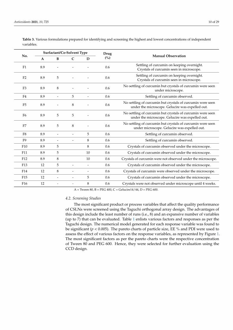
Curcumin was not found to be soluble in any of the lipids evaluated for the study.These included stearic acid, glyceryl monostearate, Compritol® ATO 888 and cetyl alcohol.Compritol® ATO 888 was, however, chosen as the lipid based on our prior experience andliterature reports, which show that it results in stable, safe, uniformly sized, and sphericalparticles which encapsulate significant amounts of hydrophobic drugs [39,49,50]. Varioussurfactants were evaluated for their property to dissolve curcumin and also to increase itssolubility in Compritol® ATO 888. Amongst a list of surfactants tested, Tween 80 was foundto show the best results. Further, curcumin was found to be highly soluble in polyethyleneglycol (PEG) 400, PEG 600 and Gelucire 14/44. Hence, they were taken as cosolvents todissolve curcumin in the lipidic phase whereas Tween 80 was selected as a surfactant forthe aqueous phase. Various formulations were then prepared by varying the concentrationof Tween 80 and the cosolvents (Table 3). The formulations were observed for stability (noseparation of phases), drug expulsion and crystallization. Based on pre-screening, Tween80 and PEG 600 were selected as the surfactant and cosolvent of choice for screening in theoptimization studies. PEG is a non-ionic glycol and acts as a solubilizer, plasticizer andpermeation enhancer. The oxygen atoms present in PEG give it hydrophilicity while theCH2–CH2 groups display lipophilicity [51]; and it seemed to have a special role in ensuringthe effective encapsulation of curcumin in the SLNs and in keeping any unencapsulateddrug in the solution. F12 did not show any drug crystals but the total concentration ofsurfactant used was very high (26.9%). So, an attempt was made to decrease the overallsurfactant concentration by increasing the concentration of Tween 80 to 12%, and furtherformulations were prepared (F13–F16) and observed for stability in terms of the separationof phases or drug expulsion and crystallization when observed under a microscope, andF16 was selected for further optimization studies.
Antioxidants 2021, 10, 725 10 of 29
Table 3. Various formulations prepared for identifying and screening the highest and lowest concentrations of independentvariables.
No.Surfactant/Co-Solvent Type Drug
(%) Manual ObservationA B C D
F1 8.9 - - - 0.6 Settling of curcumin on keeping overnight.Crystals of curcumin seen in microscope.
F2 8.9 5 - - 0.6 Settling of curcumin on keeping overnight.Crystals of curcumin seen in microscope.
F3 8.9 8 - - 0.6 No settling of curcumin but crystals of curcumin were seenunder microscope.
F4 8.9 - 5 - 0.6 Settling of curcumin observed.
F5 8.9 - 8 - 0.6 No settling of curcumin but crystals of curcumin were seenunder the microscope. Gelucire was expelled out.
F6 8.9 5 5 - 0.6 No settling of curcumin but crystals of curcumin were seenunder the microscope. Gelucire was expelled out.
F7 8.9 5 8 - 0.6 No settling of curcumin but crystals of curcumin were seenunder microscope. Gelucire was expelled out.
F8 8.9 - - 5 0.6 Settling of curcumin observed.
F9 8.9 - - 8 0.6 Settling of curcumin observed.
F10 8.9 5 8 0.6 Crystals of curcumin observed under the microscope.
F11 8.9 5 10 0.6 Crystals of curcumin observed under the microscope.
F12 8.9 8 10 0.6 Crystals of curcumin were not observed under the microscope.
F13 12 5 - - 0.6 Crystals of curcumin observed under the microscope.
F14 12 8 - - 0.6 Crystals of curcumin were observed under the microscope.
F15 12 - - 5 0.6 Crystals of curcumin observed under the microscope.
F16 12 - - 8 0.6 Crystals were not observed under microscope until 4 weeks.
A = Tween 80, B = PEG 400, C = Gelucire14/44, D = PEG 600.
4.2. Screening Studies
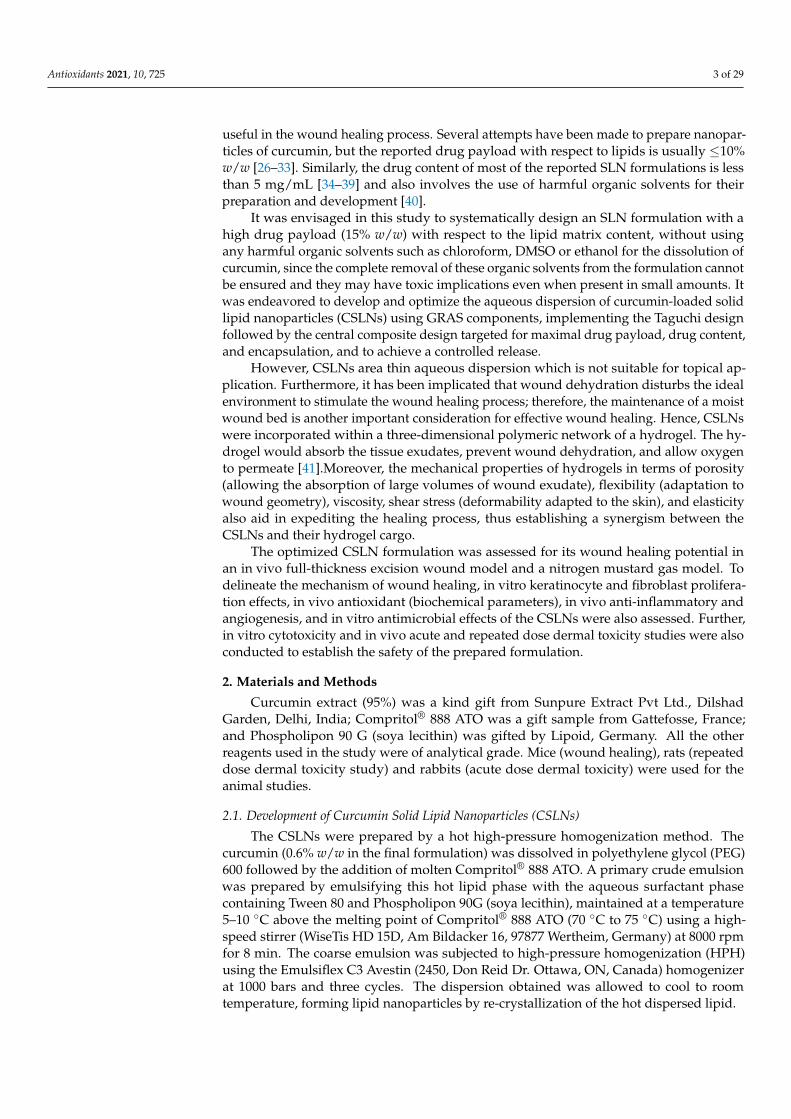
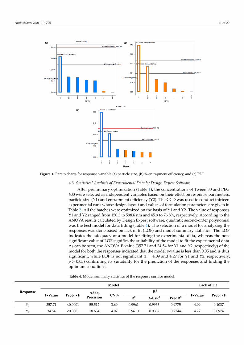
The most significant product or process variables that affect the quality performanceof CSLNs were screened using the Taguchi orthogonal array design. The advantages ofthis design include the least number of runs (i.e., 8) and an expansive number of variables(up to 7) that can be evaluated. Table 1 enlists various factors and responses as per theTaguchi design. The numerical model generated for each response variable was found tobe significant (p < 0.005). The pareto charts of particle size, EE % and PDI were used toassess the effect of various factors on the response variables, as represented by Figure 1.The most significant factors as per the pareto charts were the respective concentrationof Tween 80 and PEG 600. Hence, they were selected for further evaluation using theCCD design.
Antioxidants 2021, 10, 725 11 of 29Antioxidants 2021, 10, x FOR PEER REVIEW 12 of 30
Figure 1. Pareto charts for response variable (a) particle size, (b) % entrapment efficiency, and (c) PDI.
The suitability of the polynomial model equation for particle size (R2 = 0.9961) and % EE (R2 = 0.9610) fitting was expressed by the respective coefficients of determination. The latter indicated that the model could explain 99% and 96% variability, respectively, in the responses Y1 and Y2, and only 1% and 4% of the variability is due to noise, respectively. The predicted R-squared (Pred R2) values for the responses (Y1= 0.9775 and Y2 = 0.7754) were found to be in reasonable agreement with the adjusted R-square values (Adjs R2) (Y1= 0.9933 and Y2 = 0.9332) indicating that the model predicted the responses accurately.
The standard deviation associated with the experiment around the mean can be eval-uated from the value of the coefficient of variation (CV% = 3.69 and 4.07 for Y1 and Y2, respectively). Further, the CV (%) signifies the precision and reliability of the model used. Finally, the predicted R2 of 0.82 implies the good prediction of the particle size by the model. Adeq. Precisions for the responses Y1 and Y2 were 55.512 and 18.634, respectively (Table 4). Adeq. precision measures the signal-to-noise ratio and a ratio greater than 4 is desirable.
Table 4. Model summary statistics of the response surface model.
Response Model Lack of Fit
F-Value Prob > F Adeq. Precision
CV% R2
F-Value Prob > F R2 AdjsR2 PredR2
Y1 357.71 <0.0001 55.512 3.69 0.9961 0.9933 0.9775 4.09 0.1037 Y2 34.54 <0.0001 18.634 4.07 0.9610 0.9332 0.7744 4.27 0.0974
4.3.1. Effect on Particle Size The regression equation for the particle size in terms of coded factors is derived as:
Figure 1. Pareto charts for response variable (a) particle size, (b) % entrapment efficiency, and (c) PDI.
4.3. Statistical Analysis of Experimental Data by Design Expert Software
After preliminary optimization (Table 1), the concentrations of Tween 80 and PEG600 were selected as independent variables based on their effect on response parameters,particle size (Y1) and entrapment efficiency (Y2). The CCD was used to conduct thirteenexperimental runs whose design layout and values of formulation parameters are given inTable 2. All the batches were optimized on the basis of Y1 and Y2. The value of responsesY1 and Y2 ranged from 150.3 to 598.6 nm and 45.9 to 76.8%, respectively. According to theANOVA results calculated by Design Expert software, quadratic second-order polynomialwas the best model for data fitting (Table 4). The selection of a model for analyzing theresponses was done based on lack of fit (LOF) and model summary statistics. The LOFindicates the adequacy of a model for fitting the experimental data, whereas the non-significant value of LOF signifies the suitability of the model to fit the experimental data.As can be seen, the ANOVA F-value (357.71 and 34.54 for Y1 and Y2, respectively) of themodel for both the responses indicated that the model p-value is less than 0.05 and is thussignificant, while LOF is not significant (F = 4.09 and 4.27 for Y1 and Y2, respectively;p > 0.05) confirming its suitability for the prediction of the responses and finding theoptimum conditions.
Table 4. Model summary statistics of the response surface model.
Response
Model Lack of Fit
F-Value Prob > FAdeq.
Precision CV%R2
F-Value Prob > FR2 AdjsR2 PredR2
Y1 357.71 <0.0001 55.512 3.69 0.9961 0.9933 0.9775 4.09 0.1037
Y2 34.54 <0.0001 18.634 4.07 0.9610 0.9332 0.7744 4.27 0.0974
Antioxidants 2021, 10, 725 12 of 29
The suitability of the polynomial model equation for particle size (R2 = 0.9961) and% EE (R2 = 0.9610) fitting was expressed by the respective coefficients of determination.The latter indicated that the model could explain 99% and 96% variability, respectively, inthe responses Y1 and Y2, and only 1% and 4% of the variability is due to noise, respectively.The predicted R-squared (Pred R2) values for the responses (Y1 = 0.9775 and Y2 = 0.7754)were found to be in reasonable agreement with the adjusted R-square values (Adjs R2)(Y1 = 0.9933 and Y2 = 0.9332) indicating that the model predicted the responses accurately.
The standard deviation associated with the experiment around the mean can beevaluated from the value of the coefficient of variation (CV% = 3.69 and 4.07 for Y1 andY2, respectively). Further, the CV (%) signifies the precision and reliability of the modelused. Finally, the predicted R2 of 0.82 implies the good prediction of the particle size by themodel. Adeq. Precisions for the responses Y1 and Y2 were 55.512 and 18.634, respectively(Table 4). Adeq. precision measures the signal-to-noise ratio and a ratio greater than 4 isdesirable.
4.3.1. Effect on Particle Size
The regression equation for the particle size in terms of coded factors is derived as:
Y1 = +3969.53753 − 443.0278 ∗ X1 − 303.97911X2 + 14.775X1X2 + 13.90844X21 + 8.17056 X2
2
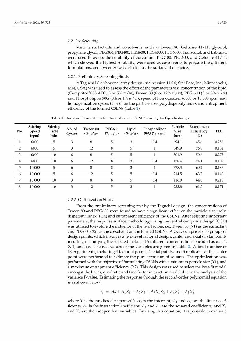
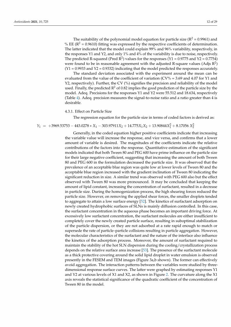
Generally, in the coded equation higher positive coefficients indicate that increasingthe variable value will increase the response, and vice versa, and confirms that a loweramount of variable is desired. The magnitudes of the coefficients indicate the relativecontributions of the factors into the response. Quantitative estimation of the significantmodels indicated that both Tween 80 and PEG 600 have prime influence on the particle sizefor their large negative coefficient, suggesting that increasing the amount of both Tween80 and PEG 600 in the formulation decreased the particle size. It was observed that theprevalence of an acceptable blue region was quite low at lower levels of Tween 80 and theacceptable blue region increased with the gradient inclination of Tween 80 indicating thesignificant reduction in size. A similar trend was observed with PEG 600 also but the effectobserved with Tween 80 was more pronounced. It may be concluded that keeping theamount of lipid constant, increasing the concentration of surfactant, resulted in a decreasein particle size. During the homogenization process, the high shearing forces reduced theparticle size. However, on removing the applied shear forces, the smaller droplets tendedto aggregate to attain a low surface energy [52]. The kinetics of surfactant adsorption onnewly created hydrophobic surfaces of SLNs is mainly diffusion controlled. In this case,the surfactant concentration in the aqueous phase becomes an important driving force. Atexcessively low surfactant concentration, the surfactant molecules are either insufficient tocompletely cover the newly created particle surface, resulting in suboptimal stabilizationof the particle dispersion, or they are not adsorbed at a rate rapid enough to match orsupersede the rate of particle–particle collisions resulting in particle aggregation. However,the molecular characteristics of the surfactant and the nature of the interface also influencethe kinetics of the adsorption process. Moreover, the amount of surfactant required tomaintain the stability of the hot SLN dispersion during the cooling/crystallization processdepends on the relative surface area increase [53]. The presence of the surfactant moleculeas a thick protective covering around the solid lipid droplet in water emulsion is observedpresently in the FESEM and TEM images (Figure 3a,b shown). The former can effectivelyavoid aggregation. The interaction patterns between the variables were studied by three-dimensional response surface curves. The latter were graphed by estimating responses Y1and Y2 at various levels of X1 and X2, as shown in Figure 2. The curvature along the X1axis reveals the statistical significance of the quadratic coefficient of the concentration ofTween 80 in the model.
Antioxidants 2021, 10, 725 13 of 29Antioxidants 2021, 10, x FOR PEER REVIEW 14 of 30
Figure 2. Response surface curves for (a) particle size, and (b) entrapment efficiency.
4.3.3. Optimized Formulation and its Validation The composition of the optimum formulation was determined as 12% (w/v) Tween
80 and 8% (w/w) PEG 600, which fulfilled the requirements of optimization for minimum particle size and maximum entrapment efficiency. At these levels, the predicted values of Y1 and Y2 were 156.6 nm and 76.5%, respectively. The check point validation of the pre-dicted values showed close observed values with % error in all cases being <±6.33 (Table 5).
Table 5. Validation of prepared formulation.
Checkpoint Conditions X1/X2
Y1 (nm) Y2 (%)
Observed Predicted Error (%)
Observed Predicted Error (%)
12/8 156.6 165.5 5.38 74.6 76.5 2.46 10/6.5 232.6 259.8 4.05 63.5 65.8 3.44 12/5 212.9 226.8 6.17 69.4 66.0 −5.13 8/8 365.5 352.1 −3.79 65.3 61.4 −6.33 8/5 614.6 590.8 −4.02 41.6 43.8 5.12
4.4.Characterization 4.4.1. Assay/TDC and EE
The total drug content of 5.8 ± 0.2 mg/mL was observed, amounting to 96% of the original amount incorporated (6 mg/mL), confirming that insignificant losses occurred during the process of preparation of the SLNs by high-pressure homogenization (n = 6). An EE of 75.55 ± 2.31% was observed (n = 12). We previously [39] reported a micro-emul-sification method for the preparation of curcumin-loaded solid lipid nanoparticles with
Figure 2. Response surface curves for (a) particle size, and (b) entrapment efficiency.
4.3.2. Effect on Entrapment Efficiency
The quadratic polynomial equation of the %EE in terms of coded factors was deter-mined and is presented in equation as below:
Y2 = −130.16712+ 15.55617 ∗X1 + 24.52045X2 − 0.59167X1X2 − 0.35281X21 − 1.071672X2
2
EE was found to increase with the increase in the concentration of both Tween 80 andPEG 600. A higher coefficient for the latter indicates its higher influence on EE, which couldbe attributed to the increased solubility of curcumin in the lipid core by the solvent actionof PEG 600. Further, PEG 600 being highly hydrophilic has a high affinity for the aqueousphase. Therefore, in addition to improving solubility of curcumin in the lipidic phase,PEG 600 having a high affinity for water will move towards the aqueous phase, in turnleaving behind/precipitating curcumin in the lipidic core. Curcumin, being hydrophobic,does not diffuse to the aqueous phase thereby improving both its payload and entrapmentefficiency. Furthermore, the surface coverage of nanoparticles with PEG 600, which is botha surfactant-supporting agent and a plasticizer [54], prevents drug leaching from the lipidmatrix [55]. An increase in the surfactant concentration increases the entrapment efficiencyof curcumin within the SLNs [38]. In Figure 2, the response surface plot shows the impactof X1 and X2 on % EE.
4.3.3. Optimized Formulation and its Validation
The composition of the optimum formulation was determined as 12% (w/v) Tween80 and 8% (w/w) PEG 600, which fulfilled the requirements of optimization for minimumparticle size and maximum entrapment efficiency. At these levels, the predicted valuesof Y1 and Y2 were 156.6 nm and 76.5%, respectively. The check point validation of thepredicted values showed close observed values with % error in all cases being <±6.33(Table 5).
Antioxidants 2021, 10, 725 14 of 29
Table 5. Validation of prepared formulation.
Checkpoint ConditionsX1/X2
Y1 (nm) Y2 (%)
Observed Predicted Error (%) Observed Predicted Error (%)
12/8 156.6 165.5 5.38 74.6 76.5 2.46
10/6.5 232.6 259.8 4.05 63.5 65.8 3.44
12/5 212.9 226.8 6.17 69.4 66.0 −5.13
8/8 365.5 352.1 −3.79 65.3 61.4 −6.33
8/5 614.6 590.8 −4.02 41.6 43.8 5.12
4.4. Characterization4.4.1. Assay/TDC and EE
The total drug content of 5.8 ± 0.2 mg/mL was observed, amounting to 96% of theoriginal amount incorporated (6 mg/mL), confirming that insignificant losses occurred dur-ing the process of preparation of the SLNs by high-pressure homogenization (n = 6). An EEof 75.55 ± 2.31% was observed (n = 12). We previously [39] reported a micro-emulsificationmethod for the preparation of curcumin-loaded solid lipid nanoparticles with an entrap-ment efficiency of 81.92 ± 2.91%. The latter, although higher than the presently obtainedvalue, is for a lower drug payload of 10% with respect to the lipid matrix (presently it is15%) and employed a much higher (>2 times) Tween 80 concentration of approximately25%. Furthermore, the microemulsion process is a less industry-amenable method. Simi-larly, a high entrapment efficiency of 84.6% is also reported elsewhere [31], but again ata much lower drug payload of 6.6% to 10% and employing high shear homogenizationfollowed by ultrasonication. The particle size of the SLNs prepared by these workers was401.9 nm.
4.4.2. Particle Size Analysis, Polydispersity Index, and Zeta Potential
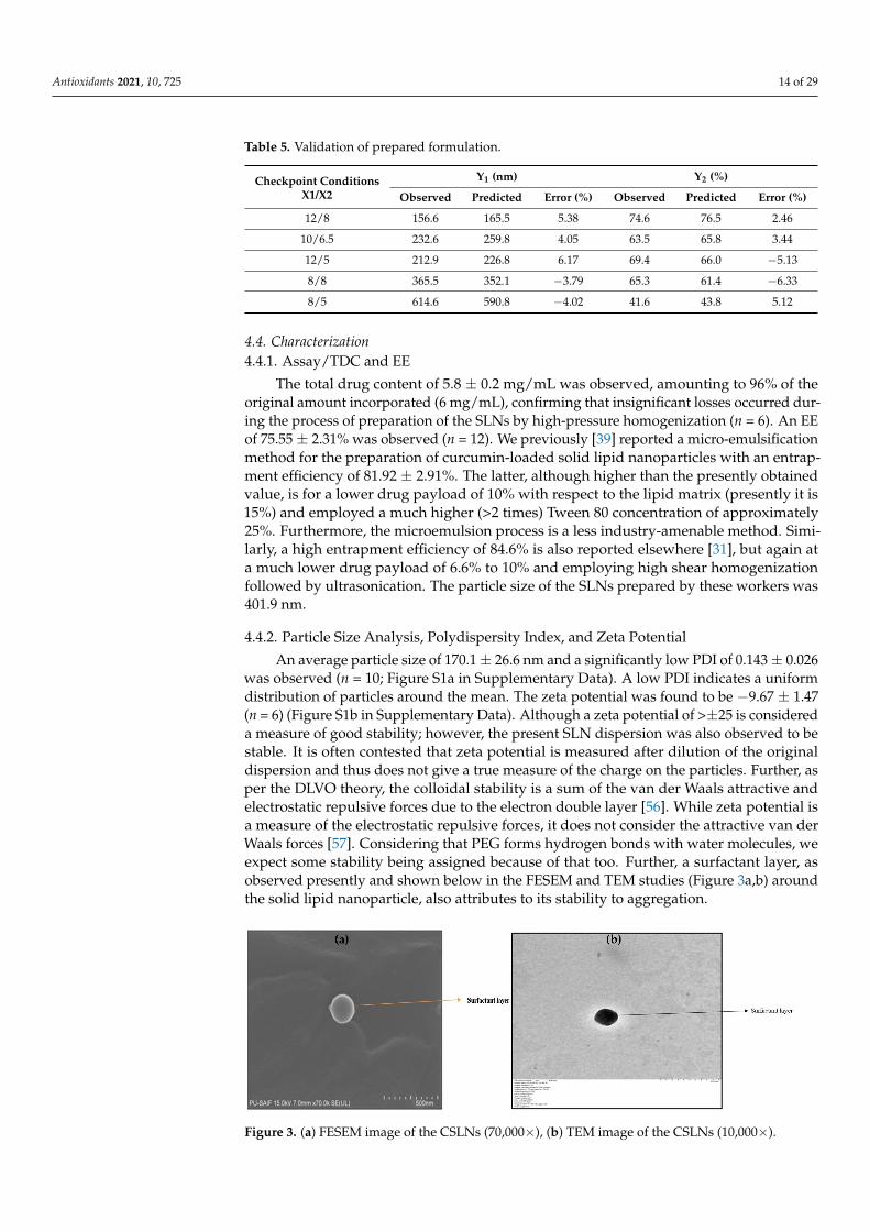
An average particle size of 170.1 ± 26.6 nm and a significantly low PDI of 0.143 ± 0.026was observed (n = 10; Figure S1a in Supplementary Data). A low PDI indicates a uniformdistribution of particles around the mean. The zeta potential was found to be −9.67 ± 1.47(n = 6) (Figure S1b in Supplementary Data). Although a zeta potential of >±25 is considereda measure of good stability; however, the present SLN dispersion was also observed to bestable. It is often contested that zeta potential is measured after dilution of the originaldispersion and thus does not give a true measure of the charge on the particles. Further, asper the DLVO theory, the colloidal stability is a sum of the van der Waals attractive andelectrostatic repulsive forces due to the electron double layer [56]. While zeta potential isa measure of the electrostatic repulsive forces, it does not consider the attractive van derWaals forces [57]. Considering that PEG forms hydrogen bonds with water molecules, weexpect some stability being assigned because of that too. Further, a surfactant layer, asobserved presently and shown below in the FESEM and TEM studies (Figure 3a,b) aroundthe solid lipid nanoparticle, also attributes to its stability to aggregation.
Antioxidants 2021, 10, x FOR PEER REVIEW 15 of 30
an entrapment efficiency of 81.92 ± 2.91%. The latter, although higher than the presently obtained value, is for a lower drug payload of 10% with respect to the lipid matrix (pres-ently it is 15%) and employed a much higher (>2 times) Tween 80 concentration of ap-proximately 25%. Furthermore, the microemulsion process is a less industry-amenable method. Similarly, a high entrapment efficiency of 84.6% is also reported elsewhere [31], but again at a much lower drug payload of 6.6% to 10% and employing high shear ho-mogenization followed by ultrasonication. The particle size of the SLNs prepared by these workers was 401.9 nm.
4.4.2. Particle Size Analysis, Polydispersity Index, and Zeta Potential An average particle size of 170.1 ± 26.6 nm and a significantly low PDI of 0.143 ± 0.026
was observed (n = 10; Figure S1a in Supplementary Data). A low PDI indicates a uniform distribution of particles around the mean. The zeta potential was found to be −9.67 ± 1.47 (n = 6) (Figure S1b in Supplementary Data). Although a zeta potential of >±25 is considered a measure of good stability; however, the present SLN dispersion was also observed to be stable. It is often contested that zeta potential is measured after dilution of the original dispersion and thus does not give a true measure of the charge on the particles. Further, as per the DLVO theory, the colloidal stability is a sum of the van der Waals attractive and electrostatic repulsive forces due to the electron double layer [56]. While zeta potential is a measure of the electrostatic repulsive forces, it does not consider the attractive van der Waals forces [57]. Considering that PEG forms hydrogen bonds with water molecules, we expect some stability being assigned because of that too. Further, a surfactant layer, as observed presently and shown below in the FESEM and TEM studies (Figure 3a,b) around the solid lipid nanoparticle, also attributes to its stability to aggregation.
4.4.3. FESEM and TEM Studies The FESEM of the CSLN (Figure 3a) formulation showed small spherical particles as
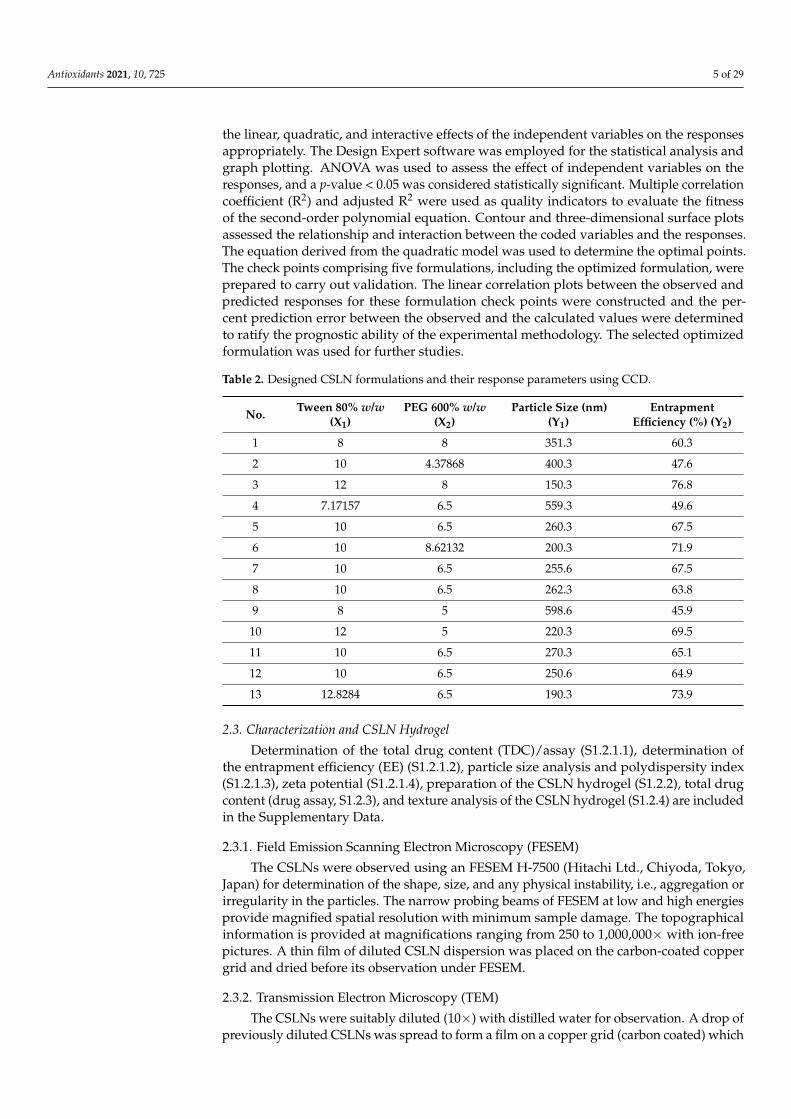
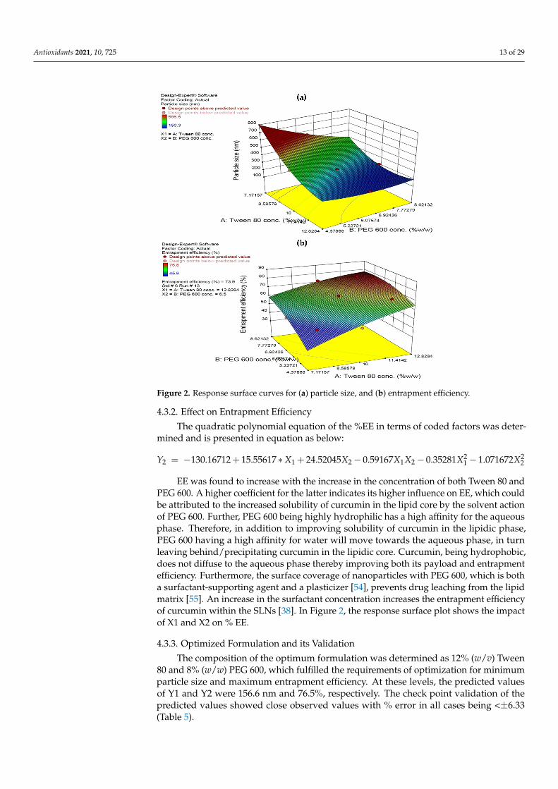
individual entities and an absence of any agglomerates indicating their stability. A coat of surfactant on the outside of SLN covering the entire surface of the particle uniformly and clearly visible in the Figure 3a, assigns it with the stability to aggregate. Morphological analysis of CSLN in aqueous dispersions, detected by TEM analysis, also indicates near-spherical particles (spindle shaped) (Figure 3b). The polymorphic form of the SLNs also reflects in the shape of the particles; particles in α modification are usually observed as circular to polyhedral [58]. The data of PXRD (S2.1.1), DSC (S2.1.2), and FTIR (S2.1.3) are included in Supplementary Data.
Figure 3. (a) FESEM image of the CSLNs (70,000×), (b) TEM image of the CSLNs (10,000×).
4.5. Stability Studies An insignificant change in TDC, EE, particle size and PDI was observed when the
CSLNs were stored at 5 ± 3 °C for a period of 12 months (Table 6). This may be attributed to the maintenance of the SLNs in their native state, in which they were produced, com-prising a suitable concentration of surfactants in the aqueous phase such that they remain
Figure 3. (a) FESEM image of the CSLNs (70,000×), (b) TEM image of the CSLNs (10,000×).
Antioxidants 2021, 10, 725 15 of 29
4.4.3. FESEM and TEM Studies
The FESEM of the CSLN (Figure 3a) formulation showed small spherical particles asindividual entities and an absence of any agglomerates indicating their stability. A coat ofsurfactant on the outside of SLN covering the entire surface of the particle uniformly andclearly visible in the Figure 3a, assigns it with the stability to aggregate. Morphologicalanalysis of CSLN in aqueous dispersions, detected by TEM analysis, also indicates near-spherical particles (spindle shaped) (Figure 3b). The polymorphic form of the SLNs alsoreflects in the shape of the particles; particles in α modification are usually observed ascircular to polyhedral [58]. The data of PXRD (S2.1.1), DSC (S2.1.2), and FTIR (S2.1.3) areincluded in Supplementary Data.
4.5. Stability Studies
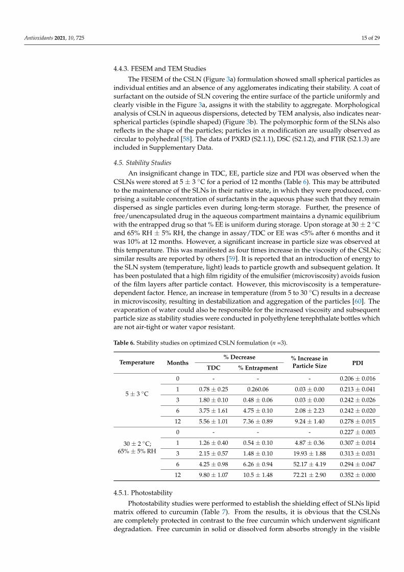
An insignificant change in TDC, EE, particle size and PDI was observed when theCSLNs were stored at 5 ± 3 ◦C for a period of 12 months (Table 6). This may be attributedto the maintenance of the SLNs in their native state, in which they were produced, com-prising a suitable concentration of surfactants in the aqueous phase such that they remaindispersed as single particles even during long-term storage. Further, the presence offree/unencapsulated drug in the aqueous compartment maintains a dynamic equilibriumwith the entrapped drug so that % EE is uniform during storage. Upon storage at 30 ± 2 ◦Cand 65% RH ± 5% RH, the change in assay/TDC or EE was <5% after 6 months and itwas 10% at 12 months. However, a significant increase in particle size was observed atthis temperature. This was manifested as four times increase in the viscosity of the CSLNs;similar results are reported by others [59]. It is reported that an introduction of energy tothe SLN system (temperature, light) leads to particle growth and subsequent gelation. Ithas been postulated that a high film rigidity of the emulsifier (microviscosity) avoids fusionof the film layers after particle contact. However, this microviscosity is a temperature-dependent factor. Hence, an increase in temperature (from 5 to 30 ◦C) results in a decreasein microviscosity, resulting in destabilization and aggregation of the particles [60]. Theevaporation of water could also be responsible for the increased viscosity and subsequentparticle size as stability studies were conducted in polyethylene terephthalate bottles whichare not air-tight or water vapor resistant.
Table 6. Stability studies on optimized CSLN formulation (n =3).
Temperature Months% Decrease % Increase in
Particle Size PDITDC % Entrapment
5 ± 3 ◦C
0 - - - 0.206 ± 0.016
1 0.78 ± 0.25 0.260.06 0.03 ± 0.00 0.213 ± 0.041
3 1.80 ± 0.10 0.48 ± 0.06 0.03 ± 0.00 0.242 ± 0.026
6 3.75 ± 1.61 4.75 ± 0.10 2.08 ± 2.23 0.242 ± 0.020
12 5.56 ± 1.01 7.36 ± 0.89 9.24 ± 1.40 0.278 ± 0.015
30 ± 2 ◦C;65% ± 5% RH
0 - - - 0.227 ± 0.003
1 1.26 ± 0.40 0.54 ± 0.10 4.87 ± 0.36 0.307 ± 0.014
3 2.15 ± 0.57 1.48 ± 0.10 19.93 ± 1.88 0.313 ± 0.031
6 4.25 ± 0.98 6.26 ± 0.94 52.17 ± 4.19 0.294 ± 0.047
12 9.80 ± 1.07 10.5 ± 1.48 72.21 ± 2.90 0.352 ± 0.000
4.5.1. Photostability
Photostability studies were performed to establish the shielding effect of SLNs lipidmatrix offered to curcumin (Table 7). From the results, it is obvious that the CSLNsare completely protected in contrast to the free curcumin which underwent significantdegradation. Free curcumin in solid or dissolved form absorbs strongly in the visible

Antioxidants 2021, 10, 725 16 of 29
wavelength range, making it predisposed to degradation and modification in daylight andartificial lighting [61,62]. The SLNs stored in amber-colored and transparent containersdid not show any degradation in terms of assay/TDC and entrapment (p > 0.05). The PDIwas also similar; however, some increase in particle size was observed (Table 7). On theother hand, 21.7% and 35% of the free curcumin was found to be degraded upon storage inamber-colored containers and transparent containers, respectively.
Table 7. Photostability studies on optimized CSLN formulation (n = 3).
Glassware DaysAssay/TDC% % Entrapment % Change in
Particle Size
Free Curcumin CSLNs CSLNs
Amber glass0 day 100.00 ± 0 100.00 ± 0 100.00 ± 0 -
10 days 78.31 ± 7.1 99.64 ± 0.02 99.82 ± 0.6 7.90
Transparent0 day 100.00 ± 0 100.00 ± 0 100.00 ± 0 -
10 days 65.51 ± 7.5 98.93 ± 0.05 99.03 ± 0.06 8.98
4.5.2. Autoclavability
The developed CSLNs were found to be stable to autoclaving with no change inentrapment efficiency. A slight increase in particle size was observed, as shown in Table 8.Similar observations are also reported by others [63]. This could be attributed to the hightemperature reached during sterilization by autoclaving, which results in the formationof hot o/w microemulsion in the autoclave and probably modifies the size of the hotnanodroplets. On subsequent slow cooling, the SLNs are reformed but some nanodropletsmay coalesce during cooling, producing larger SLNs than the initial ones [63]. Sterilizationis an important requirement for wound dressings especially if they are required to be usedon the burn or infected wounds.
Table 8. Parameters before and after autoclaving (n = 6).
Autoclaving Assay/TDC *(mg/mL)
EntrapmentEfficiency * (%)
Particle Size(nm) PDI Zeta Potential *
Before 5.8 ± 0.2 75.55 ± 2.31 170.1 ± 26.6 0.143 ± 0.026 −9.67±1.47
After 5.7±0.3 74.24± 3.6 253.7± 28.0 0.182 ± 0.032 −9.50±1.86* No significant change.
Special care has been taken to choose an appropriate sterilization technique to ensureno degradation or aggregation of the solid lipids to avoid toxicity and instability duringthe sterility of the CSLNs [64]. Further, it should not change the formulation propertiesincluding the physical and chemical stability and the drug release kinetics [64]. Oneof the advantages of SLNs over other colloidal systems is that they can be sterilized byautoclaving, a commonly used and reliable technique. SLNs melt and recrystallize in acontrolled manner at high temperatures [65].
4.6. In Vitro Studies4.6.1. Cell Proliferation Assay
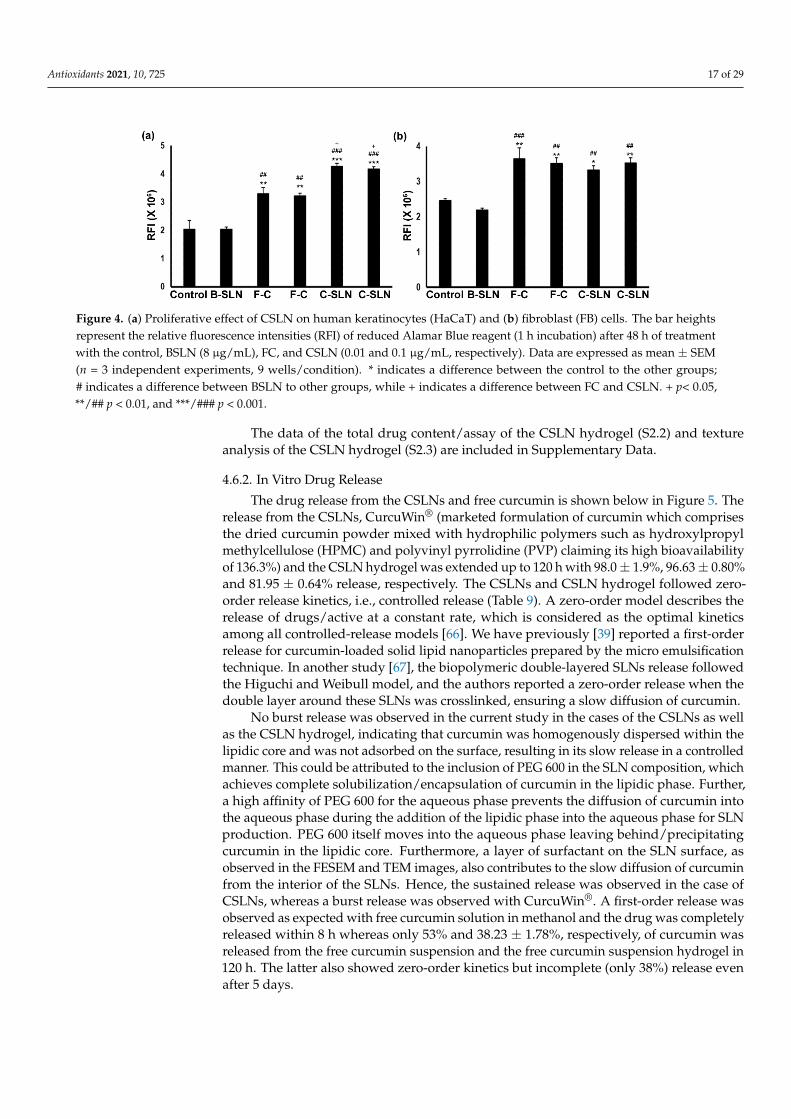
In these studies, both the cell types respond significantly to the treatments (ANOVA,F = 33.39; p < 0.0001 for HaCaT and F = 14.079; p = 0.0001 for FB) depending on theconcentration (Figure 4a,b) of different experimental conditions. The cells did not show anyeffect with the blank SLNS (BSLN) treatment up to 8 µg/mL, but both the free curcumindispersion (FC) (p < 0.001) and the CSLN (p < 0.001) significantly induced cell growth inHaCaT and FB up to a 0.1 µg/mL concentration, as compared to the control and BSLN. TheCSLN treatment induced more growth compared to the FC in HaCaT (Figure 4a, p < 0.05).The concentration of over 2 µg/mL of FC and CSLN, however, caused cell death in theseexperiments (data not shown).
Antioxidants 2021, 10, 725 17 of 29
Antioxidants 2021, 10, x FOR PEER REVIEW 17 of 30
4.5.2. Autoclavability The developed CSLNs were found to be stable to autoclaving with no change in en-
trapment efficiency. A slight increase in particle size was observed, as shown in Table 8. Similar observations are also reported by others [63]. This could be attributed to the high temperature reached during sterilization by autoclaving, which results in the formation of hot o/w microemulsion in the autoclave and probably modifies the size of the hot nanodroplets. On subsequent slow cooling, the SLNs are reformed but some nanodroplets may coalesce during cooling, producing larger SLNs than the initial ones [63]. Sterilization is an important requirement for wound dressings especially if they are required to be used on the burn or infected wounds.
Special care has been taken to choose an appropriate sterilization technique to ensure no degradation or aggregation of the solid lipids to avoid toxicity and instability during the sterility of the CSLNs [64]. Further, it should not change the formulation properties including the physical and chemical stability and the drug release kinetics [64]. One of the advantages of SLNs over other colloidal systems is that they can be sterilized by autoclav-ing, a commonly used and reliable technique. SLNs melt and recrystallize in a controlled manner at high temperatures [65].
Table 8. Parameters before and after autoclaving (n = 6).
Autoclaving Assay/TDC * (mg/mL)
Entrapment Efficiency *
(%)
Particle Size (nm)
PDI Zeta Po-tential *
Before 5.8 ± 0.2 75.55 ± 2.31 170.1 ± 26.6 0.143 ± 0.026 −9.67±1.47 After 5.7±0.3 74.24± 3.6 253.7± 28.0 0.182 ± 0.032 −9.50±1.86
* No significant change.
4.6. In Vitro Studies 4.6.1. Cell Proliferation Assay
In these studies, both the cell types respond significantly to the treatments (ANOVA, F = 33.39; p < 0.0001 for HaCaT and F = 14.079; p = 0.0001 for FB) depending on the concen-tration (Figure 4a,b) of different experimental conditions. The cells did not show any effect with the blank SLNS (BSLN) treatment up to 8 µg/mL, but both the free curcumin disper-sion (FC) (p < 0.001) and the CSLN (p < 0.001) significantly induced cell growth in HaCaT and FB up to a 0.1 µg/mL concentration, as compared to the control and BSLN. The CSLN treatment induced more growth compared to the FC in HaCaT (Figure 4a, p < 0.05). The concentration of over 2 µg/mL of FC and CSLN, however, caused cell death in these ex-periments (data not shown).
The data of the total drug content/assay of the CSLN hydrogel (S2.2) and texture analysis of the CSLN hydrogel (S2.3) are included in Supplementary Data.
Figure 4. (a) Proliferative effect of CSLN on human keratinocytes (HaCaT) and (b) fibroblast (FB) cells. The bar heights represent the relative fluorescence intensities (RFI) of reduced Alamar Blue
Figure 4. (a) Proliferative effect of CSLN on human keratinocytes (HaCaT) and (b) fibroblast (FB) cells. The bar heightsrepresent the relative fluorescence intensities (RFI) of reduced Alamar Blue reagent (1 h incubation) after 48 h of treatmentwith the control, BSLN (8 µg/mL), FC, and CSLN (0.01 and 0.1 µg/mL, respectively). Data are expressed as mean ± SEM(n = 3 independent experiments, 9 wells/condition). * indicates a difference between the control to the other groups;# indicates a difference between BSLN to other groups, while + indicates a difference between FC and CSLN. + p< 0.05,**/## p < 0.01, and ***/### p < 0.001.
The data of the total drug content/assay of the CSLN hydrogel (S2.2) and textureanalysis of the CSLN hydrogel (S2.3) are included in Supplementary Data.
4.6.2. In Vitro Drug Release
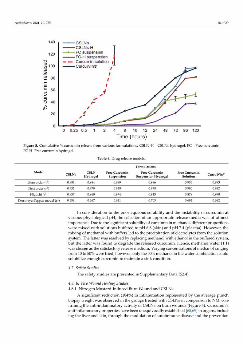
The drug release from the CSLNs and free curcumin is shown below in Figure 5. Therelease from the CSLNs, CurcuWin® (marketed formulation of curcumin which comprisesthe dried curcumin powder mixed with hydrophilic polymers such as hydroxylpropylmethylcellulose (HPMC) and polyvinyl pyrrolidine (PVP) claiming its high bioavailabilityof 136.3%) and the CSLN hydrogel was extended up to 120 h with 98.0 ± 1.9%, 96.63 ± 0.80%and 81.95 ± 0.64% release, respectively. The CSLNs and CSLN hydrogel followed zero-order release kinetics, i.e., controlled release (Table 9). A zero-order model describes therelease of drugs/active at a constant rate, which is considered as the optimal kineticsamong all controlled-release models [66]. We have previously [39] reported a first-orderrelease for curcumin-loaded solid lipid nanoparticles prepared by the micro emulsificationtechnique. In another study [67], the biopolymeric double-layered SLNs release followedthe Higuchi and Weibull model, and the authors reported a zero-order release when thedouble layer around these SLNs was crosslinked, ensuring a slow diffusion of curcumin.
No burst release was observed in the current study in the cases of the CSLNs as wellas the CSLN hydrogel, indicating that curcumin was homogenously dispersed within thelipidic core and was not adsorbed on the surface, resulting in its slow release in a controlledmanner. This could be attributed to the inclusion of PEG 600 in the SLN composition, whichachieves complete solubilization/encapsulation of curcumin in the lipidic phase. Further,a high affinity of PEG 600 for the aqueous phase prevents the diffusion of curcumin intothe aqueous phase during the addition of the lipidic phase into the aqueous phase for SLNproduction. PEG 600 itself moves into the aqueous phase leaving behind/precipitatingcurcumin in the lipidic core. Furthermore, a layer of surfactant on the SLN surface, asobserved in the FESEM and TEM images, also contributes to the slow diffusion of curcuminfrom the interior of the SLNs. Hence, the sustained release was observed in the case ofCSLNs, whereas a burst release was observed with CurcuWin®. A first-order release wasobserved as expected with free curcumin solution in methanol and the drug was completelyreleased within 8 h whereas only 53% and 38.23 ± 1.78%, respectively, of curcumin wasreleased from the free curcumin suspension and the free curcumin suspension hydrogel in120 h. The latter also showed zero-order kinetics but incomplete (only 38%) release evenafter 5 days.
Antioxidants 2021, 10, 725 18 of 29
Antioxidants 2021, 10, x FOR PEER REVIEW 19 of 30
mixing of methanol with buffers led to the precipitation of electrolytes from the solution system. The latter was resolved by replacing methanol with ethanol in the buffered sys-tem, but the latter was found to degrade the released curcumin. Hence, methanol:water (1:1) was chosen as the satisfactory release medium. Varying concentrations of methanol ranging from 10 to 50% were tried; however, only the 50% methanol in the water combi-nation could solubilize enough curcumin to maintain a sink condition.
Figure 5. Cumulative % curcumin release from various formulations. CSLN-H—CSLNs hydrogel; FC—Free curcumin; FC-H- Free curcumin hydrogel.
4.7. Safety Studies The safety studies are presented in Supplementary Data (S2.4).
4.8. In Vivo Wound Healing Studies 4.8.1. Nitrogen Mustard-Induced Burn Wound and CSLNs
A significant reduction (184%) in inflammation represented by the average punch biopsy weight was observed in the groups treated with CSLNs in comparison to NM, confirming the anti-inflammatory activity of CSLNs on burn wounds (Figure 6). Curcu-min’s anti-inflammatory properties have been unequivocally established [68,69] in or-gans, including the liver and skin, through the modulation of autoimmune disease and the prevention of injury to these organ tissues [68]. The primary mechanism by which curcumin modulates inflammation is by reducing the expression of the two main cyto-kines that are released by monocytes and macrophages [24,70]. These molecules are inter-leukin 1 (IL-1) and TNF-α, which have important roles in the regulation of the inflamma-tory response. The influence of CSLNs on TNF-α in wound healing is established below in the section on the excision wound. Curcumin inhibits the activity of the proinflamma-tory transcriptional factor, NF-κB, which is responsible for the regulation of several genes involved in the initial onset of the inflammatory response. Similar anti-inflammatory ac-tivity of curcumin has already been reported by us [68], where the application of curcumin vesicular ointment resulted in the suppression of acute inflammation as well as significant inhibition of cotton pellet-induced chronic inflammation.
Figure 5. Cumulative % curcumin release from various formulations. CSLN-H—CSLNs hydrogel; FC—Free curcumin;FC-H- Free curcumin hydrogel.
Table 9. Drug release models.
Model
Formulations
CSLNs CSLNHydrogel
Free CurcuminSuspension
Free CurcuminSuspension Hydrogel
Free CurcuminSolution CurcuWin®
Zero order (r2) 0.986 0.984 0.889 0.986 0.936 0.893
First order (r2) 0.935 0.979 0.928 0.978 0.990 0.982
Higuchi (r2) 0.957 0.960 0.974 0.913 0.878 0.990
KorsmeyerPeppas model (r2) 0.698 0.667 0.641 0.703 0.692 0.682
In consideration to the poor aqueous solubility and the instability of curcumin atvarious physiological pH, the selection of an appropriate release media was of utmostimportance. Due to the significant solubility of curcumin in methanol, different proportionswere mixed with solutions buffered to pH 6.8 (skin) and pH 7.4 (plasma). However, themixing of methanol with buffers led to the precipitation of electrolytes from the solutionsystem. The latter was resolved by replacing methanol with ethanol in the buffered system,but the latter was found to degrade the released curcumin. Hence, methanol:water (1:1)was chosen as the satisfactory release medium. Varying concentrations of methanol rangingfrom 10 to 50% were tried; however, only the 50% methanol in the water combination couldsolubilize enough curcumin to maintain a sink condition.
4.7. Safety Studies
The safety studies are presented in Supplementary Data (S2.4).
4.8. In Vivo Wound Healing Studies4.8.1. Nitrogen Mustard-Induced Burn Wound and CSLNs
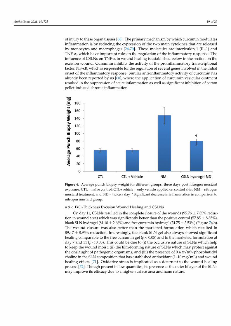
A significant reduction (184%) in inflammation represented by the average punchbiopsy weight was observed in the groups treated with CSLNs in comparison to NM, con-firming the anti-inflammatory activity of CSLNs on burn wounds (Figure 6). Curcumin’santi-inflammatory properties have been unequivocally established [68,69] in organs, includ-ing the liver and skin, through the modulation of autoimmune disease and the prevention
Antioxidants 2021, 10, 725 19 of 29
of injury to these organ tissues [68]. The primary mechanism by which curcumin modulatesinflammation is by reducing the expression of the two main cytokines that are releasedby monocytes and macrophages [24,70]. These molecules are interleukin 1 (IL-1) andTNF-α, which have important roles in the regulation of the inflammatory response. Theinfluence of CSLNs on TNF-α in wound healing is established below in the section on theexcision wound. Curcumin inhibits the activity of the proinflammatory transcriptionalfactor, NF-κB, which is responsible for the regulation of several genes involved in the initialonset of the inflammatory response. Similar anti-inflammatory activity of curcumin hasalready been reported by us [68], where the application of curcumin vesicular ointmentresulted in the suppression of acute inflammation as well as significant inhibition of cottonpellet-induced chronic inflammation.
Antioxidants 2021, 10, x FOR PEER REVIEW 20 of 30
Figure 6. Average punch biopsy weight for different groups, three days post nitrogen mustard exposure. CTL = naïve control, CTL+vehicle = only vehicle applied on control skin; NM = nitrogen mustard treatment; and BID = twice a day. * Significant decrease in inflammation in comparison to nitrogen mustard group.
4.8.2. Full-Thickness Excision Wound Healing and CSLNs On day 11, CSLNs resulted in the complete closure of the wounds (95.76 ± 7.85%
reduction in wound area) which was significantly better than the positive control (57.85 ± 8.85%), blank SLN hydrogel (81.18 ± 2.66%) and free curcumin hydrogel (74.75 ± 3.53%) (Figure 7a,b). The wound closure was also better than the marketed formulation which resulted in 89.47 ± 8.93% reduction. Interestingly, the blank SLN gel also always showed significant healing comparable to the free curcumin gel (p < 0.05) and to the marketed formulation at day 7 and 11 (p < 0.05). This could be due to (i) the occlusive nature of SLNs which help to keep the wound moist, (ii) the film-forming nature of SLNs which may protect against the onslaught of pathogenic organisms, and (iii) the presence of 0.4 w/w% phosphatidyl choline in the SLN composition that has established antioxidant (1–10 mg/mL) and wound healing effects [71]. Oxidative stress is implicated as a deterrent to the wound healing process [72]. Though present in low quantities, its presence as the outer bilayer of the SLNs may improve its efficacy due to a higher surface area and nano nature.
*
Figure 6. Average punch biopsy weight for different groups, three days post nitrogen mustardexposure. CTL = naïve control, CTL+vehicle = only vehicle applied on control skin; NM = nitrogenmustard treatment; and BID = twice a day. * Significant decrease in inflammation in comparison tonitrogen mustard group.
4.8.2. Full-Thickness Excision Wound Healing and CSLNs
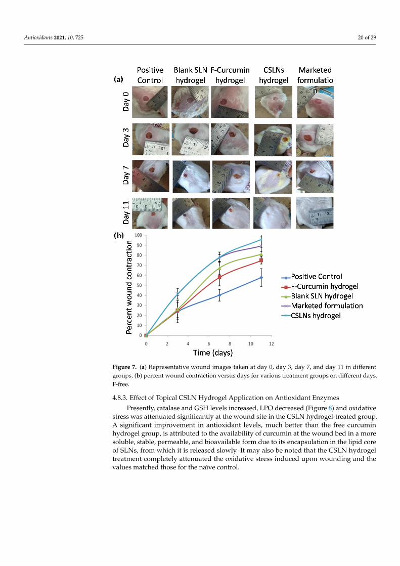
On day 11, CSLNs resulted in the complete closure of the wounds (95.76 ± 7.85% reduc-tion in wound area) which was significantly better than the positive control (57.85 ± 8.85%),blank SLN hydrogel (81.18 ± 2.66%) and free curcumin hydrogel (74.75 ± 3.53%) (Figure 7a,b).The wound closure was also better than the marketed formulation which resulted in89.47 ± 8.93% reduction. Interestingly, the blank SLN gel also always showed significanthealing comparable to the free curcumin gel (p < 0.05) and to the marketed formulation atday 7 and 11 (p < 0.05). This could be due to (i) the occlusive nature of SLNs which helpto keep the wound moist, (ii) the film-forming nature of SLNs which may protect againstthe onslaught of pathogenic organisms, and (iii) the presence of 0.4 w/w% phosphatidylcholine in the SLN composition that has established antioxidant (1–10 mg/mL) and woundhealing effects [71]. Oxidative stress is implicated as a deterrent to the wound healingprocess [72]. Though present in low quantities, its presence as the outer bilayer of the SLNsmay improve its efficacy due to a higher surface area and nano nature.
Antioxidants 2021, 10, 725 20 of 29Antioxidants 2021, 10, x FOR PEER REVIEW 21 of 30
Figure 7. (a) Representative wound images taken at day 0, day 3, day 7, and day 11 in different groups, (b) percent wound contraction versus days for various treatment groups on different days. F-free.
4.8.3. Effect of Topical CSLN Hydrogel Application on Antioxidant Enzymes Presently, catalase and GSH levels increased, LPO decreased (Figure 8) and oxidative
stress was attenuated significantly at the wound site in the CSLN hydrogel-treated group. A significant improvement in antioxidant levels, much better than the free curcumin hy-drogel group, is attributed to the availability of curcumin at the wound bed in a more soluble, stable, permeable, and bioavailable form due to its encapsulation in the lipid core of SLNs, from which it is released slowly. It may also be noted that the CSLN hydrogel treatment completely attenuated the oxidative stress induced upon wounding and the values matched those for the naïve control.
A cascade of events including inflammation, proliferation, and migration of different types of cells are responsible for the healing of injured tissues [73]. Inflammation, which constitutes a part of the acute response, results in a coordinated influx of neutrophils at the wound site. These cells, through their characteristic “respiratory burst” activity, pro-duce free radicals [74]. Wound-related non-phagocytic cells also generate free radicals by involving the non-phagocytic NAD(P)H oxidase mechanism [75]. Thus, the wound site is
Figure 7. (a) Representative wound images taken at day 0, day 3, day 7, and day 11 in differentgroups, (b) percent wound contraction versus days for various treatment groups on different days.F-free.
4.8.3. Effect of Topical CSLN Hydrogel Application on Antioxidant Enzymes
Presently, catalase and GSH levels increased, LPO decreased (Figure 8) and oxidativestress was attenuated significantly at the wound site in the CSLN hydrogel-treated group.A significant improvement in antioxidant levels, much better than the free curcuminhydrogel group, is attributed to the availability of curcumin at the wound bed in a moresoluble, stable, permeable, and bioavailable form due to its encapsulation in the lipid coreof SLNs, from which it is released slowly. It may also be noted that the CSLN hydrogeltreatment completely attenuated the oxidative stress induced upon wounding and thevalues matched those for the naïve control.
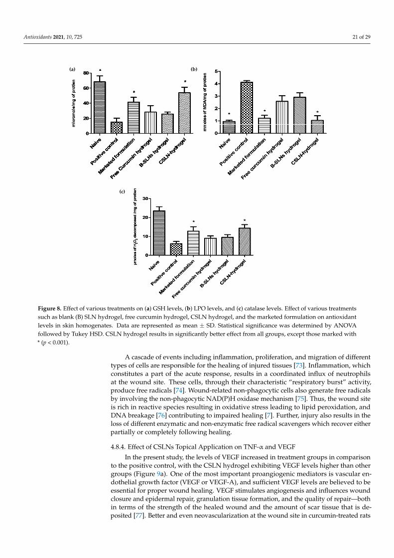
Antioxidants 2021, 10, 725 21 of 29
Antioxidants 2021, 10, x FOR PEER REVIEW 22 of 30
rich in reactive species resulting in oxidative stress leading to lipid peroxidation, and DNA breakage [76] contributing to impaired healing [7]. Further, injury also results in the loss of different enzymatic and non-enzymatic free radical scavengers which recover ei-ther partially or completely following healing.
Figure 8. Effect of various treatments on (a) GSH levels, (b) LPO levels, and (c) catalase levels. Effect of various treatments such as blank (B) SLN hydrogel, free curcumin hydrogel, CSLN hy-drogel, and the marketed formulation on antioxidant levels in skin homogenates. Data are repre-sented as mean ± SD. Statistical significance was determined by ANOVA followed by Tukey HSD. CSLN hydrogel results in significantly better effect from all groups, except those marked with * (p < 0.001).
4.8.4. Effect of CSLNs Topical Application on TNF-α and VEGF In the present study, the levels of VEGF increased in treatment groups in comparison
to the positive control, with the CSLN hydrogel exhibiting VEGF levels higher than other groups (Figure 9a). One of the most important proangiogenic mediators is vascular endo-thelial growth factor (VEGF or VEGF-A), and sufficient VEGF levels are believed to be essential for proper wound healing. VEGF stimulates angiogenesis and influences wound closure and epidermal repair, granulation tissue formation, and the quality of repair—both in terms of the strength of the healed wound and the amount of scar tissue that is deposited [77]. Better and even neovascularization at the wound site in curcumin-treated rats in comparison to the positive control using CD31 staining has been reported [78]. Further, topical application of curcumin was shown to significantly upregulate the mRNA and protein expression of VEGF and TGF-β1 until day 14 [78]. Similarly, in a recently re-ported in vitro study [79], a significant increase in the number of formed tubules, the up-regulation of VEGF expressions and the migration ability of high glucose-induced human
(a) (b)
(c)
Figure 8. Effect of various treatments on (a) GSH levels, (b) LPO levels, and (c) catalase levels. Effect of various treatmentssuch as blank (B) SLN hydrogel, free curcumin hydrogel, CSLN hydrogel, and the marketed formulation on antioxidantlevels in skin homogenates. Data are represented as mean ± SD. Statistical significance was determined by ANOVAfollowed by Tukey HSD. CSLN hydrogel results in significantly better effect from all groups, except those marked with* (p < 0.001).
A cascade of events including inflammation, proliferation, and migration of differenttypes of cells are responsible for the healing of injured tissues [73]. Inflammation, whichconstitutes a part of the acute response, results in a coordinated influx of neutrophilsat the wound site. These cells, through their characteristic “respiratory burst” activity,produce free radicals [74]. Wound-related non-phagocytic cells also generate free radicalsby involving the non-phagocytic NAD(P)H oxidase mechanism [75]. Thus, the wound siteis rich in reactive species resulting in oxidative stress leading to lipid peroxidation, andDNA breakage [76] contributing to impaired healing [7]. Further, injury also results in theloss of different enzymatic and non-enzymatic free radical scavengers which recover eitherpartially or completely following healing.
4.8.4. Effect of CSLNs Topical Application on TNF-α and VEGF
In the present study, the levels of VEGF increased in treatment groups in comparisonto the positive control, with the CSLN hydrogel exhibiting VEGF levels higher than othergroups (Figure 9a). One of the most important proangiogenic mediators is vascular en-dothelial growth factor (VEGF or VEGF-A), and sufficient VEGF levels are believed to beessential for proper wound healing. VEGF stimulates angiogenesis and influences woundclosure and epidermal repair, granulation tissue formation, and the quality of repair—bothin terms of the strength of the healed wound and the amount of scar tissue that is de-posited [77]. Better and even neovascularization at the wound site in curcumin-treated rats
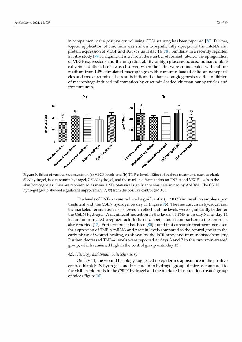
Antioxidants 2021, 10, 725 22 of 29
in comparison to the positive control using CD31 staining has been reported [78]. Further,topical application of curcumin was shown to significantly upregulate the mRNA andprotein expression of VEGF and TGF-β1 until day 14 [78]. Similarly, in a recently reportedin vitro study [79], a significant increase in the number of formed tubules, the upregulationof VEGF expressions and the migration ability of high glucose-induced human umbili-cal vein endothelial cells was observed when the latter were co-incubated with culturemedium from LPS-stimulated macrophages with curcumin-loaded chitosan nanoparti-cles and free curcumin. The results indicated enhanced angiogenesis via the inhibitionof macrophage-induced inflammation by curcumin-loaded chitosan nanoparticles andfree curcumin.
Antioxidants 2021, 10, x FOR PEER REVIEW 23 of 30
umbilical vein endothelial cells was observed when the latter were co-incubated with cul-ture medium from LPS-stimulated macrophages with curcumin-loaded chitosan nano-particles and free curcumin. The results indicated enhanced angiogenesis via the inhibi-tion of macrophage-induced inflammation by curcumin-loaded chitosan nanoparticles and free curcumin.
Figure 9. Effect of various treatments on (a) VEGF levels and (b) TNF-α levels. Effect of various treatments such as blank SLN hydrogel, free curcumin hydrogel, CSLN hydrogel, and the mar-keted formulation on TNF-α and VEGF levels in the skin homogenates. Data are represented as mean ± SD. Statistical significance was determined by ANOVA. The CSLN hydrogel group showed significant improvement (*, @) from the positive control (p< 0.05).
The levels of TNF-α were reduced significantly (p < 0.05) in the skin samples upon treatment with the CSLN hydrogel on day 11 (Figure 9b). The free curcumin hydrogel and the marketed formulation also showed an effect, but the levels were significantly better for the CSLN hydrogel. A significant reduction in the levels of TNF-α on day 7 and day 14 in curcumin-treated streptozotocin-induced diabetic rats in comparison to the control is also reported [17]. Furthermore, it has been [80] found that curcumin treatment in-creased the expression of TNF-α mRNA and protein levels compared to the control group in the early phase of wound healing, as shown by the PCR array and immunohistochem-istry. Further, decreased TNF-α levels were reported at days 3 and 7 in the curcumin-treated group, which remained high in the control group until day 12.
4.9. Histology and Immunohistochemistry On day 11, the wound histology suggested no epidermis appearance in the positive
control, blank SLN hydrogel, and free curcumin hydrogel group of mice as compared to the visible epidermis in the CSLN hydrogel and the marketed formulation-treated group of mice (Figure 10).
Figure 9. Effect of various treatments on (a) VEGF levels and (b) TNF-α levels. Effect of various treatments such as blankSLN hydrogel, free curcumin hydrogel, CSLN hydrogel, and the marketed formulation on TNF-α and VEGF levels in theskin homogenates. Data are represented as mean ± SD. Statistical significance was determined by ANOVA. The CSLNhydrogel group showed significant improvement (*, @) from the positive control (p< 0.05).
The levels of TNF-α were reduced significantly (p < 0.05) in the skin samples upontreatment with the CSLN hydrogel on day 11 (Figure 9b). The free curcumin hydrogel andthe marketed formulation also showed an effect, but the levels were significantly better forthe CSLN hydrogel. A significant reduction in the levels of TNF-α on day 7 and day 14in curcumin-treated streptozotocin-induced diabetic rats in comparison to the control isalso reported [17]. Furthermore, it has been [80] found that curcumin treatment increasedthe expression of TNF-α mRNA and protein levels compared to the control group in theearly phase of wound healing, as shown by the PCR array and immunohistochemistry.Further, decreased TNF-α levels were reported at days 3 and 7 in the curcumin-treatedgroup, which remained high in the control group until day 12.
4.9. Histology and Immunohistochemistry
On day 11, the wound histology suggested no epidermis appearance in the positivecontrol, blank SLN hydrogel, and free curcumin hydrogel group of mice as compared tothe visible epidermis in the CSLN hydrogel and the marketed formulation-treated groupof mice (Figure 10).
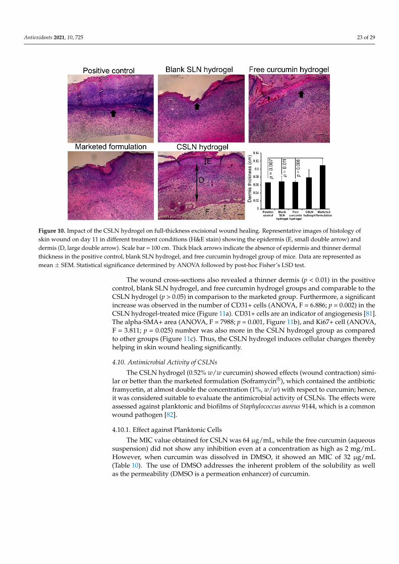
Antioxidants 2021, 10, 725 23 of 29Antioxidants 2021, 10, x FOR PEER REVIEW 24 of 30
Figure 10. Impact of the CSLN hydrogel on full-thickness excisional wound healing. Representative images of histology of skin wound on day 11 in different treatment conditions (H&E stain) showing the epidermis (E, small double arrow) and dermis (D, large double arrow). Scale bar = 100 cm. Thick black arrows indicate the absence of epidermis and thinner dermal thickness in the positive control, blank SLN hydrogel, and free curcumin hydrogel group of mice. Data are repre-sented as mean ± SEM. Statistical significance determined by ANOVA followed by post-hoc Fisher’s LSD test.
The wound cross-sections also revealed a thinner dermis (p < 0.01) in the positive control, blank SLN hydrogel, and free curcumin hydrogel groups and comparable to the CSLN hydrogel (p > 0.05) in comparison to the marketed group. Furthermore, a significant increase was observed in the number of CD31+ cells (ANOVA, F = 6.886; p = 0.002) in the CSLN hydrogel-treated mice (Figure 11a). CD31+ cells are an indicator of angiogenesis [81]. The alpha-SMA+ area (ANOVA, F = 7988; p = 0.001, Figure 11b), and Ki67+ cell (ANOVA, F = 3.811; p = 0.025) number was also more in the CSLN hydrogel group as compared to other groups (Figure 11c). Thus, the CSLN hydrogel induces cellular changes thereby helping in skin wound healing significantly.
Figure 10. Impact of the CSLN hydrogel on full-thickness excisional wound healing. Representative images of histology ofskin wound on day 11 in different treatment conditions (H&E stain) showing the epidermis (E, small double arrow) anddermis (D, large double arrow). Scale bar = 100 cm. Thick black arrows indicate the absence of epidermis and thinner dermalthickness in the positive control, blank SLN hydrogel, and free curcumin hydrogel group of mice. Data are represented asmean ± SEM. Statistical significance determined by ANOVA followed by post-hoc Fisher’s LSD test.
The wound cross-sections also revealed a thinner dermis (p < 0.01) in the positivecontrol, blank SLN hydrogel, and free curcumin hydrogel groups and comparable to theCSLN hydrogel (p > 0.05) in comparison to the marketed group. Furthermore, a significantincrease was observed in the number of CD31+ cells (ANOVA, F = 6.886; p = 0.002) in theCSLN hydrogel-treated mice (Figure 11a). CD31+ cells are an indicator of angiogenesis [81].The alpha-SMA+ area (ANOVA, F = 7988; p = 0.001, Figure 11b), and Ki67+ cell (ANOVA,F = 3.811; p = 0.025) number was also more in the CSLN hydrogel group as comparedto other groups (Figure 11c). Thus, the CSLN hydrogel induces cellular changes therebyhelping in skin wound healing significantly.
4.10. Antimicrobial Activity of CSLNs
The CSLN hydrogel (0.52% w/w curcumin) showed effects (wound contraction) simi-lar or better than the marketed formulation (Soframycin®), which contained the antibioticframycetin, at almost double the concentration (1%, w/w) with respect to curcumin; hence,it was considered suitable to evaluate the antimicrobial activity of CSLNs. The effects wereassessed against planktonic and biofilms of Staphylococcus aureus 9144, which is a commonwound pathogen [82].
4.10.1. Effect against Planktonic Cells
The MIC value obtained for CSLN was 64 µg/mL, while the free curcumin (aqueoussuspension) did not show any inhibition even at a concentration as high as 2 mg/mL.However, when curcumin was dissolved in DMSO, it showed an MIC of 32 µg/mL(Table 10). The use of DMSO addresses the inherent problem of the solubility as wellas the permeability (DMSO is a permeation enhancer) of curcumin.
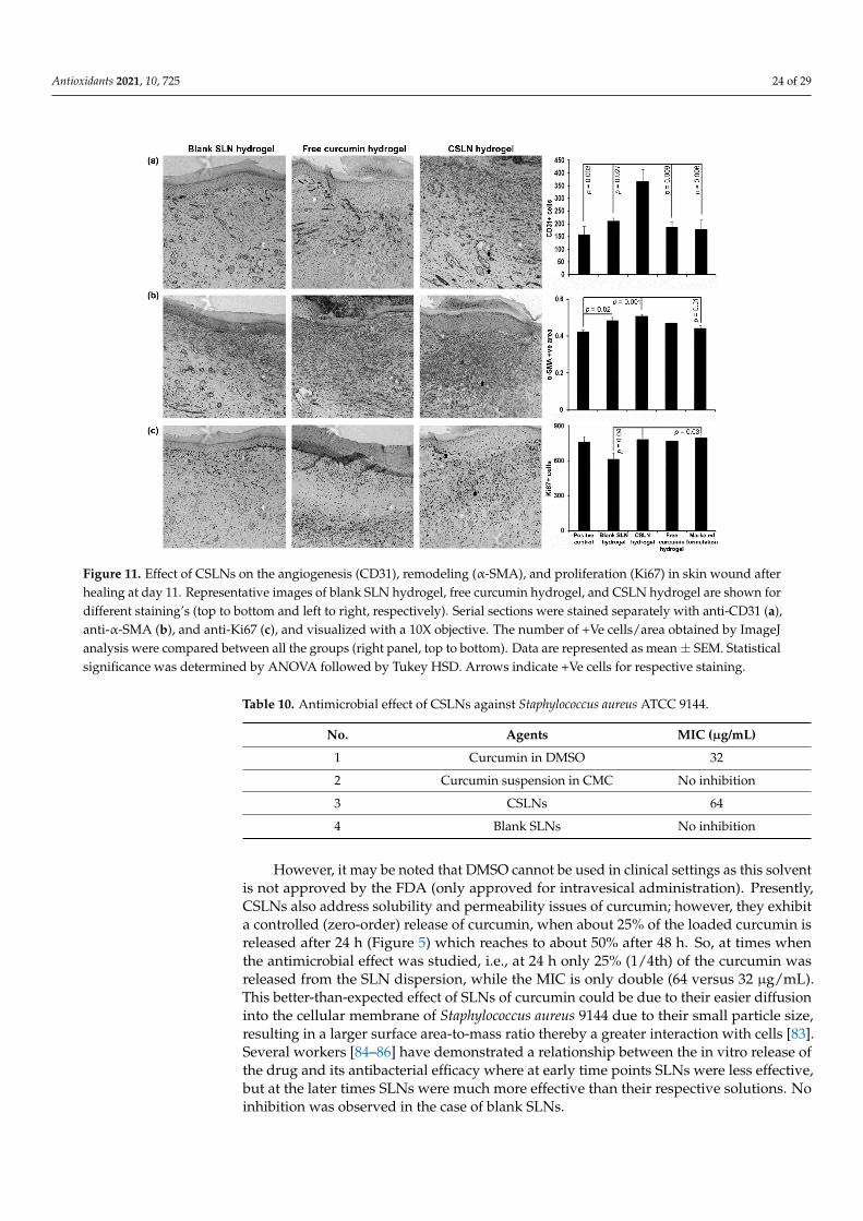
Antioxidants 2021, 10, 725 24 of 29Antioxidants 2021, 10, x FOR PEER REVIEW 25 of 30
Figure 11. Effect of CSLNs on the angiogenesis (CD31), remodeling (α-SMA), and proliferation (Ki67) in skin wound after healing at day 11. Representative images of blank SLN hydrogel, free curcumin hydrogel, and CSLN hydrogel are shown for different staining’s (top to bottom and left to right, respectively). Serial sections were stained separately with anti-CD31 (a), anti-α-SMA (b), and anti-Ki67 (c), and visualized with a 10X objective. The number of +Ve cells/area obtained by ImageJ analysis were compared between all the groups (right panel, top to bottom). Data are represented as mean ± SEM. Statistical significance was determined by ANOVA followed by Tukey HSD. Arrows indicate +Ve cells for respective staining.
4.10. Antimicrobial Activity of CSLNs The CSLN hydrogel (0.52% w/w curcumin) showed effects (wound contraction) sim-
ilar or better than the marketed formulation (Soframycin®), which contained the antibiotic framycetin, at almost double the concentration (1%, w/w) with respect to curcumin; hence, it was considered suitable to evaluate the antimicrobial activity of CSLNs. The effects were assessed against planktonic and biofilms of Staphylococcus aureus 9144, which is a common wound pathogen [82].
4.10.1. Effect against Planktonic Cells The MIC value obtained for CSLN was 64 µg/mL, while the free curcumin (aqueous
suspension) did not show any inhibition even at a concentration as high as 2 mg/mL. However, when curcumin was dissolved in DMSO, it showed an MIC of 32 µg/mL (Table 10). The use of DMSO addresses the inherent problem of the solubility as well as the per-meability (DMSO is a permeation enhancer) of curcumin.
Table 10. Antimicrobial effect of CSLNs against Staphylococcus aureus ATCC 9144.
No. Agents MIC (µg/mL) 1 Curcumin in DMSO 32 2 Curcumin suspension in CMC No inhibition 3 CSLNs 64 4 Blank SLNs No inhibition
Figure 11. Effect of CSLNs on the angiogenesis (CD31), remodeling (α-SMA), and proliferation (Ki67) in skin wound afterhealing at day 11. Representative images of blank SLN hydrogel, free curcumin hydrogel, and CSLN hydrogel are shown fordifferent staining’s (top to bottom and left to right, respectively). Serial sections were stained separately with anti-CD31 (a),anti-α-SMA (b), and anti-Ki67 (c), and visualized with a 10X objective. The number of +Ve cells/area obtained by ImageJanalysis were compared between all the groups (right panel, top to bottom). Data are represented as mean ± SEM. Statisticalsignificance was determined by ANOVA followed by Tukey HSD. Arrows indicate +Ve cells for respective staining.
Table 10. Antimicrobial effect of CSLNs against Staphylococcus aureus ATCC 9144.
No. Agents MIC (µg/mL)
1 Curcumin in DMSO 32
2 Curcumin suspension in CMC No inhibition
3 CSLNs 64
4 Blank SLNs No inhibition
However, it may be noted that DMSO cannot be used in clinical settings as this solventis not approved by the FDA (only approved for intravesical administration). Presently,CSLNs also address solubility and permeability issues of curcumin; however, they exhibita controlled (zero-order) release of curcumin, when about 25% of the loaded curcumin isreleased after 24 h (Figure 5) which reaches to about 50% after 48 h. So, at times whenthe antimicrobial effect was studied, i.e., at 24 h only 25% (1/4th) of the curcumin wasreleased from the SLN dispersion, while the MIC is only double (64 versus 32 µg/mL).This better-than-expected effect of SLNs of curcumin could be due to their easier diffusioninto the cellular membrane of Staphylococcus aureus 9144 due to their small particle size,resulting in a larger surface area-to-mass ratio thereby a greater interaction with cells [83].Several workers [84–86] have demonstrated a relationship between the in vitro release ofthe drug and its antibacterial efficacy where at early time points SLNs were less effective,but at the later times SLNs were much more effective than their respective solutions. Noinhibition was observed in the case of blank SLNs.
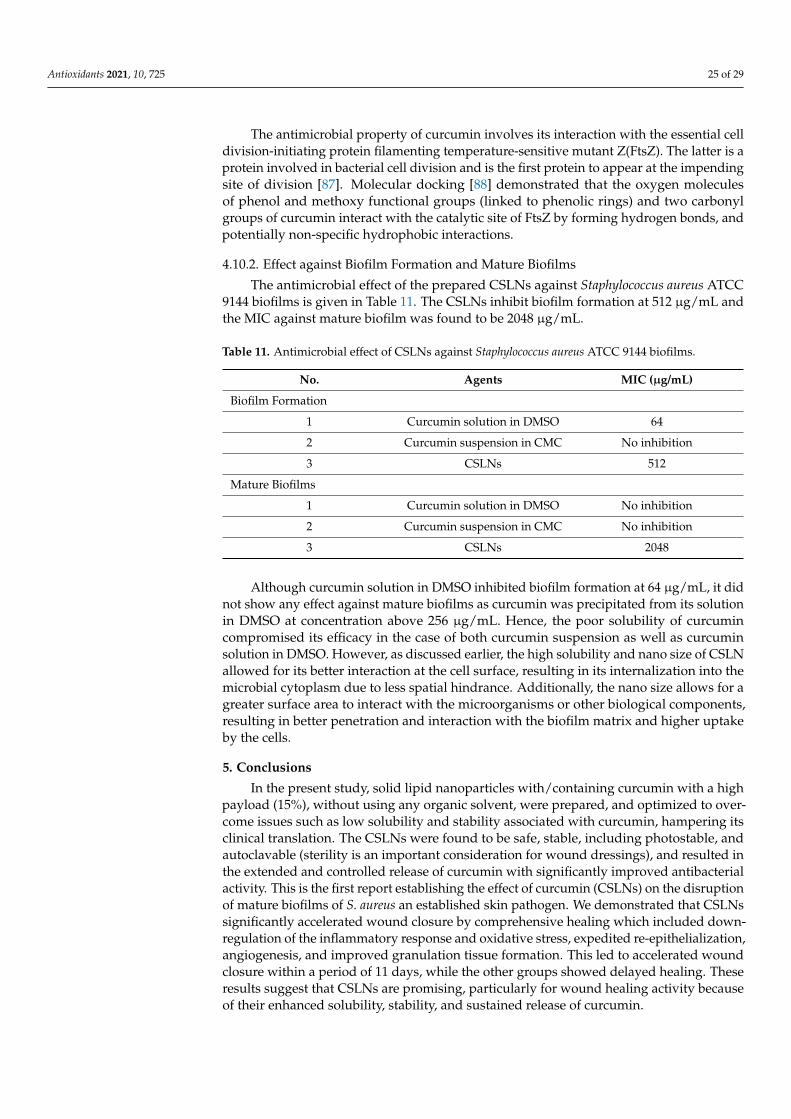
Antioxidants 2021, 10, 725 25 of 29
The antimicrobial property of curcumin involves its interaction with the essential celldivision-initiating protein filamenting temperature-sensitive mutant Z(FtsZ). The latter is aprotein involved in bacterial cell division and is the first protein to appear at the impendingsite of division [87]. Molecular docking [88] demonstrated that the oxygen moleculesof phenol and methoxy functional groups (linked to phenolic rings) and two carbonylgroups of curcumin interact with the catalytic site of FtsZ by forming hydrogen bonds, andpotentially non-specific hydrophobic interactions.
4.10.2. Effect against Biofilm Formation and Mature Biofilms
The antimicrobial effect of the prepared CSLNs against Staphylococcus aureus ATCC9144 biofilms is given in Table 11. The CSLNs inhibit biofilm formation at 512 µg/mL andthe MIC against mature biofilm was found to be 2048 µg/mL.
Table 11. Antimicrobial effect of CSLNs against Staphylococcus aureus ATCC 9144 biofilms.
No. Agents MIC (µg/mL)
Biofilm Formation
1 Curcumin solution in DMSO 64
2 Curcumin suspension in CMC No inhibition
3 CSLNs 512
Mature Biofilms
1 Curcumin solution in DMSO No inhibition
2 Curcumin suspension in CMC No inhibition
3 CSLNs 2048
Although curcumin solution in DMSO inhibited biofilm formation at 64 µg/mL, it didnot show any effect against mature biofilms as curcumin was precipitated from its solutionin DMSO at concentration above 256 µg/mL. Hence, the poor solubility of curcumincompromised its efficacy in the case of both curcumin suspension as well as curcuminsolution in DMSO. However, as discussed earlier, the high solubility and nano size of CSLNallowed for its better interaction at the cell surface, resulting in its internalization into themicrobial cytoplasm due to less spatial hindrance. Additionally, the nano size allows for agreater surface area to interact with the microorganisms or other biological components,resulting in better penetration and interaction with the biofilm matrix and higher uptakeby the cells.
5. Conclusions
In the present study, solid lipid nanoparticles with/containing curcumin with a highpayload (15%), without using any organic solvent, were prepared, and optimized to over-come issues such as low solubility and stability associated with curcumin, hampering itsclinical translation. The CSLNs were found to be safe, stable, including photostable, andautoclavable (sterility is an important consideration for wound dressings), and resulted inthe extended and controlled release of curcumin with significantly improved antibacterialactivity. This is the first report establishing the effect of curcumin (CSLNs) on the disruptionof mature biofilms of S. aureus an established skin pathogen. We demonstrated that CSLNssignificantly accelerated wound closure by comprehensive healing which included down-regulation of the inflammatory response and oxidative stress, expedited re-epithelialization,angiogenesis, and improved granulation tissue formation. This led to accelerated woundclosure within a period of 11 days, while the other groups showed delayed healing. Theseresults suggest that CSLNs are promising, particularly for wound healing activity becauseof their enhanced solubility, stability, and sustained release of curcumin.
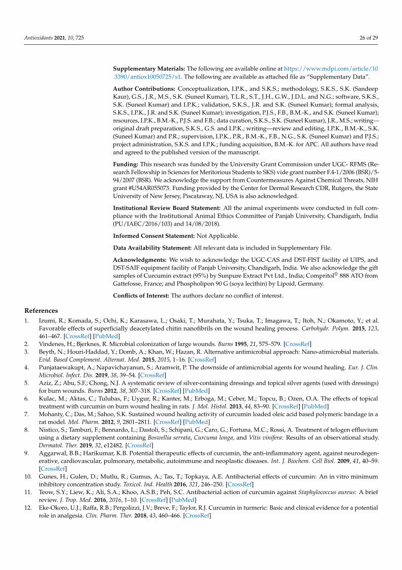
Antioxidants 2021, 10, 725 26 of 29
Supplementary Materials: The following are available online at https://www.mdpi.com/article/10.3390/antiox10050725/s1. The following are available as attached file as “Supplementary Data”.
Author Contributions: Conceptualization, I.P.K., and S.K.S.; methodology, S.K.S., S.K. (SandeepKaur), G.S., J.R., M.S., S.K. (Suneel Kumar), T.L.R., S.T., J.H., G.W., J.D.L. and N.G.; software, S.K.S.,S.K. (Suneel Kumar) and I.P.K.; validation, S.K.S., J.R. and S.K. (Suneel Kumar); formal analysis,S.K.S., I.P.K., J.R. and S.K. (Suneel Kumar); investigation, P.J.S., F.B., B.M.-K., and S.K. (Suneel Kumar);resources, I.P.K., B.M.-K., P.J.S. and F.B.; data curation, S.K.S., S.K. (Suneel Kumar), J.R., M.S.; writing—original draft preparation, S.K.S., G.S. and I.P.K.; writing—review and editing, I.P.K., B.M.-K., S.K.(Suneel Kumar) and P.R.; supervision, I.P.K., P.R., B.M.-K., F.B., N.G., S.K. (Suneel Kumar) and P.J.S.;project administration, S.K.S. and I.P.K.; funding acquisition, B.M.-K. for APC. All authors have readand agreed to the published version of the manuscript.
Funding: This research was funded by the University Grant Commission under UGC- RFMS (Re-search Fellowship in Sciences for Meritorious Students to SKS) vide grant number F.4-1/2006 (BSR)/5-94/2007 (BSR). We acknowledge the support from Countermeasures Against Chemical Threats, NIHgrant #U54AR055073. Funding provided by the Center for Dermal Research CDR, Rutgers, the StateUniversity of New Jersey, Piscataway, NJ, USA is also acknowledged.
Institutional Review Board Statement: All the animal experiments were conducted in full com-pliance with the Institutional Animal Ethics Committee of Panjab University, Chandigarh, India(PU/IAEC/2016/103) and 14/08/2018).
Informed Consent Statement: Not Applicable.
Data Availability Statement: All relevant data is included in Supplementary File.
Acknowledgments: We wish to acknowledge the UGC-CAS and DST-FIST facility of UIPS, andDST-SAIF equipment facility of Panjab University, Chandigarh, India. We also acknowledge the giftsamples of Curcumin extract (95%) by Sunpure Extract Pvt Ltd., India; Compritol® 888 ATO fromGattefosse, France; and Phospholipon 90 G (soya lecithin) by Lipoid, Germany.
Conflicts of Interest: The authors declare no conflict of interest.
References1. Izumi, R.; Komada, S.; Ochi, K.; Karasawa, L.; Osaki, T.; Murahata, Y.; Tsuka, T.; Imagawa, T.; Itoh, N.; Okamoto, Y.; et al.
Favorable effects of superficially deacetylated chitin nanofibrils on the wound healing process. Carbohydr. Polym. 2015, 123,461–467. [CrossRef] [PubMed]
2. Vindenes, H.; Bjerknes, R. Microbial colonization of large wounds. Burns 1995, 21, 575–579. [CrossRef]3. Beyth, N.; Houri-Haddad, Y.; Domb, A.; Khan, W.; Hazan, R. Alternative antimicrobial approach: Nano-atimicrobial materials.
Evid. Based Complement. Alternat. Med. 2015, 2015, 1–16. [CrossRef]4. Punjataewakupt, A.; Napavichayanun, S.; Aramwit, P. The downside of antimicrobial agents for wound healing. Eur. J. Clin.
Microbiol. Infect. Dis. 2019, 38, 39–54. [CrossRef]5. Aziz, Z.; Abu, S.F.; Chong, N.J. A systematic review of silver-containing dressings and topical silver agents (used with dressings)
for burn wounds. Burns 2012, 38, 307–318. [CrossRef] [PubMed]6. Kulac, M.; Aktas, C.; Tulubas, F.; Uygur, R.; Kanter, M.; Erboga, M.; Ceber, M.; Topcu, B.; Ozen, O.A. The effects of topical
treatment with curcumin on burn wound healing in rats. J. Mol. Histol. 2013, 44, 83–90. [CrossRef] [PubMed]7. Mohanty, C.; Das, M.; Sahoo, S.K. Sustained wound healing activity of curcumin loaded oleic acid based polymeric bandage in a
rat model. Mol. Pharm. 2012, 9, 2801–2811. [CrossRef] [PubMed]8. Nistico, S.; Tamburi, F.; Bennardo, L.; Dastoli, S.; Schipani, G.; Caro, G.; Fortuna, M.C.; Rossi, A. Treatment of telogen effluvium
using a dietary supplement containing Boswellia serrata, Curcuma longa, and Vitis vinifera: Results of an observational study.Dermatol. Ther. 2019, 32, e12482. [CrossRef]
9. Aggarwal, B.B.; Harikumar, K.B. Potential therapeutic effects of curcumin, the anti-inflammatory agent, against neurodegen-erative, cardiovascular, pulmonary, metabolic, autoimmune and neoplastic diseases. Int. J. Biochem. Cell Biol. 2009, 41, 40–59.[CrossRef]
10. Gunes, H.; Gulen, D.; Mutlu, R.; Gumus, A.; Tas, T.; Topkaya, A.E. Antibacterial effects of curcumin: An in vitro minimuminhibitory concentration study. Toxicol. Ind. Health 2016, 321, 246–250. [CrossRef]
11. Teow, S.Y.; Liew, K.; Ali, S.A.; Khoo, A.S.B.; Peh, S.C. Antibacterial action of curcumin against Staphylococcus aureus: A briefreview. J. Trop. Med. 2016, 2016, 1–10. [CrossRef] [PubMed]
12. Eke-Okoro, U.J.; Raffa, R.B.; Pergolizzi, J.V.; Breve, F.; Taylor, R.J. Curcumin in turmeric: Basic and clinical evidence for a potentialrole in analgesia. Clin. Pharm. Ther. 2018, 43, 460–466. [CrossRef]
Antioxidants 2021, 10, 725 27 of 29
13. Jayaprakasha, G.K.; Rao, L.J.; Sakariah, K.K. Antioxidant activities of curcumin, demethoxycurcumin and bisdemethoxycurcumin.Food Chem. 2006, 98, 720–724. [CrossRef]
14. Menon, V.P.; Sudheer, A.R. Antioxidant and anti-inflammatory properties of curcumin. Adv. Exp. Med. Biol. 2007, 595, 105–125.15. Gopinath, D.; Ahmed, M.; Gomathi, K.; Chitra, K.; Sehgal, P.; Jayakumar, R. Dermal wound healing processes with curcumin
incorporated collagen films. Biomaterials 2004, 25, 1911–1917. [CrossRef]16. Phan, T.T.; See, P.; Lee, S.T.; Chan, S.Y. Protective effects of curcumin against oxidative damage on skin cells in vitro: Its implication
for wound healing. J. Trauma 2001, 51, 927–931. [CrossRef]17. Kant, V.; Gopal, A.; Pathak, N.N.; Kumar, P.; Tandan, S.K.; Kumar, D. Antioxidant and anti-inflammatory potential of curcumin
accelerated the cutaneous wound healing in streptozotocin-induced diabetic rats. Int. Immunopharmacol. 2014, 20, 322–330.[CrossRef]
18. Panchatcharam, M.; Miriyala, S.; Gayathri, V.S.; Suguna, L. Curcumin improves wound healing by modulating collagen anddecreasing reactive oxygen species. Mol. Cell Biochem. 2006, 290, 87–96. [CrossRef]
19. Barzegar, A.; Moosavi-Movahedi, A.A. Intracellular ROS protection efficiency and free radical-scavenging activity of curcumin.PLoS ONE 2011, 6, e26012. [CrossRef]
20. Sreejayan, N.; Rao, M.N. Free radical scavenging activity of curcuminoids. Arzneimittelforschung 1996, 46, 146–171.21. Gupta, T.; Singh, J.; Kaur, S.; Sandhu, S.; Singh, G.; Kaur, I.P. Enhancing Bioavailability and Stability of Curcumin Using Solid
Lipid Nanoparticles (CLEN): A Covenant for Its Effectiveness. Front. Bioeng. Biotechnol. 2020, 8, 1–14. [CrossRef] [PubMed]22. Seyithanoglu, M.H.; Abdallah, A.; Kitis, S.; Güler, E.M.; Koçyigit, A.; Dündar, T.T.; Gündag Papaker, M. Investigation of cytotoxic,
genotoxic, and apoptotic effects of curcumin on glioma cells. Cell Mol. Biol. 2019, 65, 100–108. [CrossRef]23. Scharstuhl, A.; Mutsaers, H.; Pennings, S.; Szarek, W.; Russel, F.; Wagener, F. Curcumin-induced fibroblast apoptosis and in vitro
wound contraction are regulated by antioxidants and heme oxygenase: Implications for scar formation. J. Cell Mol. Med. 2009, 13,712–715. [CrossRef]
24. Akbik, D.; Ghadiri, M.; Chrzanowski, W.; Rohanizadeh, R. Curcumin as a wound healing agent. Life Sci. 2014, 116, 1–7. [CrossRef][PubMed]
25. Wissing, S.A.; Müller, R.H. The influence of solid lipid nanoparticles on skin hydration and viscoelasticity: In vivo study. Eur. J.Pharm. Biopharm. 2003, 56, 67–72. [CrossRef]
26. Ali, H.H.; Michaux, F.; Khanji, A.N.; Jasniewski, J.; Linder, M. Chitosan-shea butter solid nanoparticles assemblies for thepreparation of a novel nanoparticles in microparticles system containing curcumin. Colloids Surf. A Physicochem. Eng. Asp. 2018,553, 359–367.
27. Doktorovova, S.; Souto, E.B.; Silva, A.M. Hansen solubility parameters (HSP) for prescreening formulation of solid lipidnanoparticles (SLN): In vitro testing of curcumin-loaded SLN in MCF-7 and BT-474 cell lines. Pharm. Dev. Technol. 2018, 23,96–105. [CrossRef]
28. Lakhani, P.; Patil, A.; Taskar, P.; Ashour, E.; Majumdar, S. Curcumin-loaded nanostructured lipid carriers for ocular drug delivery:Design optimization and characterization. J. Drug Deliv. Sci. Technol. 2018, 47, 159–166. [CrossRef] [PubMed]
29. Xue, J.; Wang, T.; Hu, Q.; Zhou, M.; Luo, Y. Insight into natural biopolymer-emulsified solid lipid nanoparticles for encapsulationof curcumin: Effect of loading methods. Food Hydrocoll. 2018, 79, 110–116. [CrossRef]
30. Gumireddy, A.; Christman, R.; Kumari, D.; Tiwari, A.; North, E.J.; Chauhan, H. Preparation, characterization, and in vitroevaluation of curcumin and resveratrol-loaded solid lipid nanoparticles. AAPS PharmSciTech 2019, 20, 1–14. [CrossRef]
31. Luan, L.; Chi, Z.; Liu, C. Chinese white wax solid lipid nanoparticles as a novel nanocarrier of curcumin for inhibiting theformation of Staphylococcus aureus biofilms. Nanomaterials 2019, 9, 763. [CrossRef] [PubMed]
32. Minafra, L.; Porcino, N.; Bravatà, V.; Gaglio, D.; Bonanomi, M.; Amore, E.; Cammarata, F.P.; Russo, G.; Militello, C.; Savoca, G.;et al. Radiosensitizing effect of curcumin-loaded lipid nanoparticles in breast cancer cells. Sci. Rep. 2019, 9, 1–16. [CrossRef]
33. Ban, C.; Jo, M.; Park, Y.H.; Kim, J.H.; Han, J.Y.; Lee, K.W.; Kweon, D.H.; Choi, Y.J. Enhancing the oral bioavailability of curcuminusing solid lipid nanoparticles. Food Chem. 2020, 302, 125328. [CrossRef]
34. Kakkar, V.; Kaur, I.P. Evaluating potential of curcumin loaded solid lipid nanoparticles in aluminium induced behavioural,biochemical and histopathological alterations in mice brain. Food Chem. Toxicol. 2011, 49, 2906–2913. [CrossRef]
35. Kakkar, V.; Mishra, A.K.; Chuttani, K.; Chopra, K.; Kaur, I.P. Proof of concept studies to confirm the delivery of curcumin loadedsolid lipid nanoparticles. Int. J. Pharm. 2013, 448, 354–359. [CrossRef] [PubMed]
36. Ambarsari, L.; Nurcholis, W.; Darusman, L.K.; Mujib, M.A.; Heryanto, R. The Curcuminoids Extract of Curcuma xanthorrhizaRoxB. Loaded Solid Lipid Nanoparticles. Int. J. Sci. Res. 2014, 3, 852–856.
37. Ji, H.; Tang, J.; Li, M.; Ren, J.; Zheng, N.; Wu, L. Curcumin-loaded solid lipid nanoparticles with Brij78 and TPGS improvedin vivo oral bioavailability and in situ intestinal absorption of curcumin. Drug Deliv. 2016, 23, 459–470. [CrossRef] [PubMed]
38. Shrotriya, S.; Ranpise, N.; Satpute, P.; Vidhate, B. Skin targeting of curcumin solid lipid nanoparticles-engrossed topical gel forthe treatment of pigmentation and irritant contact dermatitis. Artif. Cells Nanomed. Biotechnol. 2018, 46, 1471–1482. [CrossRef]
39. Kakkar, V.; Singh, S.; Singla, D.; Kaur, I.P. Exploring solid lipid nanoparticles to enhance the oral bioavailability of curcumin. Mol.Nut. Food Res. 2011, 55, 495–503. [CrossRef] [PubMed]
40. Wang, W.; Chen, T.; Xu, H.; Ren, B.; Cheng, X.; Qi, R.; Liu, H.; Wang, Y.; Yan, L.; Chen, S.; et al. Curcumin-Loaded Solid LipidNanoparticles Enhanced Anticancer Efficiency in Breast Cancer. Molecules 2018, 23, 1578. [CrossRef]
Antioxidants 2021, 10, 725 28 of 29
41. Anumolu, S.S.; Menjoge, A.R.; Deshmukh, M.; Gerecke, D.; Stein, S.; Laskin, J.; Sinko, P.J. Doxycycline hydrogels with reversibledisulfide crosslinks for dermal wound healing of mustard injuries. Biomaterials 2011, 32, 1204–1217. [CrossRef]
42. Kosol, W.; Kumar, S.; Marrero-BerrÍos, I.; Berthiaume, F. Medium conditioned by human mesenchymal stromal cells reverseslow serum and hypoxia-induced inhibition of wound closure. Biochem. Biophys. Res. Commun. 2020, 522, 335–341. [CrossRef][PubMed]
43. Composto, G.M.; Laskin, J.D.; Laskin, D.L.; Gerecke, D.R.; Casillas, R.P.; Heindel, N.D.; Joseph, L.B.; Heck, D.E. Mitigation ofnitrogen mustard mediated skin injury by a novel indomethacin bifunctional prodrug. Exp. Mol. Pathol. 2016, 100, 522–531.[CrossRef] [PubMed]
44. Wills, E.D. Mechanisms of lipid peroxide formation in animal tissues. Biochem. J. 1966, 99, 667–676. [CrossRef]45. Moron, M.S.; Depierre, J.W.; Mannervik, B. Levels of glutathione, glutathione reductase and glutathione s-transferase activities in
rat lung and liver. Biochim. Biophys. Acta 1979, 582, 67–78. [CrossRef]46. Cuppett, S.L.; Wijeratne, S.S.K.; Schlegel, V.J. Hydrogen peroxide induced oxidative stress damage and antioxidant enzyme
response in caco-2 human colon cells. Agric. Food Chem. 2005, 53, 8768–8774.47. Kumar, S.; Yarmush, M.L.; Dash, B.C.; Hsia, H.C.; Berthiaume, F. Impact of complete spinal cord injury on healing of skin ulcers
in mouse models. J. Neurotrauma 2018, 15, 815–824. [CrossRef] [PubMed]48. Kumar, S.; Tan, Y.; Yarmush, M.L.; Dash, B.C.; Hsia, H.C.; Berthiaume, F. Mouse model of pressure ulcers after spinal cord injury.
J. Vis. Exp. 2019, 9, 145. [CrossRef]49. Kakkar, S.; Karuppayil, S.M.; Raut, J.S.; Giansanti, F.; Papucci, L.; Schiavone, N.; Kaur, I.P. Lipid-polyethylene glycol based
nano-ocular formulation of ketoconazole. Int. J. Pharm. 2015, 495, 276–289. [CrossRef]50. Singh, H.; Bhandari, R.; Kaur, I.P. Encapsulation of Rifampicin in a solid lipid nanoparticulate system to limit its degradation and
interaction with Isoniazid at acidic pH. Int. J. Pharm. 2013, 446, 106–111. [CrossRef]51. D’souza, A.A.; Shegokar, R. Polyethylene glycol (PEG): A versatile polymer for pharmaceutical applications. Expert Opin. Drug
Deliv. 2016, 13, 1257–1275. [CrossRef] [PubMed]52. Kaur, P.; Garg, T.; Rath, G.; Murthy, R.S.; Goyal, A.K. Development, optimization and evaluation of surfactant-based pulmonary
nanolipid carrier system of paclitaxel for the management of drug resistance lung cancer using Box-Behnken design. Drug Deliv.2016, 23, 1912–1925. [CrossRef] [PubMed]
53. Helgason, T.; Awad, T.S.; Kristbergsson, K.; McClements, D.J.; Weiss, J. Effect of surfactant surface coverage on formation of solidlipid nanoparticles (SLN). J. Colloid Interface Sci. 2009, 334, 75–81. [CrossRef]
54. Li, D.; Jiang, Y.; Lv, S.; Liu, X.; Gu, J.; Chen, Q.; Zhang, Y. Preparation of plasticized poly (lactic acid) and its influence on theproperties of composite materials. PLoS ONE 2018, 13, e0193520. [CrossRef] [PubMed]
55. Kushwaha, A.K.; Vuddanda, P.R.; Karunanidhi, P.; Singh, S.K.; Singh, S. Development and evaluation of solid lipid nanoparticlesof Raloxifene hydrochloride for enhanced bioavailability. Biomed. Res. Int. 2013, 2013, 1–9. [CrossRef] [PubMed]
56. Missana, T.; Adell, A. On the applicability of DLVO theory to the prediction of clay colloids stability. J. Colloid Interface Sci. 2000,230, 150–156. [CrossRef] [PubMed]
57. Singh, M.; Guzman-Aranguez, A.; Hussain, A.; Srinivas, C.S.; Kaur, I.P. Solid lipid nanoparticles for ocular delivery of isoniazid:Evaluation, proof of concept and in vivo safety & kinetics. Nanomedicine 2019, 14, 465–491.
58. Bunjes, H.; Westesen, K.; Koch, M.H.J. Crystallization tendency and polymorphic transitions in triglyceride nanoparticles. Int. J.Pharm. 1996, 129, 159–173. [CrossRef]
59. Freitas, C.; Müller, R.H. Effect of light and temperature on zeta potential and physical stability in solid lipid nanoparticle (SLN™)dispersions. Int. J. Pharm. 1998, 168, 221–229. [CrossRef]
60. Schuhmann, R. Physical Stability of Parenteral Fat Emulsions: Development of an Examination Scheme with a Special Focus on AnalyticalPossibilities; Freie Universität Berlin: Berlin, Germany, 1995.
61. Jankun, J.; Wyganowska-Swiatkowska, M.; Dettlaff, K.; Jelinska, A.; Surdacka, A.; Watróbska-Swietlikowska, D.; Skrzypczak-Jankun, E. Determining whether curcumin degradation/condensation is actually bioactivation. Int. J. Mol. Med. 2016, 37,1151–1158. [CrossRef] [PubMed]
62. Tønnesen, H.H. Solubility, chemical and photochemical stability of curcumin in surfactant solutions. Studies of curcumin andcurcuminoids, XXVIII. Pharmazie 2002, 57, 820–824.
63. Cavalli, R.; Caputo, O.; Carlotti, M.E.; Trotta, M.; Scarnecchia, C.; Gasco, M.R. Sterilization and freeze-drying of drug-free anddrug-loaded solid lipid nanoparticles. Int. J. Pharm. 1997, 148, 47–54. [CrossRef]
64. Mehnert, W.; Mäder, K. Solid lipid nanoparticles: Production, characterization and applications. Adv. Drug Deliv. Rev. 2001, 47,165–196. [CrossRef]
65. Schwarz, C.; Mehnert, W.; Lucks, J.S.; Müller, R.H. Solid lipid nanoparticles (SLN) for controlled drug delivery. I. Production,characterization and sterilization. J. Control. Release 1994, 30, 83–96. [CrossRef]
66. Dash, S.; Murthy, P.N.; Nath, L.; Chowdhury, P. Kinetic modeling on drug release from controlled drug delivery systems. Acta Pol.Pharm. 2010, 67, 217–223.
67. Wang, T.; Ma, X.; Lei, Y.; Luo, Y. Solid lipid nanoparticles coated with cross-linked polymeric double layer for oral delivery ofcurcumin. Colloids Surf. B 2016, 148, 1–11. [CrossRef] [PubMed]
68. Agrawal, R.; Sandhu, S.K.; Sharma, I.; Kaur, I.P. Development and evaluation of curcumin-loaded elastic vesicles as an effectivetopical anti-inflammatory formulation. AAPS PharmSciTech 2015, 16, 364–374. [CrossRef] [PubMed]
Antioxidants 2021, 10, 725 29 of 29
69. Koop, H.S.; de Freitas, R.A.; de Souza, M.M.; Savi, R.; Silveira, J.L.M. Topical curcumin-loaded hydrogels obtained usinggalactomannan from Schizolobium parahybae and xanthan. Carbohydr. Polym. 2015, 116, 229–236. [CrossRef] [PubMed]
70. Kang, D.; Li, B.; Luo, L.; Jiang, W.; Lu, Q.; Rong, M.; Lai, R. Curcumin shows excellent therapeutic effect on psoriasis in mousemodel. Biochimie 2016, 123, 73–80. [CrossRef]
71. Nasab, M.E.; Takzaree, N.; Saffaria, P.M.; Partoazar, A. In vitro antioxidant activity and in vivo wound-healing effect of lecithinliposomes: A comparative study. J. Comp. Eff. Res. 2019, 8, 633–643. [CrossRef]
72. Schafer, M.; Werner, S. Oxidative stress in normal and impaired wound repair. Pharmacol. Res. 2008, 58, 165–171. [CrossRef][PubMed]
73. Sidhu, G.S.; Mani, H.; Gaddipati, J.P.; Singh, A.K.; Seth, P.; Banaudha, K.K.; Patnaik, G.K.; Maheshwari, R.K. Curcumin enhanceswound healing in streptozotocin induced diabetic rats and genetically diabetic mice. Wound Repair Regen. 1999, 7, 362–374.[CrossRef]
74. Babior, B.M. Oxygen-dependent microbial killing by phagocytes (first of two parts). N. Engl. J. Med. 1978, 298, 659–668. [CrossRef]75. Griendling, K.K.; Sorescu, D.; Ushio-Fukai, M. NAD(P)H oxidase: Role in cardiovascular biology and disease. Circ. Res. 2000, 86,
494–501. [CrossRef]76. Wiseman, H.; Halliwell, B. Damage to DNA by reactive oxygen and nitrogen species: Role in inflammatory disease and
progression to cancer. Biochem. J. 1996, 313, 17–29. [CrossRef]77. Johnson, K.E.; Wilgus, T.A. Vascular endothelial growth factor and angiogenesis in the regulation of cutaneous wound repair.
Adv. Wound Care 2014, 3, 647–661. [CrossRef] [PubMed]78. Kant, V.; Gopal, A.; Kumar, D.; Pathak, N.N.; Ram, M.; Jangir, B.L.; Tandan, S.K.; Kumar, D. Curcumin induced angiogenesis
hastens wound healing in diabetic rat. J. Surg. Res. 2015, 193, 978–988. [CrossRef] [PubMed]79. Li, F.; Shi, Y.; Liang, J.; Zhao, L. Curcumin-loaded chitosan nanoparticles promote diabetic wound healing via attenuating
inflammation in a diabetic rat model. J. Biomater. Appl. 2019, 34, 476–486. [CrossRef]80. Yen, Y.H.; Pu, C.M.; Liu, C.W.; Chen, Y.C.; Chen, Y.C.; Liang, C.J.; Hsieh, J.H.; Huang, H.F.; Chen, Y.L. Curcumin accelerates
cutaneous wound healing via multiple biological actions: The involvement of TNF-α, MMP-9, α-SMA, and collagen. Int. WoundJ. 2018, 2018, 1–13. [CrossRef]
81. Wang, D.; Stockard, C.R.; Harkins, L.; Lott, P.; Salih, C.; Yuan, K.; Buchsbaum, D.; Hashim, A.; Zayzafoon, M.; Hardy, R.; et al.Immunohistochemistry in the evaluation of neovascularization in tumor xenografts. Biotech. Histochem. 2008, 83, 179–189.[CrossRef]
82. Roy, S.; Santra, S.; Das, A.; Dixith, S.; Sinha, M.; Ghatak, S.; Ghosh, N.; Banerjee, P.; Khanna, S.; Mathew-Steiner, S.; et al.Staphylococcus aureus biofilm infection compromises wound healing by causing deficiencies in granulation tissue collagen. Ann.Surg. 2019, 271, 1174–1185. [CrossRef]
83. Wang, L.; Hu, C.; Shao, L. The antimicrobial activity of nanoparticles: Present situation and prospects for the future. Int. J.Nanomed. 2017, 12, 1227–1249. [CrossRef]
84. Ghaffari, S.; Varshosaz, J.; Saadat, A.; Atyabi, F. Stability and antimicrobial effect of amikacin-loaded solid lipid nanoparticles. Int.J. Nanomed. 2011, 6, 35–43.
85. Kalhapure, R.S.; Mocktar, C.; Sikwal, D.R.; Sonawane, S.J.; Kathiravan, M.K.; Skelton, A.; Govender, T. Ion pairing with linoleicacid simultaneously enhances encapsulation efficiency and antibacterial activity of vancomycin in solid lipid nanoparticles.Colloids Surf. B 2014, 117, 303–311. [CrossRef] [PubMed]
86. Wang, X.F.; Zhang, S.L.; Zhu, L.Y.; Xie, S.Y.; Dong, Z.; Wang, Y.; Zhou, W.Z. Enhancement of antibacterial activity of tilmicosinagainst Staphylococcus aureus by solid lipid nanoparticles in vitro and in vivo. Vet. J. 2012, 191, 115–120. [CrossRef] [PubMed]
87. Rai, D.; Singh, J.K.; Roy, N.; Panda, D. Curcumin inhibits FtsZ assembly: An attractive mechanism for its antibacterial activity.Biochem. J. 2008, 410, 147–155. [CrossRef]
88. Kaur, S.; Modi, N.H.; Panda, D.; Roy, N. Probing the binding site of curcumin in Escherichia coli and Bacillus subtilis FtsZ—Astructural insight to unveil antibacterial activity of curcumin. Eur. J. Med. Chem. 2010, 45, 4209–4214. [CrossRef] [PubMed]