Embed Size (px)
Citation preview

SYSTEMICLUPUS
ERYTHEMATOSUS

• Systemic lupus erythematosus (SLE) is an autoimmune disease in which organs and cells undergo damage mediated by tissue-binding autoantibodies and immune complexes.
• Female : male 9 : 1 young women• 15–50 per 100,000
Definition & Prevalence

Etiology & Pathogenesis
Genes AbnormalInteraction & immune b/w Environmental
response
factors

Abnormal immune response
1. activation of innate immunity 2. lowered thresholds of adaptive immunity
cells (antigen-specific T and B lymphocytes)3. ineffective regulatory mechanism4. reduced clearance of apoptotic cells and of
immune complexes (Self-antigens: nucleosomal DNA, RNA in Sm, Ro,
La; phospholipids) 5. Activation of complement system


Multigenic factors
• C1q, MBL - clearance of apoptic cells
• FcR 2A &3A - clearance of immune complexes
• HLA-DR2,3,8 - antigen presentation
• PTPN22 – T - cell activation
• MCP-1 – chemotaxis
• IL – 10 - B cell activation

Environmental factors
• Exposure to U/V radiation
• Infections – EBV virus
• Gender – estradiol increases survival of
Lymphocytes
• Drugs – procainamide, hydralazine, D-
Pencilamaine

Environmental Triggers
Genetically Susceptible Individual
Nuleasomal proteins & Self
antigens
Activation of Th&B cells
Ig G Auto Ab
Clinical Manifestations

Clinical ManifestationsMusculoskeletalCutaneous Renal Neurological Vascular Occlusions Pulmonary Cardiac Hematologic Gastrointestinal Ocular

Musculoskeletal Manifestations
• Intermitent Polyarthritis nondeforming and nonerosive
• Myositis with muscle weakness & myalgias,
Jaccoud’s Arthopathy: Nonerosive, Reducible Deformities

Cutaneous Manifestations discoid lupus erythematosus
• Lupus dermatitis systemic rash subacute cutaneous lupus
erythematosus (SCLE)• SLE rash is photosensitive, common on
the face (particularly the cheeks and nose—the "butterfly" rash), ears, chin, V region of the neck, upper back, and extensor surfaces of the arms

Butterfly rash
Subacute Cutaneous Lupus

painful ulcerations on the oral or nasal mucosa

Renal Manifestations
• 50-70% of all lupus patients experience renal developments.
• ISN & RPS Classification of Lupus Nephritis
Minimal Lupus nephritis class – I Mesangial Lupus nephritis class – II Focal Lupus nephritis class – III Diffuse Proliferative Lupus nephritis class –IV Membranous Lupus nephritis class –V

• Class III & IV present with hematuria & proteinuria (>500mg / day)
• Nearly ½ end up in nephrotic syndrome
• ESRD occurs in DPGN after 2Yrs of disease manifestation. Leading cause of death in SLE

Lupus Nephritis
normal

Neurological Manifestations • cognitive dysfunction (memory & reasoning
diffic.)
• Headache• Seizures• Psychosis• Myelopathy
• Important to exclude other etiologies • If SLE then determine
DiffuseVascular occlusion
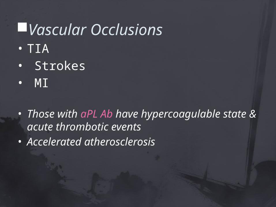
Vascular Occlusions• TIA• Strokes• MI
• Those with aPL Ab have hypercoagulable state & acute thrombotic events
• Accelerated atherosclerosis


Pulmonary Manifestations
• Pleuritis & pleural effusions
• Life-threatening – interstitial inflammation leading to fibrosis, shrinking lung syndrome, and intraalveolar hemorrhage

Cardiac Manifestations
• Pericarditis• Myocarditis• Endocarditis
(Libman – Sacks)• MI• Valvular
insufficiencies

Hematologic Manifestations• Anemia• Leukopenia• Thrombocytopenia

Gastrointestinal Manifestations• Nausea,vomiting, and diarrhea• abdominal pain -autoimmune
peritonitis• Increases (AST) and (ALT)• Vasculitis involving the intestine may be
life-threatening; perforations, ischemia, bleeding, and sepsis

Ocular Manifestations
• Sicca syndrome• Conjunctivitis• retinal vasculitis and optic neuritis

DiagnosisAMERICAN RHEUMATISM ASS. criteria based on
clinical features. ≥ 4 of these must be present

Laboratory Tests
Tests for Autoantibodies a) ANA antibodies ,DNA (dsDNA) are
specific for SLE
b) Antibodies to Sm are also specific for SLE
c) aPL their presence identify patients at increased risk for venous or arterial clotting, thrombocytopenia, and fetal loss
d) anti-Ro - neonatal lupus, sicca syndrome, and SCLE

Lupus band test

Tests for Diagnosis
• blood count• platelet count• Urinalysis• serum levels of creatinine or albumin

TreatmentNo CureAim: -to control acute flare - ↓ symptoms -prevent organ failure

Conservative Therapies for Non-Life-Threatening Disease
fatigue, pain, and autoAb of SLE, but without major organ involvement
• NSAID`S { adv effects specific for SLE:
aseptic meningitis, elevated serum transaminases, hypertension, & renal dysfunction }
ASPIRIN is da D.O.C

• Antimalarials (hydroxychloroquine, chloroquine, and quinacrine) often reduce dermatitis, arthritis, and fatigue
{Adv eff. – retinal toxicity}
• low doses of systemic glucocorticoids may be necessary
Life-Threatening SLE – Rx high-dose i.v glucocorticoid (1000mg for
3 days followed by 0.5–1 mg/kg of daily prednisone ) -standard practice
(or)
systemic glucocorticoids (0.5–2 mg/kg per day)
{ Adv eff. - infection, hyperglycemia, hypertension, osteoporosis, etc}
• maintenance dose 5 to 10 mg/ day for many years
• Cytotoxic/immunosuppressive agents
Cyclophosphamide { Adv eff. – ovarian failure}
(500–750 mg/m2 i.v, monthly for 3–6 months)
mycophenolate mofetil - safer
Azathioprine – slow acting
used in combination with GC`s
Special Conditions 1)Pregnancy Fetal demise higher in mothers with high
disease activity, aPL & Ro Ab`s . Maternal flares
• GC`s induced low birth weight, developmental abnormalities, and adult metabolic syndrome.

2)Lupus Dermatitis• prevent UV radiation• Topical GC`s & Anti malarials• Retinoic acid