Embed Size (px)
Citation preview

Systems/Circuits
State-Dependent Contribution of the Hyperpolarization-Activated Na�/K� and Persistent Na� Currents toRespiratory Rhythmogenesis In Vivo
Gaspard Montandon and Richard L. HornerDepartments of Medicine and Physiology, University of Toronto, Toronto, Ontario M5S 1A8, Canada
How rhythms are generated by neuronal networks is fundamental to understand rhythmic behaviors such as respiration, locomotion,and mastication. Respiratory rhythm is generated by the preBotzinger complex (preBotC), an anatomically and functionally discretepopulation of brainstem neurons, central and necessary for respiratory rhythm. In specific in vitro conditions, preBotC neurons dependon voltage-dependent inward currents to generate respiratory rhythm. In the mature and intact organism, where preBotC neurons aredeeply embedded in the respiratory network, the contribution of ionic currents to respiratory rhythm is unclear. We propose that a set ofionic currents plays a key role in generating respiratory rhythm in the mature organism in vivo. By microperfusing ionic current blockersinto the preBotC of adult rats, we identify the hyperpolarization-activated cation current as a critical component of the mechanismpromoting respiratory rhythm, and that this current, in combination with the persistent sodium current, is essential to respiratoryrhythm in vivo. Importantly, both currents contribute to rhythmic activity in states of anesthesia, quiet wakefulness, and sleep, but notwhen the organism is engaged in active behaviors. These data show that a set of ionic currents at the preBotC imparts the network withrhythmicity in reduced states of arousal, although the network can override their contribution to adjust its activity for nonrhythmicbehaviors in active wakefulness.
IntroductionNeuronal networks mediate essential rhythmic behaviors, such aslocomotion, mastication, and breathing (Feldman and Del Negro,2006; Zhong et al., 2007). Breathing emerges from the respiratoryrhythmic network which expresses a regular and simple rhythmicmotor activity, yet orchestrated by a complex network of brains-tem neurons. At its core is the preBotzinger complex (preBotC),an anatomically and functionally discrete population of brains-tem neurons, central and necessary for respiratory rhythm(Smith et al., 1991; Gray et al., 2001). In the preBotC, voltage-dependent ionic currents, such as the persistent sodium currentINaP or the calcium-activated cation current, play key roles inmediating respiratory rhythm (Pena et al., 2004), and their rolesare apparent in specific in vitro (Pena et al., 2004; Paton et al.,2006) and in vivo conditions (Pena and Aguileta, 2007; St-John etal., 2007). INaP often interacts with other inward currents to pro-mote rhythm (Pape, 1996). The hyperpolarization-activated cat-ion current (Ih), for instance, mediates rhythms in various
networks when combined with INaP (Biel et al., 2009). Ih wasdetected in preBotC neurons (Mironov et al., 2000) but blockadeof Ih current in immature rodents studied in vitro elicits conflict-ing results as it had either no effect on rhythm (Mironov et al.,2000) or increased respiratory rhythm (Thoby-Brisson et al.,2000). Ih and INaP expressions, however, substantially increase asthe organism matures (Bayliss et al., 1994; Cho et al., 2011),therefore limiting our understanding of their contributions in themature network. Here, we aim to understand the role of Ih andINaP in the generation of respiratory rhythm in the intact andmature organism in vivo.
Respiratory rhythm is a robust autonomic motor behaviorthat is continuously generated to maintain homeostasis. In re-duced states of brain arousal, such as sleep or anesthesia, respira-tory rhythm is generated by mechanisms within the brainstemrespiratory network, that do not require voluntary activation ofrespiratory muscles. The network has, however, the capacity tooverride automatic rhythm to coordinate its activity with behav-iors, such as grooming or feeding. In fact, respiratory rhythm istypically erratic when an organism is behaviorally active andclosely follows behaviors, but it becomes more regular when theorganism progresses from quiet wakefulness to deep sleep(Phillipson and Bowes, 1986). PreBotC neurons are likely in-volved in these state-dependent changes because their destruc-tion or inhibition induces unstable rhythm and respiratorydepression especially in states of reduced brain arousal (McKay etal., 2005; Montandon et al., 2011). Here, we propose that therobust rhythmicity observed in reduced arousal states is depen-dent on ionic currents, but that this mechanism is overridden
Received Oct. 26, 2012; revised April 4, 2013; accepted April 8, 2013.Author contributions: G.M. and R.L.H. designed research; G.M. performed research; G.M. analyzed data; G.M. and
R.L.H. wrote the paper.This work was supported by the Parker B. Francis Fellowship (G.M.), the Ontario Ministry of Research and Inno-
vation Fellowship (G.M.), the Canadian Institutes for Health Research (R.L.H.), and the Tier 1 Canada Research inSleep and Respiratory Neurobiology (R.L.H.).
The authors declare no competing financial interests.Correspondence should be addressed to either Gaspard Montandon or Richard L. Horner, University of
Toronto, 1 King’s College Circle, Toronto, ON M5S 1A8, Canada, E-mail: [email protected] [email protected].
DOI:10.1523/JNEUROSCI.5066-12.2013Copyright © 2013 the authors 0270-6474/13/338716-13$15.00/0
8716 • The Journal of Neuroscience, May 15, 2013 • 33(20):8716 – 8728
when the organism is engaged in active behaviors. By applyingagents to the preBotC with anatomical and functional specificity(Montandon et al., 2011), we show that Ih or INaP blockers re-duced respiratory rhythm and abolished breathing only whenapplied in combination. Importantly, we demonstrate that theseblockers also decreased respiratory rhythm in the freely behavingorganism, with this effect only present in quiet wakefulness andsleep, but not in active wakefulness. In summary, Ih and INaP
contribute to respiratory rhythm in states of reduced brainarousal in the intact rodent, although the respiratory network canoverride their contributions to coordinate its activity with non-respiratory behaviors in active wakefulness.
Materials and MethodsAll procedures were performed in accordance with the recommenda-tions of the Canadian Council on Animal Care, and were approved by theUniversity of Toronto Animal Care Committee.
Anesthetized preparations. To determine the contribution of Ih and INaP
at the preBotC on respiratory activities, we used reverse-microdialysis tomicroperfuse selected agents into the preBotC of anesthetized adult maleWistar rats (n � 34) weighing between 280 and 320 g. The experimentalprocedures were described previously (Montandon et al., 2011). Briefly,we recorded diaphragm and genioglossus muscle activities in isoflurane-anesthetized (2–2.5%), tracheotomized and spontaneously breathing(50% oxygen– gas mixture, balance nitrogen) adult rats while microper-fusing agents into the preBotC. Electrical signals were amplified, filtered,and averaged with a moving time window (100 ms). Raw and averagedsignals were recorded on a computer with Spike 2 software (v 6 andMicro-1401, Cambridge Electronic Design). Using a dorsal approach, amicrodialysis probe (CX-I-12– 01, diameter 200 �m, length of diffusingmembrane 1 mm, Eicom) was inserted into the brainstem 2.0 mm dorsalto the preBotC using a stereotaxic frame and micromanipulator with aresolution of 50 �m (ASI Instruments). The microdialysis probe wascontinuously perfused at 3 �l/min with freshly made artificial CSF(aCSF). The composition of the aCSF was (in mM): 125 NaCl, 3 KCl, 1
KH2PO4, 2 CaCl2, 1 MgSO4, 25 NaHCO3, and 30 glucose. The pH wasadjusted at 7.4 by bubbling CO2 in the aCSF. The probe was initiallyplaced 12.2 mm posterior, 2 mm lateral, and 8.5 mm ventral to bregma,was then progressively lowered into the brainstem while recording allphysiological variables, and was left in place when genioglossus muscleactivity decreased by �30% which typically occurred with the probe�10.5 mm ventral to bregma. We note that this response is a good initialmarker of the appropriate depth of the probe before subsequent confir-mation by postmortem histology (Montandon et al., 2011).
Baseline levels of the physiological variables were recorded for at least30 min. Following this control period, the Ih blockers ZD7288 (100 �M)and zatebradine (50 �M; Tocris Bioscience), the INaP blocker riluzole (50�M, riluzole hydrochloride; Tocris Bioscience), the voltage-gated sodiumchannel (NaV) activator veratridine (100 nM; Tocris Bioscience), and theNaV1 blocker ranolazine (100 �M, Tocris Bioscience) were added to theaCSF for microperfusion into the preBotC region. The responses toZD7288 (n � 9), zatebradine (n � 3), riluzole (n � 10), veratridine (n �5), and ranolazine (n � 4) were recorded for the next 60 min. For theriluzole experiments, riluzole microperfusion was then followed bywashout with aCSF for 60 min. In a separate set of experiments, we alsobilaterally microperfused either ZD7288 (100 �M) or a combination of50 �m riluzole and 100 �m ZD7288 into both preBotCs to determinewhether it would abolish respiratory activity altogether (n � 3). To eval-uate the frequency-dependent effect of ZD7288 and riluzole, we corre-lated the percentage change in respiratory frequency with the initial (i.e.,baseline) conditions. The initial baseline frequency varied between 30and 50 breath/min due to the inherent variation between preparationsdue to the variable sensitivity to anesthetized agents and probe place-ment. To evaluate instantaneous variability of the cyclic period ( T), weplotted Tn�1 as a function of the preceding cycle Tn and calculated theindex of cycle-by-cycle variability. This index is equivalent to the SD ofthe successive differences of cyclic periods. By comparing each cycle withits preceding cycle, this index excludes the variability due to slow degra-dation of rhythm over time.
Changes from aCSF in response to drug treatment were determinedwith one-way repeated-measure ANOVAs (drug application being the
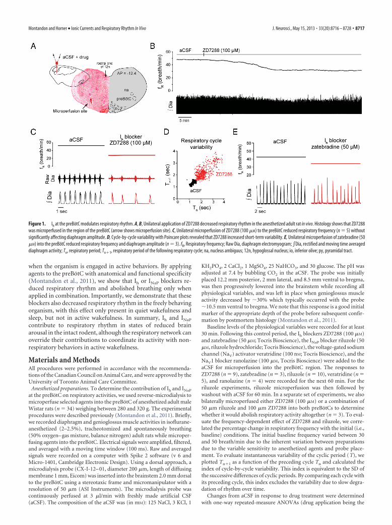
Figure 1. Ih at the preBotC modulates respiratory rhythm. A, B, Unilateral application of ZD7288 decreased respiratory rhythm in the anesthetized adult rat in vivo. Histology shows that ZD7288was microperfused in the region of the preBotC (arrow shows microperfusion site). C, Unilateral microperfusion of ZD7288 (100 �M) to the preBotC reduced respiratory frequency (n � 5) withoutsignificantly affecting diaphragm amplitude. D, Cycle-by-cycle variability with Poincare plots revealed that ZD7288 increased short-term variability. E, Unilateral microperfusion of zatebradine (50�M) into the preBotC reduced respiratory frequency and diaphragm amplitude (n � 3). fR, Respiratory frequency; Raw Dia, diaphragm electromyogram; �Dia, rectified and moving time averageddiaphragm activity; Tn, respiratory period; Tn�1, respiratory period of the following respiratory cycle; na, nucleus ambiguus; 12n, hypoglossal nucleus; io, inferior olive; py, pyramidal tract.
Montandon and Horner • Ionic Currents and Respiratory Rhythm In Vivo J. Neurosci., May 15, 2013 • 33(20):8716 – 8728 • 8717

repeated factor) followed by Dunnet’s post hoc tests for comparison witha single control (i.e., aCSF). p � 0.05 was considered statistically signifi-cant. Data are presented as means � SEM.
Freely behaving preparations. To determine the effects of Ih and INaP
blockade at the preBotC on respiratory activity across sleep–wake states,we perfused riluzole (50 �M), ZD7288 (100 �M), or a combination ofZD7288 (100 �M) and riluzole (50 �M) bilaterally into both preBotCs of
freely behaving male adult Wistar rats (n � 13). One week before theexperiments, sterile surgery was performed under isoflurane anesthesiato implant the rats with electroencephalogram (EEG) and postural(neck) muscle electrodes to identify sleep–wake states, and diaphragmand genioglossus muscle electrodes for respiratory muscle recordings aspreviously described (Montandon et al., 2011). Two microdialysis guidecannulas (AG-8, Eicom) were positioned 5.0 mm above the preBotC by
Figure 2. Sites of action of the Ih blocker ZD7288 and NK1R expression in the ventrolateral medulla. A, Sections showing NK1R expression (left hemisection) and microperfusion sites (righthemisection, red circles) in the ventrolateral medulla 12.6 –11.6 mm posterior to bregma. The perfusion sites were in the vicinity of the preBotC. Each perfusion site corresponds to one experimentwith microperfusion in the preBotC region. B, There was a significant correlation between the latencies for ZD7288 to depress frequency by 10% and the distances of the perfusion sites from thepreBotC (coordinates AP � �12.2, ML � 2.3, DV � �10.4) in anesthetized rats, showing that perfusion of drugs close to the preBotC has a faster effect on frequency than perfusion away fromit. C, Correlation map shows that the neural site most sensitive to ZD7288 corresponds to NK1R expression. D, Density of NK1R-expressing cells (see circle shown in C) from 11.6 to 12.6 mm posteriorto bregma. 7n, Facial nucleus. Values are shown as means �SEM.
8718 • J. Neurosci., May 15, 2013 • 33(20):8716 – 8728 Montandon and Horner • Ionic Currents and Respiratory Rhythm In Vivo

placing them 12.2 mm posterior, 2 mm either side, and 5.5 mm ventral tobregma with the guide cannulas secured in place with dental acrylic. Therat recovered for 1 week before the experiment.
On the day of the experiment, the rat was transiently anesthetized(isoflurane 2–2.5%) for the careful and accurate placement of the micro-dialysis probes (CX-I-12– 01, Eicom) as described above for the anesthe-tized preparation, i.e., to ensure consistent placements within andbetween preparations. After placements of the microdialysis probes, therat was connected to the recording apparatus which allowed electrophys-iological signals to be recorded while the rat moved freely in a largeopen-topped Plexiglas bowl filled with fresh bedding, food, and water.The bowl was placed on a rotating turntable (Raturn, BASi) which auto-matically adjusts its position when the rat moves to avoid entanglementsof the microdialysis tubing and recording cable. After a recovery periodfrom anesthesia of at least 120 min (with the rat apparently behavingnormally after �45 min), the EEG, neck and diaphragm muscle activitieswere recorded during bilateral perfusion of aCSF into both preBotC and
subsequent drugs. For every 10 s epoch, sleep–wake state was classified asactive wakefulness, quiet wakefulness, rapid eye movement (REM), andnon-REM sleep, and physiological values were calculated for every ep-och. Prevailing sleep–wake states were identified according to standardcriteria (Morrison et al., 2003). Briefly, EEG frequencies in the followingfrequency bands: �2 (0.5–2 Hz), �1 (2– 4 Hz), � (4 –7.5 Hz), � (7.5–13.5Hz), �1 (13.5–20 Hz), and �2 (20 –30 Hz) were calculated. Active wake-fulness was characterized by low � frequencies and high neck muscleactivity, whereas quiet wakefulness low � frequencies and low neck mus-cle activity. Non-REM sleep was characterized by high � frequencies,high EEG amplitude, and low neck muscle activity, whereas REM sleeppresented low � frequencies, high � frequencies, and low neck muscleactivity. Averaged baseline values were calculated over a 30 min periodbefore blockers were microperfused. Riluzole and/or ZD7288 were thenadded to the perfusing solution for 240 min and average values werecalculated over the last 30 min of this period. In the event that drugs didnot change rhythm, DAMGO, a �-opioid receptor agonist known to
Figure 3. INaP at the preBotC modulates respiratory rhythm. A–C, Unilateral microperfusion of the INaP blocker riluzole (50 �M) into the preBotC decreased respiratory rhythm (n � 6) in theanesthetized adult rat in vivo (arrow shows perfusion site). D–F, Mean values showed that riluzole significantly reduced respiratory frequency, increased respiratory variability, without significantlyaffecting diaphragm amplitude. This effect was reversed by washout of drugs with aCSF. G, Cycle-by-cycle variability estimated by Poincare plots revealed that riluzole increased short-term cyclevariability. Values are shown as means �SEM. *Indicates mean values significantly different from aCSF conditions with p � 0.05.
Montandon and Horner • Ionic Currents and Respiratory Rhythm In Vivo J. Neurosci., May 15, 2013 • 33(20):8716 – 8728 • 8719

depress frequency by its action on preBotC neurons (Montandon et al.,2011), was microperfused as positive control for accurate placement ofprobe. Measurements started at �11.00 and ended at �17.00. Data wereamplified, filtered, moving-time averaged, sampled, and analyzed as de-scribed previously (Montandon et al., 2011).
To determine synchronization of postural neck muscle and dia-phragm muscle activities, we computed the cross-correlation functionusing MATLAB Signal Processing Toolbox (function xcorr MATLABR12, Mathworks). This function is a measure of similarity of two wave-forms. Cross-correlation function was calculated for each 10 s segmentand associated with a specific state.
Two-way repeated-measure ANOVAs (sleep–wake states and drug appli-cation being the repeated factors) followed by Holm–Sidak post hoc testswere used to determine the state-dependent effect of manipulation of pre-BotC for each physiological variable. p � 0.05 was considered significant.
Construction of correlation maps. To determine the locations of theintervention (perfusion) sites, we cut 50 �m sections with cryostat andstained them with neutral red. Under light microscope (BX41, Olym-pus), we followed the track made by the microperfusion probe and iden-tified the end of the track as the tip of the probe. To determine the
anterior–posterior (AP) coordinate, we used the caudal end of the facialnucleus as the reference point (located 11.6 mm posterior to bregma) andcounted the number of sections caudal to the facial nucleus until theperfusion site was reached. The dorsal-ventral (DV) and medial-lateral(ML) coordinates were then identified using the nucleus ambiguus, in-ferior olive, and standard brain maps (Paxinos and Watson, 1998). Weconstructed correlation maps to relate the location of the interventionsites with the resultant effect on respiratory activity as previously de-scribed (Montandon et al., 2011). The rationale for the construction ofcorrelation maps is that for a locus of effect of drugs at any particularbrainstem site, the latency for the drug to diffuse through the tissue andto progressively change respiratory activity will vary as a function of thedistance of the probe from the effective site. We then calculated thedistance from an arbitrary set of coordinates such as the preBotC tothe perfusion site. We estimated the latency to a 10% change in respira-tory frequency in response to ZD7288 or riluzole. Using this approach,one latency and one distance was obtained for each animal/experiment.For a specific set of experiments including many rats, we then calculatedthe relationship between latency of drug effects and distances from mi-croperfusion sites to the preBotC. Each experiment in each rat contrib-
Figure 4. Anatomical sites of action of the INaP blocker riluzole and NK1R expression in the ventrolateral medulla. A, Sections showing NK1R expression (left hemisection) and microperfusion sites(right hemisection, blue circles) in the ventrolateral medulla 12.6 –11.6 mm posterior to bregma. The perfusion sites were in the vicinity of the preBotC. Each perfusion site corresponds to oneexperiment with microperfusion in the preBotC region. B, There was a significant correlation between the latencies for riluzole to depress frequency by 10% and the distances of the perfusion sitesfrom the preBotC (coordinates AP ��12.2, ML � 2.2, AP ��10.4) in anesthetized rats, showing that perfusion of drugs close to the preBotC has a faster effect on frequency than perfusion awayfrom it. C, A correlation map shows that the neural site most sensitive to riluzole corresponds to NK1R expression.
8720 • J. Neurosci., May 15, 2013 • 33(20):8716 – 8728 Montandon and Horner • Ionic Currents and Respiratory Rhythm In Vivo
uted a single point to the correlation. This approach can also be appliedto every possible set of coordinates within a 3-D grid (resolution 50 �m)spanning from 11.6 to 12.6 mm caudal to bregma. Correlation coeffi-cients were then calculated between distances and latencies to seewhether it is likely that the drugs are acting on other neural sites. Overall,this approach identifies the sites in the brainstem that respond the fastestto agent perfusion. Using MATLAB 12 software (Mathworks), correla-tion coefficients (0�r 2�1) were calculated for every set of coordinatesand plotted as color pixels (blue to red) in standard brain maps. Todetermine the relationship between the location of microperfusion siteand the latency to induce an effect on respiratory rhythm, this approachrequires to use a unilateral approach where only one neural site is pro-gressively affected by drug microperfusion. For this reason, correlationmaps were only constructed for the experiments involving unilateralmicroperfusion.
Immunohistochemistry of neurokinin-1 receptors. We used neurokinin-1receptor (NK1R) immunohistochemistry to locate preBotC neurons inbrainstem sections (Gray et al., 2001) using techniques previously described(Montandon et al., 2011). Following fixation in 4% paraformaldehyde andcryoprotection in 30% sucrose, the brains (n � 3) were frozen and cut in 50�m sections. The antibodies used were rabbit anti-NK1R (1:1000, AB-N04;Advanced Targeting Systems) and donkey anti-rabbit immunoglobin G (1:100, Jackson ImmunoResearch Laboratories). Sections were stained with achromogen diaminobenzidine solution and were mounted on slides, dried,and sealed with Cytoseal 280. Sections spanning from 11.6 to 12.6 mm pos-terior to bregma were digitized with a CCD camera (Infinity 1, Olympus)and microscope (BX-41, Olympus). We then counted the number of NK1R-positive cells in a 1-mm-diameter circle placed ventral to the nucleusambiguus on each section of the ventrolateral medulla (every 200 �m, 11.6–12.6 caudal to bregma). We then calculated the density of NK1R-positivecells as number of positive cells per mm2 and identified the preBotC as theregion with the highest density.
ResultsIh contributes to respiratory rhythm in vivoTo determine the contribution of Ih to the generation of respira-tory rhythm in vivo, we locally microperfused the Ih blockerZD7288 into the preBotC of anesthetized adult rats while record-ing respiratory activity. Unilateral microperfusion of ZD7288(100 �M) into the preBotC (Fig. 1A–C), at concentration knownto substantially block Ih in vitro (Gasparini and DiFrancesco,1997; Thoby-Brisson et al., 2000), decreased respiratory fre-quency ( fR) by 37.7% (aCSF, fR � 44.1 � 3.3 breath/min;ZD7288, fR � 28.0 � 2.6 breath/min; p � 0.029, n � 5) (Fig. 1C),but did not significantly change the amplitude of diaphragmmuscle activity (p � 0.273, n � 5). We used Poincare plots toquantify cycle-by-cycle variability and showed that ZD7288 in-creased short-term cycle variability by 180.2% compared withaCSF (p � 0.007) (Fig. 1D). Microperfusion into the preBotC ofzatebradine (50 �M), another Ih blocker (Matt et al., 2011), de-creased respiratory frequency by 25.8% (aCSF, fR � 42.2 breath/min; zatebradine, fR � 31.3 � 0.7 breath/min; p � 0.002, n � 3)(Fig. 1E), replicating the effect produced by ZD7288. There alsowas a significant decreased in diaphragm muscle amplitude (de-crease of 13.4 � 2.1%, p � 0.023, n � 3).
To overcome the spatial limitations inherent to the localizedpharmacological manipulation of selected neuronal groups inspecific brain regions, we used a functional and anatomical ap-proach to identify the sites in the medulla most sensitive to theapplied agents (Montandon et al., 2011). Using the capacity ofZD7288 to diffuse through the tissue and progressively induce areduction in respiratory frequency depending on physical prox-imity of the intervention site to the effective site, we related thelatency for ZD7288 to decrease respiratory frequency by 10% tothe proximity of the microperfusion site to the preBotC (Fig. 2A).Microperfusion of ZD7288 within the preBotC caused a relatively
fast decrease in respiratory frequency, whereas perfusion furtherfrom the preBotC induced a slower decrease in respiratory fre-quency (Fig. 2B). There was a significant relationship betweenlatency of response and distance from the preBotC to the perfu-sion sites (r � 0.836, p � 0.005, n � 9). For all the possiblecoordinates within the volume of brainstem surrounding the pre-BotC (see Materials and Methods), we calculated coefficients ofcorrelation relating the latencies for respiratory slowing with dis-tances from these coordinates to the perfusion sites (Fig. 2C).Construction of an anatomical map reveals “hotspot” regionsthat are statistically highly correlated with the latencies for respi-ratory slowing. The anatomical region most strongly associatedwith respiratory frequency slowing (illustrated in red on correla-tion map) corresponds to the preBotC as identified by highNK1R expression (Fig. 2C–D), suggesting that the sites overlap-ping with the preBotC are most rapidly sensitive to ZD7288.Importantly, this approach also showed that it is Ih blockade atthe preBotC that elicits a significant decrease in respiratory fre-
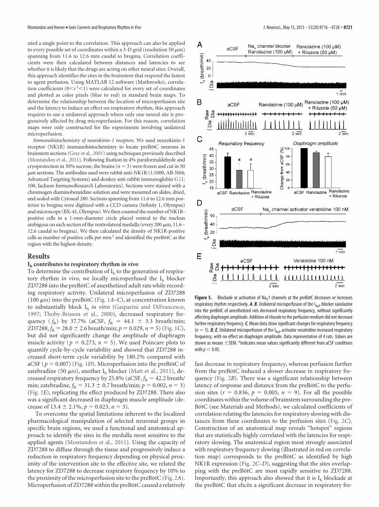
Figure 5. Blockade or activation of NaV1 channels at the preBotC decreases or increasesrespiratory rhythm respectively. A, B, Unilateral microperfusion of the INaP blocker ranolazineinto the preBotC of anesthetized rats decreased respiratory frequency, without significantlyaffecting diaphragm amplitude. Addition of riluzole to the perfusion medium did not decreasefurther respiratory frequency. C, Mean data show significant changes for respiratory frequency(n � 5). D, E, Unilateral microperfusion of the INaP activator veratridine increased respiratoryfrequency, with no effect on diaphragm amplitude. Data representative of 4 rats. Values areshown as means �SEM. *Indicates mean values significantly different from aCSF conditionswith p � 0.05.
Montandon and Horner • Ionic Currents and Respiratory Rhythm In Vivo J. Neurosci., May 15, 2013 • 33(20):8716 – 8728 • 8721

quency and that effects of ZD7288 were not due to diffusion ofthe drug to nearby respiratory nuclei.
INaP contributes to respiratory rhythm in vivoTo determine the contribution of INaP to the generation of respira-tory rhythm in vivo, we locally microperfused the INaP blocker rilu-zole into the preBotC of anesthetized adult rats. While recordingrhythmic output from the diaphragm, riluzole was unilaterally mi-croperfused at concentrations known to substantially block INaP invitro (Del Negro et al., 2002; Pena et al., 2004). Riluzole (50 �M) intothe preBotC (Fig. 3A) decreased respiratory frequency by 25.1%(p � 0.025, n � 5) (Fig. 3B–D), increased respiratory rate variabilityby 160.5% (p � 0.035, n � 5) (Fig. 3E), but did not affect diaphragmmuscle amplitude (p � 0.086, n � 5) (Fig. 3F). Riluzole increasedcycle-by-cycle short-term variability (p � 0.033, n � 5) (Fig. 3G).The riluzole effect on respiratory rhythm was not transient (Fig. 3C)as previously reported (Del Negro et al., 2005) but persisted as longas riluzole was perfused, and this effect was reversed after 45 � 5 minof aCSF washout (Fig. 3B,D).
Using a similar approach as with ZD7288, we related the la-tency for riluzole to decrease respiratory frequency by 10% to the
proximity of the perfusion sites to the preBotC (Fig. 4A). Againthere was a significant relationship between the latencies of re-sponses and the distances of the microperfusion sites to the pre-BotC (r � 0.76, p � 0.010, n � 10) (Fig. 4B). A correlation map(Fig. 4C) showed that the anatomical region most strongly asso-ciated with slowing of respiratory frequency (illustrated in red inthe correlation maps) corresponds to the preBotC as identified byNK1R expression. This result shows that the sites overlappingwith preBotC are most rapidly sensitive to riluzole and that rilu-zole effect was not due to diffusion of the drugs to nearby respi-ratory nuclei, such as the medullary raphe as previously suggested(Del Negro et al., 2010).
To determine whether the observed decrease in respiratoryfrequency is specific to INaP blockade, we used ranolazine, an-other INaP blocker known to preferentially block persistent overtransient sodium currents of NaV1 channels (Kahlig et al., 2010).These channels are highly expressed in preBotC neurons (Ptak etal., 2005) and may contribute to INaP in these cells. Microperfu-sion of ranolazine (100 �M) into the preBotC of anesthetizedadult rats decreased respiratory frequency by 25.6% (p � 0.001,n � 4) (Fig. 5A–C), without affecting diaphragm muscle ampli-
Figure 6. Frequency-dependent contribution of Ih, but not INaP, to respiratory rhythm. Relationship between baseline frequency while microperfusing aCSF into the preBotC and the changeselicited by the Ih blocker ZD7288 or the INaP blocker riluzole. A, B, There was a significant correlation between baseline frequency and the capacity of ZD7288 to reduce respiratory frequency (n �7). 95% confidence interval is indicated by gray areas. C, D, No significant correlation was found while comparing baseline frequency and blockade by riluzole (n � 7).
8722 • J. Neurosci., May 15, 2013 • 33(20):8716 – 8728 Montandon and Horner • Ionic Currents and Respiratory Rhythm In Vivo

tude (p � 0.103). Subsequent addition of riluzole (50 �M) to theperfusion medium containing ranolazine did not further de-crease respiratory frequency (Fig. 5C) suggesting that riluzoleand ranolazine are blocking similar channels.
If reduction of INaP in preBotC neurons decreases respira-tory frequency, then INaP activation should increase it. Ac-cordingly, we examined the effect on the INaP activatorveratridine (Fekete et al., 2009) applied to the preBotC onrespiratory rhythm. Veratridine targets the neurotoxin recep-tor site 2 of NaV channels and evokes a persistent sodiuminflux (Fekete et al., 2009). Microperfusion into the preBotCof veratridine at 100 nM, a concentration known to elicit INaP
in vitro, increased respiratory frequency by 18.0% (aCSF, fR �36.7 � 3.2 breath/min; veratridine, fR � 43.3 � 3.4 breath/min; p � 0.039, n � 5) (Fig. 5D–E), without affecting dia-phragm amplitude ( p � 0.207, n � 5).
Frequency-dependency of Ih but not INaP
One of the properties of Ih is that it can regulate oscillatoryactivity over a dynamic range from 0.5 to 4 Hz (Luthi andMcCormick, 1998; Biel et al., 2009). This range corresponds torespiratory frequencies �30 breath/min. On the other hand,INaP can support frequencies over a broad dynamic range in-cluding lower frequencies (Koizumi and Smith, 2008). To testthe frequency-dependent contribution of Ih and INaP to respi-ratory rhythm, we correlated the capacity of ZD7288 (100 �M)or riluzole (50 �M) to decrease respiratory frequency with theinitial respiratory frequency observed in baseline conditions,i.e., in the absence of blockers. At similar levels of anesthetic,different rats exhibited various initial baseline frequencieslikely due to variable sensitivities to anesthetics and/or vari-able placements of microperfusion probes into the preBotCregion. This inherent variability provided us with a wide rangeof initial baseline frequencies to test the frequency-dependentcontribution of Ih or INaP blockers. A significant correlation ofthe response to ZD7288 and the initial baseline frequency wasobserved (r � �0.88, p � 0.009) (Fig. 6 A, B). It showed that Ih
blockade has a substantial effect on reducing rhythm when it isinitially high, but less so when it is low and this effect fits withthe operating range of Ih. Conversely, there was no significantcorrelation between frequencies and INaP blockade (r � 0.26,p � 0.615) (Fig. 6C,D), showing that riluzole reduced respira-tory rhythm regardless of frequency.
Ih and INaP contribute to respiratory rhythmTo determine whether Ih and INaP are essential to generaterespiratory rhythm in vivo, we tested whether concomitantblockade of these currents would abolish respiratory activity.To first test the essential role of Ih, we perfused ZD7288 (100�M) bilaterally into both preBotCs and observed the conse-quent respiratory decline in anesthetized rats. After 45 min ofmicroperfusion, Ih blockade decreased significantly respira-tory frequency by 44.0% (aCSF, fR � 48.7 � 1.5 breath/min;bilateral ZD7288, fR � 24.0 � 7.3 breath/min; p � 0.049, n �3) (Fig. 7A) without significantly affecting the amplitude ofdiaphragm activity ( p � 0.184), and did not abolished breath-ing within the 2 h recording period. In a separate set of exper-iments, we microperfused the Ih and INaP blockers into bothpreBotC. ZD7288 (100 �M) and riluzole (50 �M) at the pre-BotCs (Fig. 7B) substantially decreased respiratory frequencyby 86.7% within 45 min (aCSF, fR � 43.2 � 3.9 breath/min;bilateral ZD7288�riluzole, fR � 6.7 � 6.7 breath/min, p �0.023, n � 3) and reduced diaphragm amplitude by 75.5%
( p � 0.041). Breathing was completely abolished after 43.3 �17.4 min (n � 3). This effect strongly differs from the effectsobserved with ZD7288 applied alone to both preBotCs.
State-dependent contribution of Ih and INaP torespiratory rhythmRespiratory rhythm is altered by anesthetics and modulated bystates of arousal (Phillipson and Bowes, 1986). To determinewhether Ih and INaP contribute to respiratory rhythm in the ab-sence of anesthetics and across sleep–wake states (Fig. 8A), wemicroperfused Ih and INaP blockers into preBotCs of freely behav-ing and nonanesthetized adult rats as previously described (Mon-tandon et al., 2011). Representative recordings (Fig. 8B,C) andgroup data (Fig. 8D) showed that concomitant bilateral mi-croperfusion of ZD7288 and riluzole disrupted rhythm by de-creasing respiratory frequency (p � 0.031, n � 5) (Fig. 8B,C) inquiet wakefulness (by 22.5%, p � 0.027), non-REM sleep (by28.4%, p � 0.010), and REM sleep (by 25.7%, p � 0.010), but notin active wakefulness (p � 0.720). The respiratory pattern wasalso affected by Ih and INaP blockade at the preBotCs. Inspiratorydurations were overall increased by the blockers across the sleep–wake cycle (p � 0.045) and expiratory durations were increasedonly in states of quiet wakefulness, non-REM, and REM sleep,but not in active wakefulness (p � 0.001), therefore closely fol-lowing the effect observed on frequency.
Using an approach similar to the anesthesia experiments, werelated the average distance from perfusion sites to preBotCs withthe efficacy of the blockers to depress respiratory frequency. Twomicroperfusion sites were located for each experiment and thetwo distances from each microperfusion site to its respective pre-BotC were calculated and averaged. We then correlated theseaverage distances collected in 5 experiments and the respectiveefficacy of blockers to decrease respiratory frequency in each ex-
Figure 7. Bilateral blockade of Ih and INaP, but not Ih alone, abolishes respiratory rhythm. A,Bilateral microperfusion of ZD7288 (100 �M) into both preBotCs decreased respiratory fre-quency by 44.0% (n � 3) after 45 min, without significantly affecting diaphragm amplitude. B,Bilateral microperfusion of ZD7288 (100 �M) and riluzole (50 �M) into both preBotCs signifi-cantly decreased respiratory frequency by 86.7% and diaphragm amplitude by 75.5% within 45min (n � 3). On average, breathing was abolished by ZD7288 and riluzole after 43.3 � 17.4min of microperfusion.
Montandon and Horner • Ionic Currents and Respiratory Rhythm In Vivo J. Neurosci., May 15, 2013 • 33(20):8716 – 8728 • 8723

periment. There was a significant correlation (r � 0.80, p �0.044, n � 6) (Fig. 8F) between average distances from microper-fusion sites to preBotCs and the severity of reductions byZD7288/riluzole suggesting that the closer the probes were to thepreBotC, the stronger were the frequency reductions. Inciden-tally, sleep was also disrupted by perfusion of ZD7288 and rilu-zole into preBotCs with a reduced time spent in non-REM sleep(p � 0.010) (Fig. 8G). Bilateral microperfusion of ZD7288 (100�M) or riluzole (50 �M) alone into the preBotC did not signifi-
cantly change respiratory frequency (p � 0.967, n � 5 and p �0.351, n � 3, respectively) (Fig. 9) across sleep–wake states.
Synchronization of respiratory and behavioralmotor activitiesThe data described above showed that Ih and INaP at the preBotCcontribute to rhythmogenesis in reduced states of brain arousal,such as sleep and anesthesia, but not in active wakefulness whenthe organism is engaged in complex behaviors. Active behaviors
Figure 8. State-dependent contribution of Ih and INaP to respiratory rhythm. A, States of active wakefulness, quiet wakefulness, non-REM and REM sleep are determined using electroenceph-alogram recordings and frequency spectra in freely behaving adult rats. B, Diaphragm muscle recordings and cycle-by-cycle frequencies at each sleep–wake state with bilateral application at thepreBotC of aCSF. C, Bilateral microperfusion of ZD7288 (100 �M) and riluzole (50 �M) into both preBotCs reduced significantly respiratory frequency in states of quiet wakefulness, non-REM sleep,and REM sleep, but not in active wakefulness. D, Mean data for n � 5 rats confirmed the state-dependent effect of drug microperfusion on respiratory frequency. E, Blockade of Ih and INaP did notaffect diaphragm amplitude. F, Correlation of distances of perfusion sites from preBotCs and efficacy of drugs showing that close perfusion to the preBotC produced stronger effects. G, Blockade ofIh and INaP and subsequent breathing reduction also reduced time spent in non-REM sleep. Values are shown as means �SEM. *Indicates mean values significantly different from aCSF conditionswith p � 0.05.
8724 • J. Neurosci., May 15, 2013 • 33(20):8716 – 8728 Montandon and Horner • Ionic Currents and Respiratory Rhythm In Vivo

such as grooming or feeding are not automatic behaviors gener-ated by the brainstem respiratory network, but rather arise fromdistinct cortical and cerebellar areas that impinge on the respira-tory network and influence its activity to coordinate respiratorynetwork activity to these nonrespiratory behaviors. To determinewhether respiratory network activity may be modulated by otherbrain areas involved in distinct behaviors, we evaluated synchro-nization of two separate muscle activities originating from sepa-rate brain circuits. The degree of synchronization of respiratorynetwork activity and postural motor activity was calculated usingcross-correlation functions (Katz et al., 2002) between respira-tory diaphragm and postural neck muscle activities. This measureshows whether two signals are synchronized independently oftheir intensity, which is essential to determine synchronization insignals of low amplitude such as motor activity during sleep. Weshowed that diaphragm muscle activity was tightly synchronizedto neck muscle activity when the animal was behaviorally activeor in quiet wakefulness, but not when the animal was in non-REM sleep (p � 0.002, n � 3) (Fig. 10A) or REM sleep (p �0.002). When Ih and INaP were blocked, however, postural neckand diaphragm muscles were synchronized at all states (p �0.737) (Fig. 10B,C) which differ from the state-dependent syn-chronization of muscle activities observed in control conditions(p � 0.001).
DiscussionHere, we determined the contribution of voltage-dependent in-ward currents in respiratory rhythmogenesis in vivo. We showedthat Ih and INaP, two ionic currents known to mediate rhythms inmany networks, mediate respiratory rhythm in states of anesthe-sia, quiet wakefulness, and sleep, although the network can su-
persede their contribution to comply its activity with activebehaviors that originate from distinct neuronal circuits.
The ionic playersThe unique property of Ih to activate upon hyperpolarizationbeyond resting potential makes it a potential current for providingrhythmicity in the respiratory network (Luthi and McCormick,1998). When the Ih blocker ZD7288 was applied to rhythmicallyactive brainstem sections containing the preBotC, Ih was re-duced, but either no reduction of respiratory rhythm was ob-served (Mironov et al., 2000) or respiratory rhythm increased(Thoby-Brisson et al., 2000). This shows that Ih in preBotC neu-rons does not promote rhythmicity when cells are immature andisolated from the network. Ih expression is, however, reducedunder these conditions, implying that its role may differ com-pared with mature and intact preparations (Bayliss et al., 1994;Biel et al., 2009). Here, we microperfused two distinct Ih blockersinto the preBotC, at concentrations known to substantially blockIh (Williams et al., 2002; Van Bogaert and Pittoors, 2003), and wereduced respiratory rhythm. A rhythmogenic role for Ih is furtherevidenced by the frequency range at which it operates. Ih is in-volved in rhythms from 0.5 to 4 Hz (Biel et al., 2009) correspond-ing to respiratory frequencies between 30 and 240 breath/min.ZD7288 blocked respiratory rhythm only at frequencies �30breath/min, consistent with Ih activation kinetics. There was alsoa relationship between Ih blockade and the initial baseline fre-quency before drug application. Frequency decline by ZD7288was more pronounced at relatively high than low frequencies,which may explain why Ih blockers failed to depress frequencyand even increased frequency in neonatal in vitro preparationswhere frequency is slow (Mironov et al., 2000; Thoby-Brisson etal., 2000). Similarly, respiratory rhythm was sensitive to NK1Rblockade or to locus ceruleus stimulation only when the initialfrequency was elevated (Doi and Ramirez, 2010), suggesting thatdistinct mechanisms mediate respiratory rhythm in low com-pared with high frequencies.
Surprisingly, we did not observe a significant effect of Ih block-ade alone in the freely behaving experiments. The fact that Ih isactivated by hyperpolarization suggests that, in strongly inhibitedconditions such as anesthesia, Ih expression is increased and itsphysiological role is potentiated. In freely behaving animals, ex-citatory inputs to the preBotC may raise membrane potentialabove Ih activation potentials. It is, only when INaP is blocked, thatIh activation threshold would be reached. However, Ih role maybe underestimated in our study. Although ZD7288 is the mostpotent Ih blocker currently available, it only partially blocks Ih
(Gasparini and DiFrancesco, 1997). ZD7288 also inhibits T-typecalcium channels (Sanchez-Alonso et al., 2008) and glutamatereceptor-mediated currents (Chen, 2004), and depresses synaptictransmission (Chevaleyre and Castillo, 2002). Because these ef-fects are only substantial when ZD7288 is applied at high concen-trations for long periods, it is unlikely that ZD7288 effect was dueto its action on other targets, but it cannot be excluded. Also, thefact that the Ih blocker zatebradine, which does not block calciumchannels (Satoh and Yamada, 2002), also decreased respiratoryrhythm, is consistent with a role of Ih in promoting respiratoryrhythm. Zatebradine, however, inhibits potassium currents insome cell types (Satoh and Yamada, 2002), but such blockadewould likely increase respiratory rhythm by reducing potassiumoutward current. Overall, our data reveal the importance of anessential ionic player in promoting respiratory rhythm in maturemammals in vivo, a contribution that substantially differs from itsrole in neonatal rodents in vitro.
Figure 9. Blockade of Ih or INaP alone did not modify respiratory rhythm in the freelybehaving adult rat. A, Bilateral microperfusion of riluzole (50 �M) into the preBotC ofadult rat did not affect respiratory frequency. B, Diaphragm muscle activity was notmodified by riluzole. C, Bilateral microperfusion of ZD7288 (100 �M) at the preBotCs didnot change respiratory frequency. D, Diaphragm muscle amplitude was not changed byZD7288. Values are shown as means �SEM. *Indicates mean values significantly differentfrom aCSF conditions with p � 0.05.
Montandon and Horner • Ionic Currents and Respiratory Rhythm In Vivo J. Neurosci., May 15, 2013 • 33(20):8716 – 8728 • 8725

However, Ih is not sufficient to generate respiratory rhythm byitself, because its blockade did not completely abolish rhythm. Ih
interacts with other ionic currents. INaP for instance is importantfor rhythm generation in specific configurations of the respira-tory network in hypoxia (Pena et al., 2004; Paton et al., 2006) and
in neonatal rodents in vivo (Pena and Aguileta, 2007). Here, weshow that blockade, or activation, of INaP at the preBotC, respec-tively decreased or increased, frequency. Correlation mapsstrongly suggest that riluzole is acting in the preBotC region andnot in adjacent regions, such as the medullary raphe (Del Negro
Figure 10. Synchronization of respiratory network activity and motor behavior. A, Synchronization was determined by cross-correlation function of diaphragm and neck muscle activities in freelybehaving adult rats. Gray areas indicate periods of synchronization; low to high synchronization corresponds to cross-correlation function from 0 to 1. B, When Ih and INaP were blocked at the preBotC,high synchronization was observed across states. C, Mean data show that in control conditions (aCSF, black bars), synchronization was high in active wakefulness but low in quiet wakefulness,non-REM sleep and REM sleep, whereas with Ih and INaP blockade (gray bars) synchronization occurred across states. Values are shown as means�SEM. *Indicates mean values significantly differentfrom aCSF conditions with p � 0.05.
8726 • J. Neurosci., May 15, 2013 • 33(20):8716 – 8728 Montandon and Horner • Ionic Currents and Respiratory Rhythm In Vivo

et al., 2010). In addition, low concentration of riluzole avoidsunspecific effects observed at higher concentrations (Urbani andBelluzzi, 2000). Voltage-gated sodium channels, which are pres-ent in high density in preBotC neurons (Ptak et al., 2005), con-tribute to INaP in many types of neurons (Kahlig et al., 2010).Blockade of the persistent component of sodium channels withranolazine, or activation with veratridine, substantially sup-pressed or increased respiratory rhythm, respectively, supportingthe contribution of INaP and its underlying channels to respira-tory rhythm in vivo. In the freely behaving animal, riluzole didnot affect respiratory rhythm which is consistent with previousdata in conscious mature rats (St-John et al., 2007). In immaturerodents studied in vivo, however, respiratory rhythm was in-creased after systemic administration of riluzole (Pena andAguileta, 2007), again highlighting differences between imma-ture and mature respiratory networks.
Concomitant blockade of Ih and INaP
During hyperpolarization, Ih induces slow membrane depolar-ization (Pape, 1996) which activates INaP. This mechanism hasbeen described in the cerebellum (Williams et al., 2002), in sub-threshold oscillations of the entorhinal cortex (Dickson et al.,2000), and in single-spike activity in subthalamic neurons(Beurrier et al., 2000). Consistent with this mechanism, we dem-onstrated that when Ih or INaP alone were blocked in the anes-thetized preparation, respiratory rhythm was reduced but notabolished, and only the concomitant blockade of both currentsled to rhythm cessation. Ih/INaP blockade abolishes breathingonly under anesthesia, but not in freely behaving organisms, sug-gesting either that anesthetics are essential to abolish rhythm orthat an alternative mechanism of rhythmogenesis is triggeredwhen these currents are absent in nonanesthetized animals. Sim-ilarly, reduced ventilation due to Ih/INaP blockade likely increasescarbon dioxide levels, which may stimulate rhythm, and mini-mize the effects of Ih/INaP blockade. Overall, these currents arenecessary but probably not sufficient to generate respiratoryrhythm, and the contribution of other currents cannot be ex-cluded (Pena et al., 2004; Koch et al., 2013). In summary, undernormal physiological conditions and when ionic currents and theunderlying channels are fully mature, respiratory rhythmogen-esis depends on a set of currents which includes Ih and INaP.
State dependencyThe brain can override automatic respiratory rhythm to synchro-nize breathing with other behaviors (such as feeding or groom-ing) which also engage the respiratory system (Ramirez andGarcia, 2007). Here, we showed that Ih and INaP play key roles inrespiratory rhythmogenesis, but that their contribution is over-ridden when the organism is behaviorally active. The locus ce-ruleus, a pontine structure with noradrenergic projections to thepreBotC (Doi and Ramirez, 2010), is active when the organism isengaged in behaviors and is inactive in sleep (Aston-Jones andBloom, 1981; Takahashi et al., 2010). Combined with the fact thatnoradrenaline directly inhibits Ih (Wang et al., 2007), high locusceruleus activity may therefore reduce Ih expression in preBotCcells to supersede rhythm and coordinate diaphragm activity tononrhythmic motor behaviors. When Ih and INaP are blocked,however, postural and respiratory muscles are synchronizedacross states, suggesting that a mechanism independent of Ih/INaP
maintains respiratory rhythm. Under anesthesia, Ih/INaP block-ade abolishes rhythm, and volatile anesthetics may prevent therespiratory network to recruit an alternative mechanism to sus-tain rhythm as observed in the freely behaving animal. The mech-
anism mediating synchronized activity of motor outputs isunknown and it cannot be excluded that indirect effects, such ashypercapnia may trigger their synchronization. The idea of mul-tiple mechanisms generating rhythm within the preBotC hasbeen proposed previously (Pena et al., 2004; Smith et al., 2009)and these state-dependent changes identify that there are multi-ple and redundant mechanisms that functionally cooperate tomaintain respiratory rhythm, with one or other predominating ascircumstances vary.
Mechanisms of rhythmogenesisVoltage-dependent inward currents, such as Ih and INaP, arefound in cells of various rhythmic networks and are essential totrigger spontaneous bursting (Pena et al., 2004; Biel et al., 2009).It is unknown, however, whether subthreshold voltage-activatedinward currents contribute to spontaneous pacemaker-likebursting of mature preBotC neurons. Here, we did not record thebursting of preBotC neurons in vivo, but highlight Ih/INaP impor-tance in generating respiratory rhythm in freely behaving matureorganisms. Also, Ih/INaP mediate the overall excitability of therhythmic network (Biel et al., 2009; Del Negro et al., 2010), andtheir blockade may decrease neuronal excitability which woulddecrease rhythm independently of an action on pacemaker prop-erties. Blockade of INaP abolishes respiratory rhythm in vitro(Pena et al., 2004), but rhythm can be reestablished by excitingthe respiratory network through AMPA or NK1Rs (Del Negro etal., 2010). Similarly, our study shows that Ih/INaP blockade andthe consequent rhythm inhibition may be reactivated by excit-atory inputs during active states. This is consistent with the ideathat ionic conductances are important for respiratory rhythm insome conditions or states (quiet wakefulness and sleep), but aresuperseded by excitatory inputs when the organism is behavior-ally active.
ReferencesAston-Jones G, Bloom FE (1981) Activity of norepinephrine-containing lo-
cus ceruleus neurons in behaving rats anticipates fluctuations in the sleep-waking cycle. J Neurosci 1:876 – 886. Medline
Bayliss DA, Viana F, Bellingham MC, Berger AJ (1994) Characteristics andpostnatal development of a hyperpolarization-activated inward currentin rat hypoglossal motoneurons in vitro. J Neurophysiol 71:119 –128.Medline
Beurrier C, Bioulac B, Hammond C (2000) Slowly inactivating sodium cur-rent (I(NaP)) underlies single-spike activity in rat subthalamic neurons.J Neurophysiol 83:1951–1957. Medline
Biel M, Wahl-Schott C, Michalakis S, Zong X (2009) Hyperpolarization-activated cation channels: from genes to function. Physiol Rev 89:847–885. CrossRef Medline
Chen C (2004) ZD7288 inhibits postsynaptic glutamate receptor-mediatedresponses at hippocampal perforant path-granule cell synapses. EurJ Neurosci 19:643– 649. CrossRef Medline
Chevaleyre V, Castillo PE (2002) Assessing the role of Ih channels in synap-tic transmission and mossy fiber LTP. Proc Natl Acad Sci U S A 99:9538 –9543. CrossRef Medline
Cho HJ, Furness JB, Jennings EA (2011) Postnatal maturation of thehyperpolarization-activated cation current, Ih, in trigeminal sensory neu-rons. J Neurophysiol 106:2045–2056. CrossRef Medline
Del Negro CA, Morgado-Valle C, Feldman JL (2002) Respiratory rhythm:an emergent network property? Neuron 34:821– 830. CrossRef Medline
Del Negro CA, Morgado-Valle C, Hayes JA, Mackay DD, Pace RW, CrowderEA, Feldman JL (2005) Sodium and calcium current-mediated pace-maker neurons and respiratory rhythm generation. J Neurosci 25:446 –453. CrossRef Medline
Del Negro CA, Hayes JA, Pace RW, Brush BR, Teruyama R, Feldman JL(2010) Synaptically activated burst-generating conductances may un-derlie a group-pacemaker mechanism for respiratory rhythm generationin mammals. Prog Brain Res 187:111–136. CrossRef Medline
Montandon and Horner • Ionic Currents and Respiratory Rhythm In Vivo J. Neurosci., May 15, 2013 • 33(20):8716 – 8728 • 8727

Dickson CT, Magistretti J, Shalinsky MH, Fransen E, Hasselmo ME, Alonso A(2000) Properties and role of I(h) in the pacing of subthreshold oscilla-tions in entorhinal cortex layer II neurons. J Neurophysiol 83:2562–2579.Medline
Doi A, Ramirez JM (2010) State-dependent interactions between excitatoryneuromodulators in the neuronal control of breathing. J Neurosci 30:8251– 8262. CrossRef Medline
Fekete A, Franklin L, Ikemoto T, Rozsa B, Lendvai B, Sylvester Vizi E, Zelles T(2009) Mechanism of the persistent sodium current activatorveratridine-evoked Ca elevation: implication for epilepsy. J Neurochem111:745–756. CrossRef Medline
Feldman JL, Del Negro CA (2006) Looking for inspiration: new perspectiveson respiratory rhythm. Nat Rev Neurosci 7:232–242. CrossRef Medline
Gasparini S, DiFrancesco D (1997) Action of the hyperpolarization-activated current (Ih) blocker ZD 7288 in hippocampal CA1 neurons.Pflugers Arch 435:99 –106. CrossRef Medline
Gray PA, Janczewski WA, Mellen N, McCrimmon DR, Feldman JL (2001)Normal breathing requires preBotzinger complex neurokinin-1 receptor-expressing neurons. Nat Neurosci 4:927–930. CrossRef Medline
Kahlig KM, Lepist I, Leung K, Rajamani S, George AL (2010) Ranolazineselectively blocks persistent current evoked by epilepsy-associated NaV1.1 mutations. Br J Pharmacol 161:1414 –1426. CrossRef Medline
Katz DB, Simon SA, Nicolelis MA (2002) Taste-specific neuronal ensemblesin the gustatory cortex of awake rats. J Neurosci 22:1850 –1857. Medline
Koch H, Zanella S, Elsen GE, Smith L, Doi A, Garcia AJ 3rd, Wei AD, Xun R,Kirsch S, Gomez CM, Hevner RF, Ramirez JM (2013) Stable respiratoryactivity requires both P/Q-type and N-type voltage-gated calcium chan-nels. J Neurosci 33:3633–3645. CrossRef Medline
Koizumi H, Smith JC (2008) Persistent Na� and K�-dominated leak cur-rents contribute to respiratory rhythm generation in the pre-Botzingercomplex in vitro. J Neurosci 28:1773–1785. CrossRef Medline
Luthi A, McCormick DA (1998) H-current: properties of a neuronal andnetwork pacemaker. Neuron 21:9 –12. CrossRef Medline
Matt L, Michalakis S, Hofmann F, Hammelmann V, Ludwig A, Biel M, Klep-pisch T (2011) HCN2 channels in local inhibitory interneurons con-strain LTP in the hippocampal direct perforant path. Cell Mol Life Sci68:125–137. CrossRef Medline
McKay LC, Janczewski WA, Feldman JL (2005) Sleep-disordered breathingafter targeted ablation of preBotzinger complex neurons. Nat Neurosci8:1142–1144. CrossRef Medline
Mironov SL, Langohr K, Richter DW (2000) Hyperpolarization-activatedcurrent, Ih, in inspiratory brainstem neurons and its inhibition by hyp-oxia. Eur J Neurosci 12:520 –526. CrossRef Medline
Montandon G, Qin W, Liu H, Ren J, Greer JJ, Horner RL (2011) PreBotz-inger complex neurokinin-1 receptor-expressing neurons mediateopioid-induced respiratory depression. J Neurosci 31:1292–1301.CrossRef Medline
Morrison JL, Sood S, Liu H, Park E, Liu X, Nolan P, Horner RL (2003) Roleof inhibitory amino acids in control of hypoglossal motor outflow togenioglossus muscle in naturally sleeping rats. J Physiol 552:975–991.CrossRef Medline
Pape HC (1996) Queer current and pacemaker: the hyperpolarization-activated cation current in neurons. Annu Rev Physiol 58:299 –327.CrossRef Medline
Paton JF, Abdala AP, Koizumi H, Smith JC, St-John WM (2006) Respira-tory rhythm generation during gasping depends on persistent sodiumcurrent. Nat Neurosci 9:311–313. CrossRef Medline
Paxinos G, Watson C (1998) The rat brain in stereotaxic coordinates. SanDiego: Academic.
Pena F, Aguileta MA (2007) Effects of riluzole and flufenamic acid on eu-pnea and gasping of neonatal mice in vivo. Neurosci Lett 415:288 –293.CrossRef Medline
Pena F, Parkis MA, Tryba AK, Ramirez JM (2004) Differential contributionof pacemaker properties to the generation of respiratory rhythms duringnormoxia and hypoxia. Neuron 43:105–117. CrossRef Medline
Phillipson EA, Bowes G (1986) Control of breathing during sleep. In: Hand-book of physiology, Section III, The respiratory system, vol. II (CherniakNS, Widdicombe JG, eds), pp 649 –989. Bethesda, MD: American Physi-ological Society.
Ptak K, Zummo GG, Alheid GF, Tkatch T, Surmeier DJ, McCrimmon DR(2005) Sodium currents in medullary neurons isolated from the pre-Botzinger complex region. J Neurosci 25:5159 –5170. CrossRef Medline
Ramirez JM, Garcia A 3rd (2007) Point: medullary pacemaker neurons areessential for both eupnea and gasping in mammals. J Appl Physiol 103:717–718; discussion 722. CrossRef Medline
Sanchez-Alonso JL, Halliwell JV, Colino A (2008) ZD 7288 inhibits T-typecalcium current in rat hippocampal pyramidal cells. Neurosci Lett 439:275–280. CrossRef Medline
Satoh TO, Yamada M (2002) Multiple inhibitory effects of zatebradine(UL-FS 49) on the electrophysiological properties of retinal rod photore-ceptors. Pflugers Arch 443:532–540. CrossRef Medline
Smith JC, Ellenberger HH, Ballanyi K, Richter DW, Feldman JL (1991) Pre-Botzinger complex: a brainstem region that may generate respiratoryrhythm in mammals. Science 254:726 –729. CrossRef Medline
Smith JC, Abdala AP, Rybak IA, Paton JF (2009) Structural and functionalarchitecture of respiratory networks in the mammalian brainstem. PhilosTrans R Soc Lond B Biol Sci 364:2577–2587. CrossRef Medline
St-John WM, Waki H, Dutschmann M, Paton JF (2007) Maintenance ofeupnea of in situ and in vivo rats following riluzole: a blocker of persistentsodium channels. Respir Physiol Neurobiol 155:97–100. CrossRefMedline
Takahashi K, Kayama Y, Lin JS, Sakai K (2010) Locus coeruleus neuronal activ-ity during the sleep-waking cycle in mice. Neuroscience 169:1115–1126.CrossRef Medline
Thoby-Brisson M, Telgkamp P, Ramirez JM (2000) The role of thehyperpolarization-activated current in modulating rhythmic activity inthe isolated respiratory network of mice. J Neurosci 20:2994 –3005.Medline
Urbani A, Belluzzi O (2000) Riluzole inhibits the persistent sodium currentin mammalian CNS neurons. Eur J Neurosci 12:3567–3574. CrossRefMedline
Van Bogaert PP, Pittoors F (2003) Use-dependent blockade of cardiac pace-maker current (If) by cilobradine and zatebradine. Eur J Pharmacol 478:161–171. CrossRef Medline
Wang M, Ramos BP, Paspalas CD, Shu Y, Simen A, Duque A, VijayraghavanS, Brennan A, Dudley A, Nou E, Mazer JA, McCormick DA, Arnsten AF(2007) Alpha2A-adrenoceptors strengthen working memory networksby inhibiting cAMP-HCN channel signaling in prefrontal cortex. Cell129:397– 410. CrossRef Medline
Williams SR, Christensen SR, Stuart GJ, Hausser M (2002) Membrane po-tential bistability is controlled by the hyperpolarization-activated currentI(H) in rat cerebellar Purkinje neurons in vitro. J Physiol 539:469 – 483.CrossRef Medline
Zhong G, Masino MA, Harris-Warrick RM (2007) Persistent sodium cur-rents participate in fictive locomotion generation in neonatal mouse spi-nal cord. J Neurosci 27:4507– 4518. CrossRef Medline
8728 • J. Neurosci., May 15, 2013 • 33(20):8716 – 8728 Montandon and Horner • Ionic Currents and Respiratory Rhythm In Vivo