Embed Size (px)
Citation preview
Available online http://arthritis-research.com/content/10/6/R136
Open AccessVol 10 No 6Research articleT-614, a novel immunomodulator, attenuates joint inflammation and articular damage in collagen-induced arthritisFang Du, Liang-jing Lü, Qiong Fu, Min Dai, Jia-lin Teng, Wei Fan, Shun-le Chen, Ping Ye, Nan Shen, Xin-fang Huang, Jie Qian and Chun-de Bao
Shanghai Institute of Rheumatology, Renji Hospital, Shanghai Jiao Tong University School of Medicine, Shan Dong Middle Road, Shanghai 200001, PR China
Corresponding author: Chun-de Bao, [email protected]
Received: 2 Sep 2008 Revisions requested: 9 Oct 2008 Revisions received: 29 Oct 2008 Accepted: 19 Nov 2008 Published: 19 Nov 2008
Arthritis Research & Therapy 2008, 10:R136 (doi:10.1186/ar2554)This article is online at: http://arthritis-research.com/content/10/6/R136© 2008 Du et al.; licensee BioMed Central Ltd. This is an open access article distributed under the terms of the Creative Commons Attribution License (http://creativecommons.org/licenses/by/2.0), which permits unrestricted use, distribution, and reproduction in any medium, provided the original work is properly cited.
Abstract
Introduction T-614 is a novel oral antirheumatic agent for thetreatment of rheumatoid arthritis. Whether it has immunomodulatoryor disease-modifying properties and its mechanism of action arelargely undetermined.
Methods Rats with collagen-induced arthritis (CIA) were treatedwith T-614 (5 and 20 mg/kg) daily. Animals receiving methotrexate(1 mg/kg every 3 days) and the nonsteroidal anti-inflammatory agentnimesulide (10 mg/kg per day) were used as controls. Acombination therapy group was treated with both T-614(10 mg/kgper day) and methotrexate (1 mg/kg every 3 days). Hind pawswelling was evaluated and radiographic scores calculated. Serumcytokine levels were assessed by Bio-plex analysis. QuantitativePCR was used to evaluate expression of mRNA for interferon-γ, IL-4 and IL-17. Serum IL-17 and anti-type II collagen antibodies (totalIgG, IgG1, IgG2a, IgG2b and IgM) were measured using ELISA.
Results Oral T-614 inhibited paw swelling and offered significantprotection against arthritis-induced cartilage and bone erosion,comparable to the effects of methotrexate. CIA rats treated with T-
614 exhibited decreases in both mRNA expression of IL-17 inperipheral blood mononuclear cells and lymph node cells, andcirculating IL-17 in a dose-dependent manner. T-614 also reducedserum levels of tumor necrosis factor-α, IL-1β and IL-6. Asynergistic effect was observed for the combination of methotrexateand T-614. In addition, T-614 (20 mg/kg per day) depressedproduction of anti-type II collagen antibodies and differentiallyaffected levels of IgG2a subclasses in vivo, whereas IgM level wasdecreased without any change in the IgG1 level. Together, thefindings presented here indicate that the novel agent T-614 hasdisease-modifying effects against experimental arthritis, asopposed to nimesulide.
Conclusions Our data suggested that T-614 is an effectivedisease-modifying agent that can prevent bone/cartilagedestruction and inflammation in in CIA rats. Combination withmethotrexate markedly enhances the therapeutic effect of T-614.
IntroductionT-614 (N-[7-[(methanesulfonyl)amino]-4-oxo-6-phenoxy-4H-1-benzopyran-3-yl] formamide) is a novel immunomodulator. Pre-vious research indicated that it could reduce immunoglobulinproduction by acting directly on B lymphocytes in both miceand humans, despite having no notable action on B-lym-phocyte proliferation [1]. It also suppressed inflammatorycytokine production in cultured human synovial cells induced
by tumor necrosis factor (TNF)-α by inhibiting the activity ofnuclear factor-κB [2,3]. Reflecting laboratory findings, weobserved significant improvements in rheumatoid arthritis (RA)in clinical trials [4]. The molecular mechanisms by which T-614alters an ongoing immune response in vivo are not yet clear.
Rheumatoid arthritis (RA) is a complicated and treatment-refractory autoimmune disease that is characterized by a
CIA: collagen-induced arthritis; CII: type II collagen; CT: computed tomography; ΔCT: difference cycle threshold; DMARD: disease-modifying antirheumatic drug; ELISA: enzyme-linked immunosorbent assay; IFN: interferon; IL: interleukin; MRI: magnetic resonance imaging; MTX: methotrex-ate; PBMC: peripheral blood mononuclear cell; PCR: polymerase chain reaction; RA: rheumatoid arthritis; Th: T-helper; TNF: tumor necrosis factor;
Page 1 of 11(page number not for citation purposes)
STIR: short time inversion recovery
Arthritis Research & Therapy Vol 10 No 6 Du et al.
chronic inflammatory infiltrate of immune cells, in particular Tcells, which represent approximately 40% of the synovial cel-lular infiltration and participate in a number of inflammatory anddestructive events, such as synovial hyperplasia, pannus for-mation, cartilage and bone erosion, and joint malformation [5-8]. RA was previously considered to be a T-helper (Th)1-drivendisease with a relative predominance of IFN-γ and lack of Th2cytokines, leading to induction and persistence of disease.This was challenged by the demonstration that IL-17-produc-ing T cells ('Th17' cells), and not IFN-γ CD4+ effector T cells,are pathogenic in collagen-induced arthritis (CIA) [9,10]. Liga-tion of the IL-17 receptor, which is expressed on several celltypes (including epithelial cells, endothelial cells, and fibrob-lasts), induces the secretion of IL-6, IL-8, granulocyte colony-stimulating factor, monocyte chemotactic protein-1, prostag-landin E2, TNF-α and IL-1β, as well as neutrophil chemotaxisand granulopoiesis [11-14]. IL-17 also induces the expressionof matrix metalloproteinase-1 and -13 in RA synovial cells andosteoblasts [15,16], and induces the expression of RANKL(receptor activator of nuclear factor-κB ligand), which contrib-utes to bone resorption [16].
Relative to other experimental arthritis models, CIA has beendemonstrated to resemble human RA more closely in terms ofclinical, histological and immunological features, as well asgenetic linkage [17,18]. Dysregulated Th17 cell responseshave been linked to the induction and progression of both CIAand RA. Local over-expression of IL-17 increases the severityof murine arthritis [19], and neutralizing anti-IL-17 antibodyreduces the severity of arthritis [20]. IL-17-deficient mice havereduced incidence and severity of CIA [21]. An inhibitoryeffect on Th17 cells has been demonstrated for only a fewdrugs to date, including cyclosporine A [22] and entanercept[23].
In the present work we aimed to confirm the immunoregulatoryeffect of T-614, especially on Th17 cells, in CIA in rats. As acomparator drug, we evaluated the effect of methotrexate(MTX), one of the classical disease-modifying antirheumaticdrugs (DMARDs) and the one that is most commonly used inclinical therapy, in CIA rats. We demonstrated that treatmentof rats with T-614 dramatically suppressed disease progres-sion, and markedly protected affected joints against cartilagedestruction and bone erosion in a dose-dependent manner.Alleviation of Th17 cell differentiation and serum levels of IL-17were first confirmed in CIA rats treated with T-614. The proin-flammatory cytokines IL-6, TNF-α, and IL-β were decreased bytreatment with T-614 (most significantly so for IL-6), contribut-ing to the therapeutic effect of this agent. Even at low dose, T-614 in combination with MTX was able to inhibit the develop-ment of CIA completely. In addition, a comparison of T-614with MTX suggested that T-614, but not MTX, inhibits the pro-duction of arthritogenic antibodies. In addition, nimesulide (aneffective cyclo-oxygenase [COX]-2 inhibitor) depressed theedema and soft tissue swelling markedly in early disease, but
it exhibited little inhibition of cartilage destruction and boneerosion. These findings indicate that T-614 exerts its immu-noregulatory effect by skewing responses away from Th17,and by depression of antibody formation, which illustrate itsunique character as a novel DMARD.
Materials and methodsMaterialsT-614 was kindly provided by Simcere Pharmaceutical (Nan-jing, China). Female Wistar rats (aged 6 to 7 weeks old, bodyweight 180 to 190 g) were purchased from the LaboratoryAnimal Services Center of the Shanghai Jiaotong University,School of Medicine (Shanghai, China). Animals were housedfour per cage in rooms maintained at 20 ± 1°C with an alter-nating 12-hour light-dark cycle. Food and water were providedad libitum throughout the experiments. Animals were acclima-tized to their surroundings over 1 week to eliminate the effectof stress before initiation of the experiments. All of the experi-mental protocols involving animals and their care wereapproved by the Committee on Use of Human & Animal Sub-jects in Teaching and Research of the Shanghai Jiaotong Uni-versity School of Medicine, and were carried out inaccordance with the regulations of the Department of Healthof Shanghai.
Induction of CIA in rats and T-614 treatmentCIA was induced in female Wistar rats using a methoddescribed previously [24]. Briefly, rats were subcutaneouslyinjected at the base of the tail with 200 μg bovine type II col-lagen (CII; Chondrex, Redmond, WA, USA) emulsified in com-plete Freund's adjuvant (Sigma, Redmond, WA, USA). On day7 after primary immunization, all the rats were given an intra-dermal booster injection of 100 μg CII in incomplete Freund'sadjuvant on the back (Sigma, Redmond, WA, USA). Onset ofarthritis in ankle joints usually became visually apparentbetween days 10 and 12.
In the therapeutic treatment protocol for established CIA, allrats received treatment or vehicle (orally admininstered) fromthe day after onset of arthritis (day 12) until day 36 of theexperiment. The rats received T-614 (daily dose 5 or 20 mg/kg body weight), nimesulide (Tocris Cookson, Ellisville, MO,USA; daily dose 10 mg/kg body weight), vehicle (0.5% CMCsolution [vehicle] once daily), or MTX (Sigma, St. Louis, MO,USA; 1 mg/kg body weight every 3 days). Rats in the combi-nation therapy group were administrated both MTX (1 mg/kgevery 3 days) and T-614 (5 mg/kg per day).
Evaluation of the development of arthritisClinical arthritis was observed daily and severity was assessedusing a semiqualitative clinical score [25] as follows: 0 = nor-mal, without any macroscopic signs of arthritis; 1 = mild, butdefinite redness and swelling of the ankle, or apparent rednessand swelling limited to individual digits, regardless of thenumber of affected digits; 2 = moderate redness and swelling
Page 2 of 11(page number not for citation purposes)
Available online http://arthritis-research.com/content/10/6/R136
of the ankle; 3 = redness and swelling of the entire paw includ-ing digits; or 4 = maximally inflamed limb with involvement ofmultiple joints. In these studies, the maximum score was 8,which was the sum of scores from both hind paws of each ani-mal.
Radiographic assessmentsMagnetic resonance imaging (MRI) was performed at day 21with a 1.5 T magnetic resonance scanner Excite HD (GeneralElectric Medical Systems, Milwaukee, WI, USA) using a 3-inchsurface coil to obtain coronal short time inversion recovery(STIR) sequences. The acquisition parameters were as fol-lows: repetition time 3,900 milliseconds, echo time 42.5 milli-second, field of view 60 mm, matrix 192 × 160 pixels, slicethickness 2 mm, interslice gap 0.2 mm, and scan time 2 min-utes 18 seconds. In addition, coronal T1-weighted sequenceswere obtained (repetition time 540 milliseconds, echo time16.1 milliseconds, field of view 60 mm, matrix 192 × 256 pix-els, slice thickness 2 mm, interslice gap 0.2 mm, and scan time2 minutes 18 seconds). MRI bone marrow edema was identi-fied as hyperintense lesions on STIR sequences, with lessclearly defined margins and intact trabecular structures [26].
High-resolution digital radiographs (24 kV, 40 mAs) of hindlimbs were taken on all animals on day 36. Rats were given ascore from 0 to 3 for each hind limb, with a summated maxi-mum score of six based on the extent of soft tissue swelling,joint space narrowing, bone destruction, and periosteal newbone formation (0 = normal; 1 = soft tissue swelling only; 2 =soft tissue swelling and early erosions; and 3 = severe ero-sions).
Micro-computed tomography (CT) scans were done at theShanghai Institute of Traumatology and Orthopaedics. Anklebones were exposed to nondestructive three-dimensionalimaging using a GE Medical Systems (London, Ontario) RS-9In Vivo Micro-CT Scanner. The specimens were scanned onthe micro-CT unit using the medium resolution (43.5 μm voxeldimensions in x, y, and z) scan mode. All scans were calibratedusing samples of water, air, and a bone standard in order toallow consistent gray-level settings to be used when viewingthe micro-CT images. A central sagittal section was generatedfor analysis from each mouse ankle bone image set using soft-
ware available on the scanner console. Measurements ofdefection of the ankle bone were made using the software pro-vided by the scanner manufacturer (MicroView, Waukesha,Wisconsin, USA).
RNA extraction and real-time PCR analysis of IFN-γ, IL-4 and IL-17 expressionTotal RNA was isolated from lymphocyte cells extracted withthe TRIzol reagent (Invitrogen, Carlsbad, CA, USA) andreverse-transcribed using Sensiscript RT Kit (Fermentas, Bur-lington, Canada). mRNA expression for rat β-actin, IFN-γ, IL-4and IL-17 was determined by real-time PCR using SYBRGreen Master Mix (Applied Biosystems, Foster City, Califor-nia, USA). The primers used are summarized in Table 1.
Thermocycler conditions included an initial holding at 50°C for2 minutes, then 95°C for 10 minutes. This was followed by atwo-step PCR program: 95°C for 15 seconds and 60°C for 60seconds for 40 cycles. Data were collected and quantitativelyanalyzed on an ABI PRISM 7900 sequence detection system(Applied Biosystems). The β-actin gene was used as anendogenous control. The amount of gene expression was thencalculated as the difference cycle threshold (ΔCT) betweenthe CT value of the target gene and β-actin. ΔΔCT is the differ-ence between the ΔCT values of the test sample and the con-trol. Relative expression of target genes was calculated as 2-
ΔΔCT.
Measurements of serum IL-17, TNF-α, IL-1β and IL-6 levelsLevels of the proinflammatory cytokines TNF-α, IL-1β and IL-6in blood serum were measured up to day 28 for therapeutictreatments using commercially available Bio-plex kits(Research & Development, California, USA), in accordancewith the manufacturers' recommendations. Serum specimensfor IL-17 detection were analyzed by ELISA. Microtiter plateswere coated with antibody of IL-17 (Santa Cruz Biotechnol-ogy, Santa Cruz, CA, USA) overnight at 4°C, and then blocked(0.01 mol/l phosphate-buffered saline [PBS]/0.05% bovineserum albumin; this solution was used for all further dilutions)for 2 hours at 37°C. Rat sera were diluted with PBS at 1:20and added in duplicate wells. Plates were incubated for 2hours, and subsequently horseradish peroxidase-conjugated
Table 1
Primers used
Molecule Sense Antisense
β-actin 5'-AGGCCAACCGTGAAAAGATG-3' 5'-ACCAGAGGCATAC AGGGACAA-3'
IFN-γ 5'-GAAAGACAACCAGGCCATCAG-3' 5'-TCATGAATGCATCCTTTTTTGC-3'
IL-4 5'-CCACGGAGAACGAG CTCATC-3' 5'-GAGAACCCCAGACTTGTTCTTCA-3'
IL-17 5'-GGGAAGTTGGACCACCACAT-3' 5'-TTCTCCACCCGGAAA GTGAA-3'
Page 3 of 11(page number not for citation purposes)
Arthritis Research & Therapy Vol 10 No 6 Du et al.
goat anti-rat antibody were added and incubated for 45 min-utes. At every step, plates were washed three times with 0.01mol/l PBS containing 0.05% Tween-20. 3,3',5,5'-Tetramethyl-benzidine were used for color development. Absorbance (mU)was read at 450 nm and values were expressed as mean ±standard error of the mean (Bio-Rad Laboratories, Hercules,CA, USA).
Measurement of type II collagen antibodiesAntibody titers to type II collagen were assayed by ELISA.Nunc Maxisorb plates were coated with 100 μl of bovine nasalcollagen II (5 μg/ml in PBS) overnight at 4°C, and thenblocked (0.01 mol/l PBS/0.05% bovine serum albumin; thissolution was used for all further dilutions) for 2 hours at 37°C.Serum samples were diluted 1:1,000, and 100 μl was addedto the coated 96-well plate and incubated at 37°C for 2 hours,followed by a 2-hour incubation with a horseradish peroxidase-linked goat anti-rat IgG antibody (KPL, Gaithersburg, MD,USA) and mouse anti-rat IgG1, IgG2a, IgG2b and IgM antibody(Southern Biotech, Birmingham, AL, USA). At every step,plates were washed three times with 0.01 mol/l PBS contain-ing 0.05% Tween 20. Absorbance (mU) was read at 450 nmand values were expressed as mean ± standard error of themean. Optical density was measured using Microplate compu-ter software (Bio-Rad Laboratories).
Data analysisSignificant changes in clinical arthritis as a result of drug treat-ment were determined using a dynamic modeling approach,assuming a linear fit for the slope of arthritis progression foreach individual animal (SAS Institute, Inc., Cary, NC, USA).Significant differences in serum cytokines and antibody levelswere assessed using the Student's t-test, and P < 0.05 wasconsidered statistically significant. The clinical and radiologi-cal score was analyzed using nonparametric analysis; Mann-Whitney test was used when two groups were compared. To
test for differences in trends during the study among studygroups, we used Kruskal-Wallis method followed by Dunn'stest to evaluate differences in each of the study groups fromdays 12 to day 30, adjusted to baseline values at day 12.
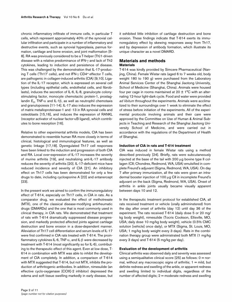
ResultsDecrease in the development of collagen induced arthritis rats treated with T-614The CIA model is characterized by aggressive synovitis, exten-sive pannus formation, cartilage degradation, and focal boneerosion. We investigated whether the protective activity of T-614 was mediated through a decrease in the severity of all ofthese clinical indices, or whether the activity of T-614 affectedonly specific pathogenetic processes. As shown in Figure 1,even after the onset of arthritis, T-614 (5 and 20 mg/kg perday) markedly reduced arthritic scores in the arthritic rats in adose-dependent manner, as compared with the vehicle-treated arthritic rats.
Progression of disease was indicated by increased edemaand erythema of one or both ankle joints, followed by involve-ment of the metatarsal and interphalangeal joints. Fully devel-oped arthritis, including red and swollen paws, was observed8 to 10 days after onset of inflammation. The clinical score inthe vehicle-treated group reached a peak approximately 20days after the first immunization (maximum arthritis score of5.75 ± 0.5; P < 0.01, versus day 12). Treatment with MTX (1mg/kg every 3 days) was efficacious and resulted in a delayedpeak (day 24) and also reduced clinical arthritis significantly atday 20 (clinical score 3.5 ± 0.57; P < 0.0286, versus vehicle).Signs of moderate arthritis were observed in rats treated witha low dose of T-614 (5 mg/kg), which became most severe atday 18 (maximal clinical score = 2.5 ± 0.6; P = 0.0286, versusday 12) and improved significantly at day 20 (clinical score =2.5 ± 1; P = 0.0286, versus vehicle). The high-dose T-614 (20mg/kg per day) and combination treatments almost completely
Figure 1
Effects of therapeutic treatment with T-614 on disease progression in rats with established CIAEffects of therapeutic treatment with T-614 on disease progression in rats with established CIA. Rats were orally treated daily with T-614 at 5 mg/kg per day or 20 mg/kg per day; MTX at 1 mg/kg every 3 days; nimesulide at 10 mg/kg per day; T-614 at 10 mg/kg per day and MTX at 1 mg/kg every 3 days; or vehicle. Treatment began on day 12 after immunization with type II collagen until day 36. Data are expressed as mean ± standard error of the mean (n = 5 to 7). *P < 0.05, **P < 0.01, versus day 12 or the vehicle-treated rats. CIA, collagen-induced arthritis; MTX, methotrexate.
Page 4 of 11(page number not for citation purposes)
Available online http://arthritis-research.com/content/10/6/R136
suppressed progression; maximal clinical scores in these ratswere 1.75 ± 0.9 at day 24 and 1.73 ± 0.8 at day 22, respec-tively (P > 0.05, versus day 12). The clinical score in the high-dose T-614 and combined treatment groups was found to bestatistically significantly lower than that in the control group atday 20; the maximal clinical scores in these two groups were1.75 ± 0.975 and 1.75 ± 0.79, respectively (P < 0.05, versusvehicle). Measurements of paw thickness and paw circumfer-ence were consistent with clinical scores (data not shown).
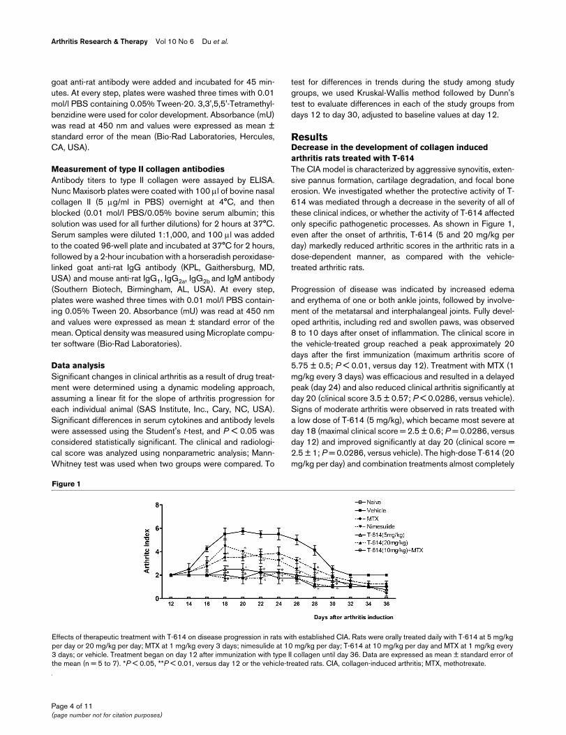
Decrease in the severity of inflammation in collagen induced arthritis rats treated with T-614The morphologic changes in the joint architecture of CIA ratswere further assessed using MRI, 21 days after the first immu-
nization. MRI soft tissue swelling is defined based on penetra-tion of subcutaneous soft tissues and bone marrow on the T1-weighted image within normal hyperintense subcutaneoussoft tissues and bone marrow. This corresponds to findings onthe STIR image (Figure 2a), in which the damage can be seenas a clearly demarcated zone of hyperintense signal within nor-mal hypointense area at this site (arrowhead).
Joints of naïve (non-CIA) rats exhibited intact joint architecture.The talus, phalanges, talocalcaneal joints, talonavicular articu-lations and cuneonavicular joints were well defined. Jointsfrom the vehicle-treated CIA group exhibited significant dam-age as well as swelling of soft tissues and marked bone mar-row edema. T-614 had a dose-related efficacy. Joints from rats
Figure 2
Effects of therapeutic treatment with T-614 on inflammation in the CIA ratsEffects of therapeutic treatment with T-614 on inflammation in the CIA rats. (a) STIR magnetic resonance images of hind paws from CIA rats. The presence of soft tissue swelling (yellow arrow) and localization of bone marrow edema (yellow triangle) are highlighted. Neither paw swelling nor bone marrow edema was seen in normal rats (subpanel a). Severe soft tissue swelling and bone erosion were seen in CIA rats treated with vehicle (subpanel b). Similar damage was observed in rats treated with nimesulide (subpanel d), but much less damage was seen in rats treated with MTX (subpanel c), T-614 (subpanels e and f), and combination treatment with T-614 and MTX (subpanel g). (b) Magnetic resonance imaging score of soft tissue swelling in treated CIA rats. Data are expressed as mean ± standard error of the mean (n = 3 to 5). *P < 0.05, **P < 0.01, versus vehicle-treated arthritic rats. CIA, collagen-induced arthritis; MTX, methotrexate.
Page 5 of 11(page number not for citation purposes)
Arthritis Research & Therapy Vol 10 No 6 Du et al.
treated with MTX (1 mg/kg every 3 days) or nimesulide (10mg/kg per day) also exhibited moderate damage, whereasnimesulide was associated with much less inhibition of bonemarrow edema. Joints from the T-614 (20 mg/kg per day)alone and combination therapy group exhibited significant inhi-bition of damage, which closely resembled the joints from thenaïve rats. As shown in Figure 2b, the mean MRI soft tissueswelling scores in vehicle-treated (5 ± 0.45) and nimesulide-treated rats (3.8 ± 0.37; P = 0.96, versus vehicle) were signif-icantly higher than those in rats treated with low-dose T-614(3.6 ± 0.4; P = 0.0479, versus vehicle), high-dose T-614 (2.8± 0.37; P = 0.0159 versus vehicle), and MTX (3.4 ± 0.25; P= 0.0318, versus vehicle). The hind paws of CIA rats receivingcombined treatment with MTX and T-614 exhibited completeprotection, with the lowest soft tissue swelling scores (1.8 ±0.38; P = 0.0079, versus vehicle).
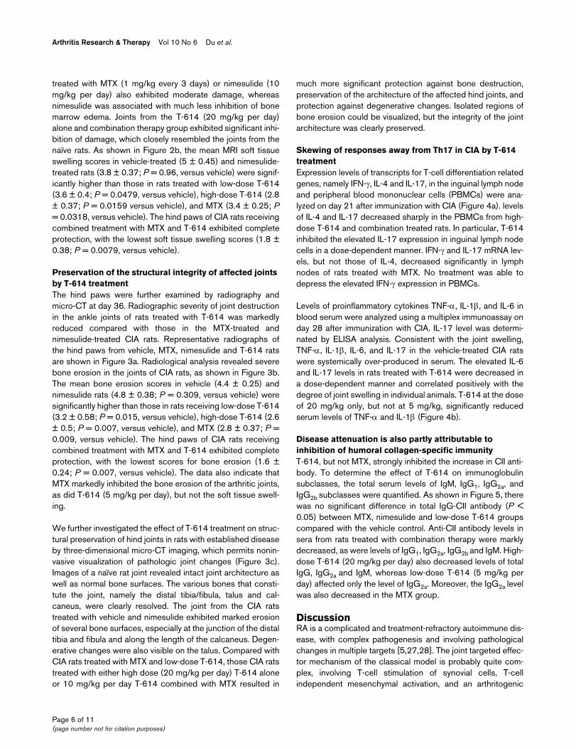
Preservation of the structural integrity of affected joints by T-614 treatmentThe hind paws were further examined by radiography andmicro-CT at day 36. Radiographic severity of joint destructionin the ankle joints of rats treated with T-614 was markedlyreduced compared with those in the MTX-treated andnimesulide-treated CIA rats. Representative radiographs ofthe hind paws from vehicle, MTX, nimesulide and T-614 ratsare shown in Figure 3a. Radiological analysis revealed severebone erosion in the joints of CIA rats, as shown in Figure 3b.The mean bone erosion scores in vehicle (4.4 ± 0.25) andnimesulide rats (4.8 ± 0.38; P = 0.309, versus vehicle) weresignificantly higher than those in rats receiving low-dose T-614(3.2 ± 0.58; P = 0.015, versus vehicle), high-dose T-614 (2.6± 0.5; P = 0.007, versus vehicle), and MTX (2.8 ± 0.37; P =0.009, versus vehicle). The hind paws of CIA rats receivingcombined treatment with MTX and T-614 exhibited completeprotection, with the lowest scores for bone erosion (1.6 ±0.24; P = 0.007, versus vehicle). The data also indicate thatMTX markedly inhibited the bone erosion of the arthritic joints,as did T-614 (5 mg/kg per day), but not the soft tissue swell-ing.
We further investigated the effect of T-614 treatment on struc-tural preservation of hind joints in rats with established diseaseby three-dimensional micro-CT imaging, which permits nonin-vasive visualization of pathologic joint changes (Figure 3c).Images of a naïve rat joint revealed intact joint architecture aswell as normal bone surfaces. The various bones that consti-tute the joint, namely the distal tibia/fibula, talus and cal-caneus, were clearly resolved. The joint from the CIA ratstreated with vehicle and nimesulide exhibited marked erosionof several bone surfaces, especially at the junction of the distaltibia and fibula and along the length of the calcaneus. Degen-erative changes were also visible on the talus. Compared withCIA rats treated with MTX and low-dose T-614, those CIA ratstreated with either high dose (20 mg/kg per day) T-614 aloneor 10 mg/kg per day T-614 combined with MTX resulted in
much more significant protection against bone destruction,preservation of the architecture of the affected hind joints, andprotection against degenerative changes. Isolated regions ofbone erosion could be visualized, but the integrity of the jointarchitecture was clearly preserved.
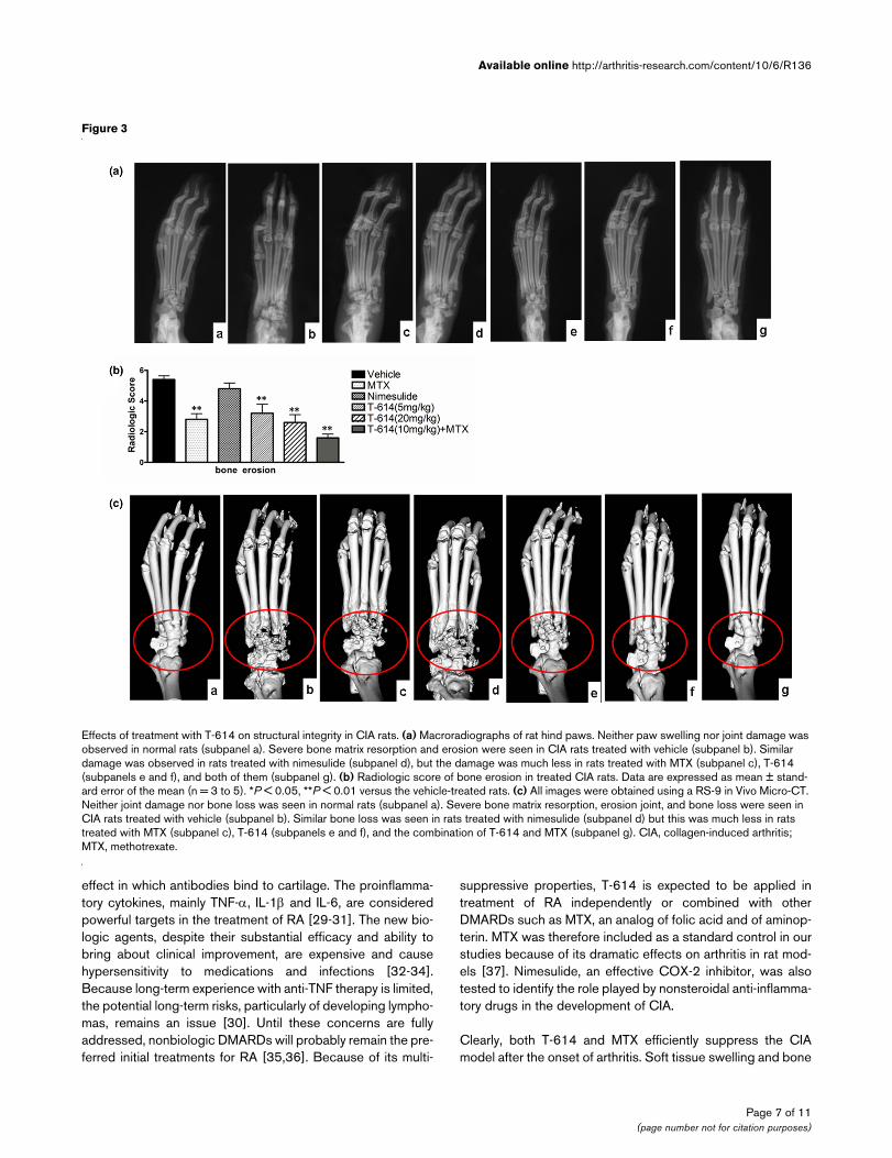
Skewing of responses away from Th17 in CIA by T-614 treatmentExpression levels of transcripts for T-cell differentiation relatedgenes, namely IFN-γ, IL-4 and IL-17, in the inguinal lymph nodeand peripheral blood mononuclear cells (PBMCs) were ana-lyzed on day 21 after immunization with CIA (Figure 4a). levelsof IL-4 and IL-17 decreased sharply in the PBMCs from high-dose T-614 and combination treated rats. In particular, T-614inhibited the elevated IL-17 expression in inguinal lymph nodecells in a dose-dependent manner. IFN-γ and IL-17 mRNA lev-els, but not those of IL-4, decreased significantly in lymphnodes of rats treated with MTX. No treatment was able todepress the elevated IFN-γ expression in PBMCs.
Levels of proinflammatory cytokines TNF-α, IL-1β, and IL-6 inblood serum were analyzed using a multiplex immunoassay onday 28 after immunization with CIA. IL-17 level was determi-nated by ELISA analysis. Consistent with the joint swelling,TNF-α, IL-1β, IL-6, and IL-17 in the vehicle-treated CIA ratswere systemically over-produced in serum. The elevated IL-6and IL-17 levels in rats treated with T-614 were decreased ina dose-dependent manner and correlated positively with thedegree of joint swelling in individual animals. T-614 at the doseof 20 mg/kg only, but not at 5 mg/kg, significantly reducedserum levels of TNF-α and IL-1β (Figure 4b).
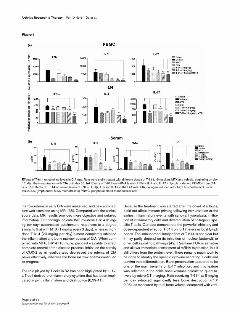
Disease attenuation is also partly attributable to inhibition of humoral collagen-specific immunityT-614, but not MTX, strongly inhibited the increase in CII anti-body. To determine the effect of T-614 on immunoglobulinsubclasses, the total serum levels of IgM, IgG1, IgG2a, andIgG2b subclasses were quantified. As shown in Figure 5, therewas no significant difference in total IgG-CII antibody (P <0.05) between MTX, nimesulide and low-dose T-614 groupscompared with the vehicle control. Anti-CII antibody levels insera from rats treated with combination therapy were marklydecreased, as were levels of IgG1, IgG2a, IgG2b and IgM. High-dose T-614 (20 mg/kg per day) also decreased levels of totalIgG, IgG2a and IgM, whereas low-dose T-614 (5 mg/kg perday) affected only the level of IgG2a. Moreover, the IgG2a levelwas also decreased in the MTX group.
DiscussionRA is a complicated and treatment-refractory autoimmune dis-ease, with complex pathogenesis and involving pathologicalchanges in multiple targets [5,27,28]. The joint targeted effec-tor mechanism of the classical model is probably quite com-plex, involving T-cell stimulation of synovial cells, T-cellindependent mesenchymal activation, and an arthritogenic
Page 6 of 11(page number not for citation purposes)
Available online http://arthritis-research.com/content/10/6/R136
effect in which antibodies bind to cartilage. The proinflamma-tory cytokines, mainly TNF-α, IL-1β and IL-6, are consideredpowerful targets in the treatment of RA [29-31]. The new bio-logic agents, despite their substantial efficacy and ability tobring about clinical improvement, are expensive and causehypersensitivity to medications and infections [32-34].Because long-term experience with anti-TNF therapy is limited,the potential long-term risks, particularly of developing lympho-mas, remains an issue [30]. Until these concerns are fullyaddressed, nonbiologic DMARDs will probably remain the pre-ferred initial treatments for RA [35,36]. Because of its multi-
suppressive properties, T-614 is expected to be applied intreatment of RA independently or combined with otherDMARDs such as MTX, an analog of folic acid and of aminop-terin. MTX was therefore included as a standard control in ourstudies because of its dramatic effects on arthritis in rat mod-els [37]. Nimesulide, an effective COX-2 inhibitor, was alsotested to identify the role played by nonsteroidal anti-inflamma-tory drugs in the development of CIA.
Clearly, both T-614 and MTX efficiently suppress the CIAmodel after the onset of arthritis. Soft tissue swelling and bone
Figure 3
Effects of treatment with T-614 on structural integrity in CIA ratsEffects of treatment with T-614 on structural integrity in CIA rats. (a) Macroradiographs of rat hind paws. Neither paw swelling nor joint damage was observed in normal rats (subpanel a). Severe bone matrix resorption and erosion were seen in CIA rats treated with vehicle (subpanel b). Similar damage was observed in rats treated with nimesulide (subpanel d), but the damage was much less in rats treated with MTX (subpanel c), T-614 (subpanels e and f), and both of them (subpanel g). (b) Radiologic score of bone erosion in treated CIA rats. Data are expressed as mean ± stand-ard error of the mean (n = 3 to 5). *P < 0.05, **P < 0.01 versus the vehicle-treated rats. (c) All images were obtained using a RS-9 in Vivo Micro-CT. Neither joint damage nor bone loss was seen in normal rats (subpanel a). Severe bone matrix resorption, erosion joint, and bone loss were seen in CIA rats treated with vehicle (subpanel b). Similar bone loss was seen in rats treated with nimesulide (subpanel d) but this was much less in rats treated with MTX (subpanel c), T-614 (subpanels e and f), and the combination of T-614 and MTX (subpanel g). CIA, collagen-induced arthritis; MTX, methotrexate.
Page 7 of 11(page number not for citation purposes)
Arthritis Research & Therapy Vol 10 No 6 Du et al.
marrow edema in early CIA were measured, and paw architec-ture was examined using MRI [38]. Compared with the clinicalscore data, MRI results provided more objective and detailedinformation. Our findings indicate that low-dose T-614 (5 mg/kg per day) suppressed autoimmune responses to a degreesimilar to that with MTX (1 mg/kg every 3 days), whereas high-dose T-614 (20 mg/kg per day) almost completely inhibitedthe inflammation and bone marrow edema of CIA. When com-bined with MTX, T-614 (10 mg/kg per day) was able to effectcomplete control of the disease process. Inhibition the activityof COX-2 by nimesulide also depressed the edema of CIApaws effectively, whereas the bone marrow edema continuedto progress.
The role played by T cells in RA has been highlighted by IL-17,a T-cell derived proinflammatory cytokine that has been impli-cated in joint inflammation and destruction [8,39-41].
Because the treatment was started after the onset of arthritis,it did not affect immune priming following immunization or theearliest inflammatory events with synovial hyperplasia, infiltra-tion of inflammatory cells and differentiation of collagen II-spe-cific T cells. Our data demonstrate the powerful inhibitory anddose-dependent effect of T-614 on IL-17 levels in local lymphnodes. The immunomodulatory effect of T-614 is not clear butit may partly depend on its inhibition of nuclear factor-κB orother cell signaling pathways [42]. Real-time PCR is sensitiveand allows immediate assessment of mRNA expression, but itstill differs from the protein level. There remains much work tobe done to identify the specific cytokine-secreting T cells andconfirm their differentiation. Bone preservation appeared to beone of the main benefits of IL-17 inhibition, and this featurewas reflected in the ankle bone volumes calculated quantita-tively by micro-CT imaging. Rats receiving T-614 at 5 mg/kgper day exhibited significantly less bone destruction (P <0.05), as measured by total bone volume, compared with vehi-
Figure 4
Effects of T-614 on cytokine levels in CIA ratsEffects of T-614 on cytokine levels in CIA rats. Rats were orally treated with different doses of T-614, nimesulide, MTX and vehicle, beginning on day 12 after the immunization with CIA until day 36. (a) Effects of T-614 on mRNA levels of IFN-γ, IL-4 and IL-17 in lymph node and PBMCs from CIA rats. (b) Effects of T-614 on serum levels of TNF-α, IL-1β, IL-6 and IL-17 in the CIA rats. CIA, collagen-induced arthritis; IFN, interferon; IL, inter-leukin; LN, lymph node; MTX, methotrexate; PBMC, peripheral blood mononuclear cell.
Page 8 of 11(page number not for citation purposes)
Available online http://arthritis-research.com/content/10/6/R136
cle-treated arthritic controls. The bone volumes of rats receiv-ing T-614 at 20 mg/kg per day and T-614 combined with MTXremained almost intact. The findings support the view that T-614 can protect the joints from damage in an inflammatoryenvironment, in concert with MTX.
Proinflammatory cytokines TNF-α, IL-1β, and IL-6 help to prop-agate the extension of a local or systemic inflammatory proc-ess. Similar to the IL-17 levels in serum, markedly low serumlevels of IL-6 were also observed in CIA rats treated with T-614, even at the dose of 5 mg/kg per day. Only MTX, high-dose T-614 (20 mg/kg per day) and not low-dose T-614 (5mg/kg per day), and combination treatment significantlyreduced serum levels of TNF-α and IL-1β. Recent studies haveshown that IL-6, in combination with transforming growth fac-tor-β, inhibits the generation of FoxP3-expressing T-regulatorycells and induces the generation of Th17 cells [43]. Th1, Th2,and Th17 cells develop from naïve T cells; in contrast, the gen-eration of T-regulatory cells and Th17 cells occurs via alterna-tive pathways, and they are selected according to thepresence or absence of IL-6, a pleiotropic cytokine that playsimportant roles in the regulation of the immune response,inflammation, and hematopoiesis. Decreased IL-6 productioncould contribute to the attenuation of Th17 responses, whichmay also explain the therapeutic effect of T-614. IL-6 alsoinduces activated B cells to differentiate into antibody-produc-ing cells [44] and promotes the production of vascularendothelial growth factor, which plays an important role in ang-iogenesis [45]. Furthermore, in terms of bone metabolism, IL-6 induces osteoclast differentiation in the presence of solubleIL-6 receptor, thereby contributing to joint destruction andosteoporosis [46]. IL-17 significantly induces the synthesis of
IL-6 by synoviocytes and macrophages. A positive feedbackloop initiates and accelerates the progression of CIA. Modula-tion of inflammatory cytokines and IL-17 by T-614 suggests itspotential therapeutic value in the treatment of other inflamma-tory diseases, such as ankylosing spondylitis and psoriaticarthritis.
During the development of CIA, increasing levels of anti-CIIantibodies bind to the collagen of the articular cartilage, acti-vate the complement system and initiate tissue damage; thisindicates that there is T-B cell cooperation and activation invivo [47,48]. More interestingly, T-614 not only suppressedCII antibody levels but also differentially modulated immu-noglobulin subclass levels; these effects suggest that it maybe useful for the treatment of lupus or other autoimmune dis-orders. Similar effects were seen in the combination therapygroup, indicating that there is synergy between T-614 andMTX. Low-dose T-614 and MTX also had an effect on the levelof IgG2a antibody, indicating that they may operate through T-cell associated antibodies in the CIA model. Because IgG2a isthe most potent activator of the classical complement cascadeand Fc receptor bearing macrophages, the present findingsadd further support to the inhibitory mechanism of T-614 andthe pathogenic role of IgG2a in rat CIA [49].
To summarize, T-614 – a novel immunomodulatory drug –appears to protect the joints from inflammation injury and oste-oclastic bone resorption through skewing the response fromprimarily a Th17-driven one, and it does so to a greater degreein combination with MTX. These findings suggest that T-614is a new candidate for use in combination therapy, which is
Figure 5
Effects of T-614 on serum IgG levels in CIA ratsEffects of T-614 on serum IgG levels in CIA rats. Serum was collected on day 36. Anti-CII (total IgG, IgM, IgG1, IgG2a, and IgG2b) levels increased as disease progressed. Combination therapy reduced the total anti-CII antibody level significantly, as well as levels of IgM, IgG1, IgG2a, and IgG2b. High dose T-614(20 mg/kg per day) also decreased levels of total IgG, IgM and IgG2a, whereas low-dose of T-614 (5 mg/kg per day) or MTX (1 mg/kg every 3 days) had an effect only on IgG2a level. Data are expressed as mean ± standard error of the mean (n = 5 to 7). *P < 0.05, versus the vehi-cle-treated rats.
Page 9 of 11(page number not for citation purposes)
Arthritis Research & Therapy Vol 10 No 6 Du et al.
increasingly being applied to the treatment of RA and otherTh17-associated inflammatory autoimmune diseases.
ConclusionIn the present experiments, T-614 significantly preventedbone/cartilage destruction and inflammation in CIA. Further-more, combination with MTX enhanced the therapeutic effectof T-614.
Competing interestsThe authors declare that they have no competing interests.
Authors' contributionsCB designed and conceived the study. FD conducted theexperimental work and drafted the manuscript. SC partici-pated in the design of the study. LL performed the statisticalanalysis. JT, MD, WF, PY, NS, XH and JQ helped with someexperimental work. All authors read and approved the finalmanuscript.
AcknowledgementsThis work was supported by grants from National Natural Science Foun-dation of China (grant no. 30873079); Doctoral Innovation Fund of Shanghai Jiao Tong University School of Medicine (grant no. BXJ0818); Shanghai Key Discipline Construction Project (grant no. T0203); and Shanghai Hospital Clinical and research resource Platform Project (grant no. SHDC12007205). The authors should like to acknowledge Simcere pharmaceutical Co., Ltd. (Nanjing, China), which provided the pharmaceutical product T-614.
References1. Tanaka K, Yamamoto T, Aikawa Y, Kizawa K, Muramoto K, Matsuno
H, Muraguchi A: Inhibitory effects of an anti-rheumatic agent T-614 on immunoglobulin production by cultured B cells andrheumatoid synovial tissues engrafted into SCID mice. Rheu-matology (Oxford) 2003, 42:1365-1371.
2. Sawada T, Hashimoto S, Tohma S, Nishioka Y, Nagai T, Sato T, ItoK, Inoue T, Iwata M, Yamamoto K: Inhibition of L-leucine methylester mediated killing of THP-1, a human monocytic cell line,by a new anti-inflammatory drug, T614. Immunopharmacology2000, 49:285-294.
3. Tanaka K, Kawasaki H, Kurata K, Aikawa Y, Tsukamoto Y, Inaba T:T-614, a novel antirheumatic drug, inhibits both the activity andinduction of cyclooxygenase-2 (COX-2) in cultured fibroblasts.Jpn J Pharmacol 1995, 67:305-314.
4. Lu LJ, Teng JL, Bao CD, Han XH, Sun LY, Xu JH, Li XF, Wu HX:Safety and efficacy of T-614 in the treatment of patients withactive rheumatoid arthritis: a double blind, randomized, pla-cebo-controlled and multicenter trial. Chin Med J (Engl) 2008,121:615-619.
5. Scrivo R, Di Franco M, Spadaro A, Valesini G: The immunologyof rheumatoid arthritis. Ann N Y Acad Sci 2007, 1108:312-322.
6. Lutzky V, Hannawi S, Thomas R: Cells of the synovium in rheu-matoid arthritis. Dendritic cells. Arthritis Res Ther 2007,9:219-231.
7. Du F, Wang L, Zhang Y, Jiang W, Sheng H, Cao Q, Wu J, Shen B,Shen T, Zhang JZ, Bao C, Li D, Li N: Role of GADD45 beta in theregulation of synovial fluid T cell apoptosis in rheumatoidarthritis. Clin Immunol 2008, 128:238-247.
8. Andreas K, Lubke C, Haupl T, Dehne T, Morawietz L, Ringe J, KapsC, Sittinger M: Key regulatory molecules of cartilage destruc-tion in rheumatoid arthritis: an in vitro study. Arthritis Res Ther2008, 10:R9-25.
9. Aarvak T, Chabaud M, Miossec P, Natvig JB: IL-17 is produced bysome proinflammatory Th1/Th0 cells but not by Th2 cells. JImmunol 1999, 162:1246-1251.
10. Lubberts E, Koenders MI, Berg WB van den: The role of T-cellinterleukin-17 in conducting destructive arthritis: lessons fromanimal models. Arthritis Res Ther 2005, 7:29-37.
11. Hwang SY, Kim JY, Kim KW, Park MK, Moon Y, Kim WU, Kim HY:IL-17 induces production of IL-6 and IL-8 in rheumatoid arthri-tis synovial fibroblasts via NF-kappaB- and PI3-kinase/Akt-dependent pathways. Arthritis Res Ther 2004, 6:R120-128.
12. Granet C, Maslinski W, Miossec P: Increased AP-1 and NF-kap-paB activation and recruitment with the combination of theproinflammatory cytokines IL-1beta, tumor necrosis factoralpha and IL-17 in rheumatoid synoviocytes. Arthritis Res Ther2004, 6:R190-198.
13. Kehlen A, Pachnio A, Thiele K, Langner J: Gene expressioninduced by interleukin-17 in fibroblast-like synoviocytes ofpatients with rheumatoid arthritis: upregulation of hyaluronan-binding protein TSG-6. Arthritis Res Ther 2003, 5:R186-192.
14. Van Bezooijen RL, Wee-Pals L Van Der, Papapoulos SE, LowikCW: Interleukin 17 synergises with tumour necrosis factoralpha to induce cartilage destruction in vitro. Ann Rheum Dis2002, 61:870-876.
15. Chabaud M, Garnero P, Dayer JM, Guerne PA, Fossiez F, MiossecP: Contribution of interleukin 17 to synovium matrix destruc-tion in rheumatoid arthritis. Cytokine 2000, 12:1092-1099.
16. Kotake S, Udagawa N, Takahashi N, Matsuzaki K, Itoh K, IshiyamaS, Saito S, Inoue K, Kamatani N, Gillespie MT, Martin TJ, Suda T:IL-17 in synovial fluids from patients with rheumatoid arthritisis a potent stimulator of osteoclastogenesis. J Clin Invest1999, 103:1345-1352.
17. Gierer P, Ibrahim S, Mittlmeier T, Koczan D, Moeller S, Landes J,Gradl G, Vollmar B: Gene expression profile and synovialmicrocirculation at early stages of collagen-induced arthritis.Arthritis Res Ther 2005, 7:R868-876.
18. Shou J, Bull CM, Li L, Qian HR, Wei T, Luo S, Perkins D, SolenbergPJ, Tan SL, Chen XY, Roehm NW, Wolos JA, Onyia JE: Identifica-tion of blood biomarkers of rheumatoid arthritis by transcriptprofiling of peripheral blood mononuclear cells from the ratcollagen-induced arthritis model. Arthritis Res Ther 2006,8:R28-42.
19. Lubberts E, Joosten LA, Oppers B, Bersselaar L van den, Coenen-de Roo CJ, Kolls JK, Schwarzenberger P, Loo FA van de, Berg WBvan den: IL-1-independent role of IL-17 in synovial inflamma-tion and joint destruction during collagen-induced arthritis. JImmunol 2001, 167:1004-1013.
20. Lubberts E, Koenders MI, Oppers-Walgreen B, Bersselaar L vanden, Coenen-de Roo CJ, Joosten LA, Berg WB van den: Treat-ment with a neutralizing anti-murine interleukin-17 antibodyafter the onset of collagen-induced arthritis reduces jointinflammation, cartilage destruction, and bone erosion. ArthritisRheum 2004, 50:650-659.
21. Nakae S, Nambu A, Sudo K, Iwakura Y: Suppression of immuneinduction of collagen-induced arthritis in IL-17-deficient mice.J Immunol 2003, 171:6173-7.
22. Cho ML, Ju JH, Kim KW, Moon YM, Lee SY, Min SY, Cho YG, KimHS, Park KS, Yoon CH, Lee SH, Park SH, Kim HY: CyclosporineA inhibits IL-15-induced IL-17 production in CD4+ T cells viadown-regulation of PI3K/Akt and NF-kappaB. Immunol Lett2007, 108:88-96.
23. Zaba LC, Cardinale I, Gilleaudeau P, Sullivan-Whalen M, SuarezFarinas M, Fuentes-Duculan J, Novitskaya I, Khatcherian A, BluthMJ, Lowes MA, Krueger JG: Amelioration of epidermal hyper-plasia by TNF inhibition is associated with reduced Th17responses. J Exp Med 2007, 204:3183-3194.
24. Rosloniec EF, Cremer M, Kang A, Myers LK: Collagen-inducedarthritis. Curr Protoc Immunol 2001, Chapter 15:. Unit 15.5.
25. Brahn E, Banquerigo ML, Firestein GS, Boyle DL, Salzman AL,Szabo C: Collagen induced arthritis: reversal by mercap-toethylguanidine, a novel antiinflammatory agent with a com-bined mechanism of action. J Rheumatol 1998, 25:1785-1793.
26. Ostergaard M, Peterfy C, Conaghan P, McQueen F, Bird P, EjbjergB, Shnier R, O'Connor P, Klarlund M, Emery P, Genant H, LassereM, Edmonds J: OMERACT Rheumatoid Arthritis Magnetic Res-onance Imaging Studies. Core set of MRI acquisitions, jointpathology definitions, and the OMERACT RA-MRI scoring sys-tem. J Rheumatol 2003, 30:1385-1386.
27. Goronzy JJ, Weyand CM: Rheumatoid arthritis. Immunol Rev2005, 204:55-73.
Page 10 of 11(page number not for citation purposes)
Available online http://arthritis-research.com/content/10/6/R136
28. Sweeney SE, Firestein GS: Signal transduction in rheumatoidarthritis. Curr Opin Rheumatol 2004, 16:231-237.
29. Chopin F, Garnero P, le Henanff A, Debiais F, Daragon A, Roux C,Sany J, Wendling D, Zarnitsky C, Ravaud P, Thomas T: Long-termeffects of infliximab on bone and cartilage turnover markers inpatients with rheumatoid arthritis. Ann Rheum Dis 2008,67:353-357.
30. Nishimoto N, Kishimoto T: Interleukin 6: from bench to bedside.Nat Clin Pract Rheumatol 2006, 2:619-626.
31. Cruyssen B Vander, Van Looy S, Wyns B, Westhovens R, DurezP, Bosch F Van den, Mielants H, De Clerck L, Peretz A, Malaise M,Verbruggen L, Vastesaeger N, Geldhof A, Boullart L, De Keyser F:Four-year follow-up of infliximab therapy in rheumatoid arthri-tis patients with long-standing refractory disease: attrition andlong-term evolution of disease activity. Arthritis Res Ther 2006,8:R112-R119.
32. Carmona L, Gomez-Reino JJ, Rodriguez-Valverde V, Montero D,Pascual-Gomez E, Mola EM, Carreno L, Figueroa M: Effective-ness of recommendations to prevent reactivation of latenttuberculosis infection in patients treated with tumor necrosisfactor antagonists. Arthritis Rheum 2005, 52:1766-1772.
33. Brassard P, Kezouh A, Suissa S: Antirheumatic drugs and therisk of tuberculosis. Clin Infect Dis 2006, 43:717-722.
34. Dixon WG, Watson K, Lunt M, Hyrich KL, Silman AJ, Symmons DP:Rates of serious infection, including site-specific and bacterialintracellular infection, in rheumatoid arthritis patients receiv-ing anti-tumor necrosis factor therapy: results from the BritishSociety for Rheumatology Biologics Register. Arthritis Rheum2006, 54:2368-2376.
35. Saag KG, Teng GG, Patkar NM, Anuntiyo J, Finney C, Curtis JR,Paulus HE, Mudano A, Pisu M, Elkins-Melton M, Outman R, AllisonJJ, Suarez Almazor M, Bridges SL Jr, Chatham WW, Hochberg M,MacLean C, Mikuls T, Moreland LW, O'Dell J, Turkiewicz AM, FurstDE: American College of Rheumatology 2008 recommenda-tions for the use of nonbiologic and biologic disease-modify-ing antirheumatic drugs in rheumatoid arthritis. ArthritisRheum 2008, 59:762-784.
36. Bathon JM, Cohen SB: The 2008 American College of Rheuma-tology recommendations for the use of nonbiologic and bio-logic disease-modifying antirheumatic drugs in rheumatoidarthritis: where the rubber meets the road. Arthritis Rheum2008, 59:757-759.
37. Lange F, Bajtner E, Rintisch C, Nandakumar KS, Sack U, HolmdahlR: Methotrexate ameliorates T cell dependent autoimmunearthritis and encephalomyelitis but not antibody induced orfibroblast induced arthritis. Ann Rheum Dis 2005, 64:599-605.
38. Jacobson PB, Morgan SJ, Wilcox DM, Nguyen P, Ratajczak CA,Carlson RP, Harris RR, Nuss M: A new spin on an old model: invivo evaluation of disease progression by magnetic resonanceimaging with respect to standard inflammatory parametersand histopathology in the adjuvant arthritic rat. Arthritis Rheum1999, 42:2060-2073.
39. Koenders MI, Lubberts E, Loo FA van de, Oppers-Walgreen B,Bersselaar L van den, Helsen MM, Kolls JK, Di Padova FE, JoostenLA, Berg WB van den: Interleukin-17 acts independently ofTNF-alpha under arthritic conditions. J Immunol 2006,176:6262-6269.
40. Chabaud M, Page G, Miossec P: Enhancing effect of IL-1, IL-17,and TNF-alpha on macrophage inflammatory protein-3alphaproduction in rheumatoid arthritis: regulation by solublereceptors and Th2 cytokines. J Immunol 2001, 167:6015-6020.
41. Lubberts E: IL-17/Th17 targeting: on the road to preventchronic destructive arthritis? Cytokine 2008, 41:84-91.
42. Aikawa Y, Yamamoto M, Yamamoto T, Morimoto K, Tanaka K: Ananti-rheumatic agent T-614 inhibits NF-kappaB activation inLPS- and TNF-alpha-stimulated THP-1 cells without interfer-ing with IkappaBalpha degradation. Inflamm Res 2002,51:188-194.
43. Zhou L, Ivanov II, Spolski R, Min R, Shenderov K, Egawa T, LevyDE, Leonard WJ, Littman DR: IL-6 programs T(H)-17 cell differ-entiation by promoting sequential engagement of the IL-21and IL-23 pathways. Nat Immunol 2007, 8:967-974.
44. Yoshizaki K, Nakagawa T, Fukunaga K, Tseng LT, Yamamura Y,Kishimoto T: Isolation and characterization of B cell differenti-ation factor (BCDF) secreted from a human B lymphoblastoidcell line. J Immunol 1984, 132:2948-2954.
45. Nakahara H, Song J, Sugimoto M, Hagihara K, Kishimoto T,Yoshizaki K, Nishimoto N: Anti-interleukin-6 receptor antibodytherapy reduces vascular endothelial growth factor productionin rheumatoid arthritis. Arthritis Rheum 2003, 48:1521-1529.
46. Tamura T, Udagawa N, Takahashi N, Miyaura C, Tanaka S, YamadaY, Koishihara Y, Ohsugi Y, Kumaki K, Taga T, Kishimoto T, Suda T:Soluble interleukin-6 receptor triggers osteoclast formationby interleukin 6. Proc Natl Acad Sci USA 1993,90:11924-11928.
47. Terato K, Hasty KA, Reife RA, Cremer MA, Kang AH, Stuart JM:Induction of arthritis with monoclonal antibodies to collagen.J Immunol 1992, 148:2103-2108.
48. Zheng B, Zhang X, Guo L, Han S: IgM plays an important role ininduction of collagen-induced arthritis. Clin Exp Immunol2007, 149:579-585.
49. Nandakumar KS, Johansson BP, Bjorck L, Holmdahl R: Blockingof experimental arthritis by cleavage of IgG antibodies in vivo.Arthritis Rheum 2007, 56:3253-3260.
Page 11 of 11(page number not for citation purposes)