Embed Size (px)
Citation preview

MRI Safety: Inherent Dangers and Preventative
Measures
Thursday, February 13th, 2014

2
SpeakerSue Dill Calloway RN, Esq.CPHRM, CCMSCPAD, BA, BSN, MSN, JDPresident Patient Safety andHealthcare Education
Board MemberEmergency Medicine PatientSafety Officer www.empsf.org

3
1. Explain the TJC sentinel event alert that was issued for preventing accidents and injuries in the MRI suite
2. Describe recommendations to improve MRI safety
Learning Objectives

4
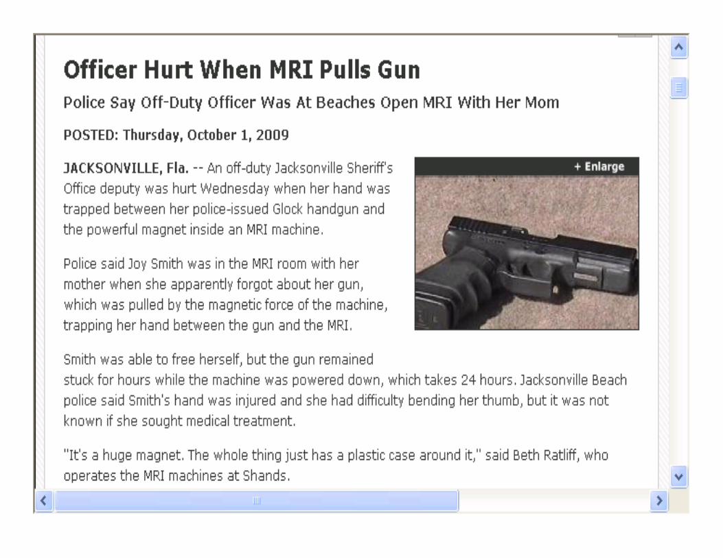
Headlines You Don’t Want to SeeHoag Hospital in California fined by state Dept of
Public Health after patient was taken to MRI on a metal gurneyPatient was pulled into the imaging machine
breaking her lower leg Leg was trapped for three minutes and spent 3
days in the hospital and magnet quenchedHad adopted new procedure and checklist before
entering MRI roomHas installed cameras for monitoringFined $50,000 in 2010

5
Headlines You Don’t Want to See

6

7
Prevention Costs Less“The costs of the safety provision to help prevent these accidents are peanuts when compared to the costs of accidents”Do you use a ferromagnet detector?Cost to restore the magnet after the quench , cost of down time, and lost revenue, lawsuit costs, fines, cost to investigate is greater than cost of prevention Source: Gurney Crashes MRI, Patient Injured, Hospital
Fined $50 K, Tobias Gilk, MRI Safety Director, Mednovus Inc., MRI Metal Detector Blog

Scissors in ForeheadFlying scissors had be
surgically removed from technologist’s forehead
This is not the real x-ray of the injury that occurred
http://mrimetaldetector.com/blog/tag/maude/
Thanks to Tobias Gilk for his input on this presentation
9

Death of Engineer by MRIField engineer called to fix blower motor due to MRI making noise
Arrived at 2100 and guard check and no response and he left after making no investigation
Found next day dead pinned by MRI machine
Reported to GE HealthcareSource FDA Maude AE Report, March 2010 at
http://www.accessdata.fda.gov/scripts/cdrh/cfdocs/cfMAUDE/detail.cfm?mdrfoi__id=1648230
10

IV Cart in MRI Machine
October 10, 2009 MRI Risk Assessment Newsletter

FDA’s Data from Maude 4th consecutive year with substantial increases in rates of
MRI accidents A 30 percent increase from last year
This was an increase in the number of reports to FDA of MRI accidents
There was a 240 percent increase from2004 to 2008
There are at least 148 reports from MRIs
One expert suggests this represents about 14,800 real world accidents
Source: http://mrimetaldetector.com/blog/?p=329
Source: http://mrimetaldetector.com/blog/?p=329 and FDA website is http://www.accessdata.fda.gov/scripts/cdrh/cfdocs/cfMAUDE/detail.cfm?mdrfoi__id=996580
12

13
MRI Scanner Eats an ICU Bed

FDA MAUDE Database
14
www.fda.gov/MedicalDevices/DeviceRegulationandGuidance/PostmarketRequirements/ReportingAdverseEvents/ucm127891.htm

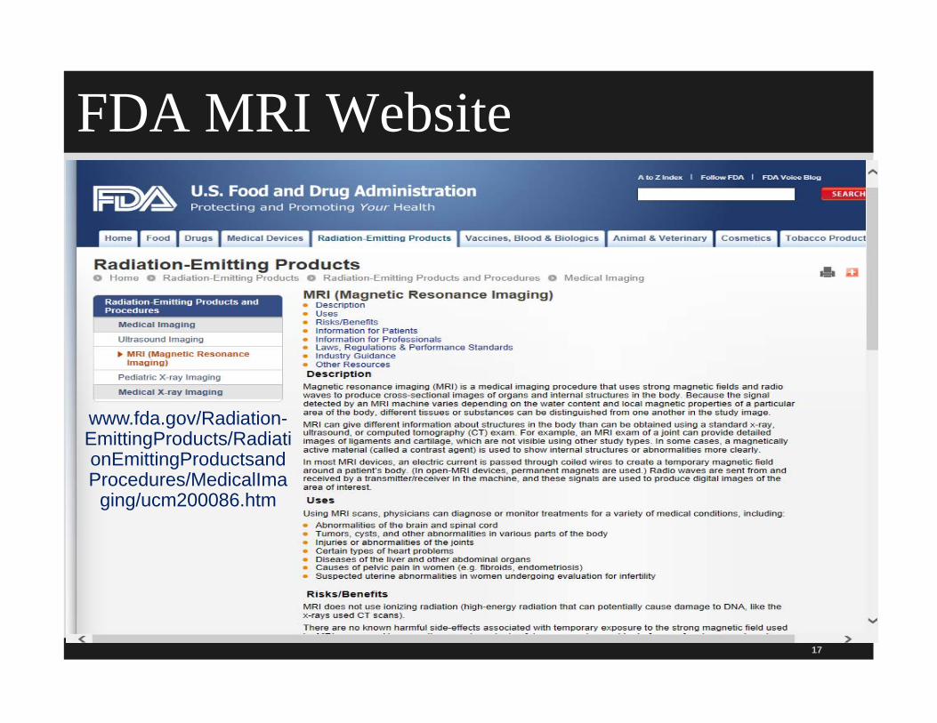
FDA MRI WebsiteThe FDA has a MRI website that includes
information on the following:
Uses and description
Risks and benefits Dyes from tattoos or tattooed eye liner
Information for patients
Information for professionals, laws, standards MRI safety, MRI contrast agents containing gadolinium, and
nephrogenic fibrosis dermopathy, injuries with implanted stimulators, burns with transdermal patches, cable and electrode burns
15
16
www.fda.gov/MedicalDevices/DeviceRegulationandGuidance/GuidanceDocuments/ucm107721.htm
FDA MRI Website
17
www.fda.gov/Radiation-EmittingProducts/RadiationEmittingProductsandProcedures/MedicalIma
ging/ucm200086.htm

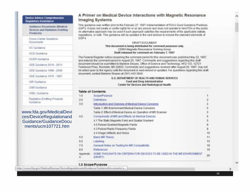
Primer on Medical Devices Interaction & MRI
18
www.fda.gov/MedicalDevices/DeviceRegulationandGuidance/GuidanceDocuments/ucm107721.htm

TJC MRI Safety StandardsTJC implements safety standards for hospitals to
manage the safety risks with MRI
Effective July 1, 2014
Must manage safety risks regarding; Patients with anxiety or claustrophobia
Patients who need urgent or emergent medical care
Patients with medical implants, devices, or imbedded FO such as shrapnel
Acoustic noise and ferromagnetic objects entering the room
19

TJC Standards July 2014 MRI
20

TJC MRI Safety StandardsHospital must restrict access to room of those not
screened or trained
Make sure restricted areas are controlled by and under direct supervision of MRI trained staff
Post signs by entrance to scanner about dangers of the magnet and that it is always on
Also statement related to PET scan, CT, or nuclear medicine services that staff dosimetry results are reviewed quarterly by the radiation safety officer or medical physicist to assess staff radiation exposure levels to be as low as possible
21

TJC MRI Safety StandardsHR must make sure technologist who does MRI
participates in on-going education that includes annual training on safe MRI practices that include the following:
Patient screening criteria that addresses ferromagnetic item, medical implants and devices
Risk for nephrogenic systemic fibrosis (NSF)
Proper positioning to prevent burns
Equipment and supplies that are acceptable to use in MRI suite
22

TJC MRI Safety StandardsAnnual training on safe MRI practices that include
the following (continued):
MRI safety response procedures for patient who require urgent or emergency medical treatment
MRI equipment emergency shutdown procedures
Patient hearing protection
Management off patients with claustrophobia, anxiety, or emotional distress
References ASTM standards
23

24
What is an MRI?Noninvasive medical imaging technique used
primarily in radiology Can be used to image anatomy in multiple
planes or slices Uses magnetic fields and not x-rays or ionizing
radiation Used to investigate the brain, spinal cord, and
vertebrae and surrounding tissueOriginally named zeugmatography In Greek it means “that which is used for joining”

25
What is an MRI? Many hospitals now have MRI information on their
website
Best noninvasive way to view abnormalities in cartilage, tendons, and ligaments
Investigating the musculoskeletal system, particularly joints
Image the eyes and sinuses
Identify tumors throughout the body and ascertain their stage of development
Evaluate large and medium-sized blood vessels

26
The Case that Everyone has HeardSix year old child had an MRI in a hospital in
Valhalla, New York on July 31, 2001
Metal oxygen cylinder brought into room
Nurse thought canister was made of nonmagnetic material like aluminum No special marking on tank
2011 was 10 year anniversary and it was MRI week
Oxygen tank became a missile and was drawn into the magnetic core while boy still in the machine in 2001

27
The Case that Everyone has Heard Tank struck Michael Colombini in the head
Caused a fractured skull
Child died of fatal cerebral hemorrhage
First fatal MRI accident of its kind
Settlement agreement wants opportunity for others to learn from this incident (settled nine years later)
Feb 2010 article states case is settled for $2.9 Million 1
2011 was 10 year anniversary and has MRI week 1 http://mrimetaldetector.com/blog/2010/02/2-9-million-settlement-closes-colombini-mri-death-case/

28

29
So What Happened? NQF Never EventStatic magnetic field generated by MR systems
attracts ferromagnetic objects with considerable force
Material can be magnetized in the presence of an external magnetic field
This causes the object, like the oxygen tank, to move toward it Called the projectile effect
2011 NQF updates never events or serious reportable errors to include from metallic object into the MRI area

30
So What Happened? Every hospital should show the video of the oxygen
cylinder crashing or the patient gurney crashing
It is said that a picture is worth a thousand wordsTwo-minute video to show the projectile effect Items pulled into MRIs IV poles, mop buckets, chair, ladder, laundry cart, floor
buffer, pulse ox transformer, tools, scissors, sand bags, and traction weights
Noted on MDR reports at the time of this occurrence (ECRI Hazard Report)
1 http://www.mri-planning.com/

31
Other CasesWoman who had an aneurysm clip in her brain died after undergoing MRI
Welder with piece of metal embedded in his eye blinded in that eye after MRI
60 year old man sustained fractures to face when oxygen canister became wedged in machine against his face (awarded $100,000 in damages)
Source: Web MD article August 1, 2001

32
Other CasesOff-duty policeman arrived to have MRI doneHe told technician he was carrying a firearm so
Tech intended to meet him in the waiting area to secure the weaponOfficer entered the MR scan room and put his gun
on the cabinet 3 feet from the 1.5 T magnet boreGun was pulled from his hand Gun hit left side of bore (inside) of MRI and fired a
round into the back wall of the scan room Luckily no one was injured
Source: Safety Concerns in the MR environment, ECRI, May 2006

33
Other CasesPatient came into ED with sweatpants and track
shoes and was sent to MRI The patient was moved to the MRI scanner
head first His legs were lifted up so his face was in the
MRI doughnut The Patient had sandbag ankle weights When his legs were lifted up, he screamed in pain The magnet was quenched (quenching can cause
asphyxiation, frostbite, fire hazard and can cost $20,000 to $500,000)
34
History First adult MRI occurred in 1977
Today more than 10 million MRIs are done in the US every year
MRI is among safest compared to many other diagnostic procedures (Strokowski, 2005)
Many believe number of actual adverse events is higher than reported Numbers of adverse events are low considering how many
are done Field of magnetic resonance imaging has seen
tremendous progress in last 20 years1
1Dr. Emanuel Kanal, University of Pittsburg Medical Center, MR Safe Practice Guidelines

35
MRI- Related EventsMany hardware and software advances and
improving contrast agents
In 2005, ECRI analyzed FDA’s data MAUDE (Manufacturer and User Facility Device Experience
Database) reported data over a 10-year time span or MRI injuries
Study found 389 reports of MRI-related events
Nine deaths from MRI studies Three related to pacemaker failure, 2 incidents of insulin
pump failure and 4 implant disturbances
One asphyxiation from a cryogenic mishap during installation of an MR imaging system

36
TJC Sentinel Event AlertThe Joint Commission issued a 3 page Sentinel
Event Alert (SEA) 38, February 14, 2008
Preventing accidents and injuries in the MRI suite
Review all SEAs and implement
Have a committee to review and address
Institute a policy based on the Alert
This and ACR MR safety are the two industry recognized MRI safety practice standards
Sign up to receive e-mail notice when Alerts are published1
www.jointcommission.org1

TJC Sentinel Event Alert MRI Safety TJC set out 10 recommendations which are
discussed in detail later Use 4 zones for safety
Use trained staff to screen any non-emergent patients twice
Provide ear plugs and hearing protection to all patients
Provide at least annual training regarding MRI safety to MR personnel– Provide non-MR staff and patients with material, such as a flier,
that explains potential for adverse events37
TJC 10 MRI Safety RecommendationsEnsure there is complete medical history to ensure
patient is safe to scan
Have specially trained MR staff who are knowledgeable about all 4 zones and the MRI safety environment issues
Take precautions to prevent burns Cold compress or ice pack on EKG leads, surgical
staples, tattoos or electrically conductive material
Ensure no electrically conductive loops are formed in the MR scanner bore
Use nonconductive foam pads to insulate patient’s skin38
TJC 10 MRI Safety RecommendationsNever code a patient in the MRI magnet room
Proactively plan for managing critically ill patients who require monitoring
This may include monitoring of life sustaining drugs or other medications
Use only MR safe equipment
Oxygen, fire extinguishers, physiologic monitors, wheelchairs etc.
The article recognized there are 3 more recommendations
39

3 Additional RecommendationsRecommended by Dr Emanuel Kanal, leading
expert on MR Safety
Appoint a patient safety officer who is responsible for enforcing safety procedures in MRI suite
Implement safe practices system such as MRI safety P&P and periodically assess compliance with P&P
Do not bring anything into the MRI room unless proven to be MR safe or MR conditional
40

TJC Sentinel Event Alerts (SEA)
41
www.jointcommission.org/sentinel_event.aspx

TJC SEA on MRI Safety
42

SEA 38 and the 2013 ACR MR Safety
43
www.acr.org/Search?q=mri safety

Proposed TJC Changes EC.02.01.01
44

Seven Injuries that Can Occur in an MRI Suite

Missile or Projectile EffectFerromagnetic or metal objects
are pulled into the MRI scanner
Patients must be instructed on what should be removed
Check under the sheets if brought to MR on cart (canes, purses etc.)
10 percent of 389 adverse events were projectile related
46
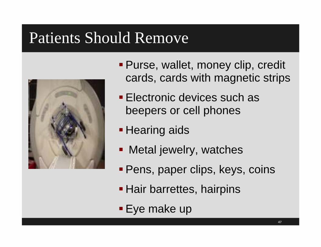
47
Patients Should RemovePurse, wallet, money clip, credit
cards, cards with magnetic strips
Electronic devices such as beepers or cell phones
Hearing aids
Metal jewelry, watches
Pens, paper clips, keys, coins
Hair barrettes, hairpins
Eye make up

Patients Should RemoveArticles of clothing that have a metal zipper, buttons, snaps, hooks, under wires, or metal threads
Shoes (especially ones with roller skates inside), belt buckles, safety pins
48

49
Projectile Objects The following have become projectiles in VA
facilities as listed in their HR Hazard Summary
Oxygen cylinder, IV pole, transport stretcher, traction weight, floor buffer, wheelchair, file cabinet, drill, and patient walker
Patient lifts, stethoscopes, infusion pumps, pulse oximeters, tools, laundry carts, scissors, pens, hair barrettes, and more
Hairpins and paper clips near a 1.5 Tesla MRI can reach speeds of 40 mph
Prevention of Missile Effect
50
www.mrisafety.com/safety_article.asp?subject=39
51
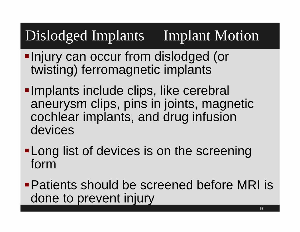
Dislodged Implants Implant MotionInjury can occur from dislodged (or twisting) ferromagnetic implantsImplants include clips, like cerebral aneurysm clips, pins in joints, magnetic cochlear implants, and drug infusion devicesLong list of devices is on the screening formPatients should be screened before MRI is done to prevent injury

Implant MotionEspecially a concern with implants that are strongly ferromagnetic Implant can move or dislodge
Determine if can wait several weeks for fibrous scarring to set in
Implants that are nonferrous in nature the risk is reduced to those resulting from Lenz’s forces alone Less important to have a waiting period
52

53
BurnsPatients have been burned from objects that can
heat up during the MR procedure Called RF heating–induces currents in electrically
conductive material
Burns from contact with conductive medical equipment cables (looped and unlooped EKG cables or pulse oximeter cable)
Other objects that can cause burns Surgical staples, the inside walls (the bore) of the MRI scanner
during the scan, site of pulse ox sensor that touches patient’s skin, Nitro patches which contain foil and other transdermal patches or those that deliver testosterone
ISMP and FDA Safety News reported

54
BurnsRare reports of burns At tattoo site (including eyeliner tattoo) since tattoos
contain iron oxide or other substances that can thermally and electrically conductive
At or near the site of implantable infusion pumps
At conductive looped formed with body, such as finger touched their thigh, patient’s arms were crossed, or thighs were touching
70 percent of the 389 adverse eventswere burns!

Guidelines to Prevent Burns with MRI
55
www.mrisafety.com/safety_article.asp?subject=17
56
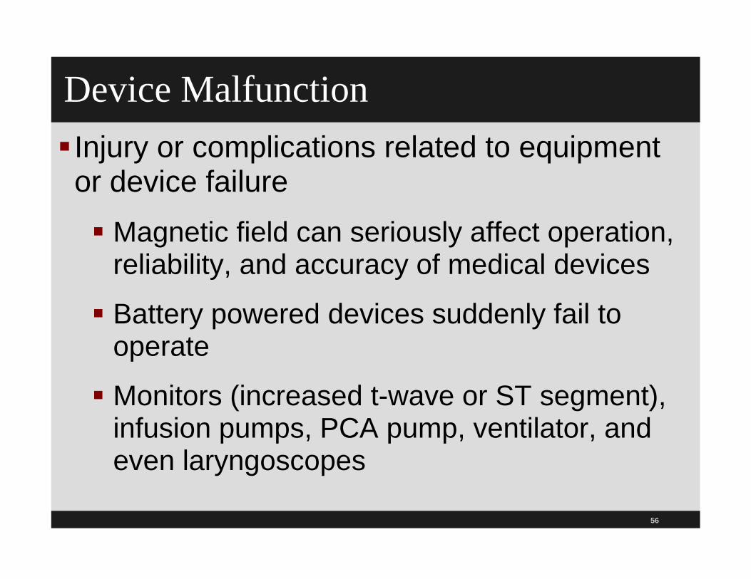
Device Malfunction Injury or complications related to equipment or device failure Magnetic field can seriously affect operation,
reliability, and accuracy of medical devices
Battery powered devices suddenly fail to operate
Monitors (increased t-wave or ST segment), infusion pumps, PCA pump, ventilator, and even laryngoscopes
57
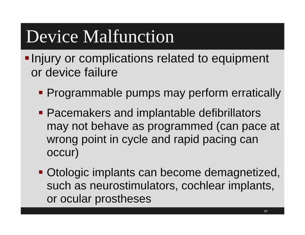
Device Malfunction Injury or complications related to equipment or device failure Programmable pumps may perform erratically
Pacemakers and implantable defibrillators may not behave as programmed (can pace at wrong point in cycle and rapid pacing can occur)
Otologic implants can become demagnetized, such as neurostimulators, cochlear implants, or ocular prostheses

58
Device Malfunction Recommendations
American College of Radiology (ACR) recommends that implanted cardiac pacemakers and implantable cardioverter/defibrillators should be considered a relative contraindication for MRI Later will cover website that lists each device and
its risks
Use ferromagnetic detectors that may help in screening patients for objects left on them Recent study shows they are about 99
percent effective

59
Acoustic InjuryCan be caused by the loud knocking of the MRI
scanner Patients should be given ear plugs or other hearing
protection
Sound can reach 130 decibels
Equivalent to jet engine take off
Four of 380 TJC adverse events were acoustic injuries FDA MR Guidance Document says manufactures must
state hearing protection is required for all patients

Acoustic Noise and MRI
60
www.mrisafety.com/safety_article.asp?subject=180

61
Image Artifacts Image artifacts can cause changes to MRI images due to: RF emission from equipment picked up by the MR RF
receiver
Examples include strips on the image or decrease in contrast
Presence of needles near imaging site (metal biopsy needles or mascara) can produce image artifacts and mask pathology
Unanticipated ferromagnetic implant or FB– Notify medical director, safety officer or physician in charge

62
Patient Support Systems Injury or complications can occur from failure to attend to patients during the procedure Especially patients who have had sedation or
anesthesia in the MRI arena
Complications include: Oxygen supply is depleted
IV solution on infusion pump is used up
Never run a code in the MRI suite

63
Adverse Events from MRI ContrastPatients with severe renal failure are at
risk for contrast-induced nephropathy
Can cause Nephrogenic Systemic Fibrosis (NSF) or Nephrogenic Fibrosing Dermopathy (NFD)
Important to know if patient has a history of renal or liver failure
Important to have kidney function test like patient in the ED has a BUN and creatinine before test done

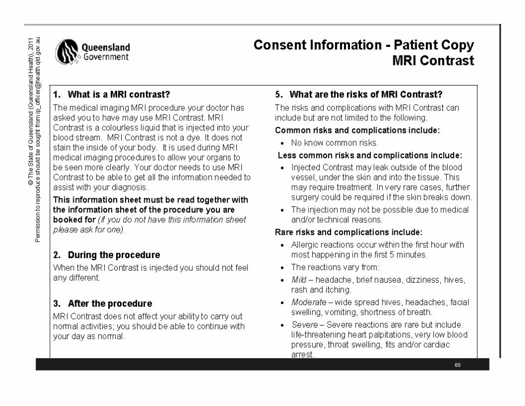
Consent Form Contrast with Renal Impairment
64
65

66
Cryogen HandlingAdverse events related to cryogen handling, storage, or inadvertent release The magnetic scanners are always left on
Turning them off or quenching is expensive and dangerous
Cryogenic gases (cooled liquid helium) can be released and are deadly– Can appear as white clouds or fog around the scanner
For superconducting systems, in event of system quench need to evacuate everyone out of the room (ACR 2013)

67
Parts of MRI and Principles of Operation Magnet creates the static magnetic field
Gradient coils are three sets of coils located inside the faceplate of the machine and allow spatial localization of the data obtained in the MRI process
Radio-frequency (RF) coils are used to transmit and receive RF radiation as part of the image acquisition. Coils are located under the thin plastic covering in the bore (inside walls) of the magnet.
Patient table and computer system and operator console
68
MR Field StrengthStrength of the static magnetic field for clinical MR
scanners is usually in the range of 0.0064 to 3.0 T
Measured from the center of the bore where the imaging occurs
Systems with field strength of 3.0 T and higher have been approved by the FDA and these are now more common (ACR 2013) Higher field strength increases the risk of injury from both
static and time varying magnetic field considerations
There are several magnet types such as permanent, resistive, superconductive, or hybrid

69
2013 ACR Guidance on Safe MR PracticesACR has 29 page document called ACR Guidance Document for Safe MR Practices: 2013 Free on their website
Published in the Journal of Magnetic Resonance Imaging 37:501-530 (2013)
Replaces 2002 , May 2004, and June 2007 edition
ACR has a website on MR safety and includes MRI safety website, safety screening form, and more at www.acr.org

ACR MR Safe Practices 2013
70
http://www.acr.org/Quality-Safety/Standards-Guidelines

ACR Practice Guidelines & Standards
71
ACR 2013 MR Guidance Includes information on the 5 G line
Need for P&P
MRI 4 zones
Training of those to enter MRI magnet room
Patient and staff assessments
What to do if a code occurs
Fight fighters, police, and security safety considerations
Device and object screening and more72

73
The 5 G Line (The Safe Line)The distance from the MR system at which the
static magnetic field is diminished sufficiently to pose no physical threat to the general public
Distance from MR imager where the static magnetic field has decreased to 5 gauss
FDA requires posting warning signs if magnetic field is more than 5 G
MRI room is shielded to protect MR system from equipment or devices that emit frequency similar to those emitted by protons in patient’s tissue

74
5 G Line Boundary

75
Zones in the MRI SuiteRestrict access to the MRI site by implementing four
zones Provide for progressive restriction in access to the MRI
scanner and discussed in ACR document
ACR has 29 page document called ACR Guidance Document for Safe MR Practices: 2013 Free on their website and in Journal of Magnetic Resonance
Imaging 37:501-530 (2013)
Replaces 2002 , May 2004, and June 2007 edition
ACR has a website on MR safety and includes MRI safety website, safety screening form, and more at www.acr.org
Discusses four zones and has MRI functional diagram

Four Zones
76

ACR MR Safe Practices 2013
77
www.acr.org/Quality-Safety/Standards-Guidelines
http://onlinelibrary.wiley.com/doi/10.1002/jmri.24011/pdf

78
Four ZonesZone I: General public
Zone II: Unscreened MRI patients
Zone III: Screened MRI patients and personnel
Zone IV: Screened MRI patients under constant direct supervision of trained MR personnel

79
Four ZonesZone II–Unscreened MRI patients–obtain information:
Patients are greeted
Patients are not free to move throughout zone II at will
Answers to MR screening questions
Patient histories and medical insurance questions

80
Four ZonesZone III–Screened MRI patients and personnel Access is strictly restricted since injury can occur from
ferromagnetic objects or equipment
Includes the control room
Physical restriction from general public entering
– Key locks, passkey locking systems or other reliable system to restrict access (not combination lock)
Prohibit physicians, non-MR personal access until trained
Area where strength exceeds 5 gauss should be clearly marked

81
Four Zones Zone IV–Screened MRI patients under constant
direct supervision of trained MR personnel
This is the MRI scanner magnet room
Clearly marked as being potentially hazardous due to the presence of very strong magnetic field
Clearly marked with red light and lighted sign that says “The Magnet is On.”
–On at all time with backup energy source in event of power loss
82
Four Zones Zone IV (continued)–MR scanner room emergency
In case of cardiac or respiratory arrest the certified MR personnel start basic life support or CPR while patient is moved to predeterminedsafe location
Do not recommend quenching the magnet (turning magnet off) since it takes more than a minute and this could be dangerous
Maintain restriction to Zones III and IV during resuscitation and other emergent situations

83
Types of PersonnelThere are three classifications and two levels of MR personnel
Non-MR personnel
MR personnel
–Level 1 MR personnel
–Level2 MR personnel

84
Non-MR PersonnelAny person (patient, visitor, staff, etc.) who has not
completed sufficient training to qualify as either level one or level two MR personnel
Includes any individual who has not had the designated training in MR safety issues in past 12 months
Must be under the immediate supervision and visual contact of a specifically identified level two MR staff member at all times when they are within MR zones III and IV

85
MR PersonnelLevel 1–Staff, including departmental office staff
and patient aides who have undergone minimal safety education efforts, sufficient to ensure their own safety as they work in Zone III areas. These staff may move freely throughout all MR zones.
These people cannot be responsible for non-MR personnel in Zone IV.
Level two–Staff, including MR technologists, radiologists, and radiology department nursing staff, who have undergone more extensive MR safety training. These staff members are also free to move throughout all
MR zones.

86
Level Two MR PersonnelMust supervise and be able to visualize or talk to
any non-MR person in zones III and IV
Should be trained and educated in broader safety programs
Include the potential for thermal loading or burns and direct neuromuscular excitation from rapidly changing gradients
Medical Director’s job is to make sure these people have education and experience to qualify as Level two MR personnel

Non-MR Personnel Nurse or PhysicianAny non-MR personnel, like a nurse or physician, who
wants to enter Zone III must first pass an MR safety screening process
Must be performed by the MR personnel before putting the non-MR person into Zone III Like a ticket to enter
Screening process and forms is identical for both patients and non-MR personnel Then physicians and nurses can enter the bore of the MR
imager during the MRI such as child who leans into the bore or anesthetist who leans into bore to bag patient if problem occurs
87

Monitoring Patients in the MRI Scanner Is sometimes necessary to monitor patients
Monitoring methods should be chosen carefully Concern about the risk of thermal injury associated with
the monitoring equipment
Sedated patients may not be about to tell you about any symptoms of injury Potential is greater for whole body scanners of 1 Tesla
and above
Use MR conditional EKG electrodes and don’t let leads touch patient during the scan Ice packs or cold compresses on electrically conductive material
88

89
http://www.imrser.org/PaperPDFRecord.asp?WebRecID=101&PgName=Guidelines&WebRecID=&sb_SummaryTitle=&

90
Screening of Non-emergent PatientsOutpatients or patients whose conditions are not life threatening are called non-emergent
Should be screened on site and by at least two separate individuals ( ACR 2013)
At least one of these people should be Level II MR personnel
At least one of these two screenings should be performed verbally or interactively

91
Conscious Non-emergent PatientsReview written MR safety screening questions prior
to their introduction to Zone III
Review questions orally with patient or family
If patient is non responsive discuss with family
They must provide yes or no to each question
Patient or family must sign these forms with no empty responses Note: Sample pre-MR forms are provided by ACR,
MRIsafety.com, www.ismrm.org, ECRI Institute, and several other sources listed in reference

92
Non-emergent Patients Must remove all metallic personal belongings including: Jewelry, cell phones, watches, contraceptive
diaphragms, body piercing (if removable), pagers, metallic drug delivery devices, hooks, zippers, metallic threads, metallic particles such as make up and eye makeup, contraceptive diaphragms, metallic drug patches, etc.
Recommend wearing hospital gown with no metal fasteners

93
Emergency PatientsEmergency department critically ill or ICU critical
patients If ED patient is unconscious and no one present to give
reliable information and test is urgent then should be physically examined by level 2 MR personnel
Looking for scars or deformities which might indicate an implant and can x-ray unless old films are available
Emergent patients and their accompanying non-MR personnel may be screened only once Level two MR personnel should provide
the screening
There should be no exceptions

94
Safe MR PracticesAny patient and non-MR personnel (nurses,
physicians) with a history of potential ferromagnetic foreign object penetration must undergo further investigation prior to being permitted entrance to Zone III Investigation includes: History, plain x-rays, prior CT or MR studies of
the questioned anatomical area, or Written documentation as to the type of implant
or foreign object that might be presentDetermine MR compatibility or MR safety of the
implant or foreign object

95
Safe MR Practices
All patients with history of orbital trauma by potential ferromagnetic FB must have their orbits cleared
This can be done by plain X-ray views of orbit films or by radiology review and assessment of contiguous cut prior CT or MR images obtained after trauma occurred
96
Metal Detectors Ferromagnetic DetectorsACR does not recommend metal detector use since
traditional metal detectors do not differentiate between ferrous and nonferrous magnetic materials They have varied sensitivity settings
Skills of operators can vary
Ferromagnetic detection systems (highly developed magnetometer type instruments) are available and simple to operate Capable of detecting very small ferromagnetic objects
external to the patient
Still need to conscientiously screen the patient

97
HIPAA Privacy If outpatient registered in radiology department,
make sure NPP is given to patient once after April 14, 2003 and again after September 23, 2013 Medical record information is called protected health
information and must be kept confidential In all four zones need to be in compliance with HIPAA
In Zone III, there should be a privacy barrier so unauthorized person cannot view control panels (ACR 2013)
If patient wants copy of MR report sent to someone outside, other than ordering physician, should sign HIPAA compliant authorization form

98
HIPAA Privacy If attending physician orders MRI, and
neurosurgeon comes into department to see film on outpatient, staff need to see documentation of physician-patient relationship
HIPAA security rules also apply, so keep back-up of MRIs and have adequate security
CMS Hospital CoPs on radiology and medical records apply to all MRIs
Patients can now sue for money damages if HIPAA violations, secretary of state can sue, HIPAA HITECH law in effect

MRI Safety
99
www.mrisafety.com/

Safety Information on MRI
100

MRI Safety Poster
101
http://www.imrser.org/
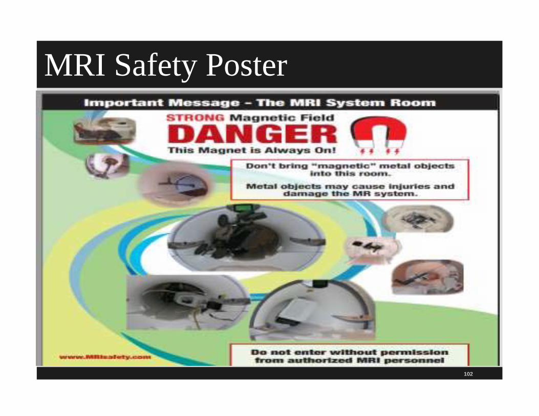
MRI Safety Poster
102

103
What Does MR Safe Mean?New classification system in 2006 was developed
by ASTM International and supported by FDA
Terminology from ASTM International
Old name was the American Society for Testing and Materials
Easy to remember because they are like colors of street light, green is go, red is stop
New MR safe and old MR safe terms have very different meanings

104
What Does MR Safe Mean? MR safe refers to an item that poses no known
hazards in all MRI environments
MR Safe means the device or implant is completely non-magnetic, non-conducting, and non-RF reactive, eliminating all primary potential threats during MRI procedure
Categories include MR safe, MR-Conditional, and MR-Unsafe This is the MR safe sign

105
MR ConditionalMR Conditional refers to a device or implant that
may contain magnetic, electrically conductive, or RF-reactive components that are safe for operations in proximity to the MRI
Provided that conditions for safe operation are defined and observed
Tested safe to 1.5 teslas, or safe in magnetic below 500 gauss in strength
RF is radio frequency–can heat the body
Yellow sign in the MR conditional sign
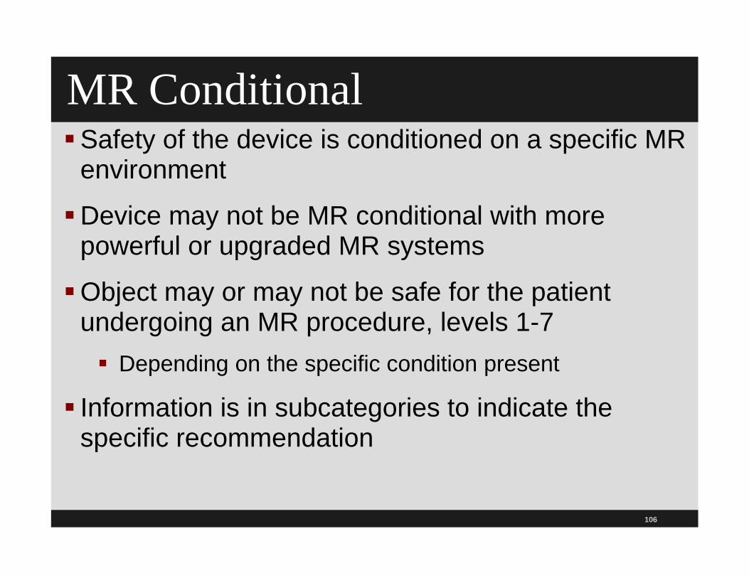
106
MR Conditional Safety of the device is conditioned on a specific MR
environment
Device may not be MR conditional with more powerful or upgraded MR systems
Object may or may not be safe for the patient undergoing an MR procedure, levels 1-7 Depending on the specific condition present
Information is in subcategories to indicate the specific recommendation
107
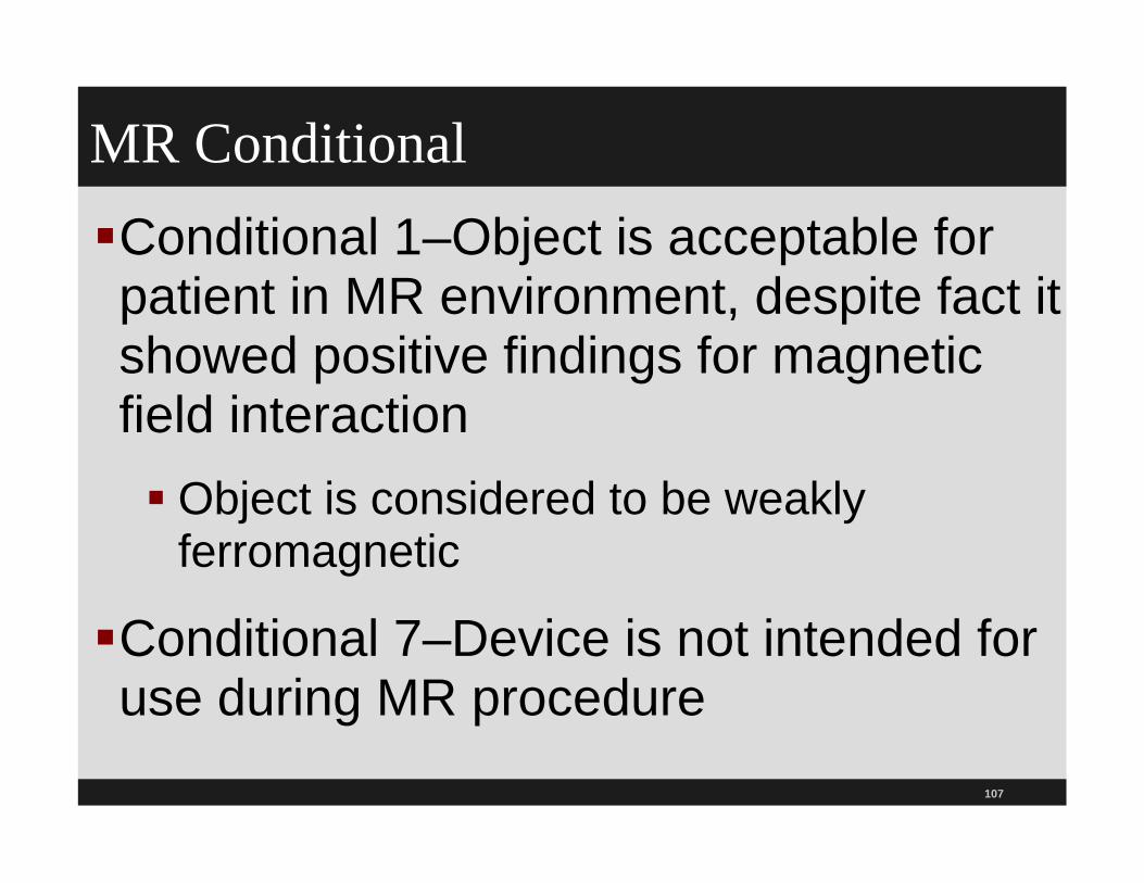
MR ConditionalConditional 1–Object is acceptable for patient in MR environment, despite fact it showed positive findings for magnetic field interaction Object is considered to be weakly
ferromagnetic
Conditional 7–Device is not intended for use during MR procedure

108
Unsafe Unsafe–Reserved for objects that are significantly
ferromagnetic and pose threat to person and equipment in room Unsafe in any MR environment
Unsafe 1–The object is considered to pose a potential or realistic risk or hazard to a patient or individual in the MR environment, primarily as the result of movement or dislodgement of the object Contraindicated for MRI Note that the “default” static magnetic field
strength for an unsafe implant or device is 1.5-Tesla

109
Unsafe 2Object displays only minor magnetic field
interactions which, in consideration of the in vivo application of this object, is unlikely to pose a hazard or risk in association with movement or dislodgment Presence of this object is considered to be a
contraindication for an MR procedure
Represents potential risks such as excessive heating or other potentially hazardous conditions
– Example: Swan-Ganz catheter melted in patientduring MRI

110
Searching the ListStatus of objects is listed as: MR-safe, Unsafe 1 and 2, and Conditional 1 through 8
List includes manufacturer name andobject category
Examples of objects on the list include: Aneurysm clips, AccuRx implantable flow pump, cochlear
implants, stents, carotid artery vascular clamps, insulin pumps, IUDs, etc.
If you click on insulin pumps it will show you it is in category of unsafe 1 status
– Provides safety information and instructions on what to do

Search the List for MRI Safety
111
www.mrisafety.com/list_search.asp

112
Follow ACR Guidelines and Standards 2013Guidelines are an educational tool designed to assist
practitioners in providing appropriate radiologic care for patients
Guidelines are not inflexible rules or requirements of practice and are not intended, nor should they be used, to establish a legal standard of care
ACR cautions against the use of these guidelines in litigation in which the clinical decisions of a practitioner are called into question
–
Source: http://www.acr.org/, see Guidelines and Standards

113
www.acr.org/Quality-Safety/Standards-Guidelines
www.acr.org/Quality-Safety/Standards-Guidelines
www.acr.org/Quality-Safety/Standards-Guidelines

114
ACR Guidelines and Standards SectionsContinuing medical education
General diagnostic radiology including:
Expert witness in radiology
Communicating findings
Use of intravascular contrast media
MRI

115
ACR SafetyACR has position statement on quality control and
improvement, safety, infection control, and patient education concernsP&P to provide for the safety of patients and staffAttention to the physical environment Proper use, storage, and disposal of
medications and hazardous equipmentMethods for responding to medical and other
emergencies1
1ACR Guidance Document for Safe MR Practice 2007, 27 pages

116
ACR Safe MR Practice 2013 Important document to review and every hospital should have a copy of this
All facilities should have MR policies Including clinical and research sites no matter the magnet
format or field strength
Review policy when any changes such as adding faster or stronger MRI machine
Consider national and international standards and recommendations when drafting and updating P&P
117
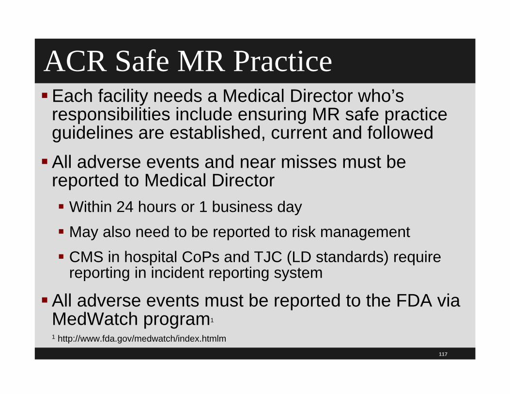
ACR Safe MR PracticeEach facility needs a Medical Director who’s
responsibilities include ensuring MR safe practice guidelines are established, current and followedAll adverse events and near misses must be
reported to Medical Director Within 24 hours or 1 business day May also need to be reported to risk management CMS in hospital CoPs and TJC (LD standards) require
reporting in incident reporting system
All adverse events must be reported to the FDA via MedWatch program1
1 http://www.fda.gov/medwatch/index.htmlm

FDA MedWatch Program
118
www.fda.gov/Safety/MedWatch/default.htm

119
ACR Safe MR Practice ACR supports this requirement
If implant is strongly ferromagnetic, concern is that of magnetic translational and rotational forces upon the implant which might move or dislodge the device from its implanted position
If implant demonstrated weak ferromagnetic forces on formal testing, may be prudent to wait several weeks for fibrous scarring to set in as this may anchor the implant in position

120
ACR Safe MR Practice It is possible to find unanticipated implant or FB during exam
May be detected by sizable field distorting artifact seen on spin-echo imaging techniques that grows more obvious on longer TE studies and expands markedly on typical moderate or long TE gradient-echo imaging sequences
Notify Medical Director, safety officer, or physician in charge of suspected findings

121
ACR Safe MR Practice
Review information and decide what course of action should be taken
If need to remove patient go slowly instraight line
Avoid temptation to have patient sit up as soon as out of bore
Wait until as far as physically possible from MRI imager

122
ACR Safe MR PracticePatients, volunteers, staff, or anyone else with implanted cardiac pacemaker, implantable cardiac defibrillator (ICD), diaphragmatic pacemaker or other electromechanically activated devices should never enter Zone IV
Should not go past 5 gauss line unless cleared in writing by Level 2 MR personnel, designated radiologist, or Medical Director of the MR site

123
PrisonersPrisoners or parolees with metallic devices such as handcuffs and shackles or RF tracking bracelets
RF ID or tracking bracelets interfere with MRI study and secondary image artifact
Bracelet can also heat up and burnthe patient
These need to be removed before doing the test

124
Firefighters, Police, Security
Persons who respond to an emergent call at the MR site
Specially designated MR personnel
Need to be on the site prior to the arrival of the firefighters or emergency response team
Firefighters cannot have free access to Zones III and IV, so educate fire marshals and others in advance

125
Firefighters, Police, Security May train security staff to be designated as MR
personnel Designated person needs to be there before others show up
In true fire, taking air tanks, crow bars, guns, and other firefighting equipment could be catastrophic
Need clearly marked, readily accessible MR-conditional or MR-safe fire extinguishing equipment physically stored in Zone III or IV
All conventional fire extinguishers and other firefighting equipment not tested and verified safe in the MR environment should be restricted from Zone III Use this section to draft your P&P

126
MR Personnel Screening and HR
All MR personnel must undergo an MR screening process as part of their employment interview
MR personnel must report to the MR Medical Director any trauma, procedure, or surgery in which ferromagnetic object or device may have been introduced
This is done to make sure it is safe for the employee to enter Zone III

127
Device and Object ScreeningDon’t let anyone bring in ferrous objects
Should have access to a strong handheld magnet (over 1000 gauss) or handheld ferromagnetic detection device
Magnet enables external and even some superficial internal testing of devices or implants Presence of grossly detectable ferromagnetic attractive forces
Document testing and include date, time, and name of tester
Oxygen cylinders must be positively identified in writing as MRI safe (non-ferromagnetic and safe or conditionally safe in the MR environment) or Unsafe

128
Device and Object Screening
Same testing for other objects such as MRI safe fire extinguishers and aneurysm clips
All portable metallic or partially metallic objects that are to brought into Zone IV must be properly identified under current FDA labeling criteria developed by ASTM
Remember the green safe sign on the label
Treat a product marked as MR safe but with metal construction as suspicious

129
Device and Object Screening Be careful about old labeling of
products with ill defined terminology
For example, “non-magnetic,” or outdated classifications such as “MR-compatible,” should not be presumed to conform to a particular current ASTM classification
If in doubt test it with handheld ferromagnetic detection device

ConsentPatient with pacemaker or ICD that is not labeled as
MR Conditional should be informed of the risk and provided informed consent
If MRI is done on patients with these devices ACR recommends a fully stocked crash cart
See additional detailed recommendations on page 517
ACR also has detailed section on patients who may have an intracranial aneurysm starting on page 515
130

131
ScreeningNeed effective screening procedure for patients
before they have an MRI
ACR has screening tool
Should be conducted by health care worker who has been specially trained in MR safety To determine if patient has an implant that may be
contraindicated for the MR procedure (e.g., a ferromagnetic aneurysm clip, pacemaker, etc.)
To determine if there is any condition that needs careful consideration (e.g., the patient is pregnant, has a disability, etc.)

132
Screening Have P&P on screening
Use a screening tool
After the preliminary screening, then the patient goes through comprehensive screening
Comprehensive patient screening uses a printed form to document this procedure
Form includes a statement that indicates hearing protection is “advised” or “required” to prevent possible problems or hazards related to acoustic noise
MR safety trained person reviews the form’s contents. If patient is unable to answer the questions, then discuss with closest family members.

133
Screening
If no family members, then with person who is most likely to know the information
Technician can also look for scars or deformities
Can use ferromagnetic detectors
Can also use plain film radiography to assist in the screening process

134
Screening Things that Create a HazardPacemaker (new pacemaker safe one)
Implantable cardioverter defibrillator (ICD)
Neurostimulators, tissue expanders, hearing aid
Aneurysm clip, surgical clips, staples
Metal implant, artificial limbs, shunts
Implanted drug infusion device, penile implant
Foreign metal objects, especially if in or near the eye, artificial heart valve, coils and stents
Radiation seeds, IUD, pessary, eyelid spring

135
Screening Things that Create a Hazard Shrapnel or bullet wounds, internal electrodes
Permanent cosmetics or tattoos, tattooed eyeliner
Dentures/teeth with magnetic keepers
Other implants that involve magnets
Medication patch (i.e., transdermal patch) that contains metal foil, cochlear implant, halo vest
Pillows may contain metal springs
Sandbags may contain iron pellets
Wigs, hair implants, body piercing, surgical mesh

ACR Safety Screening Form 2013 Page 519
136
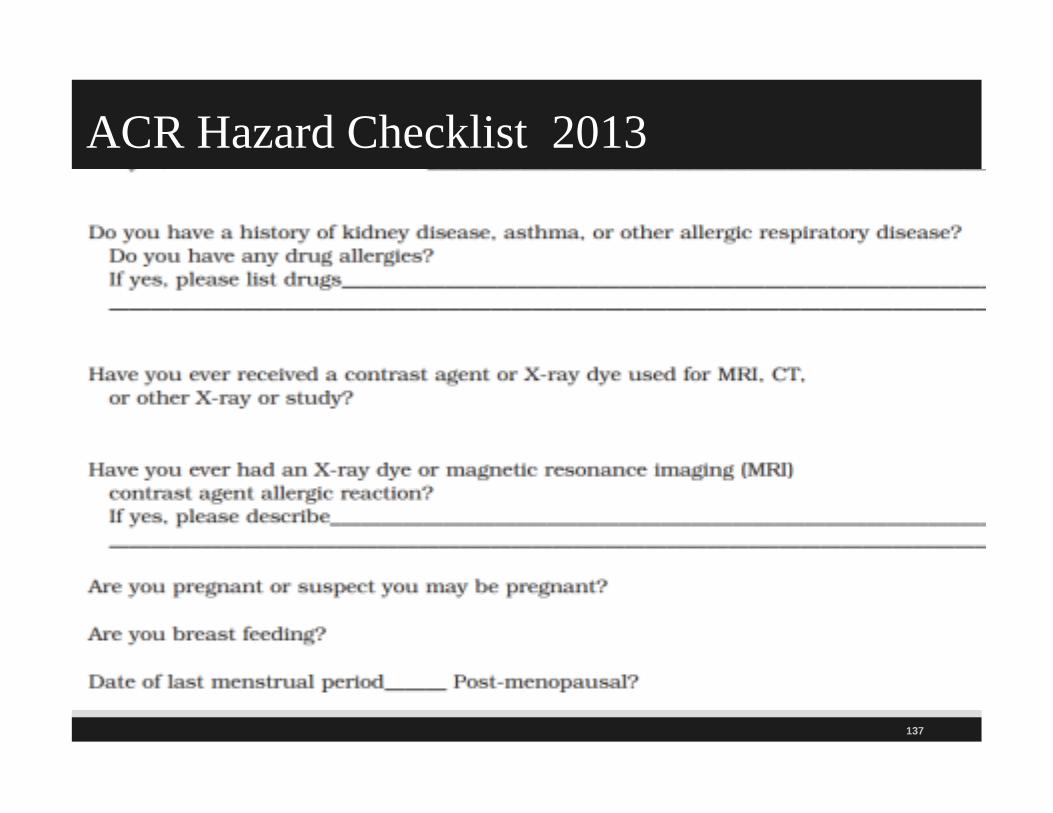
ACR Hazard Checklist 2013
137

ACR 2013 MR Hazard Checklist
138

ACR Hazard Checklist 2013
139

ACR Hazard Checklist 2013
140

ACR Hazard Checklist 2013
141

ACR Safety Screening Form 2013
142

MRI Staff Verification ACR 2013
143

144
Ferromagnetic Objects It is important to be aware of common ferromagnetic objects
Buffing machines, janitor buckets, chest tube stands, and clipboards (patient charts), chairs, canes
Gurneys, hairpins, hearing aids, identification badges, walkers
Insulin pumps, keys, and medical gas cylinders, mops, IV poles

145
Ferromagnetic ObjectsNail clippers and nail files, oxygen cylinders, pulse oximeter, pacemakers, and pagers
Paper clips, jewelry, pens, and pencils
Prosthetic limbs, shrapnel, sandbags (with metal filings)
Steel shoes, stethoscopes, scissors, staples, and tools
Vacuum cleaners, watches, and wheelchairs

146
PregnancyNo harmful effects on the fetus have been
demonstrated–does not use ionizing radiation As precaution, pregnant women should only have MRI
when essential
If they can wait until the end of pregnancy to have test, that is recommended
Gadolinium is known to cross the placenta and enter fetal bloodstream, so contrast is not routinely provided if patient is pregnant
Pregnant staff can work in and around MRI suite but asked not to remain in MRI scanner bore or Zone IV during actual scanning
147
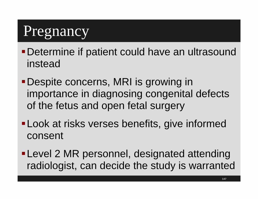
Pregnancy Determine if patient could have an ultrasound instead
Despite concerns, MRI is growing in importance in diagnosing congenital defects of the fetus and open fetal surgery
Look at risks verses benefits, give informed consent
Level 2 MR personnel, designated attending radiologist, can decide the study is warranted
148
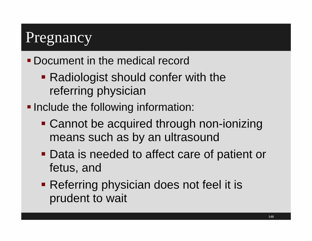
PregnancyDocument in the medical record Radiologist should confer with the
referring physician Include the following information: Cannot be acquired through non-ionizing
means such as by an ultrasound Data is needed to affect care of patient or
fetus, and Referring physician does not feel it is
prudent to wait

149
Sedation and Monitoring IssuesChildren form the largest group requiring sedation
for MRI Many are unable to stay still
Sedation protocols vary among facilities
CMS has changes to the anesthesia standards which discuss moderate sedation and deep sedation VA has moderate sedation toolkit at www.patientsafety.gov
Parent to accompany child must be screened Use hearing protection

Pediatric MR Safety Sedation & MonitoringThere is a section on pediatric safety that any
hospital that does MRIs on pediatric patients should read
Should be incorporated into P&P
Need to follow standards from the American Academy of Pediatrics, American Society of Anesthesiologist and TJC
fasting requirements, H&P, training and credentialing for staff, monitoring during and after procedure, observe child, charting, protocol for recovery and discharge, etc.
150

151
ClaustrophobiaBeing in center of long narrow tube can be
unpleasant and some patientsare claustrophobic Open MRIs and upright MRIs are an option
New scan rooms being developed with lighting, sounds and images on wall or ceiling
Sedation or general anesthesia can be used
Visualization or imagery techniques may help Holding panic button and listening to music on headphones,
or watching a movie with head mounted displays while in MRI machine may ease stress

152
Disadvantages If sedation used follow TJC, ASA, and ACR
recommendation
Discuss issue of claustrophobia and hazards Normally 20-30 minutes long but can take 60 minutes
Required to stay still and in an enclosed space can be hard for pediatric patients
Movement can create motion artifact Includes patients with tremors like Parkinson's disease
or low back pain
Patients with pain can be instructed to take pain medication prior to the procedure

153
Thermal IssuesRemove any unnecessary equipment
Unplugging unnecessary equipment is not enough
Electric current or voltage can be induced in electrically conducting materials
Can create heat which can result in a burn
MR tech must check all equipment first

154
Thermal Issues Burns If wires or leads have to remain on patient, take care that there are no large caliber conducting loops formed in the scanner
Several cases reported of coma and permanent impairment in patients with neurologic stimulators
Make sure there is insulation between the patient and the electrically conductive material (pads or air)
155
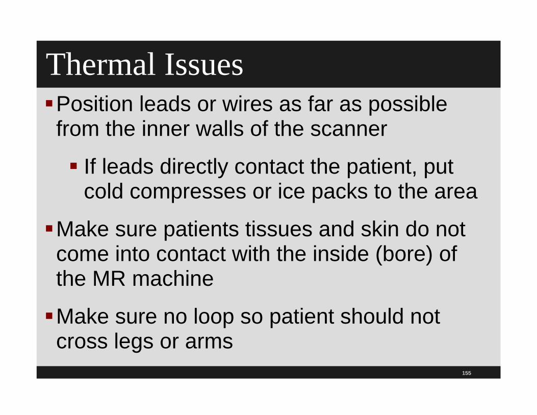
Thermal Issues Position leads or wires as far as possible from the inner walls of the scanner
If leads directly contact the patient, put cold compresses or ice packs to the area
Make sure patients tissues and skin do not come into contact with the inside (bore) of the MR machine
Make sure no loop so patient should not cross legs or arms

156
Thermal Issues Skin staples and superficial metallic sutures are okay if not ferromagnetic and not in anatomical volume of RF power deposition
Take several precautions if skin staples are ferromagnetic
Warn patient about warmth and may experience some burning along staples Can place cold compresses or ice pack along
skin staples or superficial metallic sutures

157
Thermal IssuesNotify tech immediately of heat Do not wait until end of test
Use cold compress or ice bags at site
Use ice packs for dark tattoos, including permanent eyeliner
Procedure can smear or smudge edges if new tattoo
Drug delivery patches and pads with metallic foil can result in a burn Ice bag put on patch can affect delivery of medication

158
Education of Staff
Provide education during orientation for all staff who will be involved with MRIs
Consider annual training during skills lab
Include environmental services (housekeeping) personnel, maintenance, transport, surgical, and emergency response teams for RRT and codes
159
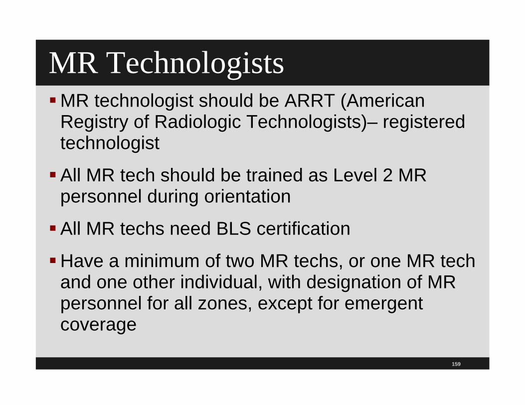
MR TechnologistsMR technologist should be ARRT (American
Registry of Radiologic Technologists)– registered technologist
All MR tech should be trained as Level 2 MR personnel during orientation
All MR techs need BLS certification
Have a minimum of two MR techs, or one MR tech and one other individual, with designation of MR personnel for all zones, except for emergent coverage

160

161
MRI Safety Policy

162
NQF 34 Safe PracticesUpdated list in 2010 and 2011 on 34
Safe Practices for Better Healthcare
Should be followed in all health care facilities to reduce risk of harm to patients
Organized into seven sections
Includes list of 29 never events that many states require tobe reported (2011 NQF changed and updated the list)

163
Resources on 34 Safe PracticesNQF has an electronic copy of the book that can
be purchased for $29.991
NQF, publication unit, 601 Thirteenth Street, NW, Suite 500 North, Washington, DC, 20052
TMIT has a website and you can listen to past presentations3
1 http://www.nqfstore.org/store/2 www.qualityforum.org3 http://www.safetyleaders.org or http://www.tmit1.org/pages/workshopsWebinars.jsp
164
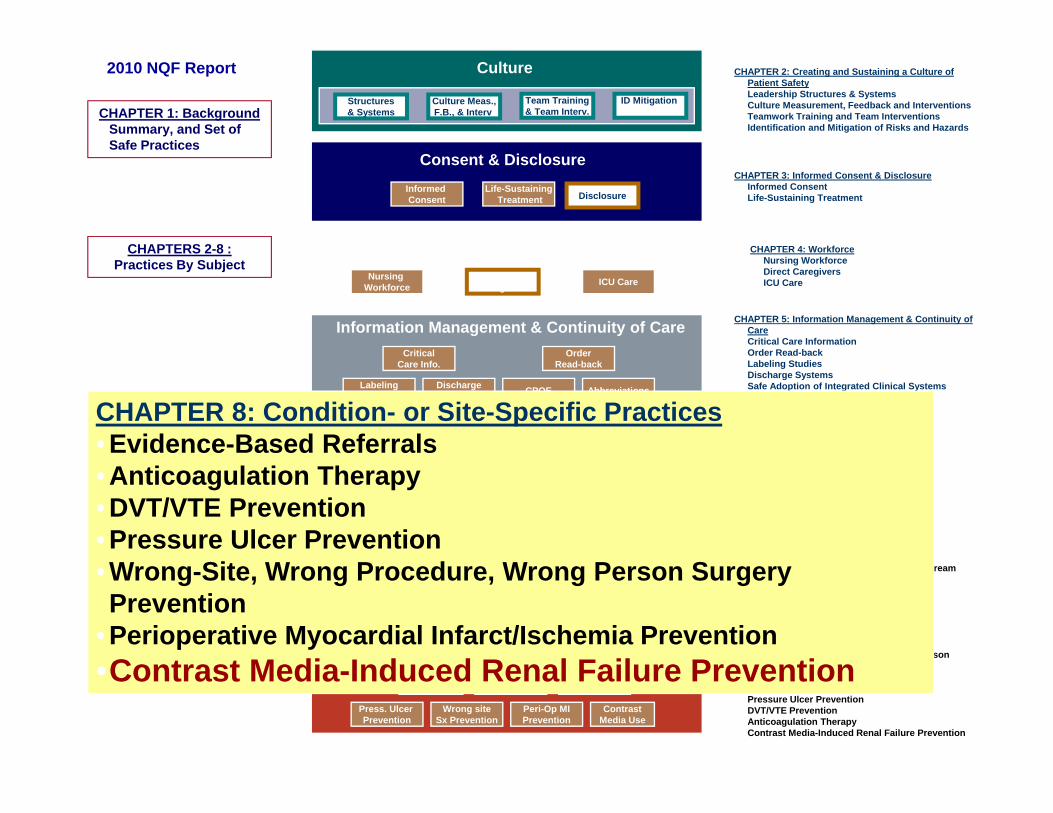
Culture SP 1
Information Management & Continuity of Care
Medication Management
Hospital Acquired Infections
Condition & Site Specific Practices
Consent & Disclosure
Wrong siteSx Prevention
Peri-Op MIPrevention
Press. Ulcer Prevention
DVT/VTE Prevention
Anticoag Therapy
Asp +VAP Prevention
Central V. CathBSI Prevention
Sx Site Inf.Prevention
Contrast Media Use
Hand Hygiene InfluenzaPrevention
PharmacistCentral Role
Med Recon.
Std. Med Labeling & Pkg
High AlertMeds
Unit DoseMedications
EvidenceBased Ref.
Culture
CPOE
OrderRead-back
AbbreviationsDischarge System
CriticalCare Info.
LabelingStudies
Culture Meas.,F.B., & Interv.
Structures& Systems
ID MitigationRisk & Hazards
Team Training& Team Interv.CHAPTER 1: Background
Summary, and Set of Safe Practices
CHAPTERS 2-8 : Practices By Subject
Nursing Workforce ICU CareDirect
Caregivers
Workforce CHAPTER 4: Workforce• Nursing Workforce• Direct Caregivers• ICU Care
CHAPTER 2: Creating and Sustaining a Culture of Patient Safety
• Leadership Structures & Systems• Culture Measurement, Feedback and Interventions• Teamwork Training and Team Interventions• Identification and Mitigation of Risks and Hazards
CHAPTER 5: Information Management & Continuity of Care
• Critical Care Information• Order Read-back• Labeling Studies• Discharge Systems• Safe Adoption of Integrated Clinical Systems
including CPOE• Abbreviations
CHAPTER 6: Medication Management• Medication Reconciliation• Pharmacist Role• Standardized Medication Labeling & Packaging• High-Alert Medications• Unit-Dose Medications
CHAPTER 7: Hospital-Acquired Infections• Prevention of Aspiration and Ventilator-
Associated Pneumonia • Central Venous Catheter-Related Blood Stream
Infection Prevention • Surgical Site Infection Prevention• Hand Hygiene• Influenza Prevention
CHAPTER 8:• Evidence-Based Referrals• Wrong-Site, Wrong Procedure, Wrong Person
Surgery Prevention • Perioperative Myocardial Infarct/Ischemia
Prevention• Pressure Ulcer Prevention• DVT/VTE Prevention • Anticoagulation Therapy• Contrast Media-Induced Renal Failure Prevention
Informed Consent
Life-SustainingTreatment Disclosure
CHAPTER 3: Informed Consent & Disclosure• Informed Consent• Life-Sustaining Treatment• Disclosure
Consent & Disclosure
CHAPTER 8: Condition- or Site-Specific Practices• Evidence-Based Referrals• Anticoagulation Therapy• DVT/VTE Prevention• Pressure Ulcer Prevention• Wrong-Site, Wrong Procedure, Wrong Person Surgery Prevention
• Perioperative Myocardial Infarct/Ischemia Prevention•Contrast Media-Induced Renal Failure Prevention
2010 NQF Report

TJC Standards EC.02.04.01: The hospital manages medical
equipment risks. EP 3 The hospital identifies the activities, in writing, for
maintaining, inspecting, and testing for all medical equipment on the inventory
Hospitals may use different strategies for different items as appropriate. For example, strategies such as predictive maintenance, reliability-centered maintenance, interval-based inspections, corrective maintenance, or metered maintenance may be selected to ensure reliable performance.
165

TJCEC.02.04.01 EP5 Hospital must monitor and report
any incidents in which medical requirement is suspected or contributed to death or serious injury or illness
Recent issue of radiation overdose and concern about increased cancer risks
See TJC SEA 47 on radiation risk of diagnostic imaging
EC.02.04.03 EP 14 Qualified staff inspect, test, and calibrate nuclear medicine equipment annually and document (DS)
166

Contrast No patient should be given contrast without a physician
order (ACR 2013) ACR MR Guidelines refer to ACR Contrast Manual-the
ACR Committee on Drugs and Contrast Material
IV injection qualified MR technologists may start a peripheral IV if had the training and are competent Make sure that the state scope of practice is consistent
IV qualified MR technologists (certified and or licensed) or radiologic nurse may administer gadolinium based contrast in peripheral IV or as bolus Must have and follow the P&P and should be consistent with
the ACR policy167

ACR Contrast Manual
168
www.acr.org/Quality-Safety/Resources/Contrast-Manual

169
Contrast Media-Induced Renal FailureUtilize validated protocols to evaluate patients who
are at risk for contrast media-induced renal failure
Use a clinically appropriate method for reducing risk of renal injury based on the patient's kidney function evaluation
Angiography, IVP, and CT scans that use contrast material containing iodine Can have allergic reaction or kidney damage
Be careful in patients with renal impairment
Do RCA on all cases of contrast media induced renal failure

170
Contrast Media-Induced Renal Failure Recommendations to prevent contrast media-
induced renal failure
Make sure patient is adequately hydrated
Use low osmolar contrast in patients withrenal failure
Check serum creatinine level prior to scheduling contrast studies
See ACR manual on Contrast Media for the use of intravascular contrast media

171
ACR Guidelines on Contrast Media
www.acr.org/SecondaryMainMenuCategories/quality_safety/RadSafety.aspx

172
Contrast Media-Induced Renal Failure Recommendations to prevent contrast media-induced renal failure (continued)
Need P&P on prevention of contrast media induced nephropathy
Document contrast media-induced renal failure assessment regarding its prevention
Double check order and make sure most current creatinine level is used

173
IV Contrast on Diabetic PatientsHave a process for diabetics on Metformin with
abnormal renal function or comorbidity
Do you hold the medication temporarily if intravascular iodinated contrast is used in category II patients?
Do you order a serum creatinine two days after the CT in category III patients?
Do you then notify the attending office to let the patient know to restart their medication?
ACR also has Manual on Contrast Media

ACR Manual 3 Categories Patients Metformin
174

ACR Manual on Contrast Media
175

176

177
Contrast Induced Nephropathy CINKidney failure can occur from iodine dye used for
x-rays (70 reports)
Hospitals should amend informed consent to include this information
10-12 percent of all renal failure cases from CIN
Most common in patients with known history of renal failure or impairment
Consider doing a FMEA Toolkit available1
1http://www.patientsafetyauthority.org/EducationalTools/PatientSafetyTools/cin/Pages/home.aspx

Contrast Induced Nephropathy Toolkit
178
http://patientsafetyauthority.org/EducationalTools/PatientSafetyTools/cin
/Pages/home.aspx

179
ToolkitToolkit includes the following:
A copy of the advisory
Brief informational video on CIN
Stand alone algorithm to identify patients at risk for CIN
Poster
Reference tables for calculating estimated glomerular filtration rate

180
Toolkit Discuss with patients the increased risk with: Nephrotoxic drugs such as chemo Certain antibiotics NSAID Acyclovir Immunosuppressants ACE inhibitors Lasix Lithium Oral phosphate bowel cleansing products

181
Gadolinium Based ContrastGadolinium is a clear, non-radioactive liquid,
approved by the FDA as an injectible contrast agent used during MRI Provides better contrast between healthy and unhealthy
tissue
Can cause nephrogenic systemic fibrosis
Screen all patients for renal dysfunction
Gadolinium is a clear, non-radioactive liquid, approved by the FDA as an injectible contrast agent used during MRI
182
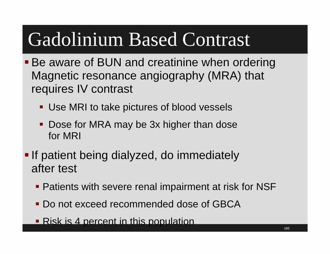
Gadolinium Based ContrastBe aware of BUN and creatinine when ordering
Magnetic resonance angiography (MRA) that requires IV contrast Use MRI to take pictures of blood vessels
Dose for MRA may be 3x higher than dosefor MRI
If patient being dialyzed, do immediatelyafter test Patients with severe renal impairment at risk for NSF
Do not exceed recommended dose of GBCA
Risk is 4 percent in this population
183
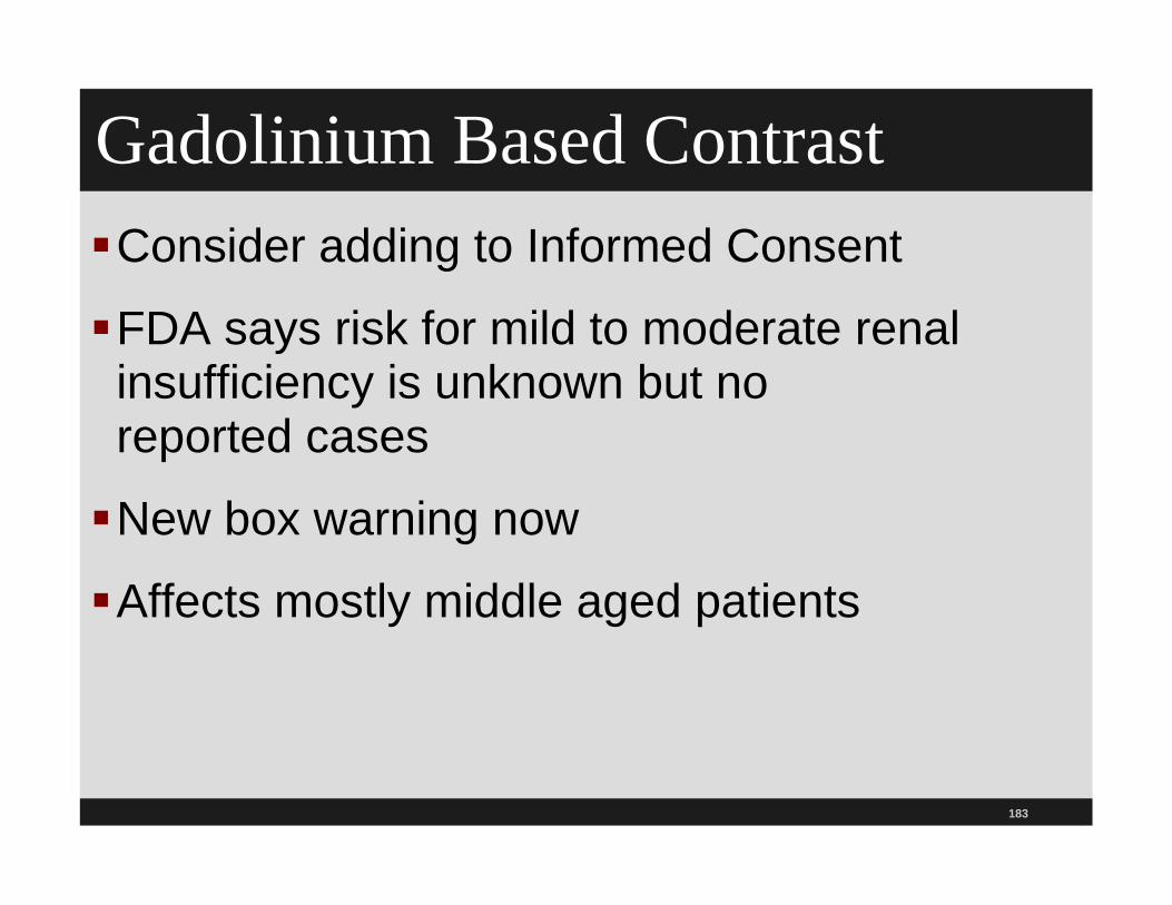
Gadolinium Based ContrastConsider adding to Informed Consent
FDA says risk for mild to moderate renal insufficiency is unknown but noreported cases
New box warning now
Affects mostly middle aged patients

FDA Gadolinium Bases Contrast
184
www.fda.gov/Drugs/DrugSafety/PostmarketDrugSafetyInformationforPatientsandProviders/ucm1428
82.htm

185
Gadolinium Based Contrast NSF (nephrogenic systemic fibrosis) is a debilitating
and sometimes fatal disease affecting skin, muscles, and internal organs
Diagnosis is confirmed by skin biopsy (thickened collagen bundles with surrounding clefts, mucin deposition, and proliferation of fibroblasts and elastic fibers)
Linked to patients with moderate or end-stage kidney disease
Note picture characterized by thickening, indurations, and hardening of the skin and distinct nodules can also be seen

186
Gadolinium Based Contrast
Symptoms can include hardening of skin, discoloration, burning, itching, joint pain and stiffness, hip pain, scarring of body organs, muscle weakness, difficult to bend joints,and death Usually develops two to four weeks, but can develop two
to three months after MRI
No known cure
Multiple sites for law firms advertising that patients may be entitled to compensation and offering free case review
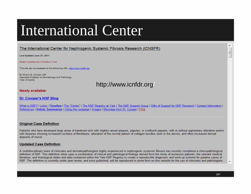
International Center
187
http://www.icnfdr.org/

188
SummaryMake sure you have a copy of the ACR MR Guidance
Document 2013
Make sure your P&P is consistent with this document
Keep door to Magnet room closed and limit and monitor access to MRI suite
Test all items for ferromagnetic properties before taking them into the MRI room
Label ferrous items that remain in the hospital so everyone knows they cannot be taken into the room
Such as sandbags

189
SummaryDo pocket check before entering, especially for
scissors, hemostats, and pens
Need to review policy and procedure annually
Consider education on MRI safety in orientation and during annual skills lab
Provide formal training for all who enter, including nursing, transport, security, environmental services, maintenance, etc.
Any nurses or physicians or other staff who enter MRI must be screened

190
Summary Routinely access compliance with these policies and procedures, especially housekeeping and maintenance and security
Have special MRI safe equipment for use in MR room such as IV pole, oxygen canister, fire extinguishers, monitors, wheelchair, etc. and have them marked for use in MRI room
Provide all patients with hearing protection

191
Don’t make assumptions about equipment such as sand bags and pillows being safe Check them out
Report all incidents to the FDAMedWatch program
Never code a patient in the MRI suite
If you buy a new MRI or upgrade the system make sure the label of “MR Conditional”still applies
Summary

192
SummaryAlways assume MR system’s static magnetic
field is on Identify 4 zones in the MRI suite and surrounding
floors Include adjacent floors where magnetic field
exceeds 5 gaussConsider doing a FMEA or RCA if
event occursCheck sedated patients periodically for heating at
sensor site

193
SummaryDon’t allow equipment and other devices past the 5 G lines unless tested by the device manufacturesMake sure these devices are labeled “MR safe” (see book at end and website with list of more than objects and implants tested at 3-Tesla or higher)1
Audit compliance with policy
1 http://www.mrisafety.com/list.asp

194
SummaryProvide patient information booklet
on MRI1
Explain it is vital to remove all metallic objects in advance of the MRI exam
Explain that scanner can dislodge clip from blood vessel, cause heart pacemaker to malfunction, or damage their external hearing aid
1 http://www.mrisafety.com/safety_article.asp?subject=170

195
SummaryUse a MRI screening form to ask about things that might cause a health risk or interfere with imaging
Refer MRI screening form on an annual basis for anything that needs to be added or amended
Remove patches that contain metal before the MRI and if unsure remove patch

196
SummaryMake sure MRI procedure is pre-certified if patient has insurance
Document information in medical record
Remember to use an ABN if no medical necessity
Check with physician for acceptable ICD code or necessity first
Patients should wear hospital gown without metallic snaps

197
Summary
Have piped medical gases in MR room
Will help prevent oxygen tanks from beingbrought in
Don’t loop cables or allow cables to cross one another and use MR compatible cables when conducting an MRI
Don’t let patients touch the wall of the magnetic bore

198
Summary
Assign a MR safety officer
Review the P&P yearly
Ensure staff are trained
Empower MR technicians to have control over access to MR environment
Screen all personnel coming intoMR environment
199
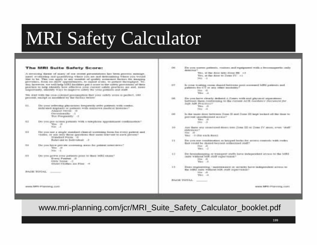
MRI Safety Calculator
www.mri-planning.com/jcr/MRI_Suite_Safety_Calculator_booklet.pdf
200
The End! Questions??Sue Dill Calloway RN, Esq.CPHRM, CCMSCPAD, BA, BSN, MSN, JDPresident Patient Safety andHealthcare Education
Board MemberEmergency Medicine PatientSafety Officer www.empsf.org

201
Resources
MRI safety website at www.mrisafety.com
Cost of MRI accidents at http://www.mri-planning.com/accidents.html
Reference Manual for Magnetic Resonance Safety, Implants and Devices, 2008 edition at above website
The Joint Commission Sentinel Event Alert 38, Issued February 14, 2008, Preventing Accidents and injuries in the MRI Suite, at www.jointcommission.org

202
Resources
Emanuel Kanal MD, Magnetic Resonance Safe Practice Guidelines of the University of Pittsburgh Medical Center, 2001, at http://www.jointcommission.org/NR/rdonlyres/8EFBFDA8-81A4-4F8C-9A14-07D1CBD55674/0/UPMC_Guidelines.pdfSafety Concerns in the MR Environment,
Healthcare Risk Control Analysis, March 2006, ECRI Institute, Vol. 4, Radiology 5,20 pagesWikipedia, MRI at http://en.wikipedia.org/wiki/MRI

203
Resources (continued)
“Fatal MRI Accident is First of Its Kind,” www.webmd.com/content/Article/34/1728_85340.htmACR Guidance Document for Safe MR
Practices: 2007, AJR:188, June 2007, http://www.acr.org/SecondaryMainMenuCategories/quality_safety/MRSafety/safe_mr07.aspx “Ensuring Safety for Infants Undergoing Magnetic
Resonance Imaging,” Medscape, http://www.medscape.com/viewarticle/499273

204
Resources (continued)
“Projectile Cylinder Accidents Resulting from the Presence of Ferromagnetic Nitrous Oxide or Oxygen Tanks in the MR Suite,” AJR:177, July 2001
Radiographic Imaging CEU Source, LLC, Part 6, MRI Safety For HealthCare Personnel

205
Resources (continued)
“What’s New in MR Safety,” Health Devices, Vol. 34, No. 10, October 2005, ECRI. Includes a “Starter List of Devices and Equipment for Use in the MR Environment.” www.ecri.org
CDRH Draft Document: "A Primer on Medical Device Interactions with Magnetic Resonance Imaging Systems"http://www.fda.gov/MedicalDevices/DeviceRegulationandGuidance/GuidanceDocuments/ucm107721.htm

206
Resources (continued)
"CDRH Guidance for Testing MR Interaction with Aneurysm Clips, Draft Document"http://www.fda.gov/
ASTM F2052-00 Standard Test Method for Measurement of Magnetically Induced Displacement Force on Passive Implants in the Magnetic Resonance Environment
ASTM F2119-01 Standard Test Method for Evaluation of MR Image Artifacts From Passive Implants at www.astm.org (standards can be purchased)

207
Resources (continued)
IEC 601-2-33 - Medical Electrical Equipment -Part 2: Particular requirements for the safety of magnetic resonance equipment for medical diagnosis at www.ansi.org (standards canbe purchased)
To see pictures of things that have flown into the MRI see Danger! Flying Objects! at http://www.simplyphysics.com/flying_objects.html#

208
Resources (continued)
MR-Technology information portal at http://www.mr-tip.com/serv1.php?type=welcome and this has links to more than 2100 publicationsThomas, S. and Kanal, E., “Ferromagnetic
Detector to Screen Patients for Metallic Foreign Bodies Prior to MR Imaging,” Abstract presented at the American Society of Neuroradiology annual meeting in Toronto, May 2005, http://www.koppdevelopment.com/articels/abstract.htm
209
Resources (continued)
American Society of Testing and Materials F 2503-05, Standard Practice for Marking Medical Devices and Other Items for Safety in the Magnetic Resonance Environment
VA National Centers for Patient Safety MR Hazard Summary and MR Hazard Summary—August 2001 Update, www.patientsafety.gov/SafetyTopics/mrihazardsummary.html

210
Resources (continued)
“Burns in MRI Patients Wearing Transdermal Patches,” ISMP Medication Safety Alert!®, April 8, 2004, www.ismp.org/Newsletters/acutecare/articles/20040408.asp?ptr=y
MRI screening form at MRIsafety.com or at www.imrser.org and www.acr.org
MRI screening tool in ACR 2007 document above
MRI safety checklist at http://mnrad.com/DOCS/MNRMRISafetyForm.pdf

211
Resources (continued)
MRI safety at GE website at http://www.gehealthcare.com/usen/mr/mrsafety/index.htmlECRI Hazard Report, Patient Death Illustrates the
Importance of Adhering to Safety Precautions in Magnetic Resonance EnvironmentsSawyer-Glover A, Shellock FG. Pre-MRI
procedure screening: recommendations and safety considerations for biomedical implants and devices. J Magn Reson Imaging 2000;12: 92-106

212
Resources (continued)
International Society for Magnetic Resonance in Medicine (ISMRM) at www.ismrm.org
American College of Radiology at www.acr.org
FDA Public Health Advisory on Nephrogenic Systemic Fibrosis (NSF) or Nephrogenic Fibrosing Dermopathy (NFD) at http://www.ismrm.org/
Shellock FG, Crues JV. MR procedures: biologic effects, safety, and patient care. Radiology, 2004;232:635-652

213
Resources (continued)
Shellock FG, Kanal E. SMRI Report. Policies, guidelines and recommendations for MR imaging safety and patient management. Questionnaire for screening patients before MR procedures. J Magn Reson Imaging 1994;4:749-751, 1994
Strokowski, Laura, Undergoing Magnetic Resonance Testing, Advances in Neonatal Care, Vol. 5, No. 1 (February), 2005; pp 14-27

214
Resources (continued)
Okada S et al. Safety of gadolinium contrast agent in hemodialysis patients. Acta Radiol 2001; 42(3): 399-341 FDA Public Health advisory on NSF at
http://google2.fda.gov/search?q=Gadolinium+based+contrast&client=FDAgov&site=FDAgov&lr=&proxystylesheet=FDAgov&output=xml_no_dtd&getfields=*&x=21&y=14FDA information for healthcare professionals at
http://www.ismrm.org/special/FDA%20gadolinium1206.pdf

215
Resources (continued)
American Society for Healthcare Engineering’s monograph Designing and Engineering MRI Safety, the Department of Veterans Affairs MRI Design Guide (see http://www.Mednovus.com/downloads/Who_recommends_FMD.pdfMR Hazard Summary, MR Primer,VA National
Center for Patient Safety at http://www.patientsafety.gov/SafetyTopics/mrihazardsummary.html

216
Lenz’s Forces
Heinrich Lenz formulated this law in 1834
Lenz’s law was a physical interpretation of Faraday’s law of induction
Lenz’s law states that the induced current in a loop is in the direction that creates a magnetic field that is parallel to the change in magnetic flux through the area enclosed by the loop. That is, the induced current tends to keep the original magnetic flux through the field from changing
It is principle of the conservation of energy

217
Lenz’s Forces (continued)
To see why, move a magnet toward the face of a closed loop of wireAnd electric current is induced in the wire
because the electrons within it are subject to an increasing magnetic field as the magnet approachesThis produces an electromagnetic field that acts
upon themDirection of current depends on whether north or
south pole of magnet is approaching (north pole is anti clockwise and south pole is clockwise)

218
Lenz’s Forces (continued)
If you take a coil and connect it to make a loop to the permanent magnet like short circuiting the coil
Basically the coil affects the magnetic field
For an easy to see video on this go to http://msdaif.googlepages.com/demo_lenz
The second object basically generated its own magnetic force
219
Lenz’s Forces (continued)
Thus, moving a large metallic but non-ferromagnetic electrical conductor toward the magnet bore will result in the induction of a voltage and associated magnetic field If, for example, one tries to move a nonferrous
oxygen tank into the bore of an MR scanner, as the scanner bore is approached, Lenz’s forces will be sufficiently strong to virtually stop forward progress of the tank
220
Lenz’s Forces (continued)
Further, the faster one moves the tank into the bore, the greater the opposing force that is created to stop this motion, implications for patients with large implants
If you move a patient /implant too fast, can result in forces on the implant, slowly move into and out of bore, some light erroneously cancel the procedure

221
This presentation is intended solely to provide general information and does not constitute legal advice. Attendance at the presentation or later review of these printed materials
does not create an attorney-client relationship with the presenter(s). You should not take any action based upon any information in this presentation without first consulting legal
counsel familiar with your particular circumstances.

222
? QUESTIONS ?
You may enter your question in the chat box in the webinar room.
OR
If you are listening to the conference via streaming audio through your computer, you must dial in on the telephone at 1-866-543-4746 to ask your question live. After dialing-in (or if you are already dialed-in):
1. Press *1 on your touchtone phone. If you are using a speaker phone, please lift the receiver and then press *1.
2. If you would like to withdraw your question, press *1.

223
The End
By: Sue Dill RN, MSN, JD CPHRM
President
5447 Fawnbrook Lane
Dublin, Ohio 43017