Embed Size (px)
Citation preview

1
Table of Contents
Introduction ...................................................................................................................................1
Note to Instructors.........................................................................................................................2
Plant Science Labs
Lab #1: The Dissecting Microscope ................................................................................3
Lab #2: The Compound Microscope, Plants Cells & Organelles ..................................10
Lab #3: Root Apical Meristems, Mitosis, Root Tissues and Modifications ..................17
Lab #4: Monocot and Dicot Stems ................................................................................24
Lab #5: Leaves ...............................................................................................................33
Lab #6: Gymnosperms I.................................................................................................40
Lab #7: Gymnosperms II – Identification of Common Conifers in Washington
State.................................................................................................................................45
Lab #8: Flower Structure ...............................................................................................53
Lab #9: Fruits and Seeds ................................................................................................61
Lab #10: Grasses ............................................................................................................69
References ...................................................................................................................................78

2
Introduction The Plant Science Laboratory Manual was made possible through a collaborative effort of Washington State Community and Technical College (CTC) faculty and the Agriculture Center of Excellence. The Agriculture Center of Excellence is one of eleven Centers of Excellence in Washington State. Centers are flagship institutions that build and sustain Washington’s competitive advantage through statewide leadership. Each Center focuses on a targeted industry that drives the state’s economy and is built upon a reputation for fast, flexible, quality education and training programs. The Agriculture Center of Excellence focuses on responding to workforce training needs and education in an industry that includes rural, urban, and related agriculture support organizations. The Center is a central hub to collect and disseminate the most current agricultural training services. In 2006, the Agriculture Center of Excellence facilitated discussions with CTC faculty across the state about common courses among multiple degrees and disciplines. Plant Science was identified as one of those courses. Additional conversations with CTC faculty who taught Plant Science determined core topics included in plant science courses taught in the CTC system and Washington State University. The CTC faculty found that the development of a common Plant Science Laboratory Manual would be beneficial. The Center then contracted with Susan McDonald, Agriculture Instructor at Walla Walla Community College, to compile the plant science labs that are found in this manual. Susan holds a Bachelor of Science in Botany and a Bachelor of Science in Psychology from the University of Washington. The Agriculture Center of Excellence supports educational efficiencies, curricula sharing, and the integration of common courses among multiple degrees. The purpose of the Plant Science Laboratory Manual is to streamline curricula sharing, promote consistency across disciplines, and provide applicable materials for CTC faculty and additional educational partners, such as high school educators. Agriculture Center of Excellence 500 Tausick Way Walla Walla, WA 99362 www.agcenterofexcellence.com Funding for this project provided by the Washington State Board of Community & Technical Colleges.

3

4
Note to Instructors The following set of labs were developed to accompany an introductory plant biology or plant science course in situations where: 1) limited lab facilities and or equipment are available; 2) the course instructor is solely responsible for setting up and taking down the labs; 3) there are no teaching assistants to help in aiding or monitoring students during the labs; and 4) students have little or no previous knowledge of general biology and thus, it is critical that labs can be successfully performed with relative ease that they maintain student interest. All of the labs, with the exception of the final lab on grasses, have been presented to students in both 3 credit and 5 credit courses. Points of confusion and cases of unsuccessful outcomes have been, hopefully, corrected. All labs have been adjusted so that they may be successfully completed within a 2 hour period. The sequence of the labs follows the sequence of topics presented in several introductory botany texts. Labs to accompany chapters dealing with metabolic processes have been omitted due to limited equipment available. Several of the labs rely heavily on fresh plant material which is totally dependent on the season in which the course is taught. The specific labs presented here accompanied a course taught winter quarter, the worst case scenario. Regardless, most plant material can be easily purchased from a nursery or grocery store or collected anytime of year. In a few instances it may be worth purchasing small potted plants that can be used throughout the quarter to illustrate various plant features. A few of the labs, especially those on gymnosperms and dry fruits and seeds, may require collecting materials in advance, when they are available. For the lab on grasses, vegetative features can be emphasized and floral features omitted. Species descriptions of specific grasses for the grass lab exercise #2 are not included as it is dependent on availability of grasses at the time the lab is taught.

5
The Dissecting Microscope

6
Lab #1 The Dissecting Microscope Name_________________________ Date__________________________ Lab Objectives 1. To become familiar with the lab and lab rules 2. To learn to use and care of the dissecting microscopes 3. To become aware of some of the morphological differences that are used to place a
plant in a specific taxonomic group Materials 1. Dissecting microscopes 2. Glass microscope slide for cutting fresh plant material 3. Scalpel or single edge razor blade 4. Fine-point tweezers 5. Fresh plant material provided by instructor Introduction The dissecting microscope is a low power microscope that allows us to view three dimensional, opaque objects. In this introductory lab we will look at several examples of fresh plant material that exhibit a number of interesting features that we are unable to see without magnification but are too thick or opaque to be seen under microscopes with higher magnification. We will revisit some of the specimens later in this course when we study the organs that make up the plant body.

7

8
Care and Use of the Dissecting Microscope 1. Always use 2 hands when retrieving a microscope from the cabinet or carrying the
microscope. One hand should be on the arm of the microscope and the other should support the base.
2. Plug in your microscope. Our microscopes have a built in light system with both direct illumination from above (reflected light) and a transmitted light source located below the stage. Refer to your diagram of the microscope.
3. Place your plant material directly on the stage unless it is messy (as in exuding sap etc.), in which case, place your specimen on a glass slide and place the slide on the stage. If you are cutting material, please use a glass slide and not the microscope stage.
4. Our microscopes have a zoom adjustment for increasing the magnification. The magnification ranges from 7X to 35X and is printed on the zoom knob. Zoom in to the highest power. Focus your specimen with the focus knobs. Now, if you zoom out to a lower power, the object will remain in focus. You can also start on the lowest power and focus your specimen and then zoom in to a higher power, but you may need to refocus. The eyepieces can be adjusted for your eyes by pushing them together or pulling them apart. Pull them apart and then look through the lenses with both eyes and slowly push them together until the 2 images merge into 1 clear image.
5. Turn on the above stage and below stage lighting. Although most of the objects that we will be looking at are opaque, the light from below does provide contrast and your specimen will be easier to see with it on.
When you are done with your microscope…. 1. Turn both light switches off. 2. Unplug your microscope and secure the cord. 3. Clean your microscope stage with a damp towel. Do not clean any lens or eyepiece.
If you feel that cleaning is necessary tell your instructor. 4. Zoom your microscope down so that the head of the microscope is in the lowest
position and replace the plastic cover. 5. Using 2 hands, place the microscope in the cabinet with the arm facing outward. Lab Exercises 1. The flowering plants are placed in the phylum, Magnoliophyta and are often referred
to as angiosperms, meaning that they produce seeds that are enclosed in a fruit. The Magnoliophyta are further subdivided into 2 major classes as follows:
Kingdom: Plantae Phylum: Magnoliophyta Class: Magnoliopsida (formerly the Dicotyledonae, or more commonly, the dicots) – flowering plants producing seeds with 2 cotyledons Class: Liliopsida (formerly the Monocotyledonae, commonly referred to as the monocots) – flowering plants producing seeds with 1 cotyledon

9
A cotyledon is an embryo or “seed leaf” that stores or absorbs food for a seedling plantlet before it produces true leaves. The ordinary kidney bean is a dicot. Each half of the bean seed is a cotyledon and a tiny, immature plantlet lies along one side between them. Seeds of monocots, such as corn grains, have only 1 seed leaf (it does not split apart into 2 halves).
a) Obtain a germinated kidney bean seed. First, look to see if there are any structures protruding from the seed. Next, carefully open the cotyledons on the convex side so they lay more or less flat without actually splitting them in half. Use the tweezers provided if necessary. View the embryonic plantlet attached to the cotyledons under the dissecting microscope. Draw, to the best of your ability, the contents of the seed. Label the cotyledons, embryonic leaves and stem, if present.
b) What part of the seedling do you think emerges from the bean seed first; the root,
stem, or leaf? 2. Monocots and dicots are distinguished from one another by several other
morphological features besides the number of cotyledons. The primary veins of monocot leaves run parallel to each other, whereas in dicots, the veins form a net-like pattern. Obtain a section of a leek leaf and coral bell leaf. View each leaf under the dissecting microscope and draw the vein pattern for each. Label your drawing as to which plant it is and indicate if it is a monocot or dicot.

10
3. Flower structure is critical for identification of flowering plants. However, there are
several other morphological features that are used to distinguish among the various plant species. One of these features is the pattern of leaf attachment to the stem. Leaves can be located opposite each other, alternate along the stem, or several leaves can radiate from the same location in a circular pattern (whorled). The leaf pattern is usually, but not always, consistent for all species in a particular genus. Obtain a stem section from both the box-leaf holly and the boxwood. These plants look very similar and yet, given the information above, they are apparently not closely related to each other. Draw a portion of the stem for each plant showing the location of the leaves. Label your drawings.
4. Many xerophytic plants (those that grow in hot, dry environments) have leaf
characteristics that help them to reduce water loss and protect them from predators. Often, the undersides of their leaves are convoluted similar to an egg carton. The “pockets” help to hold water vapor close to the leaf. In addition, many have hairs called trichomes on the upper and lower surface of the leaves. These hairs can be quite elaborate. Some have glandular balls on top of them that contain oils. Others can be star shaped or branched. Obtain a rosemary, sage, African Violet and viburnum leaf. Look at the under surface of the leaves under the dissecting microscope. Be sure to use the zoom feature on your microscope. You may have to adjust the focus up and down to make up for the reduced depth of field. For each of the 4 plants, draw a picture of the hairs. Label your drawings.
a. Which of the above plants have leaves with convoluted surfaces?

11
b. Which of the above plant(s) have hairs with glandular balls? What is the purpose of the glandular balls?
c. If you were an aphid – a very small insect with a protruding needle-like mouthpart
that pierces plant tissues and sucks up sap – which of plants on display would you be least likely to attack?

12
The Compound Microscope, Plants Cells and
Organelles

13
Lab #2 The Compound Microscope, Plants Cells and Organelles
Name_____________________________ Date______________________________ Lab Objectives 1. Learn to use and care for the compound light microscope 2. Learn how to make a wet mount slide 3. Learn how to view various plant cells and larger organelles Materials 1. Compound microscopes 2. Microscope slides 3. Cover slips 4. Fine point forceps 5. Single edge razor blades and scalpels 6. Distilled water and dropper 7. Computer printout of 8 pt. or smaller lower case letter ‘e’s 8. A red pepper 9. A red onion 10. Elodea stem
Introduction Use of the compound light microscope is necessary in order for us to see some of a plant’s anatomical features such as cells, some of their organelles and plant tissues. The compound microscope will also allow us to see plant processes such as mitosis. The resolving power of our microscopes is, at best, 2 micrometers (objects must be larger than this in order to for us to see two closely adjacent objects as separate entities) and so there will still be several cell organelles that we will not be able to see. One would need to use phase contrast microscopy or electron microscopes. Specimens to be viewed under the compound microscope must be paper thin in order for light to be transmitted through them. Thus, we need to learn how to make a microscope slide. In addition, the steps for focusing a specimen are more complicated than that of the dissecting microscope. The inability to see a specimen using the compound microscope is extremely frustrating and is usually due to material that is too thick or improperly mounted on a slide, or to improper procedure for bringing the specimen into focus. Thus, we will learn to make a slide using the letter ‘e’ as our specimen – at least then, we know ahead what we are looking for - and then we will learn, step by step, how to use the microscope.

14
15
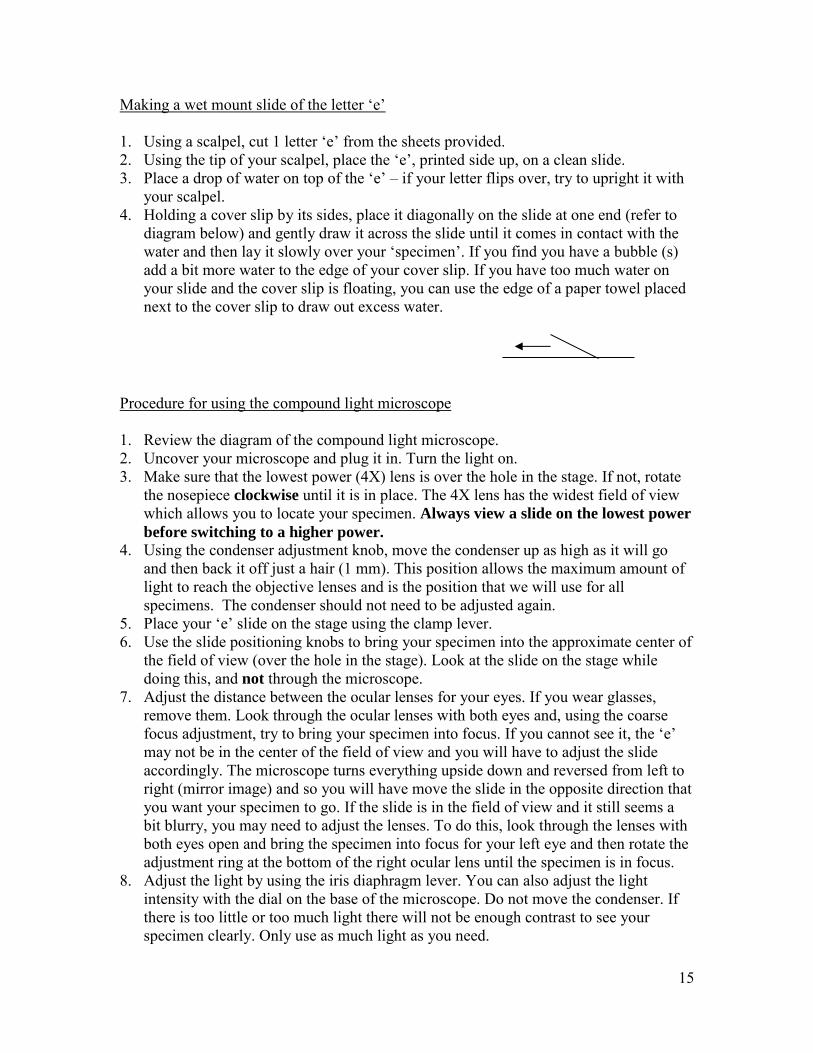
Making a wet mount slide of the letter ‘e’ 1. Using a scalpel, cut 1 letter ‘e’ from the sheets provided. 2. Using the tip of your scalpel, place the ‘e’, printed side up, on a clean slide. 3. Place a drop of water on top of the ‘e’ – if your letter flips over, try to upright it with
your scalpel. 4. Holding a cover slip by its sides, place it diagonally on the slide at one end (refer to
diagram below) and gently draw it across the slide until it comes in contact with the water and then lay it slowly over your ‘specimen’. If you find you have a bubble (s) add a bit more water to the edge of your cover slip. If you have too much water on your slide and the cover slip is floating, you can use the edge of a paper towel placed next to the cover slip to draw out excess water.
Procedure for using the compound light microscope 1. Review the diagram of the compound light microscope. 2. Uncover your microscope and plug it in. Turn the light on. 3. Make sure that the lowest power (4X) lens is over the hole in the stage. If not, rotate
the nosepiece clockwise until it is in place. The 4X lens has the widest field of view which allows you to locate your specimen. Always view a slide on the lowest power
before switching to a higher power. 4. Using the condenser adjustment knob, move the condenser up as high as it will go
and then back it off just a hair (1 mm). This position allows the maximum amount of light to reach the objective lenses and is the position that we will use for all specimens. The condenser should not need to be adjusted again.
5. Place your ‘e’ slide on the stage using the clamp lever. 6. Use the slide positioning knobs to bring your specimen into the approximate center of
the field of view (over the hole in the stage). Look at the slide on the stage while doing this, and not through the microscope.
7. Adjust the distance between the ocular lenses for your eyes. If you wear glasses, remove them. Look through the ocular lenses with both eyes and, using the coarse focus adjustment, try to bring your specimen into focus. If you cannot see it, the ‘e’ may not be in the center of the field of view and you will have to adjust the slide accordingly. The microscope turns everything upside down and reversed from left to right (mirror image) and so you will have move the slide in the opposite direction that you want your specimen to go. If the slide is in the field of view and it still seems a bit blurry, you may need to adjust the lenses. To do this, look through the lenses with both eyes open and bring the specimen into focus for your left eye and then rotate the adjustment ring at the bottom of the right ocular lens until the specimen is in focus.
8. Adjust the light by using the iris diaphragm lever. You can also adjust the light intensity with the dial on the base of the microscope. Do not move the condenser. If there is too little or too much light there will not be enough contrast to see your specimen clearly. Only use as much light as you need.
16
9. Once the specimen is in focus with the 4X lens, rotate the nosepiece counterclockwise and click the 10X lens in place. Our microscopes are parfocal, meaning that once the specimen is focused on low power it will remain approximately in focus at subsequent higher powers. You will need to bring it into
sharp focus using the fine focus adjustment only. Never use the coarse focus
adjustment unless the 4X lens is in place. You will also need to readjust the light using the iris diaphragm control.
10. Repeat step #7 for the 40X lens. If you lose sight of your specimen at any time, you
must return to the lowest power lens and start over. Depth of field refers to the distance between points furthest away and closest to the objective lens of the specimen you are viewing that will be in focus at the same time. The depth of field is substantially reduced with the higher power lenses and you will have to focus, slowly, with the fine focus, up and down to bring the point that you are interested in into focus.
11. When you are done viewing your slide, rotate the nosepiece clockwise to the lowest power lens so as to avoid the possibility of the longest (100X) lens hitting the slide. We do not use the 100X lens in this class.
Lab exercises 5) Microscope review
a) Describe what happens to the following when viewing a specimen with the compound microscope as you increase the magnification
i) the light – ii) depth of field – iii) field of view –
b) Describe 2 ways to adjust the light when viewing a slide. c) Why do you need to return to the 4X lens if you lose sight of your specimen
under higher power? d) What is the total magnification of an object under the 40X lens?

17
5) Elodea is a fresh water aquatic plant that is often used in plant biology labs because it is only 2 cells thick. This feature eliminates the hassle of trying to cut very thin slices of fresh material. Place a drop of distilled water on a clean slide and then, with tweezers or a scalpel, remove a leaf from an Elodea stem, place it on the drop of water and cover with a cover slip. Try to place it top side up (look at the orientation of the leaves on the stem).
a) Draw a picture of an Elodea cell. Label the cell wall, chloroplasts and the
approximate position of the vacuole. b) Elodea cells are ideal for observing cyclosis. Cyclosis, also called cytoplasmic
streaming, is the flowing of cytoplasm in eukaryotic cells. This movement enhances the exchange of nutrients, enzymes and other particles between organelles and between cells. It is thought that microfilaments provide the driving force for, and control the direction of the streaming. Most of the chloroplasts should appear to be moving in ‘strands’ of cytoplasm that sort of spiral around the perimeter of the cell. Why is movement restricted to the perimeter of the cell?
6) Place a drop of distilled water on a clean slide. Using tweezers, attempt to get a very
thin section of red onion epidermal (outer skin) cells (the instructor show you how to do this). Place your section in the drop of water and cover with a cover slip. View the slide under the LOWEST power on the compound scope. Draw a picture of a cell to show its shape, the cell wall and visible components. Label your drawing.
a) Why are there no chloroplasts in the red onion epidermal cells?

18
7) Make a wet mount slide of a very thin section of red pepper. Red pepper cells contain
a type of plastid called a chromoplast. These plastids contain pigments other than chlorophyll and are often red or orange. In the case of the red pepper, chromoplasts develop from chloroplasts. As the fruit (pepper) begins to ripen, the chlorophyll disappears and carotenoid pigments accumulate. a) Some pigments found in plants, such as those that affect flower color, can be
affected by the pH of the sap solution. For example, flower color may be red in acid soils, but blue or purple in more alkaline soils. Would you expect the carotenoid pigments in the pepper to be affected by pH? Why or why not?
b) How might you be able to tell, by looking at a slide of a plant cell containing
pigments, if the pigments are water soluble? (4)
c) The fruits of many angiosperms change to an attractive color as the mature. Do you think that this change is possibly related to the function of the fruit? Explain.

19
Root Apical Meristems, Mitosis, Root Tissues and
Modifications

20
Lab #3 Root Apical Meristems, Mitosis, Root Tissues and Modifications
Name _________________________________ Date __________________________________ Lab Objectives 1. To gain an understanding of plant meristems 2. To review the process of mitosis 3. To learn about the differences and similarities in distribution of mature root tissues in
monocots and dicots 4. To learn about a few root modifications seen in monocots and dicots Materials 1) Compound and Dissecting microscopes 2) Prepared slides:
a) Allium cepa l.s. root tip for mitosis b) Monocot/Dicot roots t.s. c) Salix l.s. of roots showing origin of lateral roots
3) Fine point tweezers 4) Germinated radish seeds 5) Microscope slides 6) Various edible roots 7) Clump of grass or other monocot to show fibrous root system 8) Fresh plant material to show various root modifications Introduction All plants that develop roots, stems and leaves will, under ordinary conditions, develop from a single-celled zygote into a mature organism with millions of cells. In order to go from one cell to millions of cells, cell division must occur. This process is called mitosis. Cells must also enlarge and then, differentiate into various cell types so that they can carry out specific functions. In plants, there are specific regions of growth where cell division occurs. These are called meristems. The primary meristems are located at shoot and root tips and are called apical meristems. Behind the regions of cell division are regions where the cells elongate. Following the region of elongation, cells differentiate into specific cell types that make up the mature tissues of a root. The distribution of mature root tissues differs slightly in monocot and dicot roots but there are also several similarities. In dicots, the primary root usually branches to form a taproot system. In monocots, the primary root eventually withers and a fibrous root system develops from adventitious roots that are produced from stem tissue. Dicots often produce both a taproot

21
and fibrous root system. In some plants, additional roots are produced that are modified to carry out specific functions. Lab exercises 1) View a prepared slide of an Allium (onion root tip using the compound microscope.
Start at the lowest power and get the slide in focus. Carefully bring the slide into focus under the 40x lens using the fine focus adjustment only.
a) What regions of a developing root tip can be seen on your slide? b) How can you differentiate between a cell that is in the G1 (early) interphase stage
of the cell cycle from a cell that is in the G2 (getting ready to divide) stage? c) Draw a cell that is in the metaphase stage of mitosis. Label the equator of the cell,
the poles of the cell, the cell wall, a chromosome and a chromatid. d) If your original cell has 4 chromosomes, how many chromatids are present during
metaphase of mitosis? e) Does this cell have the same amount of DNA as it did during early interphase? f) Draw a cell that is in the anaphase stage of mitosis. Label the equator of the cell,
the poles of the cell and the chromosomes. g) What structure is visible in a cell in telophase that is not present in an anaphase
cell?

22
2) View prepared slides of monocot and dicot root cross sections using the compound
microscope. Schematic diagrams of monocot and dicot root cross sections are attached at the end of the lab exercises. The diagrams show the relative distribution of tissues that make up the root.
a) Label each diagram as to whether it is a dicot or monocot root. b) For each diagram, list the root tissues in their order of appearance from the
outside of the root, progressing toward the center. c) For each diagram, draw a line from each tissue in your list to their appropriate
placement on the diagrams. d) Look at the lists that you have made. Is the relative placement of the phloem
tissue with respect the to the xylem tissue the same for both dicot and monocot roots?
e) Is the relative placement of the endodermis the same for both monocot and dicot
roots? What is the function of the endodermis? f) What tissue is present in a monocot root that is not seen in a dicot root?
3) Look at a prepared slide of a cross section of Salix (willow) roots under the
compound microscope. Draw a simple sketch (you do not have to draw all the cells) of the cross section.
a) Label the epidermis, approximate location of the pericycle, a lateral root, the
xylem and phloem (if visible) and cortex.

23
4) Using tweezers carefully place a sprouted radish seed on a glass slide. Look at the
radicle under the dissecting microscope. Draw a simple diagram of the radicle showing the location of the root hairs.
a) Do the root hairs extend the entire length of the developing root? Why or why not?
b) What is the function of the root hairs? c) Root hairs are extensions of a single cell. What kind of cell produces root hairs? d) What is the function of the root cap?
5) Look at the edible roots on display. What type of root systems do these plants have? What type of root modification do you see?
6) Using tweezers, take a small clump of the grass, gently rinse off the soil, and look at
it under the dissecting microscope.
a) Do the roots develop from the primary root? If not, from what plant organ do they arise?
b) What type of root system does a grass plant have?

24
c) What type of root makes up the root system?
7) Some plants have more than one kind of root. The primary root system absorbs water
and nutrients and anchors it to the ground. Other types of roots may be produced that are modified to perform a specific function. Look at the ivy stems and the corn stalk on display.
a) What type of modified roots does ivy produce and what is the function of these
roots? b) Are the modified ivy roots adventitious roots or taproots? c) What type of modified roots does the corn plant produce and what is the function
of these roots? d) Are the modified corn roots adventitious roots or taproots?

25

26
Monocot and Dicot Stems

27
Lab #4 Monocot and Dicot Stems Name____________________________ Date _____________________________ Lab Objectives 1) To learn the external architecture of a woody stem 2) To learn the internal anatomy of monocot and dicot stems, how they differ from each
other and how the stem anatomy differs from root anatomy 3) To learn about the growth in girth of a woody plant 4) To learn about the distribution of tissues in an old woody plant or tree 5) To look at a common stem modifications seen in plants Materials 1) Dissecting microscopes 2) Compound microscopes 3) Prepared slides
a) Dicot and Monocot stem t.s. or c.s. b) Sambucus mature bark, c.s. for lenticels c) Older woody dicot stem t.s., optional d) Cross sections of trees with visible annual rings (preferably with skewed annual
rings and less than 20 years old) 4) Birch or cherry limbs (2-4”) 5) Iris or other rhizomatous plants 6) A piece of ginger root 7) Walnut or horse chestnut branches, 3-4 years old 8) Irish potato 9) Sweet potato 10) Bermuda grass or other stoloniferous plants Introduction The main function of plant shoots, or stems, is to support and elevate the leaves, flowers and fruits. A typical woody stem is made up of a series of repeated units that include a node, internode and axillary bud. At the tips of the branches there are terminal buds which house the apical meristems. The apical meristem gives rise to these repeated units which results in growth in length of the stems. The node is the site where leaves are attached to the stem. The internode is the stem length between nodes. Axillary buds are located in the axil, or angle, that is formed between the site of leaf attachment and the stem. These buds contain meristematic tissue which may produce new lateral branches or flowers depending on environmental conditions and the development of the plant. They may remain dormant. Many plants produce modified stems that grow horizontally along

28
the soil surface or below the ground and do not have an aerial shoot system. These stems still have nodes, internodes and axillary buds. Stems are continuous with the root system and the tissues that comprise roots and shoots are similar, but there are some differences. In roots, lateral roots are produced by the pericycle. In shoots, lateral braches are produced by axillary buds and thus, stems do not have a pericycle. Stems also do not have an endodermis surrounding the vascular tissue. Why? The distribution of tissues also differs from that seen in the roots and between monocots and dicots. A woody plant not only grows in length each year, but the stems also grow in girth. This secondary growth begins with initiation of meristematic tissues called cambiums. The vascular cambium produces secondary xylem and phloem tissue and it is the production of secondary xylem tissue that makes up most of the increase in width of a tree. There is also a cork cambium which contributes to bark production. Herbaceous dicots and monocots usually produce tissues that last only a single growing season. They do not have cambiums. Grasses, which are monocots, have special terminology to describe their stem, leaf and flower structure. We will look at the structure of grasses in another lab. Lab Exercises 1) Obtain a walnut (or horse chestnut) twig. Draw a simple diagram of your twig and
label the following: nodes, internodes, axillary buds, terminal bud, bud scales and terminal bud scale scars.

29
a) How old is your walnut twig? b) How did you determine the age of your twig? c) How are the leaves, when present, arranged on the stem (alternate, opposite,
whorled)? d) What is the function of the bud scales? e) Draw a simple diagram of a leaf scar showing the location of bundle scars.
2) Look at the prepared slides of monocot and herbaceous dicot stem cross sections using the compound microscope. a) Draw a schematic diagram of a monocot stem to show the distribution of the
tissues listed below. Label your drawing.
i) vascular bundle ii) xylem iii) phloem iv) epidermis v) ground tissue
b) How does the distribution of the vascular bundles in a monocot stem differ from that of a monocot root?
c) Other than pith and cortex, which are essentially the same as ground tissue, what
tissues are absent in a monocot stem that were present a monocot roots?

30
3) On the following page are schematic diagrams of an herbaceous dicot stem and a two
year old woody dicot stem showing the relative distribution of various stem tissues. Herbaceous dicot stems and young, woody dicot stems are made up of similar tissues arranged in a similar pattern. In herbaceous dicot stems, the vascular cambium can produce some secondary tissues, but usually does not. Label the diagrams as indicated. The cambial tissues have been labeled for you.
a) What tissue is seen in a young woody stem that is not seen in an older woody
stem? b) Which tissues are responsible for increase in girth of a woody stem? c) What type of tissue is responsible for growth in length of a woody stem? d) What type of tissue gives rise to cambial cells?
4) A 2nd diagram of a 4 year woody dicot stem follows the diagram for the questions above. The dark circles are the cambiums. a) Label and indicate the location of the tissues that make up the bark of a woody
plant. b) Shade (or indicate) and label the tissue(s) that makes up wood. c) What happens to the pith and cortex tissues as a woody stem ages? d) Is secondary phloem produced in annular rings like the secondary xylem tissue?

31

32

33
5) Obtain one of the cross sections of trees on display.
a) Draw a simplified diagram of the section and label the heartwood, sapwood, bark, approximate location of the phloem and the xylem.
b) How old is the tree from which this cross section was taken? c) Using the same section of wood, can you infer any incidences in the tree’s past
history that have affected its growth? Did the tree grow more in some years than others? Explain.
d) Why are annual rings only produced by trees that grow in temperate climates? e) Why does one need to be very careful when using weed whackers near the base of
young trees?
6) Look at the prepared slides of Sambucus (elderberry) lenticels using the compound microscope. Then look at a birch limb under the dissecting microscope.
a) What are the characteristic black lines seen on the trunks of birch trees? b) Why are lenticels important for woody plants?

34
7) Look at the potatoes on display. What plant organ is represented by the Idaho spud? The sweet potato?
a) If you were cut each potato into pieces and then plant them, which potato pieces might produce a new potato plant; those from the Idaho spud, or those from the sweet potato? Explain your answer.
8) Many plants produce modified stems called rhizomes, stolons or runners. A runner usually refers to an above ground horizontal stem but sometimes these stems are also called stolons. A stolon is an underground stem that generally does not run strictly horizontally. In either case, these modified stems are usually slender and have long internodes which can give rise to a new plant. Rhizomes are modified underground stems that run horizontally, usually close to the soil surface. They are somewhat thickened affairs with compressed internodes and scale like leaves.
a) Look at the iris and ginger root on display. What type of modified stems do these
plants produce? b) Is the ginger root really a root? c) Both stolons and rhizomes give rise to new plants. Why? d) Look at the specimen of Bermuda grass on display. What type(s) of modified
stems does this grass produce?

35
Leaves

36
Lab #5 Leaves Name ____________________________ Date _____________________________ Lab Objectives 1. To learn about the anatomy of monocot and dicot leaves and how the distribution of
tissues relates to leaf function 2. To learn about the structure of a stomata and how guard cells differ from other
epidermal cells 3. To be able to tell the difference between a simple and compound leaf 4. To learn about some of the many leaf variations seen in woody, flowering plants 5. To learn about some of the various leaf modifications seen in xerophytic plants and
how these modifications function to allow these plants to survive in their environment Materials 1. Dissecting microscope 2. Compound microscope 3. Prepared slides of monocot and dicot leaf cross sections 4. Campanula (or other) leaves for looking at epidermal cells and stomata 5. Twig of laurel 6. Celery 7. Onion, cut in half 8. Twigs (preferable) of various trees and shrubs with a variety of different leaves 9. Leaves from xerophytic plants such as sage, dusty miller, lavender, succulents, cactus Introduction Leaves arise from leaf primordia in buds produced by shoot apical meristems. However, unlike plant stems and roots, they do not have apical meristems themselves, thus they exhibit determinate growth. The size of a leaf may be affected by age and the environment but, at some predetermined point, it stops growing. The variation seen in leaf size, shape, color and texture among plants is enormous. Some plants are evergreen and retain their leaves all winter and others are deciduous and loose their leaves under adverse conditions. Regardless, the function of a leaf, with some exceptions, remains constant; to convert light energy to chemical energy and to use this energy to make sugars from carbon dioxide from the air. This process is called photosynthesis. In some plants, the leaves have been modified to perform a different function and photosynthesis is carried out in stem cells. In order for photosynthesis to take place, chlorophyll pigments must be able to absorb light. The stomata must also be open in order for the plant to obtain carbon dioxide. Most of the water lost by a plant occurs through open stomata. Thus, leaf anatomy and morphology can be seen as a compromise between optimal surfaces for photosynthesis and those that minimize water loss.

37
Lab Exercises 1) Look at prepared slide of a cross section of a dicot leaf. Draw a simple diagram to
show the upper epidermis, lower epidermis, palisade mesophyll, spongy mesophyll, air space, a stoma, and a vein.
a) How does the distribution (location) of palisade and spongy mesophyll relate to leaf function?
2) Look at a prepared slide of a cross section of a monocot leaf. How does the internal makeup of the leaf differ from that of a dicot?
3) Attempt to get a very thin peel of the epidermal layer from the underside of a
Campanula (blue bell) leaf. Place in a drop of water on a slide and cover with a cover slip. Focus on the lowest power and then carefully bring into focus on the higher power.
a) Draw a picture of a stomate (stomata = plural) and a few of the surrounding
epidermal cells. Label the stoma (pit), guard cells and chloroplasts. b) Do all epidermal cells contain chloroplasts?

38
c) What is the function of chloroplasts in the guard cells? d) Why was the epidermis from the underside of a leaf used for this exercise instead
of the epidermis on the upper side of a leaf?
4) Obtain a small section of an English laurel branch. Make a simple drawing of your section. Label the stem, axillary bud, leaf petiole and leaf blade.
5) What feature of a plant stem allows you to determine if a plant has simple leaves, or if
it has compound leaves made up of leaflets? 6) In winter, if you have collected leaves from a deciduous tree or shrub, and do not
have access to the stem, what feature of deciduous leaves allows you to discern, positively, if the tree or shrub has simple or compound leaves?
7) Take a look at the celery on display. What part of the plant is the celery stalk? 8) Draw a simple diagram of the onion that has been cut in half lengthwise. Label the
leaves, stem, roots and approximate location of the apical meristem.
a) Where are the axillary buds located?

39
5) There are many taxonomic keys available that aid in identification of plants with which we are not familiar. Species determinations are made by answering yes or no to a series of questions regarding its habitat, habit, root and shoot system, its leaves and floral structures. Sometimes a microscope is needed to assess if a plant has a particular feature, but often a hand lens is all that is necessary. There are many, specific terms used to describe most plant features and leaves are no exception. Most keys include a glossary but there are also books available describing plant identification terms, often with pictures. Leaf descriptions include terms relative to the entire leaf, the leaf margins, the leaf shape, the leaf base, leaf tip, venation and so on. If the leaf is a compound leaf, the terms apply to the leaflets. In this exercise we will learn a few of the terms used for describing leaves. Diagrams and description of these terms are provided on the following page.
a) Using the list on the following page, provide a description of the leaves for each
of the flowering trees and shrubs on display. If the leaf is compound, include the number of leaflets per leaf and their approximate size. The pattern of attachment to the stem only applies to those with a branch on display. A sample description of a typical locust tree leaf is provided below.
Robinia pseudoacacia, Black Locust. Deciduous tree. Leaves alternate, pinnately compound, leaflets 11-21, 1-2” long, entire, lanceolate, pinnately veined.
Acer sp. Maple. Deciduous tree.
Aesculus hippocastanum, common Horsechestnut. Deciduous tree.
Rubus discolor, Himalayan Blackberry. Evergreen shrub.
Lonicera nitida, Boxleaf Honeysuckle. Semi-evergreen shrub.
Mahonia aquafolium, Oregon Grape. Evergreen shrub.
Quercus sp., Oak. Deciduous tree.

40

41
6) A xerophyte is a plant that lives in a hot, dry environment. These plants often have
leaf modifications which allow them to survive in their environment. Describe how each of the leaf adaptations listed below might help a plant that lives in a xeric environment.
a) Pubescent leaves b) Epidermis with thick wax deposits c) Succulent leaves d) Convoluted leaf surface(s) e) Leaves with reduced surface area f) White or light colored leaves
7) There are several xerophytic plants on display. Obtain and examine a leaf from each of these plants under the dissecting microscope. EXCEPTION: Do not touch the cactus plant; just look at the whole plant. List the leaf adaptations for each of the plants that allow it to survive in a xeric environment.
a) Kalanchoe
b) Helichrysum
c) Sage d) Cactus e) Blue Pearl plant

42
Gymnosperms I

43
Lab #6 Gymnosperms I
Name __________________________
Date ___________________________ Lab Objectives 1. To learn about the term ‘naked seed’. 2. To learn the difference between pollination and fertilization and discover that these
events initiate different processes. 3. To become familiar with features that are common to gymnosperms in the phylum
Pinophyta. 4. To understand why Ginkgo biloba is in its own phylum and why Ginkgophyta is a
gymnosperm, despite the fact that it has several features that are not seen in other gymnosperms.
Materials 1. Pine cones that have retained their seeds. 2. Pine branches with seed cones in various stages of development 3. Branches of female Ginkgo biloba 4. Dissecting microscopes 5. Glass slides for cutting 6. Single edge razor blades Introduction The gymnosperms include four phyla; the Pinophyta, Ginkgophyta, Cycadophyta and Gnetophyta. All of our common conifers including firs, spruces, larches, hemlocks, pines, douglas-firs, cedars and junipers are members of the Pinophyta .The Ginkgophyta contains only one existing species, Gingko biloba, which is widely planted in our area. The cycads and gnetophytes do not occur in our area. The sago palm, a cycad, is a common houseplant. The gymnosperms are often referred to as the conifers or simply, evergreens. However, there are gymnosperms that do not produce cones and some gymnosperms are deciduous. There are also many evergreen angiosperms. The one feature that is common to all gymnosperms is reproduction by ‘naked seeds’; seeds that are not enclosed in a fruit. In this lab we will look at some general features of a pine which is a representative member of the Pinophyta. We will also look at Ginkgo biloba, which possesses many unique features not found in other gymnosperms.

44
Lab Exercises 1) The ‘naked’ seeds produced by pines and several other conifers look similar to the
fruits produced by a number of angiosperms. The maple fruit, which encloses the seed, is a dry, winged fruit called a samara. Obtain a maple samara and separate the fruit from the seed. Now try to do the same with a pine seed. Even though both seeds have ‘wings’, is the pine seed enclosed by the winged structure? What part of the pine female reproductive structure gives rise to the wing?
2) Obtain a mature pine cone (labeled). DO NOT TURN THE CONE UPSIDE DOWN
OR SHAKE IT! Find a cone scale that has still retained the seeds.
a) Where on the cone scales are the seeds located? b) How many seeds are there per scale? c) What is the name of the female structure that gave rise to the seed? d) How does a seed differ from a spore?
3) Select one of the pine branches on display. Draw a sketch of your branch. You may
draw an oval to represent the placement of the seed cones but be sure to draw different size ovals to represent different size cones.

45
a) By looking at the reproductive structures present, estimate how long it takes for a seed cone to mature.
b) Estimate the age of the cones on your branch that have open cone scales. c) Fill in the blank in the following sentence. Seed cone scales close following ______________________.
4) Conifers that retain their foliage over the winter possess many of the same leaf adaptations as those seen in xerophytic plants. Most of our conifers grow in temperate regions with adequate soil moisture. Why do they possess leaf adaptations that allow them to survive in an arid environment?
a) List 2 visible leaf adaptations that help conifers to reduce water loss. 5) Cut a thin cross section of one of the pine needles provided. Look at the section under
the dissecting microscope. Draw a diagram of your section. Label the vein and resin ducts.
a) In the cross sections of angiosperm leaves that we saw in an earlier lab, the veins were close to the surface of the leaf and surrounded, at least in part, by air spaces. Where is the vein located in the pine leaf? Do you see many air spaces?
b) What is the function of the resin ducts?

46
6) Ginkgo biloba, the maidenhair tree, is the only living representative of the phylum Ginkgophyta. Previously, there have been many species of ginkgoes to inhabit the northern hemisphere prior to the age of the dinosaurs. They were a major component of prehistoric forests alongside tree ferns and cycads. All the species have become extinct except Ginkgo biloba, which might also have become extinct if it had not been cultivated by the Chinese. Fossils of at least one other Ginkgo species have been found in the sediments and lava flows in the Columbia River gorge near Vantage. The maidenhair tree is also widely planted as an ornamental in our area. Ginkgoes are gymnosperms (they produce naked seeds) but they are deciduous and look very different from familiar members of the Pinophyta.
a) Ginkgoes are called maidenhair trees because the shape of their leaves resembles
that of the maidenhair fern. Draw a Ginkgo leaf showing the overall shape and portion of the unique, dichotomous venation.
b) Ginkgoes are dioecious. The male and female reproductive structures are borne on
different plants. Female ginkgoes do not produce cones. Two ovules are produced at the tips of thin stalks that arise on short (fruiting) shoots. The male pollen grains are produced on catkin-like structures on short shoots and are dispersed by wind. After pollination occurs, the pollen grain germinates and produces sperm with flagella (tails) that swim to the ovule. The sperm are similar to those of non-seed plants rather than seed plants. After fertilization, a fleshy seed coat develops such that the seed looks more like a fruit with a seed inside than a naked seed. Mature Ginkgo seeds become slimy and have a terrible smell. For this reason, male plants are usually used in landscapes. Draw a portion of the Ginkgo branch to show the short shoots and (immature!) seeds.

47
Gymnosperms II – Identification of Common
Conifers in Washington State

48
Lab #7 Gymnosperms II – Identification of Common Conifers in Washington State
Name __________________________
Date ___________________________
Lab Objectives 1. To become familiar with differences among conifers and the features used to identify
and place them in different groups 2. To be able to recognize conifers native to Washington State Materials 1. Dissecting microscope 2. Glass slides to be used as a cutting platform 3. Single edge razor blades or scalpel 4. Fine point tweezers 5. Labeled samples of native conifers
Introduction The Pacific Northwest (Alaska to northern California) is known for its dense forests of coniferous trees. For many beginning botany students, any tree with needle or scale like foliage is a ‘pine’ tree. The purpose of this part of the lab is to become familiar with some of the visible differences between the several different conifers that inhabit our region and the vegetative and reproductive features that are used to classify them into different groups. The conifers that we will be examining all belong to the phylum, Pinophyta.
Within this phylum, we will be looking at conifers from 3 families; the Cupressaceae or Cypress family, the Pinaceae or Pine family, and the Taxaceae or Yew family. The general classification scheme for the conifers on display in this lab is presented below. Phylum: Pinophyta – the conifers Family: Cupressaceae – the cypress family 1. Calocedrus decurrens – Incense cedar 2. Chamaecyparis (Xanthocyparis) nootkatensis – Alaska cedar 3. Juniperus scopulorum – Rocky Mountain juniper 4. Thuja plicata – Western Red cedar

49
Family: Pinaceae – the pine family Genus: Abies – true firs 1. Abies amabilis – Pacific Silver fir 2. Abies grandis – Grand fir 3. Abies procera – noble fir 4. Abies lasiocarpa – Subalpine fir Genus: Larix - larch 5. Larix occidentalis – Western larch Genus: Picea - spruce 6. Picea sitchensis – Sitka spruce Genus: Pinus - pines 7. Pinus contorta – Lodgepole pine 8. Pinus monticola – Western white pine 9. Pinus ponderosa – Ponderosa pine Genus: Pseudotsuga – Douglas firs 10. Pseudotsuga menziesii – Douglas fir Genus: Tsuga - hemlocks 11. Tsuga heterophylla – Western hemlock 12. Tsuga mertensiana – Mountain hemlock Family: Taxaceae – yew family 1. Taxus brevifolia – Pacific yew Obtain a labeled sample of each of the conifers on display to answer the questions that follow. Cypress Family
1. Some of the conifers have needle-like foliage and others have scale-like leaves. List the 4 species that have scale-like leaves.
1. ___________________________
2. ___________________________
3. ___________________________
4. __________________________

50
2. One of the conifers with scale-like leaves has round stems versus flattened stems and foliage. The leaves are closely pressed to the stem. The twigs look like ‘braided whipcords’. Name this species below. scale-like leaves and round stems _____________________________ Note: There are several junipers that are commonly planted in our area that have very short stiff needles in 2’s or whorls of 3 that stick out from the branches. Some junipers have needle-like juvenile foliage and scale-like mature foliage on round stems. 3. Of the 3 remaining species with scale-like leaves, 1 species has leaves with decurrent leaf bases. The base of the leaf runs down the stem from the point of attachment of the leaf to the stem. Which species has decurrent leaf bases? scale-like leaves with decurrent leaf bases __________________________ 4. The leaves of the 2 remaining species with scale-like leaves have slightly different shapes. One has leaves that are rounded and wide and the other has leaves that are narrow and pointed. Label the pictures below. ______________________________ _____________________________
5) The cones of the 4 conifers with scale-like foliage differ from each other. If cones are
present, it is often easier to tell the species apart by looking at their cones, rather than the leaves. Members of the cypress family have cones that are, in general, much smaller than those of the pine or yew family and they are usually round or somewhat cylindrical.
a) Which species has a fleshy cone that is a bluish color and does not open to shed
its seeds when mature? ______________________________

51
b) Which species has a round, woody cone made up of funnel-shaped scales? ____________________________________
c) The other 2 species have dry, woody cones that look somewhat like a rosebud.
In one species, the ‘rosebud’ is more elongated and has only 4-6 scales. The other has a smaller cone with 10-12 scales.
i) Which species has the cone with 4-6 scales? ___________________________ ii) Which has the smaller cone with more scales? _________________________
Yew Family
1) All of the rest of the conifers on display have needle-like foliage. One of the species does not produce woody cones and is dioecious; the male and female reproductive structures are produced on different plants. The female reproductive structure is a red, fleshy cup that contains a single seed. This structure is called an aril. The leaves are borne singly along the branch and have decurrent leaf bases. The needles are flat and come to a point. They are arranged in 2, horizontal rows along the branches. Provide the name of this species below.
______________________________ Needle-like foliage with decurrent leaf bases, female reproductive structure an aril Pine Family
1) The remaining conifers are members of the pine family. The needles of these species occur singly along the branch, in bundles of 2, 3 or 5, or in clusters or ‘tufts’ at the tips of very short woody shoots. One of the species is deciduous and loses its needles in the fall. The new leaves are soft compared to the evergreens. Which species has soft needles in tufts at the end of short woody shoots?
______________________________
Deciduous, needles in clusters at the tips of short shoots
When the needles are not present, this species can be easily identified by the persistent woody short shoots along the branches and the cones which have a small 3-pointed bract underneath each cone scale that resemble the hind end of a rat. Note: True cedars (the genus, Cedrus) are not native to Washington State. Western Red Cedar and many members of the genus Chamaecyparis that we call cedars are not true cedars. Cedrus species look very similar to larch but the foliage is evergreen and not soft.

52
2) Three species are all in the same genus and have relatively long needles in bundles of 2, 3 or 5. The needles are actually wrapped together at their bases. These bundles are called fascicles. What is the name of this genus?
___________________________
evergreen, needles in fasicles
a) Name the 3 species in this genus.
i) Needles in bundles of 2 ________________________________
ii) Needles in bundles of 3 _________________________________
iii) Needles in bundles of 5 _________________________________ iv) Which has a 1-2” cone? ________________________________ v) Which has a cone that is 3-5”? ___________________________
3) The remaining conifers all have needles that are borne singly along their branches. Three of these species (2 genera) have needles borne on short woody pegs (much smaller than a short shoot). When the needles fall off the bare branches are rough due to the persistent woody pegs. The location of the cones also differs.
a) Which genus has larger woody pegs that stick out at right angles from the branch
and cones located behind the branch tips? ______________________________ b) Which genus has smaller woody pegs that project forward along the stem and
cones that hang from the tips of the branches? ___________________________ c) One of these conifers has large woody pegs and very stiff, sharp pointed
needles. This species is _______________________________. d) Of the 2 conifers with the small woody pegs, one has needles that are different
lengths and tend to spread horizontally from the branch. The underside of the leaves look white. The white color is due to rows of white dots that are the stomatal pores. Look at these under the dissecting microscope. The name of this species is _____________________________________.

53
e) The remaining species with small woody pegs has needles that are equal in
length and are arranged spirally around the stems. The color on the top and bottom of the needles is the same. This species is called ______________________________.
4) There are 5 conifers left to identify. These species are representatives of 2 genera: the
true firs (Abies) and the Douglas-firs (Pseudotsuga). These 2 genera are often distinguished by the type of leaf scar left on the branch when a needle is pulled off. One group has large, round leaf scars that are somewhat sunken or depressed. The other group has small leaf scars that are raised.
a) Which genus has large, round, depressed leaf scars?____________________ b) Which genus has small, raised leaf scars? _____________________________ c) Sometimes it is hard to see the leaf scars, but the cones and buds differ and are
easy to distinguish. i) Which genus has long, oval, pointed buds at the tips of branches?
_____________________________________ ii) Which genus has blunt, short buds? _________________________ iii) The genus with pointed buds has only 1 species native to our area. It can also
be easily distinguished by its cones which hang down and are woody with prominent 3-pointed bracts subtending the scales. The bracts are similar to those of larch cones, but much larger. Name this species below.
pointed buds, cones with distinctive bracts ________________________
5) All the rest of the conifers are in the genus, Abies, the true firs (Douglas-fir is not a true fir). They all have cones that are upright and they disintegrate when mature. The species can be distinguished by the arrangement of the foliage, by the presence of stomata which appear as whitish lines or by regions and the position of resin ducts in the needles.
a) Two of the species have stomata only on the undersides of their needles. These
2 species are:____________________ and_________________________

54
b) These 2 conifers have their needles arranged differently. Label the pictures below. needles distinctly 2-ranked ___________________________
a row of needles points forward
covering the upper surface of stems ___________________________
c) The remaining 2 species have stomata on both surfaces of their leaves and their shape is different.
d) Which species has straight needles with a row of stomata down the center of the
upper surface of the needles? _________________________________ e) Which one has needles that curve at the base such that they resemble hockey
sticks with 2 rows of stomata on the upper surface? ________________________________________
f) These species also differ in the position of their resin ducts in the needles. Cut a
very thin cross section of a needle from each of these conifers and look at your section under the dissecting microscope and label the pictures below.
small resin ducts close to lower epidermis ____________________________ large resin ducts midway between the upper
and lower epidermis ________________________________

55
Flower Structure

56
Lab #8 Flower Structure
Name ____________________________
Date _____________________________ Lab Objectives 1. To learn the parts of a typical flower 2. To learn the terms used to describe floral parts and structure 3. To learn about some of the common modifications seen in angiosperm flowers and
how these modifications relate to function 4. To view the structure of a highly modified angiosperm flower Materials 1. Dissecting microscopes 2. Single edge razor blades or scalpels 3. Fine point tweezers 4. Glass slides to be used as cutting boards 5. Asiatic lilies, Oriental lilies or Alstroemeria or other large flowers with typical floral
parts 6. Snapdragon or other irregular flower 7. Primrose plants or other plant to show various types of placentation 8. Sunflower, Chrysanthemum or other typical composite (with developed parts) 9. Liatris spicata – optional (or Moluccella laevis) Introduction Knowledge of floral structure is critical for assessing relationships among angiosperms and for identifying an unknown flowering plant. Flowers are the sexual reproductive organs and thus, they must function to insure that pollination, fertilization and seed dispersal takes place. Despite the tremendous variation in form, all flowers have some features in common. A flower is born on the swollen tip of a modified short shoot called a peduncle. The swollen tip is called the receptacle. A typical flower consists of 4 floral parts, some of which occur in whorls. Starting at the receptacle and working inward, the 1st whorl is made up of leaf-like structures called sepals, which collectively are called the calyx. The function of the sepals is to protect the bud. The 2nd whorl is made up of the petals and is called the corolla. The function of the corolla is to attract pollinators. The 3rd whorl is made up of stamens, the male reproductive organ. The 4th floral part is the female reproductive structure, the pistil. Flowers may be missing one or more of the parts or the parts may be modified in various ways. Monocots typically have floral parts in multiples of three, whereas dicots usually have floral parts in multiples of four or five. Much of the diversity in flower form is due to differences in how a plant is pollinated and how its seeds are dispersed.

57
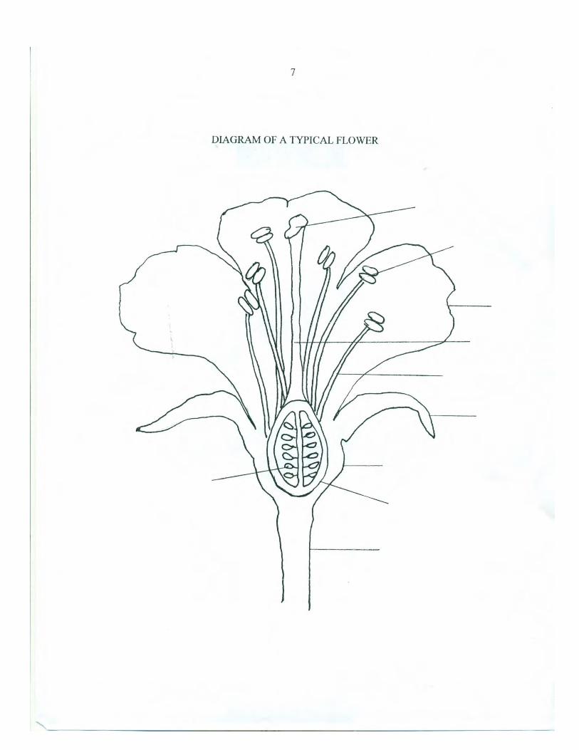
Lab Exercises 1) Label the parts of a typical flower on the diagram provided. 2) Obtain an Asiatic lily flower. Caution: Lily pollen stains so try not to get it on your
clothes or nose (it can easily be removed with alcohol if necessary). In lily flowers it is often difficult to tell the difference between the sepals and petals although the attachment point of sepals is slightly to the outside of that of the petals. Answer as many of the questions below as you can without taking the flower apart. Then, carefully, peel away the whorls one at a time. Leave the pistil intact. There is a brief glossary of terms attached that you may use.
a) Is the lily flower regular or irregular? b) Is the flower perfect or imperfect? c) How many stamens does the lily flower have? d) Is the sigma lobed? If so, how many lobes are there? e) How many sepals are there? f) How many petals are there? g) Are any of the floral parts fused together or to each other? h) Does the lily have an inferior, superior or half inferior ovary? i) Are the floral parts epigynous, hypogynous or perigynous?
3) Using a glass slide, cut a cross section of the lily ovary that is about 1/8” thick. View your cross section under the dissecting microscope. a) How many locules does the ovary have? b) How many carpels have fused to make up the lily ovary? c) Does the number of lobes of the stigma match the number of locules in the fruit? d) Is the ovary simple or compound? e) How many ovules (actually rows of ovules – we are only looking at a ‘slice’) per
locule are there? f) Does the ovary exhibit parietal or axile placentation?
g) Is the lily a monocot or dicot? Explain your answer.

58
4) Look at the snapdragon on display and then remove a single snapdragon flower.
a) What type of inflorescence does the snapdragon have?
b) Is the flower regular or irregular? c) How many sepals are there? d) Can you count the number of petals or are the petals fused?
5) Carefully remove the perianth from the snapdragon flower.
a) How many stamens are there? b) Does the stigma appear to be lobed? c) Is the flower complete or incomplete?
6) Cut the ovary of the snapdragon flower in half, crosswise. View the cut end of the ovary under the dissecting microscope.
a) Is the ovary simple or compound? b) How many locules are present? c) Does the ovary have parietal or axile placentation? d) Does the snapdragon produce only a few seeds, or many? e) Is the snapdragon a monocot or dicot? Explain your answer.

59
7) Look at the primrose plants on display. Note the leaves and general habit. Then remove a flower for a closer look at the floral structure.
a) Are the sepals fused? b) Are the sepals lobed? If so, how many lobes are there?
8) Carefully remove the sepals from the primrose flower.
a) How many petals are there? b) Are the petals fused at any point?
9) Carefully remove the corolla from the primrose flower. If it is fused, make a longitudinal cut through the corolla and carefully ‘unwrap’ it from the pistil.
a) Where are the stamens attached? b) How many stamens are there?
10) Slit the primrose ovary longitudinally. Only cut through the ovary wall, not through the entire ovary.
a) What type of placentation does a primrose flower have? b) Primroses can have floral parts in any number from 4 – 9. Keeping this in mind,
do you think this flower is a monocot or dicot?
11) Members of the aster family (Asteraceae) look like daisies or sunflowers. They are often referred to as the composites because what we consider to be a single flower is actually an inflorescence called a head. It is made up of many florets (mini flowers) with reduced floral parts that are packed tightly together on a disk. The inflorescence is subtended by a series of overlapping bracts. The term ‘sepal’ applies to a single flower, not an inflorescence. The bracts are actually modified leaves. Many of the composites have two different types of flowers; disk flowers that make up the ‘pin cushion’ like center of the “flower” and ray flowers that resemble petals, that circle the perimeter of the disk. There are composites that have only disk flowers and those that have only ray flowers. The florets either lack sepals or the sepals are modified and resemble bristles or hairs, called pappus. Pappus often acts as a parachute to carry seeds in the wind when they are mature. The fruit of a composite is an achene; a dry,

60
single seeded fruit. The sunflower ‘seed’ is an example. Cut off one of the ‘flowers’ from any of the Chrysanthemums on display. First, look at the whole inflorescence. Chrysanthemums have both disk and ray flowers. Then, cut the flower in half and look at the cut edge under the dissecting microscope. Using tweezers, carefully tease out one (or more) of each type of flower to answer the following questions. You
cannot answer the questions without looking at single florets under the
microscope.
a) Do the disk or ray flowers have sepals? b) Do the disk and ray flowers have inferior or superior ovaries? c) Are the disk flowers perfect or imperfect? If they are imperfect are they male or
female? d) Are the ray flowers perfect or imperfect? If they are imperfect, are they male or
female? e) Can you tell how many stamens a disk flower has? f) How many lobes does a disk flower stigma have? g) How many petals does a ray flower have?
12) Look at the inflorescence of the gayfeather (Liatris spicata) on display. Remove one of the flowers and, using tweezers, carefully look at each of the parts of this flower.
a) Using the information you have gathered from looking at the other flowers in this
lab, would you say that gayfeather is in the same family as a lily, snapdragon, primrose or chrysanthemum? Explain your answer.
b) How would you describe the inflorescence of the gayfeather plant? c) Describe the floral parts of this ‘flower’ as best you can. The instructor will cover
the flower structure in detail after everyone has had a chance to look at the flower.

61
GLOSSARY Regular flower – a flower that is radially symmetrical Irregular flower – a flower that is not radially symmetrical. For example, the petals on one side of the flower are a different shape or size or are joined differently than they are on the other side of the flower. Perfect flower – a flower that has both male and female parts. Imperfect flower – a flower that has only one of the sexes. Inferior ovary – an ovary that is located below the point of attachment of floral parts Superior ovary – an ovary that is above the point of attachment of floral parts Half-inferior ovary – Floral parts are usually attached to a cup shaped calyx tube that encircles the ovary but is not fused to it so that the position of the ovary seems to be neither inferior or superior but somewhere in between. Hypogynous – floral parts that are attached below and ovary. Floral parts of a flower with a superior ovary. Epigynous – floral parts attached above the ovary. Floral parts of a flower with an inferior ovary. Perigynous – floral parts that circle around the ovary. Floral parts of a flower with a half-inferior ovary. Carpel – a fertile leaf (one having ovules along the margins) that has rolled inward to form the pistil Simple ovary – comprised of a single carpel Compound ovary – comprised of more than one carpel that have become fused together. Usually, but not always, is indicated by the number stigma lobes and locules (compartments) of a fruit. Placentation – refers to how the ovules are attached in the ovary. See below. axile placentation parietal placentation free central placentation
62

63
Fruits and Seeds

64
Lab #9 Fruits and Seeds
Name ____________________________
Date _____________________________ Lab Objectives 1. To learn the various parts of a fruit and how they are used to classify different fruit
types 2. To learn about additional information that is necessary in order to classify a fruit 3. To learn the difference between simple, aggregate and multiple fruits 4. To learn about how various fruit forms are correlated with specific seed dispersal
mechanisms. Materials 1. Dissecting microscope 2. Fine point tweezers 3. Glass slides for extracting fleshy fruitlets 4. A variety of dry and fleshy simple fruits to show various fruit types and dispersal
agents (should include a few accessory fruits and an avocado) 5. A variety of dry and fleshy aggregate fruits (sweet gum, rose hips, magnolia,
strawberry, blackberry) 6. A fig or pineapple 7. Puncture vine fruits or fruitlets – optional Introduction Fertilization normally initiates both seed and fruit development. There are a few plants that are parthenocarpic. They develop fruits without fertilization and the fruits are seedless. The main function of a fruit is to aid in seed dispersal. Seeds that germinate some distance from the parent plant usually have a greater chance of survival due to reduced competition for resources such as light, water and nutrients. Seed dispersal agents are numerous and include both abiotic agents, such as wind and water, or biotic agents such as animals, insects and people. Some plants have fruits with mechanisms for flinging or shooting their seeds away. Fruits and seeds exhibit a variety of adaptations correlated with different dispersal agents and environments that a plant inhabits. Lab Exercises 1) Use the abbreviated ‘key’ to simple fruits and the description of simple fruits
provided to determine the fruit type for each of the simple fruits on display. In a few cases, the key diagram results in more than one fruit type. You will have to use the description of the fruits to decide between them. List the name of the plant and its fruit type using the lined paper provided.

65
2) List 3 things you need to know about a simple fruit in order to decide what type of fruit it is.
3) Fruits that involve tissues other than the ovary wall, such as fused sepals, petals or
receptacle tissue are called accessory fruits. Which of the simple fleshy fruits on display are accessory fruits?
4) Which of the berries on display developed from a flower with an inferior ovary? 5) Draw a simple diagram of a cucumber cross section. Label the carpels, locules, ovary,
and ovules.
a) Which part of the cucumber fruit becomes a seed? b) What are the 3 parts of a seed?
c) What type of placentation does a cucumber have?
6) How many carpels make up the ovary of a lemon? 7) Draw a simple diagram of one of the legume fruits showing where the ovules are
attached.

66
8) Draw a simple diagram of a silique.
a) How is the silique similar to a legume? b) How does the silique differ from a legume?
9) Some of the fruits that we purchase from the grocery store are not the whole fruit. For example, almonds are surrounded by a fibrous (fleshy) husk that is removed before the fruit is sold. What part of an almond fruit is the shell that we crack open? a) Given the information above, what is the difference between a true nut and the
almond?
10) For each of the 5 aggregate fruits on display, see if you can figure out what type of fruitlets make up the fruit. For each of the fleshy fruits, use tweezers to obtain a single fruitlet. Look at the fruitlets under the dissecting microscope to determine the fruit type. Blackberry – Sweet Gum – Strawberry – Magnolia – Rose hip –
11) What type of tissue makes up most of the corn grain?

67
12) Name 3 fruits on display whose seeds are dispersed by wind. For each of these fruits,
describe the adaptations for wind dispersal. 13) Look at the fig on display. Why is the fig called a multiple fruit? 14) Look at the avocado halves on display. The avocado has a single seed and appears to
arise from a simple ovary and yet, it is classified as a berry (most berries develop from a compound ovary and have several seeds). Why is it classified as a berry and not a drupe?
15) Puncture vine (Tribulus terrestris) is an obnoxious weed in our area. The puncture
vine produces a fruit called a schizocarp. Where have you seen the prefix, ‘schizo’ before? Think of a medical diagnosis. Can you guess what ‘schizo’ means? A schizocarp is a dry fruit that splits into 2 or more 1-2 seeded fruitlets. Puncture vine fruits split into 5 single carpel fruitlets. Each fruitlet has 2 projections resembling a carpet tack. The fruitlet is extremely hard and durable (it can last for years before germinating). The ‘tacks’ are not randomly placed but positioned such that if one tack punctures the ground the other sticks straight up. Take one of fruitlets (be careful!) and look at it under the dissecting scope. Puncture vine is an annual and the stems grow along the ground about 6 feet in a season. Large areas that have become covered with puncture vine are, literally, uninhabitable. How are the puncture vine seeds spread?

68

69
Description of Fruit Types on Display Fleshy Fruits 1) Drupe – a single-seeded fruit with a stony endocarp, a +/- fleshy mesocarp and skin-like
exocarp. Fruit develops from a simple, superior ovary. 2) Berry – a fruit that usually develops from a compound ovary and usually contains more than
one seed. The entire pericarp is fleshy. It is often hard to tell the difference between the endocarp and mesocarp. If the berry develops from an inferior ovary, there may be remnants of receptacle tissue present that have grown up around the ovary.
a) Hesperidium – a berry with leathery skin that contains oils (citrus fruits). b) Pepo – a berry with a thick rind (watermelon).
3) Pome - a fruit in which the fleshy part that we eat is from fused calyx, corolla and receptacle
tissue that has grown up around the ovary and not an enlargement of the ovary wall. These fruits have a ‘core’ surrounded by a papery endocarp.
Dry Fruits 1) Dry fruits that split open when they are mature. These are called dehiscent fruits.
a) Follicle – a dry fruit that develops from a simple ovary and splits along one side to expose seeds
b) Legume – a dry fruit that develops from a simple ovary and splits along two sides to
expose seeds c) Silique – a dry fruit that splits along two sides but the seeds are attached to a central
partition located between the two halves d) Capsule – a dry fruit that develops from a compound ovary and splits along more than
two sides.
2) Dry fruits that do not split open when they are mature. These are called indehiscent fruits.
a) Achene – a one-seeded fruit. The pericarp separates from the seed. b) Nut – a one-seeded fruit similar to an achene, but the pericarp is much thicker and harder. c) Samara – a one or two-seeded fruit where the ovary wall has grown out to form a wing. d) Grain – a one-seeded fruit in which the seed coat and pericarp are fused and cannot be
separated from each other.

70
Complete list of fruits used in this lab Viola sp.
Magnolia soulangiana
Pennisetum setaceum
Aesculus hippocastanum
Ailanthus altissima
Acer sp.
Koelreuteria paniculata
Cercis canadensis
Liquidambar styraciflua
Kniphofia sp.
Tribulus terrestris
Lunaria annua
Rose hips Acorns Almonds (in shell) Corn seed Dandelion Avocado Tomato Lemon Cucumber Strawberry Blackberry Blueberry Peach or olive Fig Apple Snap peas Sunflower seeds

71
Grasses

72
Lab #10 Grasses
Name________________________________
Date_________________________________ Lab Objectives 1. To become familiar with some of the unique vegetative and reproductive features of
grasses. 2. To learn some of the terminology used to describe grass features. 3. To become familiar with some of the features used to identify a grass species. Materials 1. Small clumps of various grasses (with flowers if possible) labeled #1, #2 etc. 2. Dissecting microscope and/or 10X hand lens 3. Single edge razor blades 4. Glass slides for cutting platform Note: The format and some of the text and description of terms were adapted for use in this lab courtesy of Offwell Woodland & Wildlife Trust (http://www.offwell.free-online.co.uk/grass_id/ident.htm). Introduction Eastern Washington is home to many grass species, including cultivated grasses such as wheat and barley. Some knowledge about how grass plants grow is important for establishing desired grasses such as turf grasses or pasture grasses and, of course, agricultural grass crops. Identification of grasses as well as knowledge of how, when and where they grow is critical for determining how to eliminate undesirable species. How grasses develop is somewhat different than that of other flowering plants and, although they do share many of the same features as other angiosperms, they have several morphological and reproductive features that are unique and thus, a whole different and rather extensive set of terms is used to describe them. Grasses are members of the phylum Magnoliophyta; they are flowering plants. They are also in the class Liliopsida the monocots. They have one seed leaf (cotyledon), leaves with parallel veins, fibrous roots, and scattered vascular bundles in their stems. There are 3 main groups (families) of grasses: the Poaceae, which are the true grasses, the Cyperaceae or sedges and the Juncaceae, the rushes. Below is a diagram of the classification scheme.

73
Grasses in the order Graminales have inconspicuous flowers arranged in spikelets and enclosed in papery bracts. They do not have sepals or petals. The fruit, called a caryopsis, is one seeded and the pericarp does not separate from the seed. Rushes produce capsules and have flowers with 3 sepals and 3 petals. Both the rushes and sedges are often found in moist habitats along stream banks, alpine bogs and such. The rushes often, but not always, have cylindrical stems with very reduced leaves. Luzula have flat hairy leaves and look more like the true grasses. True grasses typically have cylindrical stems that are hollow. Sedges have solid, triangular stems. There is a short poem that is often recited to tell the difference between grasses, sedges and rushes (there are several variations of this poem):
Sedges have wedges, Rushes are round, Grasses have holes,
What have you found? The lab exercises that follow will concentrate on members of the Poaceae, the true grasses. Grasses are most easily identified when they are in flower using a combination of floral and vegetative features. The structure of the flower heads is quite complex and must be clearly understood in order to be able to identify grasses. It is possible (although more challenging) to identify grasses even when they are not flowering. In this case, the structure of the leaves and the growth form of the overall plant become the most important features. Diagrams and descriptions of the vegetative and reproductive parts of a grass plant are shown below.

74
Courtesy of University of Georgia, College of Agricultural and Environmental Sciences

75

76
Description of Parts of a Grass Plant
Leaf sheath – The leaf sheath is the part of the leaf which encircles the grass stem before it opens out into the leaf blade. It may be smooth (glabrous), or hairy. The edges of the sheath may be joined to form a tube around the stem (closed) or the edges may overlap (open). The leaf sheath may be round or have a distinct ridge (keeled). Leaf blade – The leaf blade is the upper portion of the leaf where it leaves the stem. Leaf blades can be smooth or hairy, flat or rolled and their tips can be pointed or hooded where the sides of the blade are joined together like the bow of a boat. New young leaves emerging from inside the sheathing bases of older leaves may be rolled or folded. Ligule – The ligule is a small flap of tissue located between the base of the leaf blade (the junction of the blade and sheath) and stem. It can be membrane like, a fringe of hairs, or absent. If present, the ligule may be long or short, and the edge may be jagged or smooth. Auricles - Auricles are appendages that extend from either side of the collar. They may be long and claw-like or short and stubby. Some grasses have auricles and others do not. Collar - The collar refers to the area on the outside of the leaf at the junction of the blade and sheath. This area is usually lighter in color than the rest of the leaf and may be continuous or divided by a midrib. The shape of the collar also varies. Floret – A floret (mini flower) is a single grass flower including the lemma and palea; papery bracts which enclose the flower. A grass flower does not have sepals or petals. They are usually perfect (are bisexual) with 3 stamens, 2 feathery stigmas, and 1 ovule per ovary. Spikelet – A spikelet contains one to several florets arranged on a central axis called a rachilla and set of papery bracts which enclose the spikelet, called glumes. Inflorescence – This term refers to the overall arrangement of spikelets. Grass infloresences can be spikes, racemes or panicles. Glumes - The glumes are a set of papery bracts that enclose (protect) a spikelet. The lower glume is located slightly below the upper glume. Lemma and Palea – The lemma and palea are a set of papery bracts that enclose (protect) a floret. The lower bract is called the lemma and the upper bract, the palea. Awn – An awn is a bristle or needle-like appendage that may project from the glumes and/or the lemmas of some grasses.

77
Lab Exercises 1. Obtain a clump of each of the labeled grasses on display. Descriptions of where the
grasses were found have been provided for you. For each of the grasses, fill out a “Species Description” sheet (provided). If a question does not apply for the species, write, NA.
2. After you have completed the description sheets, compare your description to the descriptions of various grasses provided to determine the grass species and enter below.
a. Grass #1 is _________________________________ b. Grass #2 is _________________________________ c. Grass #3 is _________________________________

78
Species Description Sheet for Grass Lab
Name _________________________
Date __________________________
1) Describe the habitat where the grass was found. 2) Is the grass an annual or perennial? Annual grasses will only have very fine roots,
whereas perennial grasses will have some tougher, thicker roots. 3) Does the grass appear to grow in a localized clump, or does it appear to spread via
rhizomes, stolons, or both? 4) Are the leaf sheaths:
a) Hairy or smooth? b) Closed or open? c) Round or keeled?
5) Are the leaf blades:
a) Hairy or smooth? b) Flat or round? c) Have pointed or hooded tips? d) Is the arrangement of the leaf in the buds rolled or folded? You will have to cut an
emerging new leaf crosswise and look at the cut end under the dissecting microscope to see this.
6) Are ligules:
a) Present or absent? b) If present, are they membranous or a fringe of hairs?

79
c) If present, is the margin of the ligule smooth, or jagged? d) If present, is the ligule long and quite conspicuous, or short?
7) Are auricles:
a) Present or absent? b) If present, are they long and claw-like or short and stubby?
8) Is the collar:
a) Continuous or divided?
9) Is the inflorescence a panicle, spike or raceme? 10) How many florets does a spikelet have? 11) Are the glumes:
a) Hairy, awned, or both? b) Are both of the glumes the same size? c) How many nerves do the glumes have? A nerve is a conspicuous, longitudinal
vein.
12) Are the lemmas:
a) Hairy, awned, or both? b) How many nerves does a lemma have?
13) If awns are present, are the long and easily visible, or short and inconspicuous?

80
References
Brockman, C. F., R. Merrilees and H. S. Zim. Trees of North America: A Guide to Field
Identification. New York: Golden Press, 1968. Greenlee, J. The Encyclopedia of Ornamental Grasses: How to Grow and Use Over 250
Beautiful and Versatile Plants. Emmaus, PA: Rodale Press, 1992. Harrington, H. D. and L. W. Durrell. How To Identify Plants. Chicago: The Swallow
Press Inc., 1957. Hitchcock, C. L. and A. Cronquist. Flora of the Pacific Northwest. Seattle and London:
University of Washington Press, 1973. Liberty Hyde Bailey Hortorium. Hortus Third. A Concise Dictionary of Plants Cultivated
in the United States and Canada. New York: Macmillan, 1976. Rost, T. L., M. G. Barbour, C. R. Stocking and T. M. Murphy. Plant Biology, 2nd ed.
Belmont, CA: Thomson Brooks/Cole, 2006. Stern, K. R., J. E. Bidlack and S. H. Jansky. Introductory Plant Biology, 11th ed. New
York: McGraw-Hill, 2008.


![By: Leslie Harper. Classification [1] Kingdom: Plantae Subkingdom: Tracheobionta Superdivision: Spermatophyta Division: Magnoliophyta Class: Magnoliopsida](https://img.pdfslide.net/doc/110x75/56649ce15503460f949aca27/by-leslie-harper-classification-1-kingdom-plantae-subkingdom-tracheobionta.jpg)