Embed Size (px)
Citation preview
Background Materials for Frampton (2002)
TABLE OF CONTENTS
1. Table of Contents……………………………………………………………………………………….. p. 1
2. Article: Frampton (2002)……………...………………………………………………………….. pp. 2‐12
3. EPA Science Review of Frampton…………………………………………………………………… pp. 13‐28
4. EPA Ethics Review of Frampton……………………………………………………………………… pp. 29‐33
a. Attachment 1: Email from Mark Frampton, MD………………………………… pp. 34‐35
b. Attachment 2: Records of IRB review ……………………………………………….. pp. 36‐51
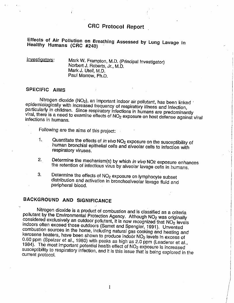
Nitrogen dioxide exposure: effectson airway and blood cells
MARK W. FRAMPTON,1,2 JOSEPH BOSCIA,1 NORBERT J. ROBERTS, JR.,3 MITRA AZADNIV,1
ALFONSO TORRES,1 CHRISTOPHER COX,4 PAUL E. MORROW,2 JOAN NICHOLS,3
DAVID CHALUPA,1 LAUREN M. FRASIER,1 F. RAYMOND GIBB,1
DONNA M. SPEERS,1 YING TSAI,1 AND MARK J. UTELL1,2
Departments of 1Medicine, 2Environmental Medicine, and 4Biostatistics, Universityof Rochester School of Medicine, Rochester, New York 14642-8692; and 3Divisionof Infectious Diseases, University of Texas Medical Branch, Galveston, Texas 77555-0835Received 4 June 2001; accepted in final form 17 September 2001
Frampton, Mark W., Joseph Boscia, Norbert J. Rob-erts, Jr., Mitra Azadniv, Alfonso Torres, ChristopherCox, Paul E. Morrow, Joan Nichols, David Chalupa, Lau-ren M. Frasier, F. Raymond Gibb, Donna M. Speers, YingTsai, and Mark J. Utell. Nitrogen dioxide exposure: effectson airway and blood cells. Am J Physiol Lung Cell MolPhysiol 282: L155–L165, 2002.—This study examined theeffects of nitrogen dioxide (NO2) exposure on airway inflam-mation, blood cells, and antiviral respiratory defense. Twenty-one healthy volunteers were exposed on separate occasions toair and 0.6 and 1.5 ppm NO2 for 3 h with intermittentmoderate exercise. Phlebotomy and bronchoscopy were per-formed 3.5 h after each exposure, and recovered cells werechallenged with respiratory viruses in vitro. Blood studiesrevealed a 4.1% NO2 dose-related decrease in hematocrit(P � 0.003). Circulating total lymphocytes (P � 0.024) and Tlymphocytes (P � 0.049) decreased with NO2 exposure. Ex-posure to NO2 increased the blood lymphocyte CD4�-to-CD8� ratio from 1.74 � 0.11 to 1.85 � 0.12 in males butdecreased it from 1.88 � 0.19 to 1.78 � 0.19 in females (P �0.001 for gender difference). Polymorphonuclear leukocytesin bronchial lavage increased with NO2 exposure (P � 0.003).Bronchial epithelial cells obtained after exposure to 1.5 ppmNO2 released 40% more lactate dehydrogenase after chal-lenge with respiratory syncytial virus than with air exposure(P � 0.024). In healthy subjects, exposures to NO2 at levelsfound indoors cause mild airway inflammation, effects onblood cells, and increased susceptibility of airway epithelialcells to injury from respiratory viruses.
air pollution; influenza virus; respiratory syncytial virus;blood; epithelial cells
NITROGEN DIOXIDE (NO2), a byproduct of oxidation andcombustion, is a primary outdoor and indoor air pol-lutant (14). Because of its oxidative potential and lim-ited solubility, NO2 is a deep lung irritant, and acci-dental exposures to high concentrations can causeacute lung injury and death (13, 49). NO2 interactswith the lung epithelial lining fluid and epithelial cellmembranes, with local production of reactive oxygen
and nitrogen species (47). A National Ambient AirQuality Standard has been established for NO2 as anannual mean of 0.053 ppm (100 �g/m3), and the Stateof California has established a short-term NO2 stan-dard at 0.25 ppm for 1 h (6). Indoor NO2 concentrationsare often greater than those found outdoors, with peaklevels exceeding 2.0 ppm (29) in homes with unventedsources of combustion.
Epidemiological studies have linked NO2 exposurewith increased respiratory illness in children (20, 28,30, 33). A meta-analysis found that a 16-ppb increasein indoor NO2 levels was associated with a 20% in-creased risk of respiratory illness in children (21).Exposure to NO2 inside ice hockey arenas, from oper-ation of natural gas-fueled ice resurfacing machines inthe presence of inadequate ventilation, has been asso-ciated with “epidemics” of acute respiratory illness inexposed players and fans. Concentrations as high as4–5 ppm have been measured in ice arenas (22). Re-cent studies have linked ambient NO2 exposure withincreased mortality (16), increases in cardiac arrhyth-mias in patients with implantable defibrillators (35),and with increased intrauterine mortality (34). How-ever, it is difficult in epidemiology studies to separateeffects of NO2 exposure from other combustion-relatedpollutants.
Human clinical studies have generally found no ef-fects of NO2 exposure on pulmonary function at con-centrations �2.0 ppm. However, exposures to 1.5–2.0ppm for 1–3 h increased nonspecific airway responsive-ness (11, 32), and recent studies suggest that expo-sures as low as 0.26 ppm NO2 for 30 min at rest induceincreased responsiveness to specific allergen challengein patients with asthma (25, 46). Exposures to 2.0 ppmfor 6 h with intermittent exercise caused a very mildairway inflammatory response in healthy subjects,with no changes in lung function (1) or alveolar mac-rophage (AM) phenotype (17). These data suggest thatthere are effects on airway epithelium at concentra-
Address for reprint requests and other correspondence: M. W.Frampton, Univ. of Rochester Medical Center, 601 Elmwood Ave.,Box 692, Rochester, NY 14642-8692 (E-mail: [email protected]).
The costs of publication of this article were defrayed in part by thepayment of page charges. The article must therefore be herebymarked ‘‘advertisement’’ in accordance with 18 U.S.C. Section 1734solely to indicate this fact.
Am J Physiol Lung Cell Mol Physiol282: L155–L165, 2002.
.
1040-0605/02 $5.00 Copyright © 2002 the American Physiological Societyhttp://www.ajplung.org L155
tions below those associated with pulmonary functionchanges or inflammation.
Animal exposure studies have indicated that NO2may increase susceptibility to infection. For example,rodents exposed to NO2 at levels only 5- to 10-foldhigher than peak indoor levels showed impaired re-sponses to infectious challenges, in part through im-pairment of AM function (19, 24, 41). Damji and Rich-ters (9) found alterations in circulating and spleniclymphocyte subsets after exposure to NO2 for 8 h atlevels as low as 4 ppm. These findings suggest thatNO2 exposure may alter both local and systemic hostdefenses. However, clinical studies have been inconclu-sive. Goings et al. (18) exposed healthy volunteers to1–3 ppm NO2 or air for 2 h/day for three consecutivedays. A live, attenuated cold-adapted influenza A vac-cine virus was administered nasally to all subjectsafter exposure on day 2. Volunteers exposed in thethird year of the three-year study became infectedmore frequently in association with NO2, but the effectwas not statistically significant.
The health effects of NO2 exposure may thereforeresult both from the direct oxidant effects of the pol-lutant and from increasing airway susceptibility toother challenges, including respiratory virus infection.We hypothesized that NO2 causes a cascade of events,beginning with injury and inflammation of the distalairway epithelium, recruitment of T lymphocytes fromblood to the airways, and increased susceptibility of theinjured epithelial cells to viral infection.
METHODS AND STUDY DESIGN
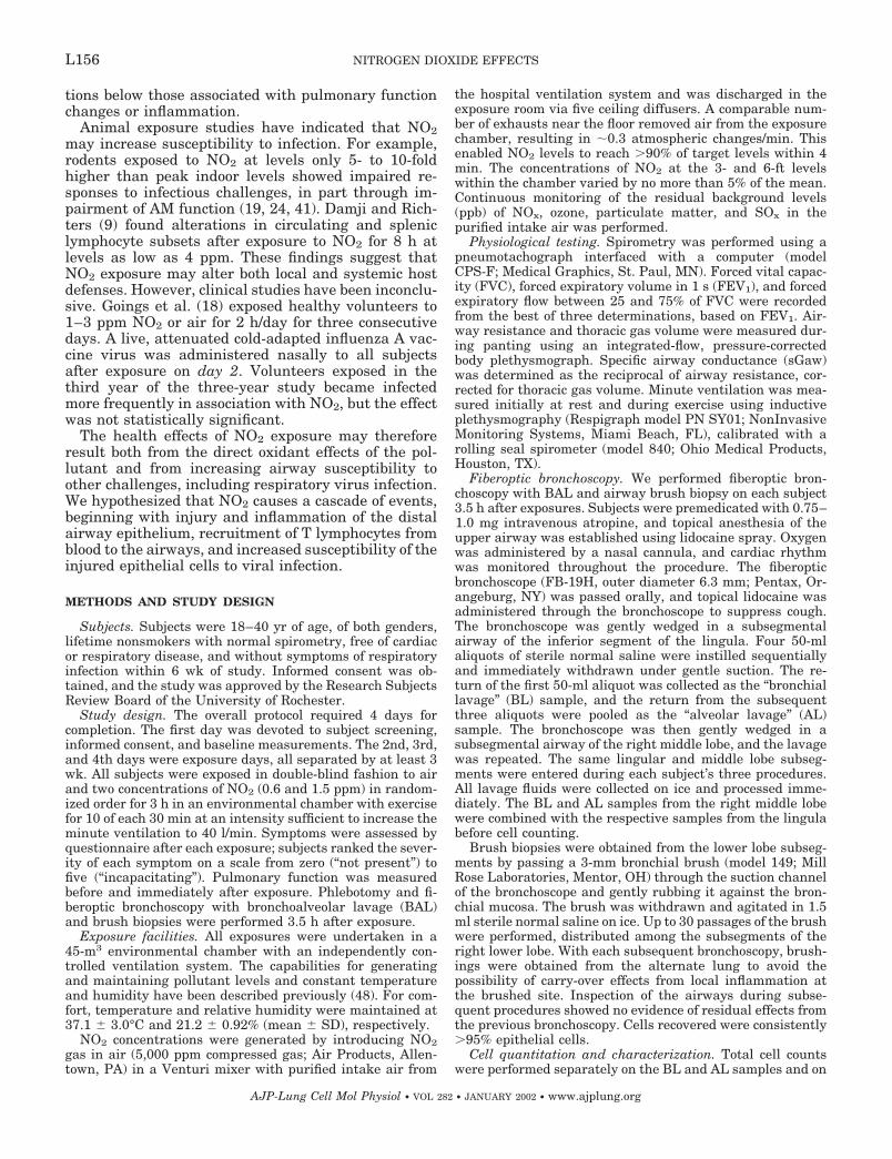
Subjects. Subjects were 18–40 yr of age, of both genders,lifetime nonsmokers with normal spirometry, free of cardiacor respiratory disease, and without symptoms of respiratoryinfection within 6 wk of study. Informed consent was ob-tained, and the study was approved by the Research SubjectsReview Board of the University of Rochester.
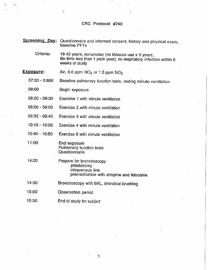
Study design. The overall protocol required 4 days forcompletion. The first day was devoted to subject screening,informed consent, and baseline measurements. The 2nd, 3rd,and 4th days were exposure days, all separated by at least 3wk. All subjects were exposed in double-blind fashion to airand two concentrations of NO2 (0.6 and 1.5 ppm) in random-ized order for 3 h in an environmental chamber with exercisefor 10 of each 30 min at an intensity sufficient to increase theminute ventilation to 40 l/min. Symptoms were assessed byquestionnaire after each exposure; subjects ranked the sever-ity of each symptom on a scale from zero (“not present”) tofive (“incapacitating”). Pulmonary function was measuredbefore and immediately after exposure. Phlebotomy and fi-beroptic bronchoscopy with bronchoalveolar lavage (BAL)and brush biopsies were performed 3.5 h after exposure.
Exposure facilities. All exposures were undertaken in a45-m3 environmental chamber with an independently con-trolled ventilation system. The capabilities for generatingand maintaining pollutant levels and constant temperatureand humidity have been described previously (48). For com-fort, temperature and relative humidity were maintained at37.1 � 3.0°C and 21.2 � 0.92% (mean � SD), respectively.
NO2 concentrations were generated by introducing NO2
gas in air (5,000 ppm compressed gas; Air Products, Allen-town, PA) in a Venturi mixer with purified intake air from
the hospital ventilation system and was discharged in theexposure room via five ceiling diffusers. A comparable num-ber of exhausts near the floor removed air from the exposurechamber, resulting in �0.3 atmospheric changes/min. Thisenabled NO2 levels to reach �90% of target levels within 4min. The concentrations of NO2 at the 3- and 6-ft levelswithin the chamber varied by no more than 5% of the mean.Continuous monitoring of the residual background levels(ppb) of NOx, ozone, particulate matter, and SOx in thepurified intake air was performed.
Physiological testing. Spirometry was performed using apneumotachograph interfaced with a computer (modelCPS-F; Medical Graphics, St. Paul, MN). Forced vital capac-ity (FVC), forced expiratory volume in 1 s (FEV1), and forcedexpiratory flow between 25 and 75% of FVC were recordedfrom the best of three determinations, based on FEV1. Air-way resistance and thoracic gas volume were measured dur-ing panting using an integrated-flow, pressure-correctedbody plethysmograph. Specific airway conductance (sGaw)was determined as the reciprocal of airway resistance, cor-rected for thoracic gas volume. Minute ventilation was mea-sured initially at rest and during exercise using inductiveplethysmography (Respigraph model PN SY01; NonInvasiveMonitoring Systems, Miami Beach, FL), calibrated with arolling seal spirometer (model 840; Ohio Medical Products,Houston, TX).
Fiberoptic bronchoscopy. We performed fiberoptic bron-choscopy with BAL and airway brush biopsy on each subject3.5 h after exposures. Subjects were premedicated with 0.75–1.0 mg intravenous atropine, and topical anesthesia of theupper airway was established using lidocaine spray. Oxygenwas administered by a nasal cannula, and cardiac rhythmwas monitored throughout the procedure. The fiberopticbronchoscope (FB-19H, outer diameter 6.3 mm; Pentax, Or-angeburg, NY) was passed orally, and topical lidocaine wasadministered through the bronchoscope to suppress cough.The bronchoscope was gently wedged in a subsegmentalairway of the inferior segment of the lingula. Four 50-mlaliquots of sterile normal saline were instilled sequentiallyand immediately withdrawn under gentle suction. The re-turn of the first 50-ml aliquot was collected as the “bronchiallavage” (BL) sample, and the return from the subsequentthree aliquots were pooled as the “alveolar lavage” (AL)sample. The bronchoscope was then gently wedged in asubsegmental airway of the right middle lobe, and the lavagewas repeated. The same lingular and middle lobe subseg-ments were entered during each subject’s three procedures.All lavage fluids were collected on ice and processed imme-diately. The BL and AL samples from the right middle lobewere combined with the respective samples from the lingulabefore cell counting.
Brush biopsies were obtained from the lower lobe subseg-ments by passing a 3-mm bronchial brush (model 149; MillRose Laboratories, Mentor, OH) through the suction channelof the bronchoscope and gently rubbing it against the bron-chial mucosa. The brush was withdrawn and agitated in 1.5ml sterile normal saline on ice. Up to 30 passages of the brushwere performed, distributed among the subsegments of theright lower lobe. With each subsequent bronchoscopy, brush-ings were obtained from the alternate lung to avoid thepossibility of carry-over effects from local inflammation atthe brushed site. Inspection of the airways during subse-quent procedures showed no evidence of residual effects fromthe previous bronchoscopy. Cells recovered were consistently�95% epithelial cells.
Cell quantitation and characterization. Total cell countswere performed separately on the BL and AL samples and on
L156 NITROGEN DIOXIDE EFFECTS
AJP-Lung Cell Mol Physiol • VOL 282 • JANUARY 2002 • www.ajplung.org
cells recovered by airway brush biopsy, using a hemocytom-eter. Viability was assessed using trypan blue dye exclusion.Cytospin slides (Shandon, Pittsburgh, PA) were preparedfrom aliquots of BL, AL, and epithelial cells of sufficientvolume to contain 5 � 104 cells. Slides were stained withDiff-Quick (American Scientific Products, McGraw Park, IL)for differential counts; at least 500 cells from each slide werecounted. A separate slide of cells from AL was stained withMayer’s hematoxylin and toluidine blue for enumeration ofmast cells.
Venous blood was analyzed for hematocrit, hemoglobin,red blood cell indexes, and total and differential leukocytecounts in the clinical hematology laboratories of Strong Me-morial Hospital (Rochester, NY). Differential cell counts aspercentages were multiplied by the total white blood cellcount and expressed as concentrations of cells.
Immunofluorescence analysis. Flow cytometry was used asa sensitive method for evaluating cell differential counts, andfor assessing changes in phenotype and expression of activa-tion markers, for both blood and lung lymphocytes. Freshheparinized whole blood or cells from AL fluid that had beenwashed one time in cold PBS were stained with fluoro-chrome-labeled monoclonal antibodies (Becton-Dickinson,Mountain View, CA) with appropriate isotype control anti-bodies. Red blood cells were lysed, and cells were analyzed ona FACScan flow cytometer (Becton-Dickinson) equipped witha 15-mW argon ion laser at 488 nm. Data (forward scatter,linear scale; wide angle light scatter, log scale; and fluores-cence emission at 500–560 nm and 543–627 nm, log scale)were collected in list mode for subsequent analysis. Irrele-vant antibodies of the appropriate subclass showed no non-specific binding. The lymphocyte gate was selected based onlight scattering properties, and lymphocyte subsets weredetermined as a percentage of gated cells. These percentageswere then multiplied by the concentration of lymphocytes toexpress lymphocyte subsets as concentrations of cells.
Infection with influenza and respiratory syncytial virus invitro. Influenza A/AA/Marton/43 H1N1 virus was grown inallantoic cavities of 10-day-old embryonated hens’ eggs (8)and was stored at 70°C until use. The long strain of respi-ratory syncytial virus (RSV) was grown in HEp-2 cell mono-layers cultured in Eagle’s MEM (Biowhittaker, Walkersville,MD) with 2% heat-inactivated FBS in 5% CO2 at 37°C. Thevirus was harvested after cytopathological changes involved90% of the HEp-2 monolayer. The cells were scraped andsonicated to release cell-associated virus. The cell-free super-natant containing the virus was stored at 70°C until use.
Aliquots (2 � 105) of BL, AL, and epithelial cells wereresuspended in serum-free growth medium (LHC-8; Biowhit-taker), plated in 24-well culture dishes, and exposed to influ-enza virus or RSV for 1 h at 37°C at a virus-to-cell ratio of 1:1.Control wells contained virus without cells and cells withoutvirus. After infection, virus was removed by washing twotimes with PBS. Cells were then incubated in LHC-8 mediumat 37°C in a 5% CO2 atmosphere for 4 days; supernatantfluids were removed each 24 h and stored at 70°C untilassay for infectious influenza and RSV. Viability of BL, AL,and epithelial cells, with and without virus infection, wasassessed by measuring lactate dehydrogenase (LDH) in cul-ture supernatants on day 1 and day 4 of culture and bytrypan blue dye exclusion after 4 days in culture. To providedirect comparison with our previous studies of the effects ofNO2 exposure on AM inactivation of influenza virus (16), andbecause we expected to see less infectious RSV released byAM than by epithelial cells, separate samples of AL cells (1 �106) were infected with influenza and RSV at a virus-to-cell
ratio of 10:1. Supernatant fluids were removed each 24 h andstored for subsequent analysis of infectious virus.
Assay of infectious virus. All samples from a given subjectwere analyzed simultaneously without knowledge of the ex-posure. For influenza virus, confluent monolayers of Madin-Darby canine kidney cells were grown in 24-well clusterdishes in MEM containing 10% FBS, 100 U/ml penicillin, 50�g/ml streptomycin, and 2.5 �g/ml amphotericin B. Aliquotsof lavage cell culture fluids to be assayed for virus wereserially diluted 10-fold in Earle’s balanced salt solution,inoculated in quadruplicate (0.2 ml/well), and incubated at37°C for 1 h with agitation every 15 min to ensure an evendistribution of inoculum and to maintain moisture on cellsurfaces. The cell monolayers were then washed with PBSand were overlaid with agarose containing MEM, antibiotics,and trypsin (2.5%; Biowhittaker), without FBS. After 2 daysof further incubation, the cells were fixed with 10% formalin,the agarose layer was removed, and the wells were washedwith water. Methylene blue stain (0.3%) was added for 15min, the cultures were washed and air-dried, and the viralplaques were counted. Results were recorded as the meanvalues of quadruplicate determinations for each assay.
For RSV assay, HEp-2 cells were grown in 24-well tissueculture plates in MEM plus 5% FBS, 100 U/ml penicillin, and100 �g/ml streptomycin in a 5% CO2 atmosphere. When theHEp-2 cell monolayers were between 30 and 50% confluent,quadruplicate wells were inoculated with virus. Wells werefirst washed with warm, serum-free medium and then inoc-ulated with 0.2 ml of diluted cell-free culture supernatantsplus 1.8 ml MEM containing 1% FBS. After 2 h of incubationat 37°C in 5% CO2 to allow the virus to absorb to the cells, 0.8ml of medium was added to each well. Plates were thenincubated at 37°C and examined daily for the cytopathiceffect from day 3 to day 7. Plates showing no cytopathic effectby day 7 were recorded as negative for the presence of virus.The virus titer was calculated by end-point dilution, usingthe method originally described by Reed and Muench (37).
Data handling and statistical methods. The study wasdesigned as a standard, three-period cross-over design withthree different treatments. The statistical analysis was astandard ANOVA (26) that included both period and carry-over effects in addition to an effect of treatments and gender(a between-subjects effect). Each ANOVA included an exam-ination of residuals as a check on the assumptions ofnormally distributed errors with constant variance. If therequired assumptions were not satisfied then data transforma-tions such as the logarithm were considered. A P value �0.05was required for significance. Data means shown in RESULTS
include all study subjects, even though statistical outliers wereexcluded for the ANOVA.
RESULTS
Subject characteristics. Twenty-one subjects werestudied (9 females, 12 males). Table 1 shows their ageand baseline pulmonary function. All subjects com-pleted all exposures and procedures without significantadverse effects.
Exposure data. Actual achieved NO2 concentrationswere 0.61 � 0.02 and 1.50 � 0.02 (SD) ppm. Minuteventilation during rest and exercise, and estimatedtotal NO2 intake, are shown in Table 2. Total NO2
intake did not differ significantly between males andfemales at either exposure level using the Student’st-test.
L157NITROGEN DIOXIDE EFFECTS
AJP-Lung Cell Mol Physiol • VOL 282 • JANUARY 2002 • www.ajplung.org
Pulmonary function. There were no significant ef-fects of NO2 exposure on FVC, FEV1, their ratio, orsGaw in either males or females.
Symptoms. Most subjects did not experience symp-toms during any of the three exposures. Occasionallysubjects reported mild respiratory symptoms duringNO2 exposure; respiratory symptom scores were gen-erally highest after exposure to 1.5 ppm NO2, butdifferences between NO2 and air exposure were notsignificant for any single symptom, or for total symp-tom scores, by Wilcoxon analysis.
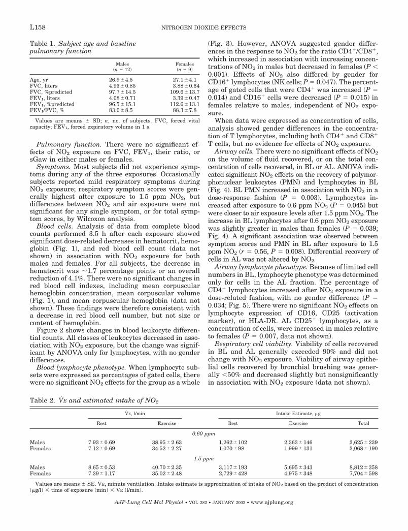
Blood cells. Analysis of data from complete bloodcounts performed 3.5 h after each exposure showedsignificant dose-related decreases in hematocrit, hemo-globin (Fig. 1), and red blood cell count (data notshown) in association with NO2 exposure for bothmales and females. For all subjects, the decrease inhematocrit was �1.7 percentage points or an overallreduction of 4.1%. There were no significant changes inred blood cell indexes, including mean corpuscularhemoglobin concentration, mean corpuscular volume(Fig. 1), and mean corpuscular hemoglobin (data notshown). These findings were therefore consistent witha decrease in red blood cell number, but not size orcontent of hemoglobin.
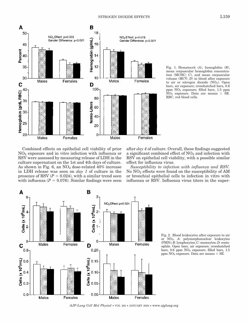
Figure 2 shows changes in blood leukocyte differen-tial counts. All classes of leukocytes decreased in asso-ciation with NO2 exposure, but the change was signif-icant by ANOVA only for lymphocytes, with no genderdifferences.
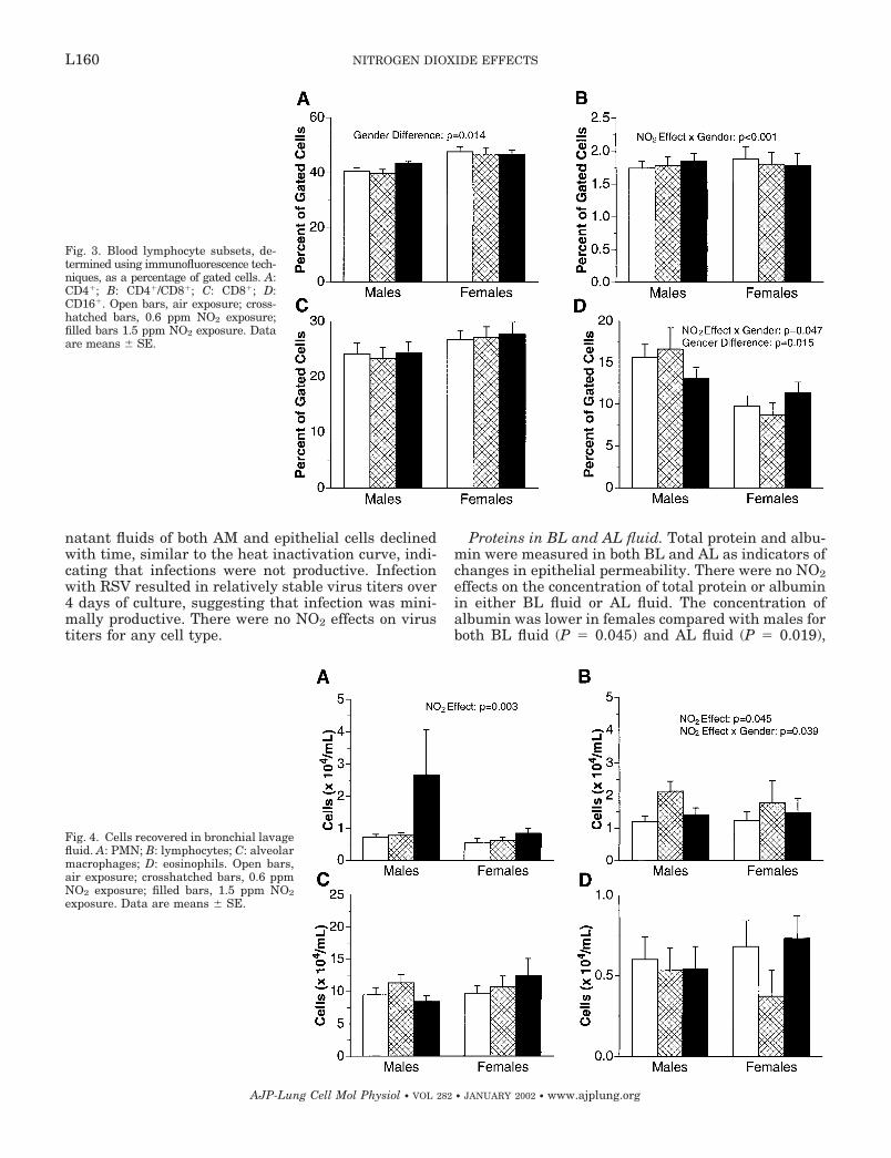
Blood lymphocyte phenotype. When lymphocyte sub-sets were expressed as percentages of gated cells, therewere no significant NO2 effects for the group as a whole
(Fig. 3). However, ANOVA suggested gender differ-ences in the response to NO2 for the ratio CD4�/CD8�,which increased in association with increasing concen-trations of NO2 in males but decreased in females (P �0.001). Effects of NO2 also differed by gender forCD16� lymphocytes (NK cells; P � 0.047). The percent-age of gated cells that were CD4� was increased (P �0.014) and CD16� cells were decreased (P � 0.015) infemales relative to males, independent of NO2 expo-sure.
When data were expressed as concentration of cells,analysis showed gender differences in the concentra-tion of T lymphocytes, including both CD4� and CD8�
T cells, but no evidence for effects of NO2 exposure.Airway cells. There were no significant effects of NO2
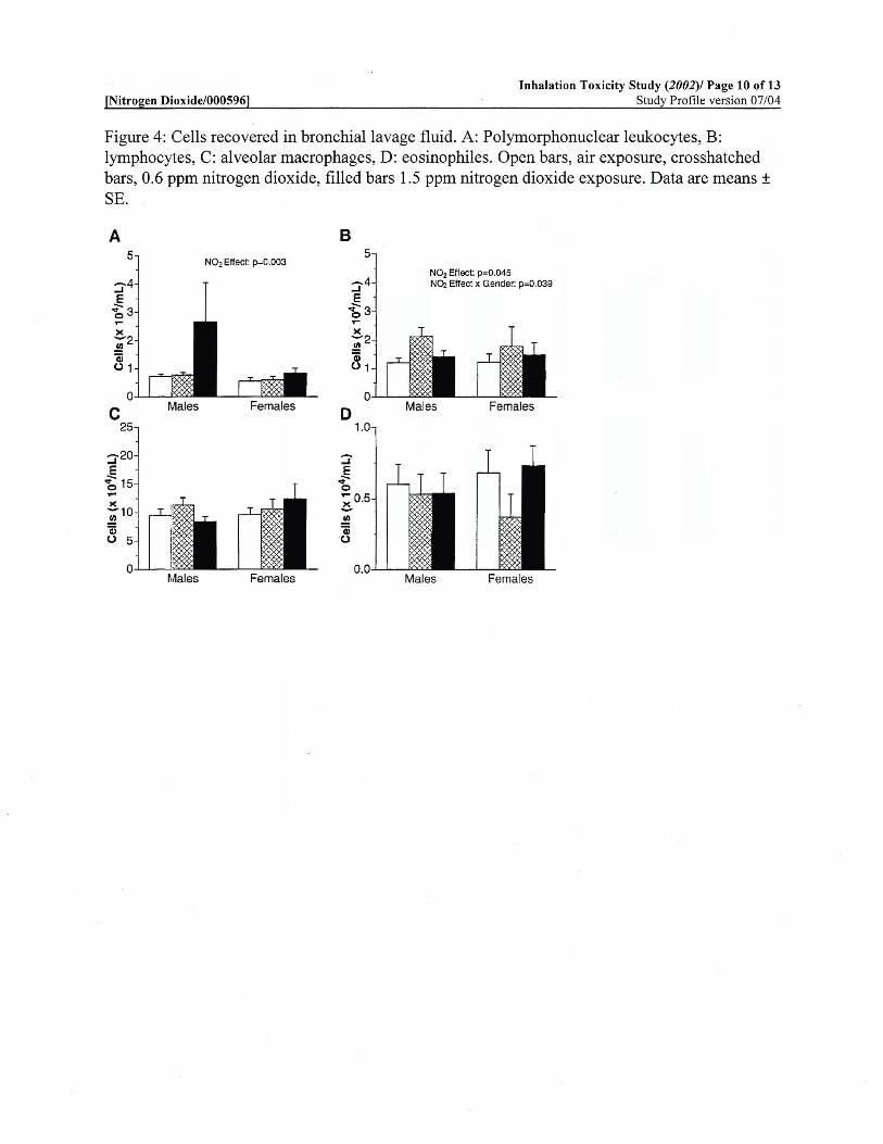
on the volume of fluid recovered, or on the total con-centration of cells recovered, in BL or AL. ANOVA indi-cated significant NO2 effects on the recovery of polymor-phonuclear leukocytes (PMN) and lymphocytes in BL(Fig. 4). BL PMN increased in association with NO2 in adose-response fashion (P � 0.003). Lymphocytes in-creased after exposure to 0.6 ppm NO2 (P � 0.045) butwere closer to air exposure levels after 1.5 ppm NO2. Theincrease in BL lymphocytes after 0.6 ppm NO2 exposurewas slightly greater in males than females (P � 0.039;Fig. 4). A significant association was observed betweensymptom scores and PMN in BL after exposure to 1.5ppm NO2 (r � 0.56, P � 0.008). Differential recovery ofcells in AL was not altered by NO2.
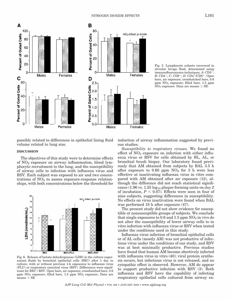
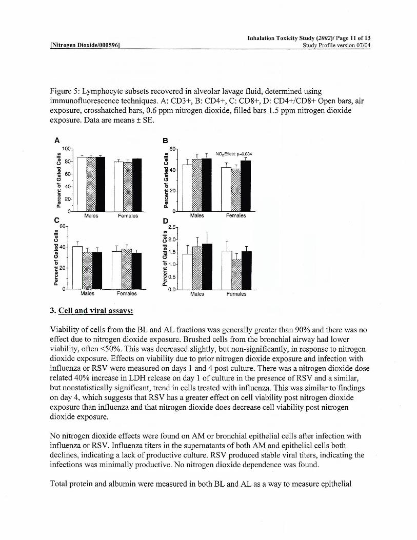
Airway lymphocyte phenotype. Because of limited cellnumbers in BL, lymphocyte phenotype was determinedonly for cells in the AL fraction. The percentage ofCD4� lymphocytes increased after NO2 exposure in adose-related fashion, with no gender difference (P �0.034; Fig. 5). There were no significant NO2 effects onlymphocyte expression of CD16, CD25 (activationmarker), or HLA-DR. AL CD25� lymphocytes, as aconcentration of cells, were increased in males relativeto females (P � 0.007, data not shown).
Respiratory cell viability. Viability of cells recoveredin BL and AL generally exceeded 90% and did notchange with NO2 exposure. Viability of airway epithe-lial cells recovered by bronchial brushing was gener-ally �50% and decreased slightly but nonsignificantlyin association with NO2 exposure (data not shown).
Table 2. VE and estimated intake of NO2
VE, l/min Intake Estimate, �g
Rest Exercise Rest Exercise Total
0.60 ppm
Males 7.93�0.69 38.95�2.63 1,262�102 2,363�146 3,625�239Females 7.12�0.69 34.52�2.27 1,070�98 1,999�131 3,068�190
1.5 ppm
Males 8.65�0.53 40.70�2.35 3,117�193 5,695�343 8,812�358Females 7.39�1.17 35.02�2.48 2,729�428 4,975�348 7,704�598
Values are means � SE. VE, minute ventilation. Intake estimate is approximation of intake of NO2 based on the product of concentration(�g/l) � time of exposure (min) � VE (l/min).
Table 1. Subject age and baselinepulmonary function
Males(n � 12)
Females(n � 9)
Age, yr 26.9�4.5 27.1�4.1FVC, liters 4.93�0.85 3.88�0.64FVC, %predicted 97.7�14.5 109.6�13.7FEV1, liters 4.08�0.71 3.39�0.47FEV1, %predicted 96.5�15.1 112.6�13.1FEV1/FVC, % 83.0�8.5 88.3�7.8
Values are means � SD; n, no. of subjects. FVC, forced vitalcapacity; FEV1, forced expiratory volume in 1 s.
L158 NITROGEN DIOXIDE EFFECTS
AJP-Lung Cell Mol Physiol • VOL 282 • JANUARY 2002 • www.ajplung.org
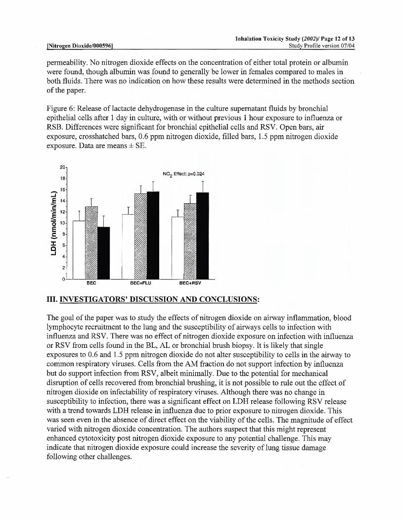
Combined effects on epithelial cell viability of priorNO2 exposure and in vitro infection with influenza orRSV were assessed by measuring release of LDH in theculture supernatant on the 1st and 4th days of culture.As shown in Fig. 6, an NO2 dose-related 40% increasein LDH release was seen on day 1 of culture in thepresence of RSV (P � 0.024), with a similar trend seenwith influenza (P � 0.076). Similar findings were seen
after day 4 of culture. Overall, these findings suggesteda significant combined effect of NO2 and infection withRSV on epithelial cell viability, with a possible similareffect for influenza virus.
Susceptibility to infection with influenza and RSV.No NO2 effects were found on the susceptibility of AMor bronchial epithelial cells to infection in vitro withinfluenza or RSV. Influenza virus titers in the super-
Fig. 1. Hematocrit (A), hemoglobin (B),mean corpuscular hemoglobin concentra-tion (MCHC; C), and mean corpuscularvolume (MCV; D) in blood after exposureto air or nitrogen dioxide (NO2). Openbars, air exposure; crosshatched bars, 0.6ppm NO2 exposure; filled bars, 1.5 ppmNO2 exposure. Data are means � SE.RBC, red blood cells.
Fig. 2. Blood leukocytes after exposure to airor NO2. A: polymorphonuclear leukocytes(PMN); B: lymphocytes; C: monocytes; D: eosin-ophils. Open bars, air exposure; crosshatchedbars, 0.6 ppm NO2 exposure; filled bars, 1.5ppm NO2 exposure. Data are means � SE.
L159NITROGEN DIOXIDE EFFECTS
AJP-Lung Cell Mol Physiol • VOL 282 • JANUARY 2002 • www.ajplung.org
natant fluids of both AM and epithelial cells declinedwith time, similar to the heat inactivation curve, indi-cating that infections were not productive. Infectionwith RSV resulted in relatively stable virus titers over4 days of culture, suggesting that infection was mini-mally productive. There were no NO2 effects on virustiters for any cell type.
Proteins in BL and AL fluid. Total protein and albu-min were measured in both BL and AL as indicators ofchanges in epithelial permeability. There were no NO2effects on the concentration of total protein or albuminin either BL fluid or AL fluid. The concentration ofalbumin was lower in females compared with males forboth BL fluid (P � 0.045) and AL fluid (P � 0.019),
Fig. 3. Blood lymphocyte subsets, de-termined using immunofluorescence tech-niques, as a percentage of gated cells. A:CD4�; B: CD4�/CD8�; C: CD8�; D:CD16�. Open bars, air exposure; cross-hatched bars, 0.6 ppm NO2 exposure;filled bars 1.5 ppm NO2 exposure. Dataare means � SE.
Fig. 4. Cells recovered in bronchial lavagefluid. A: PMN; B: lymphocytes; C: alveolarmacrophages; D: eosinophils. Open bars,air exposure; crosshatched bars, 0.6 ppmNO2 exposure; filled bars, 1.5 ppm NO2
exposure. Data are means � SE.
L160 NITROGEN DIOXIDE EFFECTS
AJP-Lung Cell Mol Physiol • VOL 282 • JANUARY 2002 • www.ajplung.org
possibly related to differences in epithelial lining fluidvolume related to lung size.
DISCUSSION
The objectives of this study were to determine effectsof NO2 exposure on airway inflammation, blood lym-phocyte recruitment to the lung, and the susceptibilityof airway cells to infection with influenza virus andRSV. Each subject was exposed to air and two concen-trations of NO2 to assess exposure-response relation-ships, with both concentrations below the threshold for
induction of airway inflammation suggested by previ-ous studies.
Susceptibility to respiratory viruses. We found noeffect of NO2 exposure on infection with either influ-enza virus or RSV for cells obtained by BL, AL, orbronchial brush biopsy. Our laboratory found previ-ously that AM obtained from subjects by BAL 3.5 hafter exposure to 0.60 ppm NO2 for 3 h were lesseffective at inactivating influenza virus in vitro com-pared with AM obtained after air exposure (12), al-though the difference did not reach statistical signifi-cance (1.96 vs. 1.25 log10 plaque-forming units on day 2of incubation, P � 0.07). Effects were seen in four ofnine subjects, suggesting differences in susceptibility.No effects on virus inactivation were found when BALwas performed 18 h after exposure (47).
The present study did not show evidence for suscep-tible or nonsusceptible groups of subjects. We concludethat single exposures to 0.6 and 1.5 ppm NO2 in vivo donot alter the susceptibility of lower airway cells to invitro infection with influenza virus or RSV when testedunder the conditions used in this study.
Influenza virus infection of bronchial epithelial cellsor of AL cells (mostly AM) was not productive of infec-tious virus under the conditions of our study, and RSVwas at best minimally productive. Previous studieshave found that human AM become abortively infectedwith influenza virus in vitro (40); viral protein synthe-sis occurs, but infectious virus is not released, and nocytopathic effect is observed. However, AM do appearto support productive infection with RSV (3). Bothinfluenza and RSV have the capability of infectingrespiratory epithelial cells cultured from airway ex-
Fig. 5. Lymphocyte subsets recovered inalveolar lavage fluid, determined usingimmunofluorescence techniques. A: CD3�;B: CD4�; C: CD8�; D: CD4�/CD8�. Openbars, air exposure; crosshatched bars, 0.6ppm NO2 exposure; filled bars, 1.5 ppmNO2 exposure. Data are means � SE.
Fig. 6. Release of lactate dehydrogenase (LDH) in the culture super-natant fluids by bronchial epithelial cells (BEC) after 1 day inculture, with or without previous 1-h exposures to influenza virus(FLU) or respiratory syncytial virus (RSV). Differences were signif-icant for BEC�RSV. Open bars, air exposure; crosshatched bars, 0.6ppm NO2 exposure; filled bars, 1.5 ppm NO2 exposure. Data aremeans � SE.
L161NITROGEN DIOXIDE EFFECTS
AJP-Lung Cell Mol Physiol • VOL 282 • JANUARY 2002 • www.ajplung.org
plants (2, 7, 39). It is possible that cells obtained bybronchial brushing did not yield productive infectionsbecause of disruption of membrane viral receptors dur-ing processing or because the cells obtained were su-perficial cells that were terminally differentiated.These findings do not rule out the possibility of NO2effects on viral infectivity in vivo.
Although NO2 exposure did not alter susceptibilityto infection with viruses in this study, there was asignificant effect of prior NO2 exposure on LDH releaseby airway epithelial cells exposed to RSV, with a sim-ilar trend for influenza virus (Fig. 6). This was seen inthe absence of any direct effect of NO2 on the viabilityof epithelial cells. The magnitude of the effect variedwith NO2 concentration and was seen after both 1 and4 days of culture. This finding may not be specific forviral challenge but may represent enhanced cytotoxic-ity from a variety of infectious or noninfectious chal-lenges. Our data suggest that NO2 exposure enhancesthe cytotoxic effects of respiratory viruses or otherchallenges on epithelial cells and could thereby in-crease the severity of epithelial injury after such chal-lenges.
The mechanism responsible for this effect is un-known but may involve injury to the epithelial cellmembrane by reactive oxygen and nitrogen speciesgenerated from NO2. Alternatively, epithelial suscep-tibility may have been enhanced by the small increasein airway PMN observed after exposure to 1.5 ppmNO2.
Leukocyte subsets and activation. NO2 exposure ap-peared to have an effect on lymphocyte recovery in bothblood and BL fluid. In blood, NO2 exposure resulted ina decrease in lymphocytes in both males and females(Fig. 2). In BL fluid, lymphocytes increased after expo-sure to 0.60 ppm NO2 (Fig. 4). There were no signifi-
cant effects on lymphocyte recovery in AL. These find-ings suggest that NO2 exposure may induce therecruitment of lymphocytes from the blood to the con-ducting airways.
There were gender differences in blood lymphocyteresponses to NO2. The ratio of CD4� to CD8� lympho-cytes increased slightly in males in response to NO2but decreased in females. This may be in part becausemales and females differed in the concentration ofblood T cells at baseline in our study, consistent withpublished findings (38). There was also a marginalgender difference in the response to NO2 with regard tothe percentage of CD16� cells (NK cells) in the blood.There were no NO2-related changes in the expressionof the activation markers CD25 or HLA-DR.
Analysis of lymphocyte subsets in AL fluid revealedsignificant increases in the percentage of CD4� T cellsafter NO2 exposure (Fig. 5). There were no NO2 effectson other lymphocyte subsets or activation markers inAL fluid. There were insufficient numbers of cells forlymphocyte subset characterization in BL fluid, wheresignificant effects on total lymphocytes were seen.
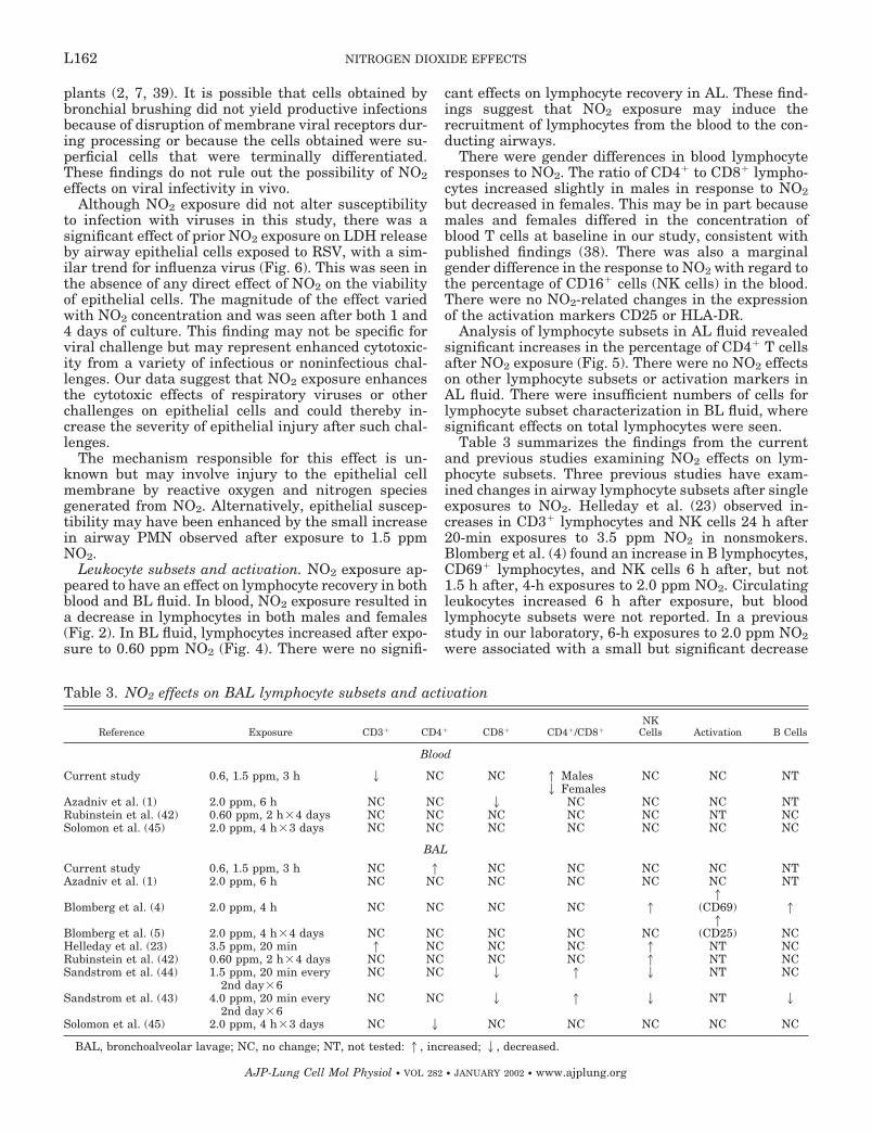
Table 3 summarizes the findings from the currentand previous studies examining NO2 effects on lym-phocyte subsets. Three previous studies have exam-ined changes in airway lymphocyte subsets after singleexposures to NO2. Helleday et al. (23) observed in-creases in CD3� lymphocytes and NK cells 24 h after20-min exposures to 3.5 ppm NO2 in nonsmokers.Blomberg et al. (4) found an increase in B lymphocytes,CD69� lymphocytes, and NK cells 6 h after, but not1.5 h after, 4-h exposures to 2.0 ppm NO2. Circulatingleukocytes increased 6 h after exposure, but bloodlymphocyte subsets were not reported. In a previousstudy in our laboratory, 6-h exposures to 2.0 ppm NO2were associated with a small but significant decrease
Table 3. NO2 effects on BAL lymphocyte subsets and activation
Reference Exposure CD3� CD4� CD8� CD4�/CD8�NK
Cells Activation B Cells
Blood
Current study 0.6, 1.5 ppm, 3 h 2 NC NC 1 Males NC NC NT2 Females
Azadniv et al. (1) 2.0 ppm, 6 h NC NC 2 NC NC NC NTRubinstein et al. (42) 0.60 ppm, 2 h�4 days NC NC NC NC NC NT NCSolomon et al. (45) 2.0 ppm, 4 h�3 days NC NC NC NC NC NC NC
BAL
Current study 0.6, 1.5 ppm, 3 h NC 1 NC NC NC NC NTAzadniv et al. (1) 2.0 ppm, 6 h NC NC NC NC NC NC NT
Blomberg et al. (4) 2.0 ppm, 4 h NC NC NC NC 11
(CD69) 1
Blomberg et al. (5) 2.0 ppm, 4 h�4 days NC NC NC NC NC1
(CD25) NCHelleday et al. (23) 3.5 ppm, 20 min 1 NC NC NC 1 NT NCRubinstein et al. (42) 0.60 ppm, 2 h�4 days NC NC NC NC 1 NT NCSandstrom et al. (44) 1.5 ppm, 20 min every
2nd day�6NC NC 2 1 2 NT NC
Sandstrom et al. (43) 4.0 ppm, 20 min every2nd day�6
NC NC 2 1 2 NT 2
Solomon et al. (45) 2.0 ppm, 4 h�3 days NC 2 NC NC NC NC NC
BAL, bronchoalveolar lavage; NC, no change; NT, not tested: 1, increased; 2, decreased.
L162 NITROGEN DIOXIDE EFFECTS
AJP-Lung Cell Mol Physiol • VOL 282 • JANUARY 2002 • www.ajplung.org
in the percentage of CD8� T lymphocytes in blood, withno changes in BAL fluid, 18 h after exposure (1).
Thus effects of NO2 exposure on lymphocytes in BALfluid are small and not consistent among studies. Thismay reflect differing exposure protocols, subject selec-tion, or differences in sampling times among the stud-ies. It is also possible that NO2 exposure alters lym-phocyte populations predominantly in the conductingairways rather than in the alveoli; lymphocyte subsetsin the bronchial fraction of lavage were not assessed inany of these studies. In the current study, the dataappear consistent with an NO2-induced reduction incirculating T lymphocytes. In addition, the currentfindings suggest the possibility of gender differences inthe lymphocyte responses to NO2, and this possibilityneeds to be considered in the design of future studies.
Airway inflammation. Exposure to NO2 was followedby an exposure-related increase in PMN recovered inBL 3.5 h after exposure in this study. Azadniv et al. (1)observed a small increase in PMN in unfractionatedBAL fluid both immediately and 18 h after exposure to2.0 ppm for 6 h. Blomberg et al. (4) found a 2.5-foldincrease in PMN in bronchial wash 6 h, but not 1.5 h,after 4-h exposures to 2.0 ppm NO2, with an increase ininterleukin-8 levels 1.5 h after exposure. Thus singleexposures to NO2 at concentrations as low as 2.0 ppminduce a mild airway inflammatory response inhealthy subjects, which may persist at least 18 h. Inthe current study, a weak association was observedbetween respiratory symptoms and PMN in BL afterexposure to 1.5 ppm NO2, suggesting that these lowconcentrations of NO2 may be associated with clinicaleffects in some subjects.
Red blood cell effects. Analysis of data from completeblood counts revealed a small but highly significantdecrease in red blood cell number and hemoglobinconcentration in association with NO2 exposure (Fig.1). The mean reduction in hematocrit was �4.1% andwas similar for males and females despite the expecteddifference at baseline, with no change in red blood cellsize or hemoglobin content. There was also an overalltrend toward a decrease in the white blood count (Fig.2), although this change was not statistically signifi-cant.
The decrease in hematocrit and hemoglobin in asso-ciation with NO2 exposure was an unexpected finding.However, this effect has been observed previously inhuman studies of NO2 exposure. Posin et al. (36) ob-served small but significant decreases in hemoglobinand hematocrit immediately after 2.5-h exposures to 1or 2 ppm NO2. In addition, mice exposed to 5 ppm NO2for 1 h demonstrated reductions in hemoglobin anderythrocyte counts along with increases in bilirubinand methemoglobin concentrations, suggesting a mildhemolytic anemia (10). Other studies in animals pro-vide evidence supporting increased red blood cell turn-over after exposure to low concentrations of NO2 (27,31). Considered in light of these previous findings, itappears possible that NO2 exposure, even at the lowconcentrations used in this study, leads to small reduc-tions in circulating red blood cells. The duration of this
effect is unknown. Mechanisms may involve red bloodcell membrane changes, methemoglobin formation, orcellular redistribution within the circulation. We be-lieve hemodilution effects are an unlikely explanationbecause subjects performed a similar intensity of exer-cise on each exposure day.
The magnitude of these changes is small and un-likely to be of clinical significance for most individuals.However, in a 70-kg male, this drop in hematocritwould be equivalent to removal of �200 ml of blood.Therefore, clinical consequences are possible for indi-viduals with cardiovascular compromise or for compet-ing athletes. Future studies of NO2 exposure shouldconsider assessment of red blood cell membranes, re-ticulocyte counts, and methemoglobin levels.
We conclude that, in healthy subjects, single expo-sures to NO2 with exercise, at levels found indoors inhomes with unvented combustion sources, induce thefollowing effects: 1) mild airway inflammation; 2) mildrespiratory symptoms in some subjects; 3) small reduc-tions in hematocrit and hemoglobin; 4) possible smallreductions in circulating T lymphocytes; and 5) possi-ble increased susceptibility of airway epithelial cells toinjury from exposure to respiratory viruses.
In addition, there may be gender differences in theeffects of NO2 on blood and/or airway lymphocytes. Wefound no effects of NO2 on pulmonary function or onthe susceptibility of airway epithelial cells or AM toproductive infection by influenza virus or RSV in vitro.
With the relatively large number of statistical testsin this study, some significant P values could haveoccurred by chance. Our a priori approach was toevaluate findings based on the level of significance,consistency with the primary hypotheses and otherstudy findings, and biological plausibility. The reduc-tions in hematocrit and hemoglobin were the only find-ings not consistent with the primary hypotheses; herethe highly significant P value (P � 0.003) from theANOVA, and the similar effect in males and femalesdespite baseline differences, makes the finding un-likely to be a chance occurrence.
All NO2 effects observed in this study were small andunlikely to be of clinical significance for healthy sub-jects. However, young children, the elderly, and indi-viduals with underlying respiratory or cardiovasculardisease may be more susceptible to such effects. In-deed, time-series epidemiology studies indicate thatthese groups are at risk for adverse health effects fromeven modest increases in ambient air pollution (15). Itis possible that a combination of airway and bloodeffects of exposure to NO2 could exacerbate underlyingairway disease, particularly after infections with respi-ratory viruses or other respiratory challenges.
This work was supported by Contract 93–07 from the Center forIndoor Air Research and National Institutes of Health Grants RR-00044 and ES-01247.
REFERENCES
1. Azadniv M, Utell MJ, Morrow PE, Gibb FR, Nichols J,Roberts NJ Jr, Speers DM, Torres A, Tsai Y, Abraham MK,Voter KZ, and Frampton MW. Effects of nitrogen dioxide
L163NITROGEN DIOXIDE EFFECTS
AJP-Lung Cell Mol Physiol • VOL 282 • JANUARY 2002 • www.ajplung.org
exposure on human host defense. Inhal Toxicol 10: 585–602,1998.
2. Becker S, Reed W, Henderson FW, and Noah TL. RSVinfection of human airway epithelial cells causes production ofthe -chemokine RANTES. Am J Physiol Lung Cell Mol Physiol272: L512–L520, 1997.
3. Becker S, Soukup J, and Yankaskas JR. Respiratory syncy-tial virus infection of human primary nasal and bronchial epi-thelial cell cultures and bronchoalveolar macrophages. Am JRespir Cell Mol Biol 6: 369–374, 1992.
4. Blomberg A, Krishna MT, Bocchino V, Biscione GL, ShuteJK, Kelly FJ, Frew AJ, Holgate ST, and Sandstrom T. Theinflammatory effects of 2 ppm NO2 on the airways of healthysubjects. Am J Respir Crit Care Med 156: 418–424, 1997.
5. Blomberg A, Krishna MT, Helleday R, Soderberg M, LedinMC, Kelly FJ, Frew AJ, Holgate ST, and Sandstrom T.Persistent airway inflammation but accommodated antioxidantand lung function responses after repeated daily exposure tonitrogen dioxide. Am J Respir Crit Care Med 159: 536–543, 1999.
6. California Code of Regulations. Title 17. Table of Standards.Sacramento, CA: 1997, p. 70200.
7. Choi AMK and Jacoby DB. Influenza virus A infection inducesinterleukin-8 gene expression in human airway epithelial cells.FEBS 309: 327–329, 1992.
8. Chonmaitree T, Roberts NJ Jr, Douglas RG Jr, Hall CB,and Simons RL. Interferon production by human mononuclearleukocytes: differences between respiratory syncytial virus andinfluenza viruses. Infect Immun 32: 300–303, 1981.
9. Damji KS and Richters A. Reduction in T lymphocyte sub-populations following acute exposure to 4 ppm nitrogen dioxide.Environ Res 49: 217–224, 1989.
10. Ehrman RA, Treshow M, and Lytle IM. The hematology ofmice exposed to nitrogen dioxide. Am Ind Hyg Assoc J 33:751–755, 1972.
11. Frampton MW, Morrow PE, Gibb FR, Speers DM, andUtell MJ. Effects of nitrogen dioxide exposure on pulmonaryfunction and airway reactivity in normal humans. Am Rev Re-spir Dis 143: 522–527, 1991.
12. Frampton MW, Smeglin AM, Roberts NJ Jr, FinkelsteinJN, Morrow PE, and Utell MJ. Nitrogen dioxide exposure invivo and human alveolar macrophage inactivation of influenzavirus in vitro. Environ Res 48: 179–192, 1989.
13. Frampton MW and Utell MJ. Inhalation injuries due to acci-dental and environmental exposures. Curr Opin Crit Care 1:246–252, 1995.
14. Frampton MW and Utell MJ. Air pollution and human hostdefense: the role of nitrogen dioxide. In: Disease and Exposure toAir Pollution, edited by Mohr U. Washington, DC: ILSI, 1998, p.91–98.
15. Frampton MW, Utell MJ, and Samet JM. Cardiopulmonaryconsequences of particle inhalation. In: Particle-Lung Interac-tions, edited by Gehr P and Heyder J. New York: Dekker, 2000,p. 653–670.
16. Garcia-Aymerich J, Tobias A, Anto JM, and Sunyer J. Airpollution and mortality in a cohort of patients with chronicobstructive pulmonary disease: a time series analysis. J Epide-miol Community Health 54: 73–74, 2000.
17. Gavras JB, Frampton MW, Ryan DH, Levy PC, Looney RJ,Cox C, Morrow PE, and Utell MJ. Expression of membraneantigens on human alveolar macrophages after exposure to ni-trogen dioxide. Inhal Toxicol 6: 633–646, 1994.
18. Goings SAJ, Kulle TJ, Bascom R, Sauder LR, Green DJ,Hebel JR, and Clements ML. Effect of nitrogen dioxide expo-sure on susceptibility to influenza A virus infection in healthyadults. Am Rev Respir Dis 139: 1075–1081, 1989.
19. Goldstein E, Eagle MC, and Hoeprich PD. Effect of nitrogendioxide on pulmonary bacterial defense mechanisms. Arch En-viron Health 26: 202–204, 1973.
20. Hajat S, Haines A, Goubet SA, Atkinson RW, and Ander-son HR. Association of air pollution with daily GP consultationsfor asthma and other lower respiratory conditions in London.Thorax 54: 597–605, 1999.
21. Hasselblad V, Kotchmar DJ, and Eddy DM. Synthesis ofenvironmental evidence: nitrogen dioxide epidemiology studies.J Air Waste Management 42: 662–671, 1992.
22. Hedberg K, Hedberg CW, Iber C, White KE, Osterholm MT,Jones DBW, Flink JR, and MacDonald KL. An outbreak ofnitrogen dioxide-induced respiratory illness among ice hockeyplayers. JAMA 262: 3014–3017, 1989.
23. Helleday R, Sandstrom T, and Stjernberg N. Differences inbronchoalveolar cell response to nitrogen dioxide exposure be-tween smokers and nonsmokers. Eur Respir J 7: 1213–1220,1994.
24. Jakab GJ. Modulation of pulmonary defense mechanisms byacute exposures to nitrogen dioxide. Environ Res 42: 215–228,1987.
25. Jenkins HS, Devalia JL, Mister RL, Bevan AM, Rusznak C,and Davies RJ. The effect of exposure to ozone and nitrogendioxide on the airway response of atopic asthmatics to inhaledallergen. Am J Respir Crit Care Med 160: 33–39, 1999.
26. Jones B and Kenward MG. Design and Analysis of Cross-overTrials. New York, NY: Chapman and Hall, 1989.
27. Kaya K, Miura T, and Kubota K. Effects of nitrogen dioxide onred blood cells of rats: changes in components of red cell mem-branes during in vivo exposure to NO2. Environ Res 23: 397–409,1980.
28. Kramer U, Koch T, Ranft U, Ring J, and Behrendt H.Traffic-related air pollution is associated with atopy in childrenliving in urban areas. Epidemiology 11: 64–70, 2000.
29. Leaderer BP, Stolwijk JAJ, Zagraniski RT, and Quing-Shan M. A field study of indoor air contaminant levels associ-ated with unvented combustion sources. 77th Ann Meet AirPollution Control Assoc San Francisco, CA 1984, vol. 84, p. 33.3.
30. McConnell R, Berhane K, Gilliland F, London SJ, Vora H,Avol E, Gauderman WJ, Margolis HG, Lurmann F,Thomas DC, and Peters JM. Air pollution and bronchiticsymptoms in Southern California children with asthma. EnvironHealth Perspect 107: 757–760, 1999.
31. Mochitate K and Miura T. In vivo effect of nitrogen dioxide onthe activities of glycolytic enzymes in red blood cells of rats.Toxicol Lett 22: 315–321, 1984.
32. Mohsenin V. Airway responses to 2.0 ppm nitrogen dioxide innormal subjects. Arch Environ Health 43: 242–246, 1988.
33. Neas LM, Dockery DW, Ware JH, Spengler JD, Speizer FE,and Ferris BG Jr. Association of indoor nitrogen dioxide withrespiratory symptoms and pulmonary function in children. Am JEpidemiol 134: 204–219, 1991.
34. Pereira LAA, Loomis D, Conceicao GMS, Braga ALF, Ar-cas RM, Kishi HS, Singer JM, Bohm GM, and Saldiva PHN.Association between air pollution and intrauterine mortality inSao Paulo, Brazil. Environ Health Perspect 106: 325–329, 1998.
35. Peters A, Liu E, Verrier RL, Schwartz J, Gold DR, Mittle-man M, Baliff J, Oh JA, Allen G, Monahan K, and DockeryDW. Air pollution and incidence of cardiac arrhythmia. Epide-miology 11: 11–17, 2000.
36. Posin C, Clark K, Jones MP, Patterson JV, Buckley RD,and Hackney JD. Nitrogen dioxide inhalation and humanblood biochemistry. Arch Environ Health 33: 318–324, 1978.
37. Reed LJ and Muench H. A simple method of estimating fiftypercent endpoints (Abstract). Am J Hygiene 27: 493, 1938.
38. Reichert T, DeBruyere M, Deneys V, Totterman T, Ly-dyard P, Yuksel F, Chapel H, Jewell D, Van Hove L, andLinden J. Lymphocyte subset reference ranges in adult cauca-sians. Clin Immunol Immunopathol 60: 190–208, 1991.
39. Reiss TF, Gruenert DC, Nadel JA, and Jacoby DB. Infectionof cultured human airway epithelial cells by influenza A virus.Life Sci 49: 1173–1181, 1991.
40. Rodgers BC and Mims CA. Influenza virus replication inhuman alveolar macrophages. J Med Virol 9: 177–184, 1982.
41. Rose RM, Fuglestad JM, Skornik WA, Hammer SM,Wolfthal SF, Beck BD, and Brain JD. The pathophysiology ofenhanced susceptibility to murine cytomegalovirus respiratoryinfection during short-term exposure to 5 ppm nitrogen dioxide.Am Rev Respir Dis 137: 912–917, 1988.
42. Rubinstein I, Reiss TF, Bigby BG, Stites DP, and BousheyJr HA. Effects of 0.60 PPM nitrogen dioxide on circulating and
L164 NITROGEN DIOXIDE EFFECTS
AJP-Lung Cell Mol Physiol • VOL 282 • JANUARY 2002 • www.ajplung.org
bronchoalveolar lavage lymphocyte phenotypes in healthy sub-jects. Environ Res 55: 18–30, 1991.
43. Sandstrom T, Helleday R, Bjermer L, and Stjernberg N.Effects of repeated exposure to 4 ppm nitrogen dioxide on bron-choalveolar lymphocyte subsets and macrophages in healthymen. Eur Respir J 5: 1092–1096, 1992.
44. Sandstrom T, Ledin MC, Thomasson L, Helleday R, andStjernberg N. Reductions in lymphocyte subpopulations afterrepeated exposure to 1.5 ppm nitrogen dioxide. Br J Ind Med 49:850–854, 1992.
45. Solomon C, Christian DL, Chen LL, Welch BS, KleinmanMT, Dunham E, Erle DJ, and Balmes JR. Effect of serial-day exposure to nitrogen dioxide on airway and blood leuko-cytes and lymphocyte subsets. Eur Respir J 15: 922–928,2000.
46. Strand V, Svartengren M, Rak S, Barck C, and Bylin G.Repeated exposure to an ambient level of NO2 enhances asth-matic response to a nonsymptomatic allergen dose. Eur Respir J12: 6–12, 1998.
47. Utell MJ and Frampton MW. Oxides of nitrogen. In: Toxicol-ogy of the Respiratory System, edited by Sipes IG, McQueen CA,and Gandolfi AJ. Oxford, UK: Elsevier, 1997, p. 303–312.
48. Utell MJ, Frampton MW, Roberts NJ Jr, Finkelstein JN,Cox C, and Morrow PE. Mechanisms of nitrogen dioxide tox-icity in humans. Health Effects Institute Research Report 43:1–44, 1991.
49. Utell MJ, Morrow PE, Hyde RW, and Schreck RM. Exposurechamber for studies of pollutant gases and aerosols in humansubjects: design considerations. J Aerosol Sci 15: 219–221, 1984.
50. Zwemer FL, Pratt DS, and May JJ. Silo filler’s disease in NewYork State. Am Rev Respir Dis 146: 650–653, 1992.
L165NITROGEN DIOXIDE EFFECTS
AJP-Lung Cell Mol Physiol • VOL 282 • JANUARY 2002 • www.ajplung.org
UNITED STATES ENVIRONMENTAL PROTECTION AGENCY WASHINGTON, D.C. 20460
OFFICE OF CHEMICAL SAFETY AND POLLUTION PREVENTION
October 7th, 2014
MEMORANDUM
Subject:
From:
Thru:
To:
Nitrogen Dioxide: Evaluation of inhalation toxicity study, Frampton et al 2002
PC Code: 000596 DP Barcode: 421314 Decision No.: N/A Registration No.: N/A Petition No.: N/A Regulatory Action: Data Evaluation Record
(DER), Toxicology Review for Product Registration, Section 3
Risk Assess Type: Single chemical, non Case No.: 4045 aggregate TXR No.: 1003341 CAS No.: 10102-44-0 MRlD No.: 49420001 40 CFR: N/A
Jonathan Leshin, PhD, Toxicologist --::7~-------Risk Assessment and Science Support Branch (~ ~ Antimicrobials Division (751 OP)
Tim McMahon, PhD, Senior Sci Steven H. Weiss, Branch Chi Risk Assessment and Science pport Branch (RA Antimicrobials Division (751 OP)
Jaqueline Hardy, Product Manager Regulatory Management Branch II (RMB2) Antimicrobials Division (751 OP)
-
Agency Conclusion: The Agency has reviewed the following journal article reporting about a study conducted with nitrogen dioxide. The study was not conducted according to the 870.3465 guidelines, but is acceptable for regulatory purposes.
Frampton, M.W., Boscia, J., Roberts, J.R., et al (2002) Nitrogen dioxide exposure: effects on airway and blood cells Department of Medicine, Environmental medicine and Biostatistics (University of Rochester, School ofMedicine) MRID 49420001 American Journal of Physiology -Lung, Cellular and Molecular Physiology, 282: L155-165
EXECUTIVE SUMMARY: In an inhalation toxicity study (MRID 49420001) nitrogen dioxide was administered to 21 people aged 18-40 years (9 females, 12 males) in an environmental chamber at concentrations of 0, (room air), 0.6 ppm or 1.5 ppm nitrogen dioxide for a total of three hours. Exercise was performed for 1 0 minutes out of each thirty minutes at a sufficient level to raise minute ventilation to 40 1/min. Each exposure period was separated by at least three weeks from the previous exposure period. The subjects were assessed for pulmonary function before and after each exposure. Phlebotomy and bronchoscopy with bronchoalveolar lavage and
brush biopsies were performed 3.5 hours post exposure. Recovered cells were challenged with respiratory viruses in vitro.
There was no effect of nitrogen dioxide exposure on infection with influenza or respiratory syncytial virus (RSV) from cells found in the bronchial lavage (BL), alveolar lavage (AL) or bronchial brush biopsy. It is likely that single exposures to 0.6 and 1.5 ppm nitrogen dioxide do not alter susceptibility to cells in the airway to common respiratory viruses. Cells from the alveolar macrophage (AM) fraction do not support infection by influenza but do support infection from RSV, albeit minimally.
Although there was no change in susceptibility to infection, there was a significant effect on lactate dehydrogenase (LDH) release following RSV release with a trend towards LDH release in influenza due to prior exposure to nitrogen dioxide. This was seen even in the absence of a direct effect on the viability of the cells. The magnitude of effect varied with nitrogen dioxide concentration.
Nitrogen dioxide exposure had an effect on lymphocyte recovery from both blood and BL fluid. In blood, nitrogen dioxide exposure resulted in a decrease of lymphocytes, while in BL fluid, there was an increase in lymphocytes following exposure to .6 ppm nitrogen dioxide. There was no effect on lymphocytes in AL.
There are differences between genders of the blood lymphocyte response to nitrogen dioxide. The ratio of CD4+ to CD8+ increased slightly in males in response to nitrogen dioxide but decreased in females. This may be due to differences in initial blood t cell concentration, which is consistent with previously published work. There was a marginal gender difference in response to nitrogen dioxide in terms of percentage ofCD16+ cells in blood. CD25 and HLADR exhibited no nitrogen dioxide dependent changes. Lymphocytes in AL fluid revealed increases in the percentage of CD4+ cells post nitrogen dioxide exposure. No other changes in lymphocyte subsets were seen in AL fluid. BL fluid could not be measured for lymphocyte subsets due to insufficient fluid volume. There was an increase in polymorphonuclear leukocytes after exposure to nitrogen dioxide in the BL. This indicates a mild inflammatory response to nitrogen dioxide. Overall, there appear to be some gender differences in lymphocyte subsets expression due to nitrogen dioxide exposure and a decrease in lymphocytes in the blood due to nitrogen dioxide exposure.
There was a small but statistically significant decrease in red blood cell number and hemoglobin concentration in association with nitrogen dioxide. This change was similar for both males and females and occurred despite no change in hemoglobin content or red blood cell size. It is unclear the reason for this. This change would unlikely be clinically significant in a healthy individual but indicates there may be some effect in persons with cardiovascular compromise, children or the elderly.
This study is listed as quantitative and acceptable/nonguideline. The LOAEL is 600 ppb. There is no NOAEL for this study.
2
ATTACHMENT
Data Evaluation Record (DER)
Inhalation Toxicity
For
Nitrogen Dioxide
PC Code: 000596
3
[Nitrogen Dioxide/000596)
EPA Reviewer: Jonathan Leshin, Ph.D. RASSB, Antimicrobials Division (7510P) Secondary Review: Tim McMahon, Toxicologist RASSB, Antimicrobials Division (7510P)
Inhalation Toxicity Study (2002)1 Page 1 of 13 Study Profile version 07/04
II Data Evaluation Record II STUDY TYPE: Inhalation Toxicity- [human]
PC CODE: 000596 DP BARCODE: 421314
TXR#: 1003341
TEST MATERIAL (PURITY): Nitrogen Dioxide
SYNONYMS: None
CITATION: Frampton, M.W., Boscia, J., Roberts, J.R., et al (2002) Nitrogen dioxide exposure: effects on airway and blood cells Department of Medicine, Environmental medicine and Biostatistics (University of Rochester, School of Medicine) MRID 49420001 American Journal of Physiology- Lung, Cellular and Molecular Physiology, 282: L155-165
SPONSOR: Center for Indoor Air Research and National Institutes of Health
INVESTIGATORS' EXECUTIVE SUMMARY:
In an inhalation toxicity study (MRID 49420001) nitrogen dioxide was administered to 21 people aged 18-40 years (9 females, 12 males) in an environmental chamber at concentrations ofO, (room air), 0.6 ppm or 1.5 ppm nitrogen dioxide for a total ofthree hours. Exercise was performed for 10 minutes out of each thirty minutes at a sufficient level to raise minute ventilation to 40 1/min. Each exposure period was separated by at least three weeks from the previous exposure period. The subjects were assessed for pulmonary function before and after each exposure. Phlebotomy and bronchoscopy with bronchoalveolar lavage and brush biopsies were performed 3.5 hours post exposure. Recovered cells were challenged with respiratory viruses in vitro.
There was no effect of nitrogen dioxide exposure on infection with influenza or respiratory syncytial virus (RSV) from cells found in the bronchial lavage (BL), alveolar lavage (AL) or bronchial brush biopsy. It is likely that single exposures to 0.6 and 1.5 ppm nitrogen dioxide do not alter susceptibility to cells in the airway to common respiratory viruses. Cells from the alveolar macrophage (AM) fraction do not support infection by influenza but do support
[Nitrogen Dioxide/000596]
infection from RSV, albeit minimally.
Inhalation Toxicity Study (2002)1 Page 2 of 13 Study Profile version 07/04
Although there was no change in susceptibility to infection, there was a significant effect on lactate dehydrogenase (LDH) release following RSV release with a trend towards LDH release in influenza due to prior exposure to nitrogen dioxide. This was seen even in the absence of a direct effect on the viability of the cells. The magnitude of effect varied with nitrogen dioxide concentration.
Nitrogen dioxide exposure had an effect on lymphocyte recovery from both blood and BL fluid. In blood, nitrogen dioxide exposure resulted in a decrease of lymphocytes, while in BL fluid, there was an increase in lymphocytes following exposure to .6 ppm nitrogen dioxide. There was no effect on lymphocytes in AL.
There are differences between genders of the blood lymphocyte response to nitrogen dioxide. The ratio of CD4+ to CD8+ increased slightly in males in response to nitrogen dioxide but decreased in females. This may be due to differences in initial blood t cell concentration, which is consistent with previously published work. There was a marginal gender difference in response to nitrogen dioxide in terms of percentage ofCD16+ cells in blood. CD25 and HLA-DR exhibited no nitrogen dioxide dependent changes. Lymphocytes in AL fluid revealed increases in the percentage of CD4+ cells post nitrogen dioxide exposure. No other changes in lymphocyte subsets were seen in AL fluid. BL fluid could not be measured for lymphocyte subsets due to insufficient fluid volume. There was an increase in polymorphonuclear leukocytes after exposure to nitrogen dioxide in the BL. This indicates a mild inflammatory response to nitrogen dioxide. Overall, there appear to be some gender differences in lymphocyte subsets expression due to nitrogen dioxide exposure and a decrease in lymphocytes in the blood due to nitrogen dioxide exposure.
There was a small but statistically significant decrease in red blood cell number and hemoglobin concentration in association with nitrogen dioxide. This change was similar for ,both males and females and occurred despite no change in hemoglobin content or red blood cell size. It is unclear the reason for this. This change would unlikely be clinically significant in a healthy individual but indicates there may be some effect in persons with cardiovascular compromise, children or the elderly.
This study is listed as quantitative and acceptable/nonguideline. The LOAEL is 600 ppb. There is no NOAEL for this study.
I. MATERIALS AND METHODS
A. MATIB::RIALS:
1. Test Material: Description: Lot/Batch #:
Purity:
Nitrogen dioxide Gas
Unknown Unknown
[Nitrogen Dioxide/000596)
Compound Stability: CAS # of TGAI:
Unknown 10102-44-0
2. Vehicle and/or positive control: Room air
3. Baseline characteristics of study:
Inhalation Toxicity Study (2002)1 Page 3 of 13 Study Profile version 07/04
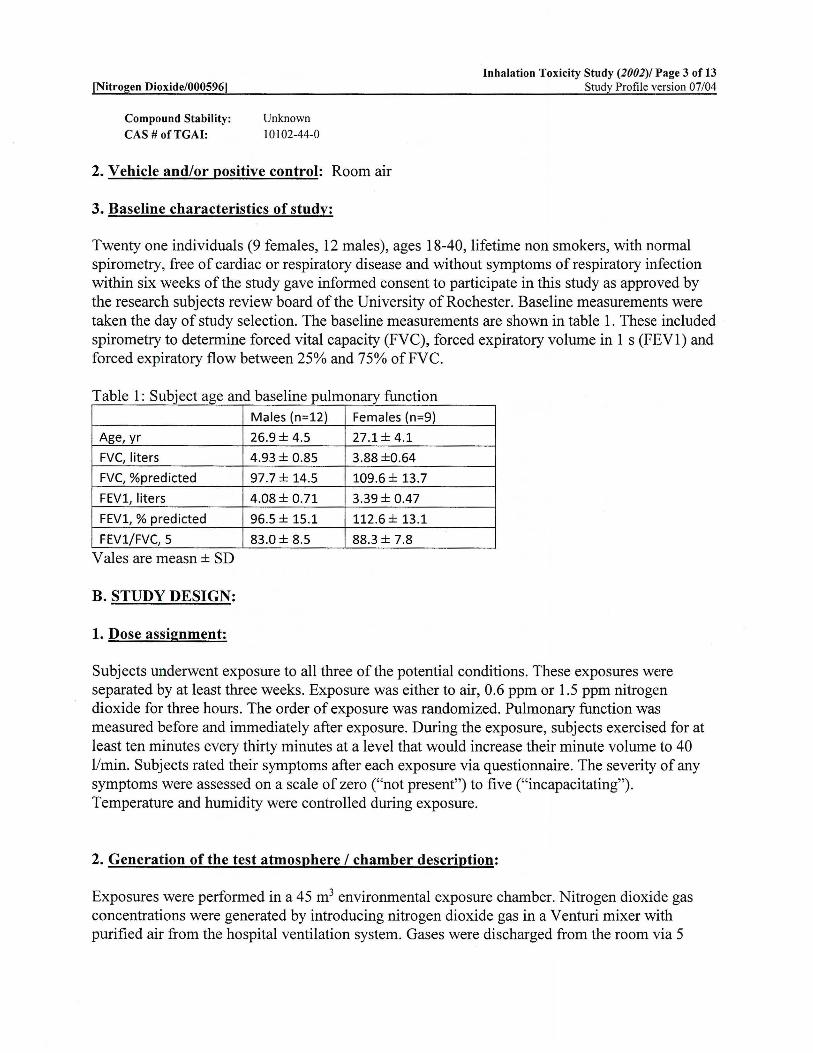
Twenty one individuals (9 females, 12 males), ages 18-40, lifetime non smokers, with normal spirometry, free of cardiac or respiratory disease and without symptoms of respiratory infection within six weeks of the study gave informed consent to participate in this study as approved by the research subjects review board of the University of Rochester. Baseline measurements were taken the day of study selection. The baseline measurements are shown in table 1. These included spirometry to determine forced vital capacity (FVC), forced expiratory volume in 1 s (FEV1) and forced expiratory flow between 25% and 75% ofFVC.
T bl 1 S b' a e u >Ject age an db r ase me pu monary fu nct10n Males (n=12) Females (n=9)
Age, yr 26.9 ± 4.5 27.1 ± 4.1
FVC, liters 4.93 ± 0.85 3.88 ±0.64
FVC, %predicted 97.7 ± 14.5 109.6 ± 13.7
FEV1, liters 4.08± 0.71 3.39 ± 0.47
FEV1, % predicted 96.5 ± 15.1 112.6 ± 13.1
FEV1/FVC, 5 83.0± 8.5 88.3 ± 7.8
Vales are measn ± SD
B. STUDY DESIGN:
1. Dose assignment:
Subjects underwent exposure to all three of the potential conditions. These exposures were separated by at least three weeks. Exposure was either to air, 0.6 ppm or 1.5 ppm nitrogen dioxide for three hours. The order of exposure was randomized. Pulmonary function was measured before and immediately after exposure. During the exposure, subjects exercised for at least ten minutes every thirty minutes at a level that would increase their minute volume to 40 1/min. Subjects rated their symptoms after each exposure via questionnaire. The severity of any symptoms were assessed on a scale of zero ("not present") to five ("incapacitating"). Temperature and humidity were controlled during exposure.
2. Generation of the test atmosphere I chamber description:
Exposures were performed in a 45m3 environmental exposure chamber. Nitrogen dioxide gas concentrations were generated by introducing nitrogen dioxide gas in a Venturi mixer with purified air from the hospital ventilation system. Gases were discharged from the room via 5
[Nitrogen Dioxide/000596) Inhalation Toxicity Study (2002)1 Page 4 of 13
Study Profile version 07/04
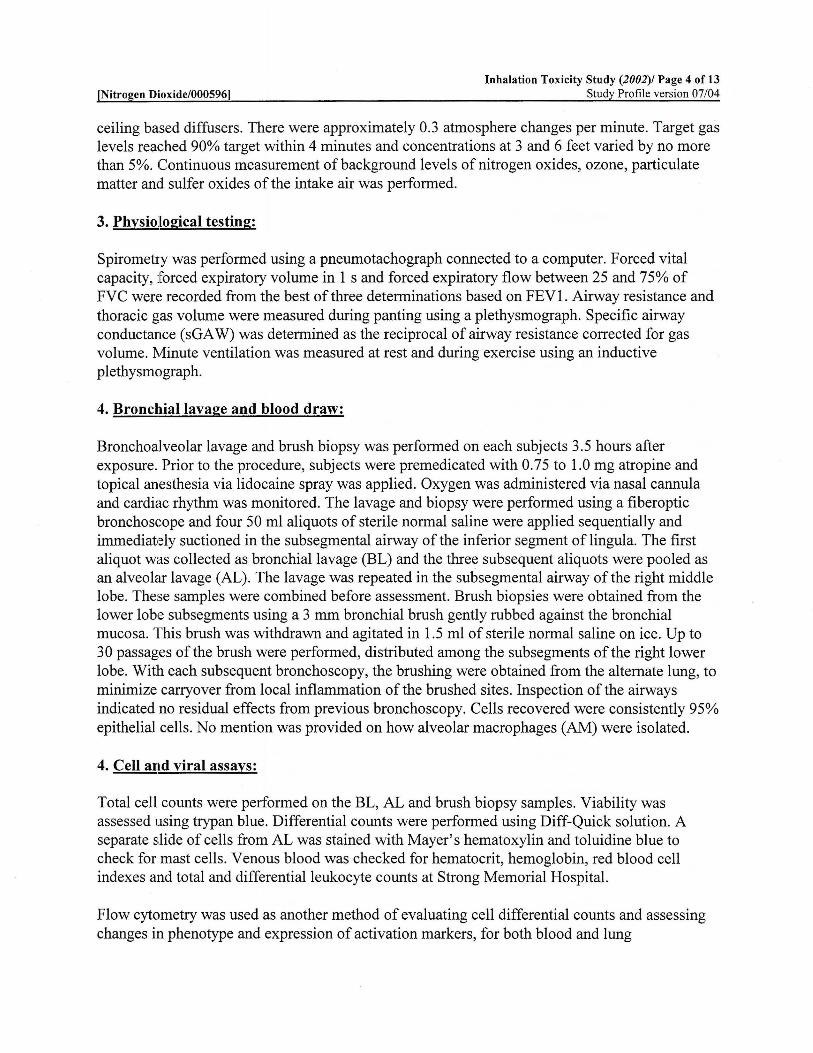
ceiling based diffusers. There were approximately 0.3 atmosphere changes per minute. Target gas levels reached 90% target within 4 minutes and concentrations at 3 and 6 feet varied by no more than 5%. Continuous measurement ofbackground levels of nitrogen oxides, ozone, particulate matter and sulfer oxides of the intake air was performed.
3. Physiollogical testing:
Spirometry was performed using a pneumotachograph connected to a computer. Forced vital capacity, forced expiratory volume in 1 sand forced expiratory flow between 25 and 75% of FVC wen: recorded from the best ofthree determinations based on FEVl. Airway resistance and thoracic gas volume were measured during panting using a plethysmograph. Specific airway conductance (sGAW) was determined as the reciprocal of airway resistance corrected for gas volume. Minute ventilation was measured at rest and during exercise using an inductive plethysmograph.
4. Bronchial lavage and blood draw:
Bronchoalveolar lavage and brush biopsy was performed on each subjects 3.5 hours after exposure. Prior to the procedure, subjects were premedicated with 0.75 to 1.0 mg atropine and topical anesthesia via lidocaine spray was applied. Oxygen was administered via nasal cannula and cardiac rhythm was monitored. The lavage and biopsy were perfortned using a fiberoptic bronchoscope and four 50 ml aliquots of sterile normal saline were applied sequentially and immediately suctioned in the subsegmental airway of the inferior segment of lingula. The first aliquot was collected as bronchial lavage (BL) and the three subsequent aliquots were pooled as an alveolar lavage (AL). The lavage was repeated in the subsegmental airway of the right middle lobe. These samples were combined before assessment. Brush biopsies were obtained from the lower lobe subsegments using a 3 mm bronchial brush gently rubbed against the bronchial mucosa. This brush was withdrawn and agitated in 1.5 ml of sterile normal saline on ice. Up to 30 passages of the brush were performed, distributed among the subsegments of the right lower lobe. With each subsequent bronchoscopy, the brushing were obtained from the alternate lung, to minimize carryover from local inflammation of the brushed sites. Inspection of the airways indicated no residual effects from previous bronchoscopy. Cells recovered were consistently 95% epithelial cells. No mention was provided on how alveolar macrophages (AM) were isolated.
4. Cell and viral assays:
Total cell counts were performed on the BL, ALand brush biopsy samples. Viability was assessed using trypan blue. Differential counts were performed using Diff-Quick solution. A separate slide of cells from AL was stained with Mayer' s hematoxylin and toluidine blue to check for mast cells. Venous blood was checked for hematocrit, hemoglobin, red blood cell indexes and total and differential leukocyte counts at Strong Memorial Hospital.
Flow cytometry was used as another method of evaluating cell differential counts and assessing changes in phenotype and expression of activation markers, for both blood and lung
[Nitrogen Dioxide/000596) Inhalation Toxicity Study (2002)/ Page 5 of 13
Study Profil e version 07/04
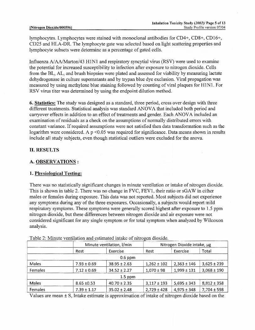
lymphocytes. Lymphocytes were stained with monoclonal antibodies for CD4+, CD8+, CD16+, CD25 and HLA-DR. The lymphocyte gate was selected based on light scattering properties and lymphocyte subsets were determine as a percentage of gated cells.
Influenza A/AA/Marton/43 HINI and respiratory syncytial virus (RSV) were used to examine the potential for increased susceptibility to infection after exposure to nitrogen dioxide. Cells from the BL, AL, and brush biopsies were plated and assessed for viability by measuring lactate dehydrogenase in culture supernatants and by trypan blue dye exclusion. Viral propagation was measured by using methylene blue staining followed by counting ofviral plaques for HlNl. For RSV virus titer was determined by using the endpoint dilution method.
6. Statistics: The study was designed as a standard, three period, cross over design with three different treatments. Statistical analysis was standard ANOV A that included both period and carryover effects in addition to an effect oftreatments and gender. Each ANOVA included an examination of residuals as a check on the assumptions of normally distributed errors with constant variance. If required assumptions were not satisfied then data transformation such as the logarithm were considered. A p <0.05 was required for significance. Data means shown in results include all study subjects, even though statistical outliers were excluded for the anova.
II. RESULTS
A. OBSERVATIONS:
1. Physiological Testing:
There was no statistically significant changes in minute ventilation or intake of nitrogen dioxide. This is shown in table 2. There was no change in FVC, FEVI, their ratio or sGA Win either males or females during exposure. This data was not reported. Most subjects did not experience any symptoms during any of the three exposures. Occasionally, a subjects would report mild respiratory symptoms. These symptoms were generally scored highest after exposure to 1.5 ppm nitrogen dioxide, but these differences between nitrogen dioxide and air exposure were not considered significant for any single symptom or for total symptom when analyzed by "Wilcoxon analysis.
T bl 2 M. t t"lf a e mu e ven 1 a wn an d f td " tak ft es 1ma e m eo m ro gen d" "d wx1 e. Minute ventilation, 1/min Nitrogen Dioxide intake, jlg
Rest Exercise Rest Exercise Total
0.6 ppm
Males 7.93 ± 0.69 38.95 ± 2.63 1,262 ± 102 2,363 ± 146 3,625 ± 239
Females 7.12 ± 0.69 34.52 ± 2.27 1,070 ± 98 1,999 ± 131 3,068 ± 190
1.5 ppm
Males 8.65 ±0.53 40.70 ± 2.35 3,117 ± 193 5,695 ± 343 8,812 ± 358
Females 7.39 ± 1.17 35.02 ± 2.48 2,729 ± 428 4,975 ± 348 7J04 ± 598
Values are mean ± S, Intake estimate is approximation of intake of nitrogen dioxide based on the
[Nitrogen Dioxide/000596) Inhalation Toxicity Study (2002)1 Page 6 of 13
Study Profile version 07/04
product of concentration (~g/1) X time of exposure (min) X minute ventilation
2. Bronchial lavage and blood draw:
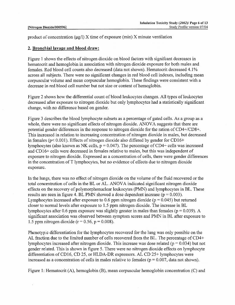
Figure 1 shows the effects of nitrogen dioxide on blood factors with signifcant decreases in hematocrit and hemoglobin in association with nitrogen dioxide exposure for both males and females. Red blood cell counts also decreased (data not shown). Hematocrit decreased 4.1% across all subjects. There were no significant changes in red blood cell indexes, including mean corpuscular volume and mean corpuscular hemoglobin. These findings were consistent with a decrease in red blood cell number but not size or content of hemoglobin.
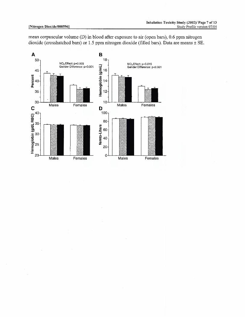
Figure 2 shows how the differential count of blood leukocytes changes. All types of leukocytes decreased after exposure to nitrogen dioxide but only lymphocytes had a statistically significant change, with no difference based on gender.
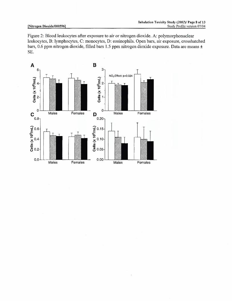
Figure 3 describes the blood lymphocyte subsets as a percentage of gated cells. As a group as a whole, there were no significant effects of nitrogen dioxide. ANOV A suggests that there are potential gender differences in the response to nitrogen dioxide for the ration of CD4+/CD8+. This increased in relation to increasing concentration of nitrogen dioxide in males, but decreased in females (p< 0.001). Effects of nitrogen dioxide also differed by gender for CD16+ lymphocytes (also known as NK cells, p = 0.047). The percentage ofCD4+ cells was increased and CD 16+ cells were decreased in females relative to males, but this was independent of exposure to nitrogen dioxide. Expressed as a concentration of cells, there were gender differences in the concentration ofT lymphocytes, but no evidence of effects due to nitrogen dioxide exposure.
In the lungs, there was no effect of nitrogen dioxide on the volume of the fluid recovered or the total concentration of cells in the BL orAL. ANOV A indicated significant nitrogen dioxide effects on the recovery of polymorphonuclear leukocytes (PMN) and lymphocytes in BL. These results are seen in figure 4. BL PMN showed a dose dependent increase (p = 0.003). Lymphocytes increased after exposure to 0.6 ppm nitrogen dioxide (p = 0.045) but returned closer to normal levels after exposure to 1.5 ppm nitrogen dioxide. The increase in BL lymphocytes after 0.6 ppm exposure was slightly greater in males than females (p = 0.039). A significant association was observed between symptom scores and PMN in BL after exposure to 1.5 ppm nitrogen dioxide (r = 0.56, p = 0.008).
Phenotypic differentiation for the lymphocytes recovered for the lung was only possible on the AL fraction due to the limited number of cells recovered from the BL. The percentage of CD4+ lymphocytes increased after nitrogen dioxide. This increase was dose related (p = 0.034) but not gender rellated. This is shown in figure 5. There were no nitrogen dioxide effects on lymphocyte differentiation ofCD16, CD 25, or HLDA-DR expressors. AL CD 25+ lymphocytes were increased as a concentration of cells in males relative to females (p = 0.007, data not shown).
Figure 1: Hematocrit (A), hemoglobin (B), mean corpuscular hemoglobin concentration (C) and
[Nitrogen Dioxide/000596) Inhalation Toxicity Study (2002)/ Page 7 of 13
Study Profile version 07/04
mean corpuscular volume (D) in blood after exposure to air (open bars), 0.6 ppm nitrogen dioxide (crosshatched bars) or 1.5 ppm nitrogen dioxide (filled bars). Data are means± SE.
A 50
c Males
Males
N02 Effect: p=0.003 Gender Difference: p<0.001
Females
Females
B
:J ~1 c :c 14 0 c, 0
~ 1 J:
D 100
80
20
Males
N02Effect: P=0.015 Gender Difference: p<0.001
Females
Males Females
[Nitrogen Dioxide/000596] Inhalation Toxicity Study (2002)/ Page 8 of 13
Study Profile version 07/04
Figure 2: Blood leukocytes after exposure to air or nitrogen dioxide. A: polymorphonuclear leukocytes, B: lymphocytes, C: monocytes, D: eosinophils. Open bars, air exposure, crosshatched bars, 0.6 ppm nitrogen dioxide, filled bars 1.5 ppm nitrogen dioxide exposure. Data are means ± SE.
A B 6 3
:::J :::J E ,!.2 ~ 0 .... .... ~ ~
~2 ~ 1 Q) Q) (.) (.)
0 0 c Males Females D Males Females
0.
:go. :g 0.1 cO"" cO"" 0 0
-;; 0 . :;_o.1 ._. .!!! .!!! ~0. Qi
(.)
0. Males Females Males Females
[Nitrogen Dioxide/000596) Inhalation Toxicity Study (2002)1 Page 9 of 13
Study Profile version 07/04
Figure 3: Blood lymphocyte subsets, determined using immunofluorescence techniques as a percentage of gated cells. A: CD4+, B: CD4+/CD8+, C: CD8+, D: CD16+. Open bars, air exposure, crosshatched bars, 0.6 ppm nitrogen dioxide, filled bars 1.5 ppm nitrogen dioxide exposure. Data are means ± SE.
A 8
c
[Nitrogen Dioxide/000596] Inhalation Toxicity Study (2002)1 Page 10 of 13
Study Profile version 07/04
Figure 4: Cells recovered in bronchial lavage fluid. A: Polymorphonuclear leukocytes, B: lymphocytes, C: alveolar macrophages, D: eosinophiles. Open bars, air exposure, crosshatched bars, 0.6 ppm nitrogen dioxide, filled bars 1.5 ppm nitrogen dioxide exposure. Data are means ± SE.
A 5
c Males Females
8 5
N02 Effect: p=0.045 ::J NO. Effect x Gender: p=0.039
~
D
::J E ~ 0
1
;. 0.5
.!!!. Cii 0
Males Females
Males Females
[Nitrogen Dioxide/000596] Inhalation Toxicity Study (2002)1 lf>age 11 of 13
Study Profile version 07/04
Figure 5: Lymphocyte subsets recovered in alveolar lavage fluid, determined using immunofluorescence techniques. A: CD3+, B: CD4+, C: CD8+, D: CD4+/CD8+ Open bars, air exposure, crosshatched bars, 0.6 ppm nitrogen dioxide, filled bars 1.5 ppm nitrogen dioxide exposure. Data are means ± SE.
A B
D
3. Cell and viral assays:
Viability of cells from the BL and AL fractions was generally greater than 90% and there was no effect due to nitrogen dioxide exposure. Brushed cells from the bronchial airway had lower viability, often <50%. This was decreased slightly, but non-significantly, in response to nitrogen dioxide exposure. Effects on viability due to prior nitrogen dioxide exposure and infection with influenza or RSV were measured on days 1 and 4 post culture. There was a nitrogen dioxide dose related 40% increase in LDH release on day 1 of culture in the presence of RSV and a similar, but nonstatistically significant, trend in cells treated with influenza. This was similar to findings on day 4, which suggests that RSV has a greater effect on cell viability post nitrogen dioxide exposure than influenza and that nitrogen dioxide does decrease cell viability post nitrogen dioxide exposure.
No nitrogen dioxide effects were found on AM or bronchial epithelial cells after infection with influenza or RSV. Influenza titers in the supernatants of both AM and epithelial cells both declines, indicating a lack of productive culture. RSV produced stable viral titers, indicating the infections was minimally productive. No nitrogen dioxide dependence was found.
Total protein and albumin were measured in both BLand ALas a way to measure epithelial
[Nitrogen Dioxide/000596) Inhalation Toxicity Study (2002)1 Page 12 of 13
Study Profile version 07/04
permeabililty. No nitrogen dioxide effects on the concentration of either total protein or albumin were found, though albumin was found to generally be lower in females compared to males in both fluids. There was no indication on how these results were determined in the methods section of the paper.
Figure 6: Release of lactacte dehydrogenase in the culture supernatant fluids by bronchial epithelial cells after 1 day in culture, with or without previous 1 hour exposure to influenza or RSB. Differences were significant for bronchial epithelial cells and RSV. Open bars, air exposure, crosshatched bars, 0.6 ppm nitrogen dioxide, filled bars, 1.5 ppm nitrogen dioxide exposure. Data are means± SE.
20
18
-.....1 E1 :E E .... '01 E 1: -:X: c
...1
N02 Effect: p:0.024
III. INVESTIGATORS' DISCUSSION AND CONCLUSIONS:
The goal of the paper was to study the effects of nitrogen dioxide on airway inflammation, blood lymphocyte recruitment to the lung and the susceptibility of airways cells to infection with influenza and RSV. There was no effect of nitrogen dioxide exposure on infection with influenza or RSV from cells found in the BL, AL or bronchial brush biopsy. It is likely that single exposures to 0.6 and 1.5 ppm nitrogen dioxide do not alter susceptibility to cells in the airway to common respiratory viruses. Cells from the AM fraction do not support infection by influenza but do support infection from RSV, albeit minimally. Due to the potential for mechanical disruption of cells recovered from bronchial brushing, it is not possible to rule out the effect of nitrogen dioxide on infectability of respiratory viruses. Although there was no change in susceptibility to infection, there was a significant effect on LDH release following RSV release with a trend towards LDH release in influenza due to prior exposure to nitrogen dioxide. This was seen c;:ven in the absence of direct effect on the viability of the cells. The magnitude of effect varied with nitrogen dioxide concentration. The authors suspect that this might represent enhanced cytotoxicity post nitrogen dioxide exposure to any potential challenge. This may indicate that nitrogen dioxide exposure could increase the severity of lung tissue damage following other challenges.
[Nitrogen Dioxide/0005961 Inhalation Toxicity Study (2002)/ll'age 13 of 13
Study Profile version 07/04
Nitrogen dioxide exposure had an effect on lymphocyte recovery from both blood and BL fluid. In blood, nitrogen dioxide exposure resulted in a decrease of lymphocytes, while in BL fluid, there was an increase in lymphocytes following exposure to .6 ppm nitrogen dioxide. There was no effect on lymphocytes in AL. This suggests that nitrogen dioxide induces the recruitment of lymphocytes from blood to the airways.
There are differences between genders of the blood lymphocyte response to nitrogen dioxide. The ratio of CD4+ to CD8+ increased slightly in males in response to nitrogen dioxide but decreased in females. This may be due to differences in initial blood T cell concentration, which is consistent with previously published work. There was a marginal gender difference in response to nitrogen dioxide in terms of percentage ofCD16+ cells in blood. CD25 and HLA-DR exhibited no nitrogen dioxide dependent changes. Lymphocytes in AL fluid revealed increases in the percentage of CD4+ cells post nitrogen dioxide exposure. No other changes in lymphocyte subsets were seen in AL fluid. BL fluid could not be measured for lymphocyte subsets due to insufficient fluid volume. There was an increase in polymorphonuclear leukocytes after exposure to nitrogen dioxide in the BL. This indicates a mild inflammatory response to nitrogen dioxide. Overall, there appear to be some gender differences in lymphocyte subsets expression due to nitrogen dioxide exposure and a decrease in lymphocytes in the blood due to nitrogen dioxide exposure.
There was a small but statistically significant decrease in red blood cell number and hemoglobin concentration in association with nitrogen dioxide. This change was similar for both males and females and occurred despite no change in hemoglobin content or red blood cell size. It is unclear the reason for this. This change would unlikely be clinically significant in a healthy individual but indicates there may be some effect in persons with cardiovascular compromise, children or the elderly.
In general, these effects are small and unlikely to be of clinical significance to healthy individuals. These effects occur in a largely dose dependent fashion, with more significant effects present at the 1.5 ppm dose level. However, individuals with underlying respiratory or heart conditions, the young or old or persons who are immunocompromised may be at risk at these concentrations.
This study is listed as quantitative and acceptable/nonguideline. The LOAEL is 600 ppb. There is no NOAEL for this study.
UNITED STATES ENVIRONMENTAL PROTECTION AGENCY
WASHINGTON D.C., 20460
OFFICE OF CHEMICAL SAFETY AND POLLUTION PREVENTION
October 9, 2014
MEMORANDUM SUBJECT: Ethics Review of Human Toxicity Study with Nitrogen Dioxide FROM: Kelly Sherman, Human Studies Ethics Review Officer Office of the Director Office of Pesticide Programs TO: Steven Weiss, Chief Risk Assessment Science Support Branch Antimicrobials Division Office of Pesticide Programs REF: Frampton, M.W. et al. Nitrogen Dioxide Exposure: Effects on Airway and Blood
Cells. Am J Physiol Lung Cell Mol Physiol. 282: L155-L165, 2002. (MRID 49420001)
I have reviewed the referenced human toxicity study with nitrogen dioxide. I conclude that if the study is determined to be scientifically valid and relevant, there is no regulatory barrier to EPA relying on this research in actions taken under FIFRA or §408 of FFDCA.
Summary Characteristics of the Research
In this study, nitrogen dioxide was administered to 21 people aged 18-40 years (9 females, 12 males) in an environmental chamber at concentrations of 0 (room air), 0.6 ppm or 1.5 ppm nitrogen dioxide for a total of three hours. Exercise was performed for 10 minutes out of every thirty minute period at a sufficient level to raise minute ventilation to 40 1/min. Each exposure period was separated from the previous exposure period by at least three weeks. The subjects were assessed for pulmonary function before and after each exposure. Phlebotomy and bronchoscopy with bronchoalveolar lavage and brush biopsies were performed 3.5 hours after exposure. Recovered cells were challenged with respiratory viruses in vitro.
To supplement the ethics information provided in the journal article, Jonathan Leshin and
I (both of EPA) spoke to Dr. Mark Frampton by telephone on August 20, 2014. After that conversation, I emailed Dr. Frampton with some specific questions about the ethics of the study.
2
Dr. Frampton replied via email on September 11, 2014. A copy of the questions and Dr. Frampton’s responses are provided as Attachment 1.
In addition, I contacted the reviewing institutional review board, the Research Subjects
Review Board of the University of Rochester, and requested copies of the available records about this study. On September 25, 2014, I received copies of the protocol, consent form, and approval letter for this study via email. These documents are provided as Attachment 2. 1. Value of the Research to Society:
The objective of this study was to examine the effects of nitrogen dioxide (NO2) exposure on airway inflammation, blood cells, and antiviral respiratory defense. The study was conducted at the University of Rochester School of Medicine. It was funded in part by the Center for Indoor Air Research and National Institutes of Health grants. The results were published in the American Journal of Physiology, Lung Cellular and Molecular Physiology in 2002. EPA is proposing to use the study in its risk assessment for nitrogen dioxide as medical equipment sterilant.
2. Subject Selection:
a. Demographics. Twenty one subjects aged 18-40 years (9 females, 12 males) participated in the study. The subjects were lifetime non-smokers with normal spirometry, no cardiac or respiratory disease, and without symptoms of respiratory infection within 6 weeks of the study. (Frampton et al., p. L156)
b. Pregnancy and Nursing Status. Dr. Frampton stated that none of the subjects were pregnant and that subjects were tested for pregnancy prior to each exposure. (Attachment 1)
c. Recruitment. The subjects were recruited from the University of Rochester community.
As part of the recruitment and consent process, the study coordinator described the study and initially screened interested individuals by telephone. Next, each potential subject met with one of the researchers and received a copy of the consent form to take home and review prior to deciding whether to sign the consent form and enroll. (Attachments 1, 2). No vulnerable subjects were recruited or participated in the study (Attachment 1).
3. Risks and Benefits:
a. Risks. The risks to subjects are discussed briefly in the consent form. Risks were minimized by enrolling only healthy subjects and closely monitoring the subjects during and after the procedure. (Attachment 1) Medical oversight and assistance from nursing staff at the hospital also minimized potential risks to the subjects. (Attachment 2)
b. Benefits. There are no benefits to the subjects. The consent form states “personal therapeutic benefit may not result from participation in this study, although knowledge gained may be beneficial to others.” (Attachment 2)
3
c. Risk-Benefit Balance. The consent form and areticle are silent regarding the risk-
benefit balance. Dr. Frampton stated that “the potential societal benefits of increased understanding of NO2 toxicity and effects outweighed the small risks associated with the study.” (Attachment 1)
4. Independent Ethics Review: The study was reviewed and approved by the University of Rochester Research Subjects Review Board. (Attachment 2)
5. Informed Consent: Dr. Frampton stated: “The study coordinator described the study and
screened interested subjects by telephone. The study coordinator explained all aspects of the study during a visit to the medical center and provided the consent form, with a copy to take home. Consent was obtained by a study physician.” (Attachment 1) Each subject provided written informed consent before participating. (Attachment 1) A copy of the consent form is included in Attachment 2.
6. Respect for Subjects. Subjects were paid $50 for completing the first exposure and
bronchoscopy, another $50 for completing the second exposure and bronchoscopy, and $450 for the third exposure and bronchoscopy, for a total of $550. (Attachment 2) The consent form states that subjects were free to withdraw from the study at any time. However, it appears that subjects were only partially compensated if they withdrew early. The subjects’ identifies are not revealed in the study report.
Applicable Standards Standards Applicable to the Conduct of the Research
This research was conducted in 1995-1996, before EPA’s Rule for Protection of Human
Subjects of Research became effective in 2006. Thus, 40 CFR part 26 did not apply when this research was conducted.
The prevailing ethical standards for medical research conducted in 1995-1996 are
articulated in the 1989 and 1996 revisions of the Declaration of Helsinki. Key elements are:
1. Research must be scientifically sound and conducted by qualified personnel 2. There must be a clear purpose and protocol, reviewed and approved by an
independent ethics committee 3. The interests of science and society should never take precedence over considerations
related to the well-being of the subject 4. Participants should give prior, informed, voluntary consent
Standards Applicable to EPA’s Reliance on the Research
The Agency’s rule (40 CFR part 26 subpart Q) defines standards for EPA to apply in deciding whether to rely on research—like this study—involving intentional exposure of human subjects. The applicable acceptance standards from 40 CFR part 26 subpart Q are these:
4
§26.1703. Except as provided in §26.1706, EPA must not rely on data from any research subject to this subpart involving intentional exposure of any human subject who is a pregnant woman (and therefore her fetus), a nursing woman, or a child. §26.1704 EPA must not rely on data from any research subject to this section if there is clear and convincing evidence that: (1) The conduct of the research was fundamentally unethical (e.g., the research was intended to seriously harm participants or failed to obtain informed consent); or (2) The conduct of the research was deficient relative to the ethical standards prevailing at the time the research was conducted in a way that placed participants at increased risk of harm (based on knowledge available at the time the study was conducted) or impaired their informed consent. EPA has submitted this study for review by the Human Studies Review Board (HSRB)
because 40 CFR §26.1602 requires HSRB review for pre-2006 studies intended for EPA reliance that were conducted for the purpose of identifying or measuring a toxic effect. This study meets those criteria. Compliance with Applicable Standards
All of the subjects in this study were adults. The nine female subjects were not pregnant. Therefore, this research did not involve intentional exposure of any pregnant or nursing female subjects or any children. EPA’s reliance on the research is not prohibited by 40 CFR §26.1703.
The subjects provided written informed consent and the protocol underwent independent ethics review and approval. Based on these facts, and the absence of any information suggesting that the research was fundamentally unethical or intended to harm participants, I conclude that reliance on the research is not prohibited by 40 CFR §26.1704(1).
With regard to the study’s compliance with prevailing ethical standards, I considered all
available information in the published article, the information provided by Dr. Frampton, and the documents provided by the reviewing institutional review board. The potentials risks of the study were explained to subjects and detailed in the consent form. All of the subjects provided written informed consent. Some of the subjects may have been employees or students at the University, so it is possible that some of the subjects may have had a subordinate relationship with one or more of the researchers. However, recruiting among employees and students was common practice at the time of this study, and there is no clear and convincing evidence to suggest undue influence or lack of fully informed, fully voluntary consent. The research was reviewed and approved by the University of Rochester Research Subjects Review Board.
Based on these facts, I conclude that the study was not deficient relative to the prevailing
ethical standards in a way that placed participants at increased risk of harm or impaired their informed consent. Therefore, reliance on this study is not prohibited by 40 CFR §26.1704(2).
5
Conclusion
I find no barrier in law or regulation to reliance on MRID 49420001 in EPA actions taken under FIFRA or §408 of FFDCA. I defer to others for a full review of the scientific validity of this study. If it were determined not to have scientific validity, it would also not be ethically acceptable.

ATTACHMENT 1
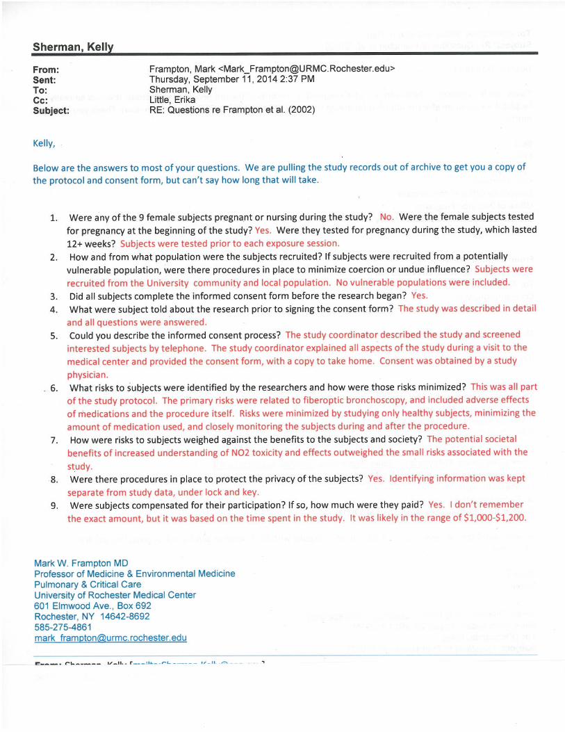

ATTACHMENT 2