Embed Size (px)
Citation preview
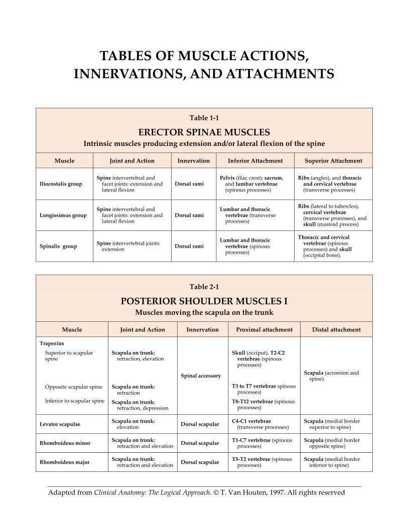
Adapted from Clinical Anatomy: The Logical Approach. © T. Van Houten, 1997. All rights reserved
TABLES OF MUSCLE ACTIONS,
INNERVATIONS, AND ATTACHMENTS
Table 1-1
ERECTOR SPINAE MUSCLES Intrinsic muscles producing extension and/or lateral flexion of the spine
Muscle Joint and Action Innervation Inferior Attachment Superior Attachment
Iliocostalis group
Spine intervertebral and facet joints: extension and lateral flexion
Dorsal rami Pelvis (iliac crest); sacrum,
and lumbar vertebrae (spinous processes)
Ribs (angles), and thoracic and cervical vertebrae (transverse processes)
Longissimus group Spine intervertebral and
facet joints: extension and lateral flexion
Dorsal rami Lumbar and thoracic
vertebrae (transverse processes)
Ribs (lateral to tubercles), cervical vertebrae (transverse processes), and skull (mastoid process)
Spinalis group Spine intervertebral joints:
extension Dorsal rami
Lumbar and thoracic vertebrae (spinous processes)
Thoracic and cervical vertebrae (spinous processes) and skull (occipital bone).
Table 2-1
POSTERIOR SHOULDER MUSCLES I Muscles moving the scapula on the trunk
Muscle Joint and Action Innervation Proximal attachment Distal attachment
Trapezius
Superior to scapular spine
Opposite scapular spine
Inferior to scapular spine
Scapula on trunk: retraction, elevation
Scapula on trunk: retraction
Scapula on trunk: retraction, depression
Spinal accessory
Skull (occiput), T2-C2 vertebrae (spinous processes)
T3 to T7 vertebrae spinous processes)
T8-T12 vertebrae (spinous processes)
Scapula (acromion and spine)
Levator scapulae Scapula on trunk:
elevation Dorsal scapular
C4-C1 vertebrae (transverse processes)
Scapula (medial border superior to spine)
Rhomboideus minor Scapula on trunk:
retraction and elevation Dorsal scapular
T1-C7 vertebrae (spinous processes)
Scapula (medial border opposite spine)
Rhomboideus major Scapula on trunk:
retraction and elevation Dorsal scapular
T5-T2 vertebrae (spinous processes)
Scapula (medial border inferior to spine)
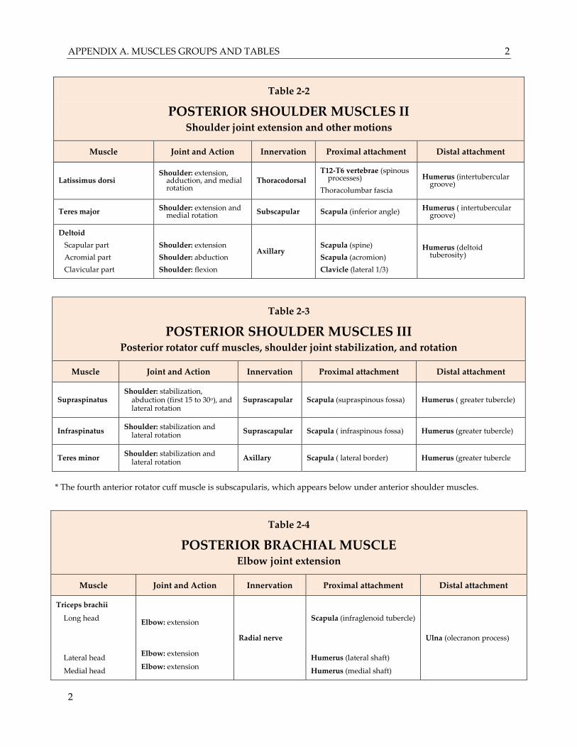
APPENDIX A. MUSCLES GROUPS AND TABLES 2
2
Table 2-2
POSTERIOR SHOULDER MUSCLES II Shoulder joint extension and other motions
Muscle Joint and Action Innervation Proximal attachment Distal attachment
Latissimus dorsi Shoulder: extension,
adduction, and medial rotation
Thoracodorsal
T12-T6 vertebrae (spinous processes)
Thoracolumbar fascia
Humerus (intertubercular groove)
Teres major Shoulder: extension and medial rotation Subscapular Scapula (inferior angle) Humerus ( intertubercular
groove)
Deltoid
Scapular part
Acromial part
Clavicular part
Shoulder: extension
Shoulder: abduction
Shoulder: flexion
Axillary
Scapula (spine)
Scapula (acromion)
Clavicle (lateral 1/3)
Humerus (deltoid tuberosity)
Table 2-3
POSTERIOR SHOULDER MUSCLES III Posterior rotator cuff muscles, shoulder joint stabilization, and rotation
Muscle Joint and Action Innervation Proximal attachment Distal attachment
Supraspinatus Shoulder: stabilization,
abduction (first 15 to 30o), and lateral rotation
Suprascapular Scapula (supraspinous fossa) Humerus ( greater tubercle)
Infraspinatus Shoulder: stabilization and
lateral rotation Suprascapular Scapula ( infraspinous fossa) Humerus (greater tubercle)
Teres minor Shoulder: stabilization and
lateral rotation Axillary Scapula ( lateral border) Humerus (greater tubercle
* The fourth anterior rotator cuff muscle is subscapularis, which appears below under anterior shoulder muscles.
Table 2-4
POSTERIOR BRACHIAL MUSCLE Elbow joint extension
Muscle Joint and Action Innervation Proximal attachment Distal attachment
Triceps brachii
Long head
Lateral head
Medial head
Elbow: extension
Elbow: extension
Elbow: extension
Radial nerve
Scapula (infraglenoid tubercle)
Humerus (lateral shaft)
Humerus (medial shaft)
Ulna (olecranon process)
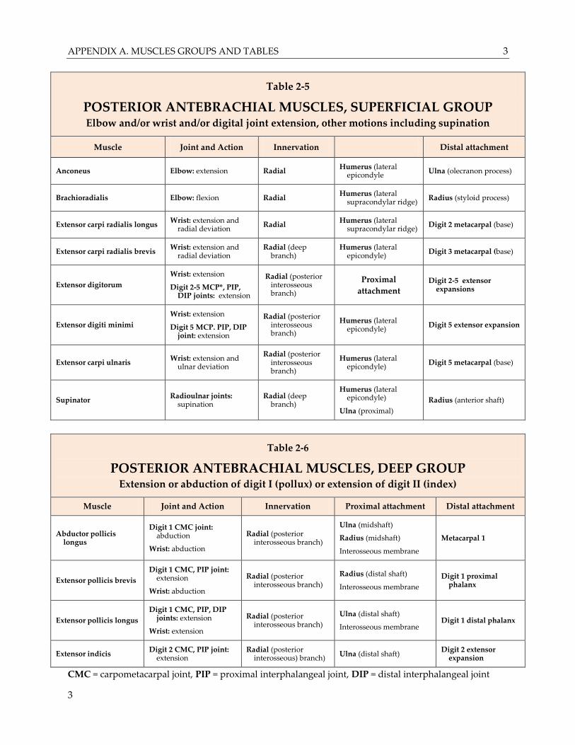
APPENDIX A. MUSCLES GROUPS AND TABLES 3
3
Table 2-5
POSTERIOR ANTEBRACHIAL MUSCLES, SUPERFICIAL GROUP
Elbow and/or wrist and/or digital joint extension, other motions including supination
Muscle Joint and Action Innervation Distal attachment
Anconeus Elbow: extension Radial Humerus (lateral
epicondyle Ulna (olecranon process)
Brachioradialis Elbow: flexion Radial Humerus (lateral
supracondylar ridge) Radius (styloid process)
Extensor carpi radialis longus Wrist: extension and
radial deviation Radial
Humerus (lateral supracondylar ridge)
Digit 2 metacarpal (base)
Extensor carpi radialis brevis Wrist: extension and
radial deviation Radial (deep
branch) Humerus (lateral
epicondyle) Digit 3 metacarpal (base)
Extensor digitorum
Wrist: extension
Digit 2-5 MCP*, PIP, DIP joints: extension
Radial (posterior interosseous branch)
Proximal
attachment Digit 2-5 extensor
expansions
Extensor digiti minimi
Wrist: extension
Digit 5 MCP. PIP, DIP joint: extension
Radial (posterior interosseous branch)
Humerus (lateral epicondyle)
Digit 5 extensor expansion
Extensor carpi ulnaris Wrist: extension and
ulnar deviation
Radial (posterior interosseous branch)
Humerus (lateral epicondyle)
Digit 5 metacarpal (base)
Supinator Radioulnar joints:
supination Radial (deep
branch)
Humerus (lateral epicondyle)
Ulna (proximal)
Radius (anterior shaft)
Table 2-6
POSTERIOR ANTEBRACHIAL MUSCLES, DEEP GROUP Extension or abduction of digit I (pollux) or extension of digit II (index)
Muscle Joint and Action Innervation Proximal attachment Distal attachment
Abductor pollicis longus
Digit 1 CMC joint: abduction
Wrist: abduction
Radial (posterior interosseous branch)
Ulna (midshaft)
Radius (midshaft)
Interosseous membrane
Metacarpal 1
Extensor pollicis brevis
Digit 1 CMC, PIP joint: extension
Wrist: abduction
Radial (posterior interosseous branch)
Radius (distal shaft)
Interosseous membrane
Digit 1 proximal phalanx
Extensor pollicis longus
Digit 1 CMC, PIP, DIP joints: extension
Wrist: extension
Radial (posterior interosseous branch)
Ulna (distal shaft)
Interosseous membrane Digit 1 distal phalanx
Extensor indicis Digit 2 CMC, PIP joint:
extension Radial (posterior
interosseous) branch) Ulna (distal shaft)
Digit 2 extensor expansion
CMC = carpometacarpal joint, PIP = proximal interphalangeal joint, DIP = distal interphalangeal joint
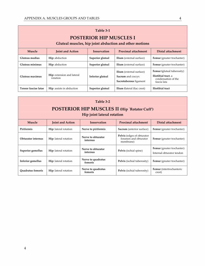
APPENDIX A. MUSCLES GROUPS AND TABLES 4
4
Table 3-1
POSTERIOR HIP MUSCLES I Gluteal muscles, hip joint abduction and other motions
Muscle Joint and Action Innervation Proximal attachment Distal attachment
Gluteus medius Hip: abduction Superior gluteal Ilium (external surface) Femur (greater trochanter)
Gluteus minimus Hip: abduction Superior gluteal Ilium (external surface) Femur (greater trochanter)
Gluteus maximus Hip: extension and lateral
rotation Inferior gluteal
Ilium (external surface)
Sacrum and coccyx
Sacrotuberous ligament
Femur (gluteal tuberosity)
Iliotibial tract, a condensation of the fascia lata
Tensor fasciae latae Hip: assists in abduction Superior gluteal Ilium (lateral iliac crest) Iliotibial tract
Table 3-2
POSTERIOR HIP MUSCLES II (Hip `Rotator Cuff’) Hip joint lateral rotation
Muscle Joint and Action Innervation Proximal attachment Distal attachment
Piriformis Hip: lateral rotation Nerve to piriformis Sacrum (anterior surface) Femur (greater trochanter)
Obturator internus Hip: lateral rotation Nerve to obturator
internus
Pelvis (edges of obturator foramen and obturator membrane)
Femur (greater trochanter)
Superior gemellus Hip: lateral rotation Nerve to obturator
internus Pelvis (ischial spine)
Femur (greater trochanter)
Internal obturator tendon
Inferior gemellus Hip: lateral rotation Nerve to quadratus
femoris Pelvis (ischial tuberosity) Femur (greater trochanter)
Quadratus femoris Hip: lateral rotation Nerve to quadratus
femoris Pelvis (ischial tuberosity)
Femur (intertrochanteric crest)
APPENDIX A. MUSCLES GROUPS AND TABLES 5
5
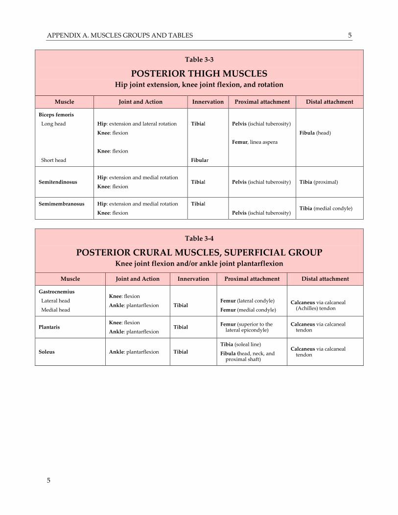
Table 3-3
POSTERIOR THIGH MUSCLES Hip joint extension, knee joint flexion, and rotation
Muscle Joint and Action Innervation Proximal attachment Distal attachment
Biceps femoris
Long head
Short head
Hip: extension and lateral rotation
Knee: flexion
Knee: flexion
Tibial
Fibular
Pelvis (ischial tuberosity)
Femur, linea aspera
Fibula (head)
Semitendinosus Hip: extension and medial rotation
Knee: flexion
Tibial
Pelvis (ischial tuberosity) Tibia (proximal)
Semimembranosus Hip: extension and medial rotation
Knee: flexion
Tibial
Pelvis (ischial tuberosity) Tibia (medial condyle)
Table 3-4
POSTERIOR CRURAL MUSCLES, SUPERFICIAL GROUP Knee joint flexion and/or ankle joint plantarflexion
Muscle Joint and Action Innervation Proximal attachment Distal attachment
Gastrocnemius
Lateral head
Medial head
Knee: flexion
Ankle: plantarflexion
Tibial
Femur (lateral condyle)
Femur (medial condyle)
Calcaneus via calcaneal (Achilles) tendon
Plantaris Knee: flexion
Ankle: plantarflexion Tibial
Femur (superior to the lateral epicondyle)
Calcaneus via calcaneal tendon
Soleus Ankle: plantarflexion Tibial
Tibia (soleal line)
Fibula (head, neck, and proximal shaft)
Calcaneus via calcaneal tendon
APPENDIX A. MUSCLES GROUPS AND TABLES 6
6
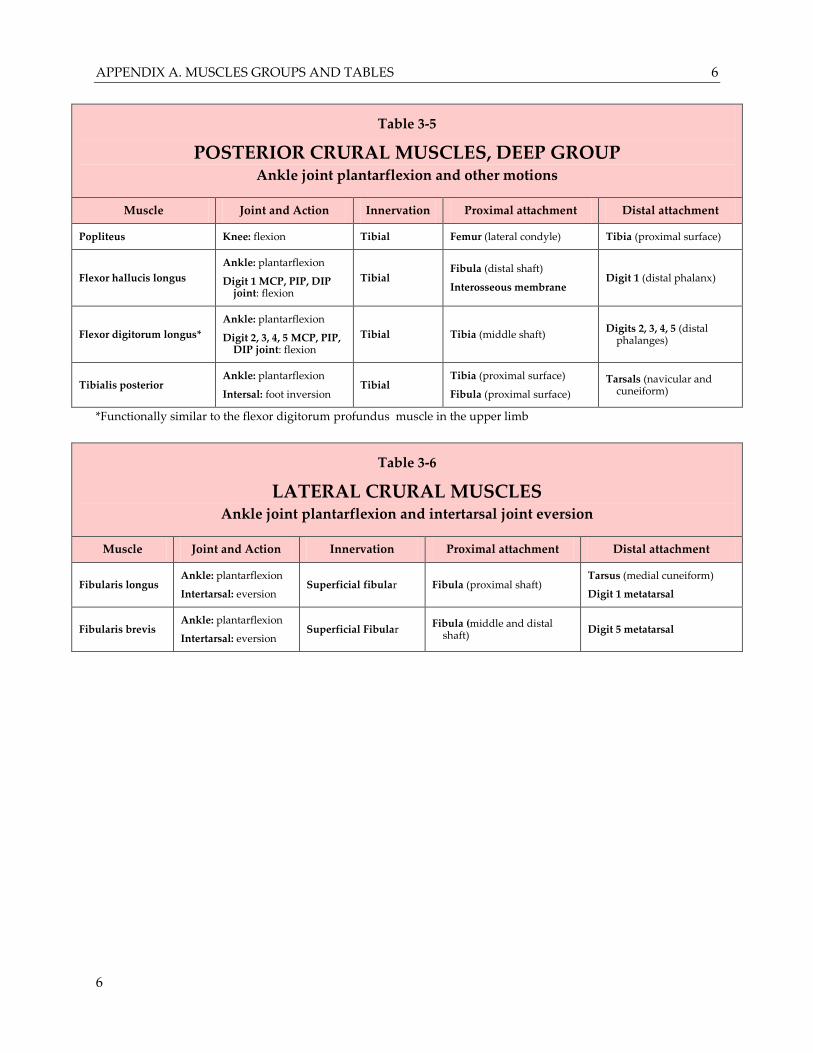
Table 3-5
POSTERIOR CRURAL MUSCLES, DEEP GROUP Ankle joint plantarflexion and other motions
Muscle Joint and Action Innervation Proximal attachment Distal attachment
Popliteus Knee: flexion Tibial Femur (lateral condyle) Tibia (proximal surface)
Flexor hallucis longus
Ankle: plantarflexion
Digit 1 MCP, PIP, DIP joint: flexion
Tibial Fibula (distal shaft)
Interosseous membrane Digit 1 (distal phalanx)
Flexor digitorum longus*
Ankle: plantarflexion
Digit 2, 3, 4, 5 MCP, PIP, DIP joint: flexion
Tibial Tibia (middle shaft) Digits 2, 3, 4, 5 (distal
phalanges)
Tibialis posterior Ankle: plantarflexion
Intersal: foot inversion Tibial
Tibia (proximal surface)
Fibula (proximal surface)
Tarsals (navicular and cuneiform)
*Functionally similar to the flexor digitorum profundus muscle in the upper limb
Table 3-6
LATERAL CRURAL MUSCLES Ankle joint plantarflexion and intertarsal joint eversion
Muscle Joint and Action Innervation Proximal attachment Distal attachment
Fibularis longus Ankle: plantarflexion
Intertarsal: eversion Superficial fibular Fibula (proximal shaft)
Tarsus (medial cuneiform)
Digit 1 metatarsal
Fibularis brevis Ankle: plantarflexion
Intertarsal: eversion Superficial Fibular
Fibula (middle and distal shaft)
Digit 5 metatarsal
APPENDIX A. MUSCLES GROUPS AND TABLES 7
7
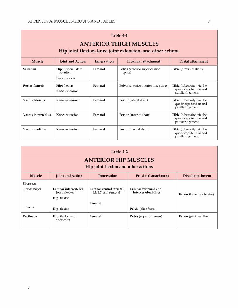
Table 4-1
ANTERIOR THIGH MUSCLES Hip joint flexion, knee joint extension, and other actions
Muscle Joint and Action Innervation Proximal attachment Distal attachment
Sartorius Hip: flexion, lateral rotation
Knee: flexion
Femoral Pelvis (anterior superior iliac spine)
Tibia (proximal shaft)
Rectus femoris Hip: flexion
Knee: extension
Femoral Pelvis (anterior inferior iliac spine) Tibia (tuberosity) via the quadriceps tendon and patellar ligament
Vastus lateralis Knee: extension Femoral Femur (lateral shaft) Tibia (tuberosity) via the quadriceps tendon and patellar ligament
Vastus intermedius Knee: extension Femoral Femur (anterior shaft) Tibia (tuberosity) via the quadriceps tendon and patellar ligament
Vastus medialis Knee: extension Femoral Femur (medial shaft) Tibia (tuberosity) via the quadriceps tendon and patellar ligament
Table 4-2
ANTERIOR HIP MUSCLES Hip joint flexion and other actions
Muscle Joint and Action Innervation Proximal attachment Distal attachment
Iliopsoas
Psoas major
Iliacus
Lumbar intervertebral joint: flexion
Hip: flexion
Hip: flexion
Lumbar ventral rami (L1, L2, L3) and femoral
Femoral
Lumbar vertebrae and intervertebral discs
Pelvis ( iliac fossa)
Femur (lesser trochanter)
Pectineus Hip: flexion and adduction
Femoral Pubis (superior ramus) Femur (pectineal line)
APPENDIX A. MUSCLES GROUPS AND TABLES 8
8
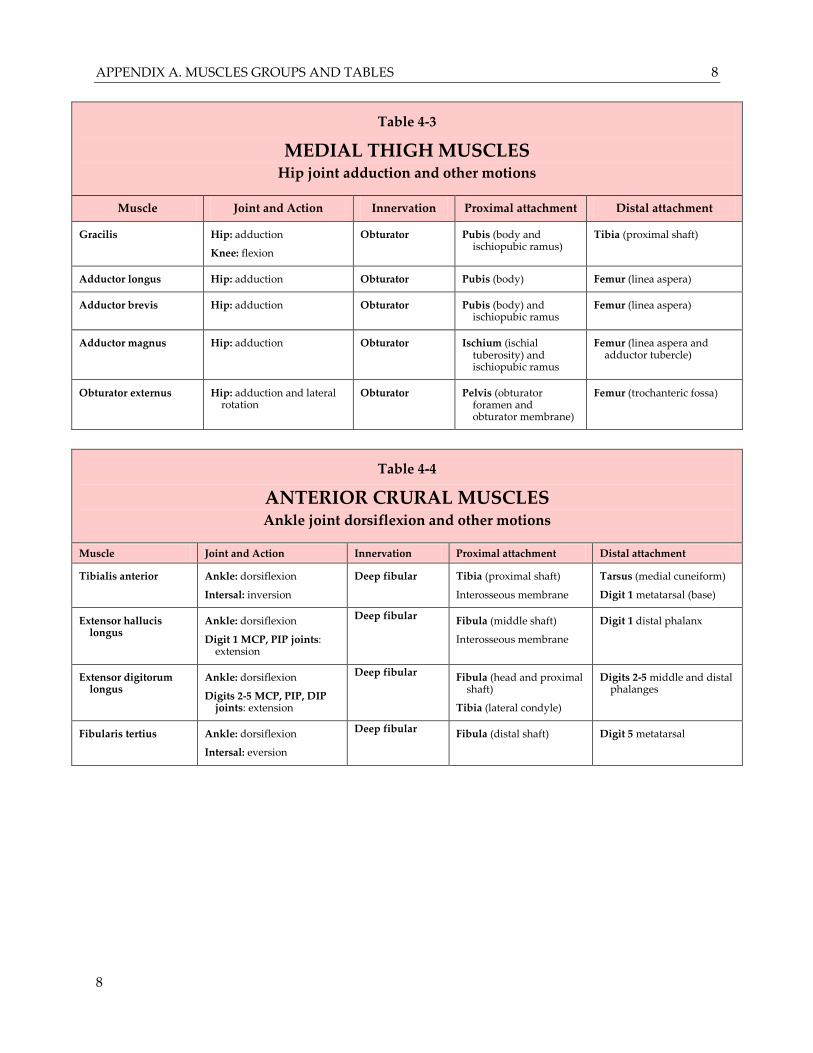
Table 4-3
MEDIAL THIGH MUSCLES Hip joint adduction and other motions
Muscle Joint and Action Innervation Proximal attachment Distal attachment
Gracilis Hip: adduction
Knee: flexion
Obturator Pubis (body and ischiopubic ramus)
Tibia (proximal shaft)
Adductor longus Hip: adduction Obturator Pubis (body) Femur (linea aspera)
Adductor brevis Hip: adduction Obturator Pubis (body) and ischiopubic ramus
Femur (linea aspera)
Adductor magnus Hip: adduction Obturator Ischium (ischial tuberosity) and ischiopubic ramus
Femur (linea aspera and adductor tubercle)
Obturator externus Hip: adduction and lateral rotation
Obturator Pelvis (obturator foramen and obturator membrane)
Femur (trochanteric fossa)
Table 4-4
ANTERIOR CRURAL MUSCLES Ankle joint dorsiflexion and other motions
Muscle Joint and Action Innervation Proximal attachment Distal attachment
Tibialis anterior Ankle: dorsiflexion
Intersal: inversion
Deep fibular Tibia (proximal shaft)
Interosseous membrane
Tarsus (medial cuneiform)
Digit 1 metatarsal (base)
Extensor hallucis longus
Ankle: dorsiflexion
Digit 1 MCP, PIP joints: extension
Deep fibular Fibula (middle shaft)
Interosseous membrane
Digit 1 distal phalanx
Extensor digitorum longus
Ankle: dorsiflexion
Digits 2-5 MCP, PIP, DIP joints: extension
Deep fibular Fibula (head and proximal shaft)
Tibia (lateral condyle)
Digits 2-5 middle and distal phalanges
Fibularis tertius Ankle: dorsiflexion
Intersal: eversion
Deep fibular Fibula (distal shaft) Digit 5 metatarsal
APPENDIX A. MUSCLES GROUPS AND TABLES 9
9
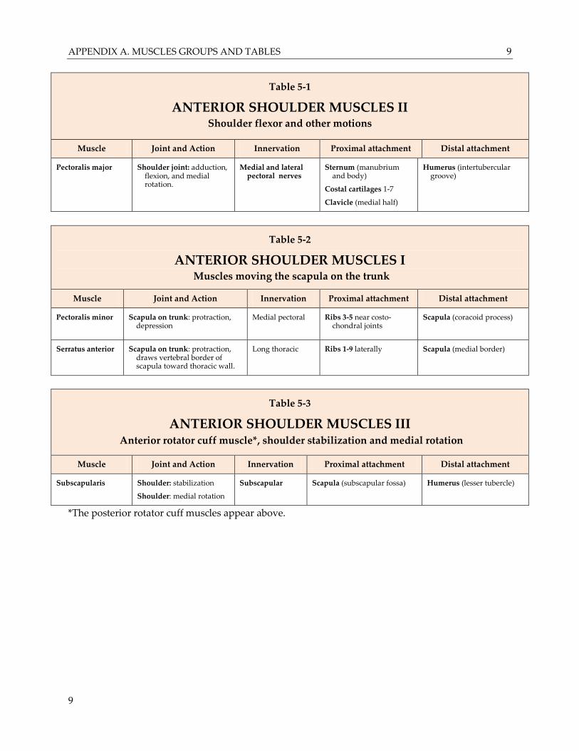
Table 5-1
ANTERIOR SHOULDER MUSCLES II Shoulder flexor and other motions
Muscle Joint and Action Innervation Proximal attachment Distal attachment
Pectoralis major
Shoulder joint: adduction, flexion, and medial rotation.
Medial and lateral pectoral nerves
Sternum (manubrium and body)
Costal cartilages 1-7
Clavicle (medial half)
Humerus (intertubercular groove)
Table 5-2
ANTERIOR SHOULDER MUSCLES I Muscles moving the scapula on the trunk
Muscle Joint and Action Innervation Proximal attachment Distal attachment
Pectoralis minor Scapula on trunk: protraction, depression
Medial pectoral
Ribs 3-5 near costo-chondral joints
Scapula (coracoid process)
Serratus anterior Scapula on trunk: protraction, draws vertebral border of scapula toward thoracic wall.
Long thoracic Ribs 1-9 laterally Scapula (medial border)
Table 5-3
ANTERIOR SHOULDER MUSCLES III Anterior rotator cuff muscle*, shoulder stabilization and medial rotation
Muscle Joint and Action Innervation Proximal attachment Distal attachment
Subscapularis Shoulder: stabilization
Shoulder: medial rotation
Subscapular Scapula (subscapular fossa) Humerus (lesser tubercle)
*The posterior rotator cuff muscles appear above.
APPENDIX A. MUSCLES GROUPS AND TABLES 10
10
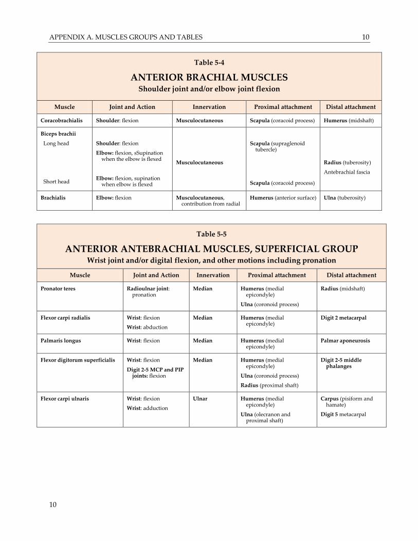
Table 5-4
ANTERIOR BRACHIAL MUSCLES Shoulder joint and/or elbow joint flexion
Muscle Joint and Action Innervation Proximal attachment Distal attachment
Coracobrachialis Shoulder: flexion Musculocutaneous Scapula (coracoid process) Humerus (midshaft)
Biceps brachii
Long head
Short head
Shoulder: flexion
Elbow: flexion, sSupination when the elbow is flexed
Elbow: flexion, supination when elbow is flexed
Musculocutaneous
Scapula (supraglenoid tubercle)
Scapula (coracoid process)
Radius (tuberosity)
Antebrachial fascia
Brachialis Elbow: flexion Musculocutaneous, contribution from radial
Humerus (anterior surface) Ulna (tuberosity)
Table 5-5
ANTERIOR ANTEBRACHIAL MUSCLES, SUPERFICIAL GROUP Wrist joint and/or digital flexion, and other motions including pronation
Muscle Joint and Action Innervation Proximal attachment Distal attachment
Pronator teres Radioulnar joint: pronation
Median Humerus (medial epicondyle)
Ulna (coronoid process)
Radius (midshaft)
Flexor carpi radialis Wrist: flexion
Wrist: abduction
Median Humerus (medial epicondyle)
Digit 2 metacarpal
Palmaris longus Wrist: flexion Median Humerus (medial epicondyle)
Palmar aponeurosis
Flexor digitorum superficialis Wrist: flexion
Digit 2-5 MCP and PIP joints: flexion
Median Humerus (medial epicondyle)
Ulna (coronoid process)
Radius (proximal shaft)
Digit 2-5 middle phalanges
Flexor carpi ulnaris Wrist: flexion
Wrist: adduction
Ulnar Humerus (medial epicondyle)
Ulna (olecranon and proximal shaft)
Carpus (pisiform and hamate)
Digit 5 metacarpal
APPENDIX A. MUSCLES GROUPS AND TABLES 11
11
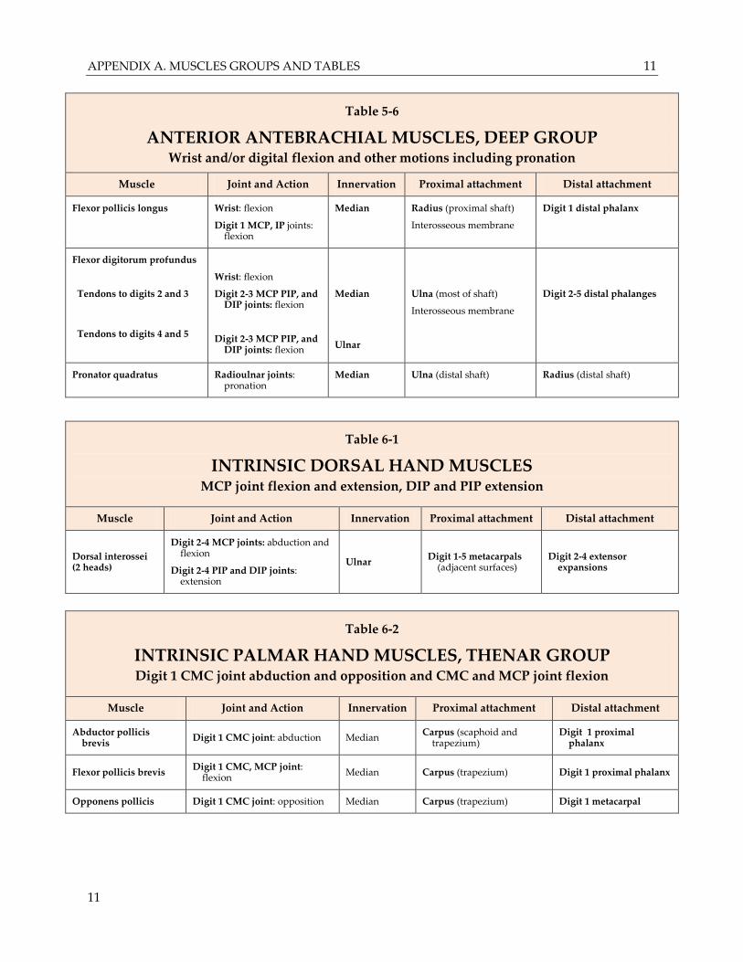
Table 5-6
ANTERIOR ANTEBRACHIAL MUSCLES, DEEP GROUP Wrist and/or digital flexion and other motions including pronation
Muscle Joint and Action Innervation Proximal attachment Distal attachment
Flexor pollicis longus Wrist: flexion
Digit 1 MCP, IP joints: flexion
Median Radius (proximal shaft)
Interosseous membrane
Digit 1 distal phalanx
Flexor digitorum profundus
Tendons to digits 2 and 3
Tendons to digits 4 and 5
Wrist: flexion
Digit 2-3 MCP PIP, and DIP joints: flexion
Digit 2-3 MCP PIP, and DIP joints: flexion
Median
Ulnar
Ulna (most of shaft)
Interosseous membrane
Digit 2-5 distal phalanges
Pronator quadratus Radioulnar joints: pronation
Median Ulna (distal shaft) Radius (distal shaft)
Table 6-1
INTRINSIC DORSAL HAND MUSCLES MCP joint flexion and extension, DIP and PIP extension
Muscle Joint and Action Innervation Proximal attachment Distal attachment
Dorsal interossei (2 heads)
Digit 2-4 MCP joints: abduction and flexion
Digit 2-4 PIP and DIP joints: extension
Ulnar Digit 1-5 metacarpals
(adjacent surfaces) Digit 2-4 extensor
expansions
Table 6-2
INTRINSIC PALMAR HAND MUSCLES, THENAR GROUP Digit 1 CMC joint abduction and opposition and CMC and MCP joint flexion
Muscle Joint and Action Innervation Proximal attachment Distal attachment
Abductor pollicis brevis
Digit 1 CMC joint: abduction Median Carpus (scaphoid and
trapezium) Digit 1 proximal
phalanx
Flexor pollicis brevis Digit 1 CMC, MCP joint:
flexion Median Carpus (trapezium) Digit 1 proximal phalanx
Opponens pollicis Digit 1 CMC joint: opposition Median Carpus (trapezium) Digit 1 metacarpal
APPENDIX A. MUSCLES GROUPS AND TABLES 12
12
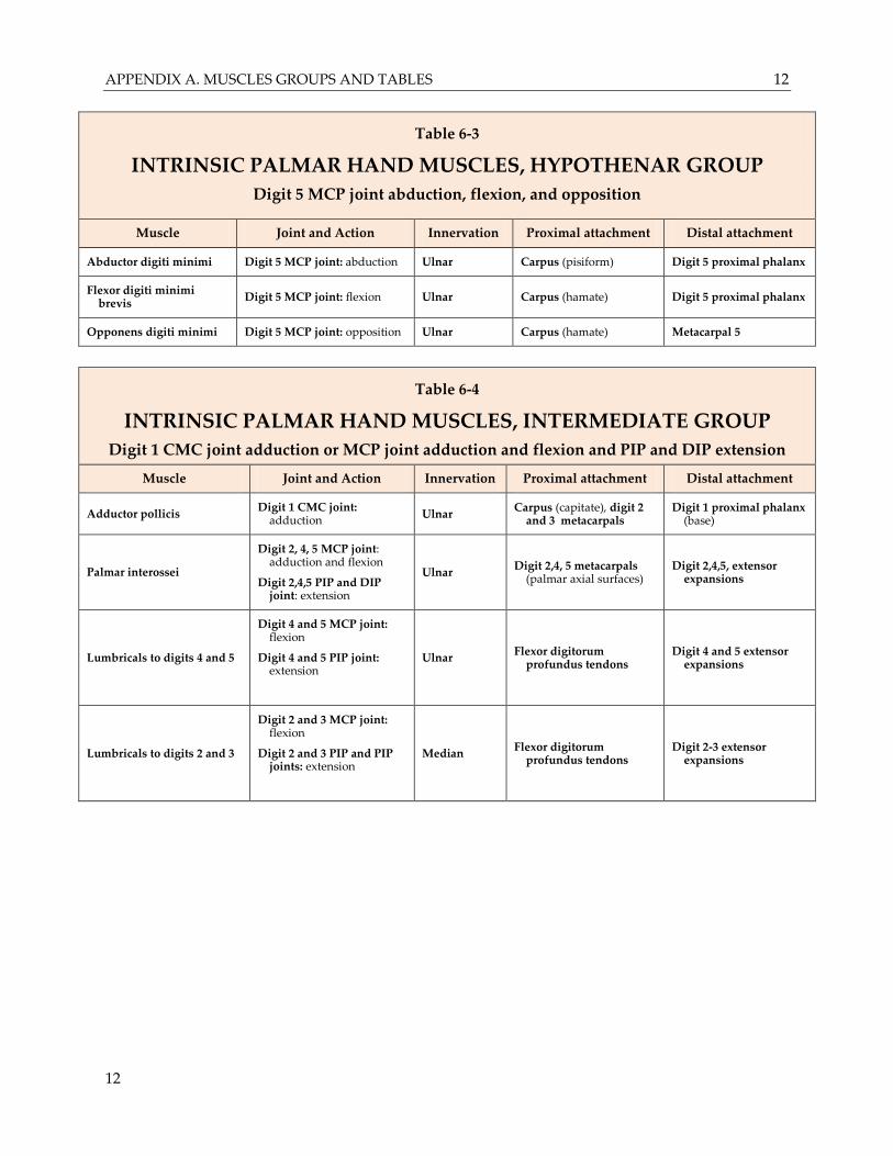
Table 6-3
INTRINSIC PALMAR HAND MUSCLES, HYPOTHENAR GROUP
Digit 5 MCP joint abduction, flexion, and opposition
Muscle Joint and Action Innervation Proximal attachment Distal attachment
Abductor digiti minimi Digit 5 MCP joint: abduction Ulnar Carpus (pisiform) Digit 5 proximal phalanx
Flexor digiti minimi brevis
Digit 5 MCP joint: flexion Ulnar Carpus (hamate) Digit 5 proximal phalanx
Opponens digiti minimi Digit 5 MCP joint: opposition Ulnar Carpus (hamate) Metacarpal 5
Table 6-4
INTRINSIC PALMAR HAND MUSCLES, INTERMEDIATE GROUP
Digit 1 CMC joint adduction or MCP joint adduction and flexion and PIP and DIP extension
Muscle Joint and Action Innervation Proximal attachment Distal attachment
Adductor pollicis Digit 1 CMC joint:
adduction Ulnar
Carpus (capitate), digit 2 and 3 metacarpals
Digit 1 proximal phalanx (base)
Palmar interossei
Digit 2, 4, 5 MCP joint: adduction and flexion
Digit 2,4,5 PIP and DIP joint: extension
Ulnar Digit 2,4, 5 metacarpals
(palmar axial surfaces) Digit 2,4,5, extensor
expansions
Lumbricals to digits 4 and 5
Digit 4 and 5 MCP joint: flexion
Digit 4 and 5 PIP joint: extension
Ulnar Flexor digitorum
profundus tendons Digit 4 and 5 extensor
expansions
Lumbricals to digits 2 and 3
Digit 2 and 3 MCP joint: flexion
Digit 2 and 3 PIP and PIP joints: extension
Median Flexor digitorum
profundus tendons Digit 2-3 extensor
expansions
APPENDIX A. MUSCLES GROUPS AND TABLES 13
13
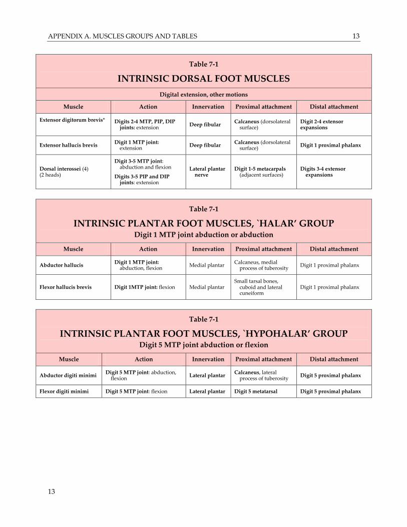
Table 7-1
INTRINSIC DORSAL FOOT MUSCLES
Digital extension, other motions
Muscle Action Innervation Proximal attachment Distal attachment
Extensor digitorum brevis*
Digits 2-4 MTP, PIP, DIP joints: extension
Deep fibular Calcaneus (dorsolateral
surface) Digit 2-4 extensor expansions
Extensor hallucis brevis Digit 1 MTP joint:
extension Deep fibular
Calcaneus (dorsolateral surface)
Digit 1 proximal phalanx
Dorsal interossei (4) (2 heads)
Digit 3-5 MTP joint: abduction and flexion
Digits 3-5 PIP and DIP joints: extension
Lateral plantar nerve
Digit 1-5 metacarpals (adjacent surfaces)
Digits 3-4 extensor expansions
Table 7-1
INTRINSIC PLANTAR FOOT MUSCLES, `HALAR’ GROUP Digit 1 MTP joint abduction or abduction
Muscle Action Innervation Proximal attachment Distal attachment
Abductor hallucis Digit 1 MTP joint:
abduction, flexion Medial plantar
Calcaneus, medial process of tuberosity
Digit 1 proximal phalanx
Flexor hallucis brevis Digit 1MTP joint: flexion Medial plantar Small tarsal bones,
cuboid and lateral cuneiform
Digit 1 proximal phalanx
Table 7-1
INTRINSIC PLANTAR FOOT MUSCLES, `HYPOHALAR’ GROUP Digit 5 MTP joint abduction or flexion
Muscle Action Innervation Proximal attachment Distal attachment
Abductor digiti minimi Digit 5 MTP joint: abduction,
flexion Lateral plantar
Calcaneus, lateral process of tuberosity
Digit 5 proximal phalanx
Flexor digiti minimi Digit 5 MTP joint: flexion Lateral plantar Digit 5 metatarsal Digit 5 proximal phalanx
APPENDIX A. MUSCLES GROUPS AND TABLES 14
14
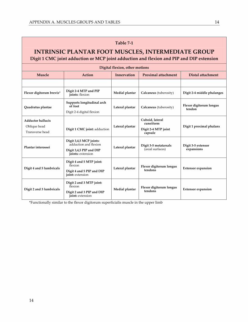
Table 7-1
INTRINSIC PLANTAR FOOT MUSCLES, INTERMEDIATE GROUP Digit 1 CMC joint adduction or MCP joint adduction and flexion and PIP and DIP extension
Digital flexion, other motions
Muscle Action Innervation Proximal attachment Distal attachment
Flexor digitorum brevis* Digit 2-4 MTP and PIP
joints: flexion Medial plantar Calcaneus (tuberosity) Digit 2-4 middle phalanges
Quadratus plantae
Supports longitudinal arch of foot
Digit 2-4 digital flexion
Lateral plantar Calcaneus (tuberosity) Flexor digitorum longus
tendon
Adductor hallucis
Oblique head
Transverse head
Digit 1 CMC joint: adduction Lateral plantar
Cuboid, lateral cuneiform
Digit 2-4 MTP joint capsule
Digit 1 proximal phalanx
Plantar interossei
Digit 3,4,5 MCP joints: adduction and flexion
Digit 3,4,5 PIP and DIP joints: extension
Lateral plantar Digit 3-5 metatarsals
(axial surfaces) Digit 3-5 extensor
expansions
Digit 4 and 5 lumbricals
Digit 4 and 5 MTP joint: flexion
Digit 4 and 5 PIP and DIP joint: extension
Lateral plantar Flexor digitorum longus
tendons Extensor expansion
Digit 2 and 3 lumbricals
Digit 2 and 3 MTP joint: flexion
Digit 2 and 3 PIP and DIP joint: extension
Medial plantar Flexor digitorum longus
tendons Extensor expansion
*Functionally similar to the flexor digitorum superficialis muscle in the upper limb