Embed Size (px)
Citation preview
CASE REPORT Open Access
Tablets at the bedside - iPad-based visualfield test used in the diagnosis of IntrasellarHaemangiopericytoma: a case reportNisha Nesaratnam1* , Peter B. M. Thomas1, Ramez Kirollos2, Algis J. Vingrys3, George Y. X. Kong1
and Keith R. Martin1
Abstract
Background: In the assessment of a pituitary mass, objective visual field testing represents a valuable means ofevaluating mass effect, and thus in deciding whether surgical management is warranted.
Case presentation: In this vignette, we describe a 73 year-old lady who presented with a three-week history offrontal headache, and ‘blurriness’ in the left side of her vision, due to a WHO grade III anaplastic haemangiopericytomacompressing the optic chiasm. We report how timely investigations, including an iPad-based visual field test(Melbourne Rapid Field, (MRF)) conducted at the bedside aided swift and appropriate management of the patient.
Conclusions: We envisage such a test having a role in assessing bed-bound patients in hospital where access toformal visual field testing is difficult, or indeed in rapid testing of visual fields at the bedside to screen for post-operative complications, such as haematoma.
Keywords: Melbourne rapid field (MRF), Intrasellar haemangiopericytoma, Bitemporal hemianopia, Translationaltechnology, Case report
BackgroundHistory, examination and investigation of a patient witha suspected pituitary mass should aim not only to iden-tify the cause of the mass, but also to ascertain if there iscompression of adjacent structures, or clinical featuresof pituitary hormone abnormality. In any patient withsigns of mass effect, such as a visual field defect or cra-nial nerve neuropathy, urgent MRI imaging and consid-eration of surgical decompression is warranted [1].Formal assessment of visual fields using standard au-
tomated perimetry is routinely performed in patientswith pituitary tumours to determine the degree ofimpairment caused by optic chiasm compression. Suchassessment is necessary in planning the urgency of sur-gical intervention. However, formal standard automatedperimetry cannot be performed in patients who arebed-bound, or in situations when visual field testing
equipment is unavailable. Here we report a case inwhich a novel visual field test using a portable tabletdevice (Melbourne Rapid Field, (MRF)) was used to as-sess a patient with a pituitary tumour at the bedside.
Case presentationA 73 year-old presented to Addenbrooke’s Hospital inCambridge with a three-week history of frontal headacheand ‘blurriness’ in the left side of her vision. She had nonausea, vomiting, diplopia, facial pain or paraesthesia,and had no symptoms of pituitary hormone abnormalityon admission. She had, however, presented 7 monthsprior with nausea and vomiting. On this previous admis-sion, she was found to be hyponatraemic, with a reducedcortisol of 26 nmol/L and impaired cortisol response tosynacthen (peak 119 nmol/L), and was commenced onoral hydrocortisone. FSH and LH were within normallimits and an MRI head showed no obvious mass lesionwithin the sella. Her past medical history included long-standing hypothyroidism, for which she was takinglevothyroxine, iron-deficiency anaemia, and left sacroilitis.
* Correspondence: [email protected] of Ophthalmology, Addenbrooke’s Hospital, CambridgeUniversity Hospitals NHS Foundation Trust, Hills Road, Cambridge CB20QQ, UKFull list of author information is available at the end of the article
© The Author(s). 2017 Open Access This article is distributed under the terms of the Creative Commons Attribution 4.0International License (http://creativecommons.org/licenses/by/4.0/), which permits unrestricted use, distribution, andreproduction in any medium, provided you give appropriate credit to the original author(s) and the source, provide a link tothe Creative Commons license, and indicate if changes were made. The Creative Commons Public Domain Dedication waiver(http://creativecommons.org/publicdomain/zero/1.0/) applies to the data made available in this article, unless otherwise stated.
Nesaratnam et al. BMC Ophthalmology (2017) 17:53 DOI 10.1186/s12886-017-0445-z
At presentation, pertinent examination findingsincluded a reduced visual acuity (6/18 in both eyes giv-ing 6/12 with pinhole) and bitemporal hemianopia toconfrontation. Further neurological examinations wereunremarkable, and there were no other cranial nerveabnormalities. Urgent MRI head, pituitary hormone as-says and Ophthalmology review for formal visual acuityand visual field testing were requested.MRI imaging revealed an intrasellar mass with con-
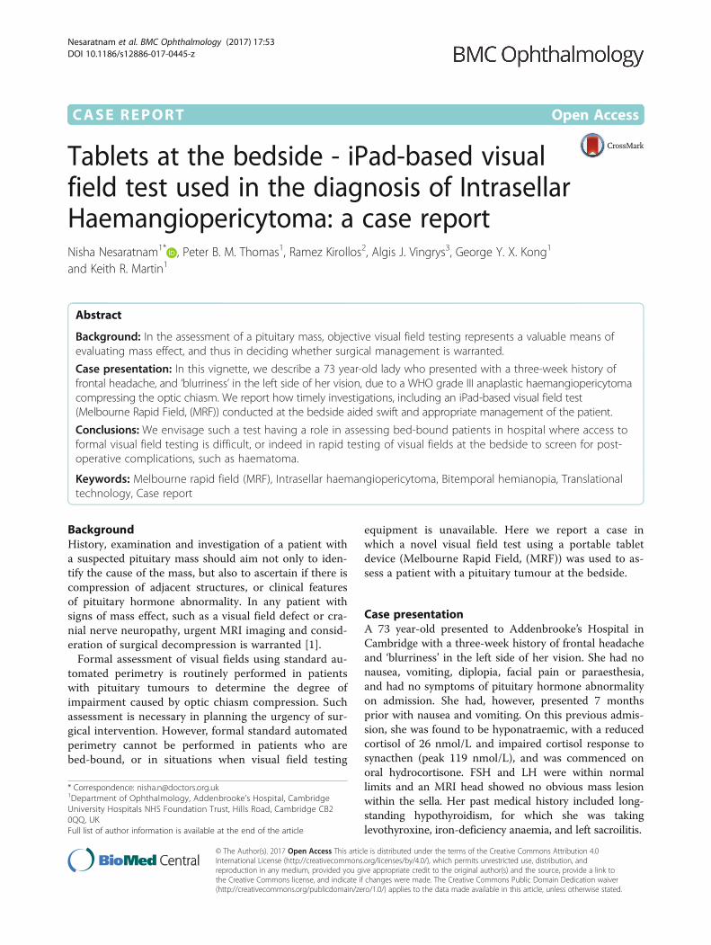
trast enhancement (Fig. 1), which showed significantenlargement since the previous MRI head carried out 7months earlier. Pre- and post-contrast images throughthe pituitary fossa showed the mass extending super-iorly into the suprasellar region, where it appeared tocompress the optic chiasm, and inferiorly into thesphenoid sinus. It had a lobular margin, and measured34×12×15mm, with homogeneous enhancement post-contrast. It appeared to extend laterally into the cavern-ous sinus to lie inseparable from the carotid arteries.No pituitary tissue could be seen separately from themass.Ophthalmology review took place out-of-hours, when
formal visual field testing was unavailable. Fundoscopyrevealed healthy appearances of the optic disc. Visualacuity was 6/18 in both eyes. Visual field testing was
performed on an iPad-based tangent perimeter,(Melbourne Rapid Field (MRF)), which performs fastthresholding at various locations within 30° of fixation,and has been validated in a patients with glaucoma [2].With the iPad tablet screen, measuring 195 × 150 mm,viewed 33 cm away from the patient, the patient isinstructed to fixate upon a target, and tap their finger onthe screen or keyboard when they see a stimulus.The MRF test was performed at the bedside of the
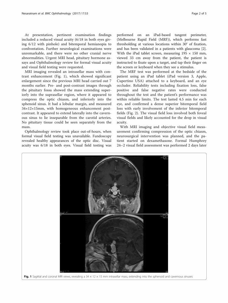
patient using an iPad tablet (iPad version 3, Apple,Cupertino USA) attached to a keyboard, and an eyeoccluder. Reliability tests including fixation loss, falsepositive and false negative rates were conductedthroughout the test and the patient’s performance waswithin reliable limits. The test lasted 4.5 min for eacheye, and confirmed a dense superior bitemporal fieldloss with early involvement of the inferior bitemporalfields (Fig. 2). The visual field loss involved both fovealvisual fields and likely accounted for the drop in visualacuity.With MRI imaging and objective visual field meas-
urement confirming compression of the optic chiasm,neurosurgical intervention was planned, and the pa-tient started on dexamethasone. Formal Humphrey24–2 visual field assessment was performed 2 days later
Fig. 1 Sagittal and coronal MRI views, revealing a 34 × 12 × 15 mm intrasellar mass, extending into the sphenoid and cavernous sinuses
Nesaratnam et al. BMC Ophthalmology (2017) 17:53 Page 2 of 5
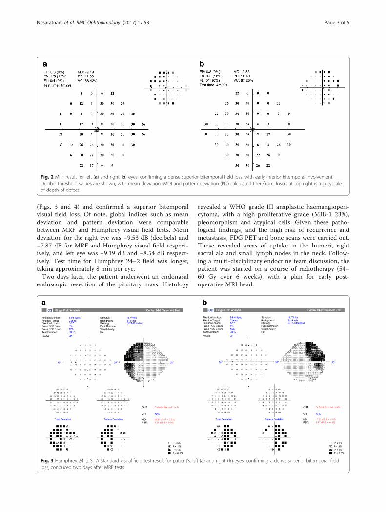
(Figs. 3 and 4) and confirmed a superior bitemporalvisual field loss. Of note, global indices such as meandeviation and pattern deviation were comparablebetween MRF and Humphrey visual field tests. Meandeviation for the right eye was −9.53 dB (decibels) and−7.87 dB for MRF and Humphrey visual field respect-ively, and left eye was −9.19 dB and −8.54 dB respect-ively. Test time for Humphrey 24–2 field was longer,taking approximately 8 min per eye.Two days later, the patient underwent an endonasal
endoscopic resection of the pituitary mass. Histology
revealed a WHO grade III anaplastic haemangioperi-cytoma, with a high proliferative grade (MIB-1 23%),pleomorphism and atypical cells. Given these patho-logical findings, and the high risk of recurrence andmetastasis, FDG PET and bone scans were carried out.These revealed areas of uptake in the humeri, rightsacral ala and small lymph nodes in the neck. Follow-ing a multi-disciplinary endocrine team discussion, thepatient was started on a course of radiotherapy (54–60 Gy over 6 weeks), with a plan for early post-operative MRI head.
Fig. 2 MRF result for left (a) and right (b) eyes, confirming a dense superior bitemporal field loss, with early inferior bitemporal involvement.Decibel threshold values are shown, with mean deviation (MD) and pattern deviation (PD) calculated therefrom. Insert at top right is a greyscaleof depth of defect
Fig. 3 Humphrey 24–2 SITA-Standard visual field test result for patient’s left (a) and right (b) eyes, confirming a dense superior bitemporal fieldloss, conduced two days after MRF tests
Nesaratnam et al. BMC Ophthalmology (2017) 17:53 Page 3 of 5
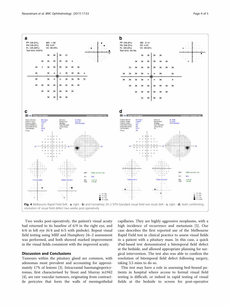
Two weeks post-operatively, the patient’s visual acuityhad returned to its baseline of 6/9 in the right eye, and6/6 in left eye (6/4 and 6/5 with pinhole). Repeat visualfield testing using MRF and Humphrey 24–2 assessmentwas performed, and both showed marked improvementin the visual fields consistent with the improved acuity.
Discussion and ConclusionsTumours within the pituitary gland are common, withadenomas most prevalent and accounting for approxi-mately 17% of lesions [3]. Intracranial haemangiopericy-tomas, first characterised by Stout and Murray in1942[4], are rare vascular tumours, originating from contract-ile pericytes that form the walls of meningothelial
capillaries. They are highly aggressive neoplasms, with ahigh incidence of recurrence and metastasis [5]. Ourcase describes the first reported use of the MelbourneRapid Field test in clinical practice to assess visual fieldsin a patient with a pituitary mass. In this case, a quickiPad-based test demonstrated a bitemporal field defectat the bedside, and allowed appropriate planning for sur-gical intervention. The test also was able to confirm theresolution of bitemporal field defect following surgery,taking 3.5 mins to do so.This test may have a role in assessing bed-bound pa-
tients in hospital where access to formal visual fieldtesting is difficult, or indeed in rapid testing of visualfields at the bedside to screen for post-operative
Fig. 4 Melbourne Rapid Field (left - a, right - b) and Humphrey 24–2 SITA-Standard visual field test result (left - c, right - d), both confirmimgresolution of visual field defect two weeks post-operatively
Nesaratnam et al. BMC Ophthalmology (2017) 17:53 Page 4 of 5

complications, such as haematoma. Use of a keyboardas in this case, which the patient can tap when they seea stimulus, limits the visual dexterity needed to per-form the test, and avoids changes in the alignment anddistance of the iPad relative to the eye.We envisage the test also being utilised in settings where
formal visual field testing is not readily available, such asrural areas in developing countries [6], or in the home set-ting, where visual fields could be monitored in patientswith slow growing pituitary adenomas. Such a setup mayimprove the overall experience of regular visual field test-ing, and alleviate the current difficulties associated withformal testing currently reported by patients [7].
AbbreviationsFDG PET: Fludeoxyglucose positron emission tomography; FSH: Follicle-stimulating hormone; LH: Luteinising hormone; MRF: Melbourne rapid field;MRI: Magnetic resonance imaging; WHO: World Health Organisation
AcknowledgementsNot applicable.
FundingNone.
Availability of data and materialsAll data generated or analysed during this study are included in thispublished article.
Authors’ contributionsNN drafted the manuscript, and interpreted patient data. PT and RK aided indata gathering and interpretation, as well as review of manuscript. AV, GKand KM critically reviewed the manuscript. All authors read and approvedthe final manuscript.
Competing interestsAV and GK are founding directors of GLANCE Optical Pty Ltd., theproduction company of the Melbourne Rapid Field app.
Consent for publicationWritten consent for publication obtained.
Ethics approval and consent to participateNot applicable.
Publisher’s NoteSpringer Nature remains neutral with regard to jurisdictional claims inpublished maps and institutional affiliations.
Author details1Department of Ophthalmology, Addenbrooke’s Hospital, CambridgeUniversity Hospitals NHS Foundation Trust, Hills Road, Cambridge CB20QQ, UK. 2Department of Neurosurgery, Addenbrooke’s Hospital,Cambridge University Hospitals NHS Foundation Trust, Hills Road,Cambridge, UK. 3Department of Optometry & Vision Sciences Melbourne Schoolof Health Sciences, University of Melbourne, Melbourne, VIC 3010, Australia.
Received: 22 September 2016 Accepted: 20 April 2017
References1. Freda PU, Beckers AM, Katznelson L, Molitch ME, Montori VM, Post KD, et al.
Pituitary incidentaloma: an endocrine society clinical practice guideline.J Clin Endocrinol Metab. 2011;96:894–904.
2. Kong YXG, He M, Crowston JG, Vingrys AJ. A comparison of perimetricresults from a tablet perimeter and Humphrey field analyzer in glaucomapatients. Transl Vis Sci Technol. 2016;5:2.
3. Ezzat S, Asa SL, Couldwell WT, Barr CE, Dodge WE, Vance ML, et al. Theprevalence of pituitary adenomas: a systematic review. Cancer. 2004;101:613–9.
4. Stout AP, Murray MR. Hemangiopericytoma: a vascular tumor featuringZimmermann's Pericytes. Ann Surg. 1942;116:26–33.
5. Dufour H, Métellus P, Fuentes S, Murracciole X, Régis J, Figarella-Branger D,et al. Meningeal hemangiopericytoma: a retrospective study of 21 patientswith special review of postoperative external radiotherapy. Neurosurgery.2001;48:756–762; discussion 762–763.
6. Johnson CA, Thapa S, Robin AL. Visual field screening to detect glaucomaand diabetic retinopathy in Nepal using an iPad application program. AmAcad Optom. 2014. Available at: http://www.aaopt.org/visual-field-screening-detect-glaucoma-and-diabetic-retinopathy-nepal-using-ipad-application-program. Accessed 13 Aug 2016.
7. Glen FC, Baker H, Crabb DP. A qualitative investigation into patients’ viewson visual field testing for glaucoma monitoring. BMJ Open. 2014;4:e003996.
• We accept pre-submission inquiries
• Our selector tool helps you to find the most relevant journal
• We provide round the clock customer support
• Convenient online submission
• Thorough peer review
• Inclusion in PubMed and all major indexing services
• Maximum visibility for your research
Submit your manuscript atwww.biomedcentral.com/submit
Submit your next manuscript to BioMed Central and we will help you at every step:
Nesaratnam et al. BMC Ophthalmology (2017) 17:53 Page 5 of 5