Embed Size (px)
Citation preview

Drug-Carrying NanoparticlesDOI: 10.1002/anie.201204663
Targeted Cargo Delivery in Senescent Cells Using Capped MesoporousSilica Nanoparticles**Alessandro Agostini, Laura Mondrag�n, Andrea Bernardos, Ram�n Mart�nez-M�Çez,*M. Dolores Marcos, F�lix Sancen�n, Juan Soto, Ana Costero, Cristina Manguan-Garc�a,Rosario Perona,* Marta Moreno-Torres, Rafael Aparicio-Sanchis, and Jos� Ram�n Murgu�a*
Normal somatic cells invariably enter a state of irreversiblyarrested growth and altered function after a finite number ofdivisions, called cellular senescence. Senescent cells displaya radically altered phenotype that is thought to impair tissuefunction and predispose tissues to disease development and/or progression. Despite the fact that the immune systemdestroys many senescent cells, it becomes much less effectiveat this task during aging. As a consequence, senescent or“death resistant” cells accumulate in tissues, thus acceleratingaging and contributing to disease development. Senescent-cell accumulation alters neighboring cell behavior, favorsdegradation of the extracellular matrix, decreases the pool ofmitotic-competent cells, and stimulates cancer.[1] Moreover
a number of pathologies are associated with acceleratedcellular aging such as Werner adult progeria syndrome (WS),Hutchinson–Gilford syndrome (HGS), and Rothmund–Thompson syndrome (RTS).[2] In WS or RTS, the shorteningof telomeres is observed in most tissues, even if defectivetelomerase is not the main cause of the disease.[3, 4] Otherdiseases are more related to tissue-specific accelerated agingsuch as Dyskeratosis congenita (DC) and idiopathic pulmo-nary fibrosis (IPF).[5] In these conditions, the replicativecapacity of cells is impaired by defective telomerase activity inthe stem-cell compartment of high turnover tissues such asskin, pulmonary epithelium, and bone marrow. A frequentlyassociated secondary effect in these diseases is the inductionof cancer, especially in those that involve a shortening of thetelomeres.[6] To combat this problem, strategies to prevent,replace, or remove senescent cells are of fundamental interestboth for basic research and clinical applications. In particular,the design of such therapies could contribute to the treatmentof accelerated cellular-aging diseases and may boost the long-term possibility of human rejuvenation. In fact, as a proof-of-concept, two recent studies supported the fundamentalconcept that senescent cells can drive the aging process andthat their elimination can be therapeutic. First, inducibleremoval of p16-positive senescent cells in a geneticallyengineered progeroid-mouse background arrested virtuallyall of the accelerated aging phenotypes.[7] Second, tissuedegeneration was reversed by reactivation of telomeraseexpression in aged telomerase-deficient mice.[8] However,these examples were applied to transgenic mice and effectivestrategies involving the pharmacological reactivation,removal, or replacement of senescent cells for treatingaging-related conditions in human patients are currentlyunavailable. A first approach to achieve this goal would be todevelop selective-delivery carriers that are able to releasetheir cargo in the senescent cells. However, as far as we know,such targeted release systems have not been described.
Nanotechnology has proven to be an innovative approachfor drug-delivery therapies. Drug-delivery systems able torelease active molecules to certain cells in a controlledmanner have recently gained much attention. Microcapsules,polymers,[9] dendrimers,[10] micelles,[11] and nanoparticles[12]
have been used as potential drug-delivery systems. Alterna-tively, mesoporous silica nanoparticles (MSNs) have beenwidely used as reservoirs for drug storage[13] because of theirunique mesoporous structure, large specific volume, and easyfunctionalization. Moreover MSNs are in general biocompat-ible and have been reported to undergo cellular uptake by
[*] A. Agostini,[+] Dr. L. Mondrag�n,[+] Dr. A. Bernardos,Prof. R. Mart�nez-M�Çez, Dr. M. D. Marcos, Dr. F. Sancen�n,Dr. J. SotoCentro de Reconocimiento Molecular y Desarrollo Tecnol�gico(IDM), Unidad Mixta Universitat Polit�cnica de Val�ncia-Universitatde Val�ncia. CIBER de Bioingenier�a, Biomateriales y Nanomedicina(CIBER-BBN)Camino de Vera s/n, 46022 Valencia (Spain)E-mail: [email protected]
Prof. A. CosteroCentro de Reconocimiento Molecular y Desarrollo Tecnol�gico(IDM), Unidad Mixta Universitat Polit�cnica de Val�ncia-Universitatde Val�ncia. Departamento de Qu�mica Org�nica, Facultad deCiencias Qu�micas, Universidad de ValenciaDoctor Moliner 50, 46100 Burjassot, Valencia (Spain)
Dr. C. Manguan-Garc�a, Prof. R. PeronaInstituto de Investigaciones Biom�dicas CSIC/UAMIDIPaz, Madrid (Spain)andCIBER de Enfernedades Raras (CIBERER)E-mail: [email protected]
Dr. M. Moreno-Torres, Dr. R. Aparicio-Sanchis, Prof. J. R. Murgu�aInstituto Universitaio Mixto de Biolog�a Molecular y Celular dePlantasCamino de Vera s/n, 46071 Valencia (Spain)E-mail: [email protected]
[+] These authors contributed equally to this work.
[**] We thank the Spanish Government (projects MAT2009-14564-C04and FIS PI11-0949) the Generalitat Valencia (project PROMETEO/2009/016), and the Universidad Polit�cnica de Valencia (UPV PAID-05-09) for support. We thank Fundaci�n Ram�n Areces for support.C.M.-G. is supported by CIBERER. L.M. thanks the GeneralitatValenciana for her VALI +D postdoctoral contract.
Supporting information for this article (experimental details) isavailable on the WWW under http://dx.doi.org/10.1002/anie.201204663.
AngewandteChemie
1Angew. Chem. Int. Ed. 2012, 51, 1 – 6 � 2012 Wiley-VCH Verlag GmbH & Co. KGaA, Weinheim
These are not the final page numbers! � �

way of endocytosis. Additionally MSNs can be functionalizedwith molecular/supramolecular ensembles on their externalsurface to develop gated MSNs showing “zero delivery” (thatis, the hybrid material alone is unable to release the payload)and are capable of releasing their cargo in response toexternal stimuli. Using this concept, MSNs displaying con-trolled release using several different stimuli have beenreported.[14–25]
Given the need to develop new ways of preventing theappearance of senescence-related human impairment anddisease, we present herein the design of nanoparticles that areable to display selective and controlled cargo delivery insenescent cells. Our strategy involves the use of MSNs cappedwith a galacto-oligosaccharide (GOS) and the presence ofsenescence associated b-galactosidase (SA-b-gal) specificallyin senescent cells. The existence of SA-b-gal in these cells wasdescribed in 1995[26] and its presence explained by theoverexpression of the endogenous lysosomal b-galactosidasethat specifically occurs in senescent cells.[27] The source of SA-b-gal activity in senescent cells is encoded by the GLB1gene[27] and its presence is a surrogate marker for increasedlysosome number or activity, which has long been associatedwith replicative senescence[29,30] and organismalaging.[31,32] We reasoned that if easily derivat-ized with GOS, the gated mesoporous nano-devices would show “zero release”, yet wouldselectively release their cargo senescent cellsbecause of b-galactosidase-mediated hydrolysisof the cap (Figure 1A).
For this study, MCM-41-based MSNs wereselected as the inorganic scaffold.[33] The struc-ture of the mesoporous starting material wasconfirmed by X-ray diffraction and TEM (Fig-ure 1B). For the preparation of the final cappednanodevice (S1), the calcined MSNs were firstloaded with Rhodamine-B as a model drug andthen reacted with the capping oligosaccharidederivative 3 (see Supporting Information). 3was prepared starting from a commerciallyavailable galacto-oligosaccharide polymer(GOS) which was first brought to pH 7.0 andthen reacted with 3-aminopropyltriethoxysi-lane to give the corresponding alkylglucon-amine derivative (see Supporting Informationfor details).[34] The mesoporous structure of S1was confirmed by XRD and TEM studies. Thefinal nanoparticles were roughly sphericalhaving a diameter of approximately 100 nmand an average pore diameter of 2.5 nm (Fig-ure 1B). The N2 adsorption–desorption iso-therm of S1 (see Supporting Information) wastypical of mesoporous systems with cappedmesopores, and a significant decrease in the N2
volume adsorbed and surface area(228.4 m2 g�1) was observed when comparedwith the starting MCM-41-based MSNs(999.6 m2 g�1). The maximum loading of theRhodamine-B dye from the final material S1amounted to 0.14 grams per gram SiO2. More-
over the content of the anchored 3 in S1 amounted to0.28 grams per gram SiO2.
In vitro studies of the delivery of the Rhodamine-B cargofrom the MSN S1 in water in the presence and absence of b-gal were performed (Figure 1C). The amount of dye releasedwas determined by monitoring the emission of the Rhod-amine-B in the solution as a function of time (lex = 550 nm,lem = 580 nm). In the absence of the enzyme b-gal a flatbaseline was found indicating that the Rhodamine-B cargoremained in the nanoparticles without release. In contrast, inthe presence of b-gal, release of the Rhodamine-B was shownas an increase of the dye fluorescence as a function of time.This behavior was assigned to the galactosidase-inducedhydrolysis of the glycosidic bonds of the anchored GOSderivative, which results in a reduction of the size of theattached groups and allows delivery of the entrapped cargo.To confirm this, the supernatant of different aliquots asa function of time of an S1 and b-gal mixture in water wereanalyzed by MALDI-TOF-MS spectroscopy. At zero minutesno saccharide fragments were observed, whereas in subse-quent aliquots (increasing hydrolysis time) peaks correspond-ing to the galactose monomer appeared. In parallel, we
Figure 1. Synthesis and characterization of MSN S1 nanoparticles. A) Representation ofthe gated material S1 capped with a galacto-oligosaccharide (GOS) and the selectivedelivery mechanism in the presence of b-gal enzyme. B) Powder X-ray diffractionpatterns of MCM-41 as synthesized, calcined MCM-41 and the final S1. TEM images ofcalcined MCM-41 and solid S1 showing the typical porosity of the MCM-41 meso-porous matrix. C) Release profiles of Rhodamine-B dye from MSN S1 in the absence(&) and in the presence (^) of b-gal enzyme in water at pH 7.5 at room temperature.
.AngewandteCommunications
2 www.angewandte.org � 2012 Wiley-VCH Verlag GmbH & Co. KGaA, Weinheim Angew. Chem. Int. Ed. 2012, 51, 1 – 6� �
These are not the final page numbers!
confirmed that free galactose increased with timeusing a galactose-detection kit (Deltaclon).
Moreover, to demonstrate that b-gal is respon-sible for the release observed two additionalexperiments were performed. In one of them solidS1 was incubated in the presence of the proteasepepsin, whereas in a second experiment the b-galenzyme was denatured by heating. In both experi-ments no release of the dye was observed from S1.In addition, we also studied the release of theRhodamine-B dye from S1 suspended in cellularmedia and found no dye release in the absence of b-gal.
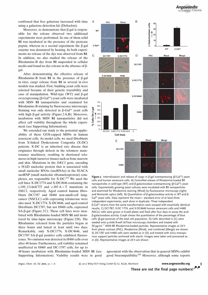
After demonstrating the effective release ofRhodamine-B from S1 in the presence of b-galin vitro, cargo release from S1 in several in vivomodels was studied. First, budding yeast cells wereselected because of their genetic tractability andease of manipulation. Wild-type (WT) and b-galoverexpressing (b-Galoe) yeast cells were incubatedwith MSN S1 nanoparticles and examined forRhodamine-B staining by fluorescence microscopy.Staining was only detected in b-Galoe yeast cellswith high b-gal activity (Figure 2A,B). Moreover,incubation with MSN S1 nanoparticles did notaffect cell viability throughout the whole experi-ment (see Supporting Information).
We extended our study to the potential applic-ability of these GOS-capped MSNs in humansenescent cells. As model cells, we used fibroblastsfrom X-linked Dyskeratosis Congenita (X-DC)patients. X-DC is an inherited rare disease thatoriginates through defects in the telomere main-tenance machinery, resulting in shortened telo-meres in high-turnover tissues such as bone marrowand skin. Mutations in the DKC1 gene, encodinga 58 kD nucleolar protein that is associated withsmall nucleolar RNAs (snoRNAs) in the H/ACAsnoRNP (small nucleolar ribonucleoprotein) com-plexes, are responsible for X-DC.[35] We used thecell lines X-DC1774 and X-DC4646 containing thec.109_111delCTT and c.385 A>T mutations inDKC1, respectively. Aged control human fibro-blasts DC1787 and H460 non-small-cell lung-cancer (NSCLC) cells expressing telomerase werealso used. X-DC1774, X-DC4646, and aged controlfibroblasts DC1787, but not H460 cells, expressedSA-b-gal (Figure 2C). These cell lines were incu-bated with Rhodamine-loaded MSN S1 and moni-tored by time-lapse microscopy (Figure 2D). TheRhodamine released from the particles began atthree hours and lasted at least until two days.Remarkably, only X-DC1774, X-DC4646, andDC1787 SA-b-gal positive cells exhibited fluores-cence. No emission was detected in H460 cells evenafter 48 hours. Furthermore, cell viability remainedunaffected in H460 and DC-1787 cells, for up to48 hours incubation with Rhodamine-loaded MSN S1 (seeSupporting Information). Viability results were in good
agreement with the observation that in general MSNs exhibitgood biocompatibility.[36] Moreover, although some reports
Figure 2. Internalization and release of cargo in b-gal overexpressing (b-Galoe) yeastcells and human senescent cells. A) Controlled release of Rhodamine-loaded S1nanoparticles in wild-type (WT) and b-galactosidase overexpressing (b-Galoe) yeastcells. Exponentially growing yeast cultures were incubated with S1 nanoparticlesand examined for Rhodamine staining (Rhod) by fluorescence microscopy (right)and Nomarski optics (left). B) Quantitation of b-galactosidase activity in WT and b-Galoe yeast cells. Data represent the mean� standard error of at least threeindependent experiments, each done in duplicate. Three independentb-Galoe strains from the same transformation were assayed with essentially identicalresults. C) DC1787, X-DC 1774, and X-DC4646 human senescent cells and H460NSCLC cells were grown in 6-well plates and fixed after four days to assay the acid-b-galactosidase activity. Graph shows the quantitation of the percentage of bluecells (b-gal positive) of the total cell population. D) Cells described in (C) wereseeded onto m-slide 8-well ibiTreat microscopy chambers and treated with50 mgmL�1 MSN S1 Rhodamine-loaded particles. Representative images at 24 hfrom phase contrast (PhC), Rhodamine (Rhod), and combined (Merge) are shown.E) DC1787 and H460 cells were seeded as in (D) and treated with silica mesopo-rous support particles anchored with starch. Images were taken and processed asin (D). Representative images at 24 h are shown.
AngewandteChemie
3Angew. Chem. Int. Ed. 2012, 51, 1 – 6 � 2012 Wiley-VCH Verlag GmbH & Co. KGaA, Weinheim www.angewandte.org
These are not the final page numbers! � �

suggested that, depending on the route of administrationin vivo, MSNs might become toxic,[37] our own data in humancells, are encouraging from the perspective of the potentialuse of S1 in in vivo models.
The experiments shown above clearly and remarkablyshow that despite the fact that GOS-capped S1 nanoparticlesare internalized in all cell types studied, they only releasedtheir cargo in b-gal overexpressing cells. Additionally, todemonstrate that the lack of Rhodamine staining in H460cells was not an artifact of the conditions used, additionalcontrol experiments were performed. Rhodamine-loadedMSN nanoparticles capped with hydrolyzed starch wereprepared (solid S2). MSNs of S2 are similar to S1 but containan oligosaccharide that is hydrolyzed by the amylase enzymein lysosomes.[38] When using S2 in internalization studiesunder similar conditions to those described above, clearlydetectable staining was observed in both of the cell linesstudied (Figure 2 E), indicating that the release of Rhodaminefrom S1 can be ascribed to a selective cellular b-gal enzyme-mediated mechanism.
In summary, the MSN S1 nanoparticles described hereinhave proven to be suitable nanodevices to selectively releasetheir cargo in senescent cells with high specificity and a lack ofdetectable toxicity. Nanoparticles S1 were able to selectivelydeliver their cargo in SA-b-gal positive b-Galoe yeast cells, inaged human fibroblasts DC1787, and in X-DC1774 and X-DC4646 cells from human Dyskeratosis Congenita patients,whereas no cargo release from S1 was observed in controlexperiments with H460 non-small-cell lung-cancer cells andwild-type yeast cells. To our knowledge, this is the first timethat a nanotherapy has been targeted to senescent cells. Theseresults suggest that by choosing an appropriate cargo (that isa telomerase reactivation drug or a cytotoxic drug) preven-tion and removal/replacement of senescent cells could bepossible. Despite the fact that the road from these results tosenescent-cell removal or rejuvenation therapies remainslong and uncertain, we believe that our findings might openup new avenues for developing innovative therapeuticapplications to treat or delay age-related diseases.
Received: June 14, 2012Revised: July 31, 2012Published online: && &&, &&&&
.Keywords: controlled release · drug delivery ·dyskeratosis congenita · gated mesoporous materials ·nanoparticles
[1] D. G. Burton, Cellular senescence, ageing and disease, Vol. 31,Age, Dordrecht, Netherlands, 2009, pp. 1 – 9.
[2] C. R. Burtner, B. K. Kennedy, Nat. Rev. 2010, 11, 567 – 578.[3] N. Ishikawa, K. Nakamura, N. Izumiyama-Shimomura, J. Aida,
A. Ishii, M. Goto, Y. Ishikawa, R. Asaka, M. Matsuura, A.Hatamochi, M. Kuroiwa, K. Takubo, Aging 2011, 3, 417 – 429.
[4] A. K. Ghosh, M. L. Rossi, D. K. Singh, C. Dunn, M. Rama-moorthy, D. L. Croteau, Y. Liu, V. A. Bohr, J. Biol. Chem. 2012,287, 196 – 209.
[5] R. T. Calado, N. S. Young, N. Engl. J. Med. 2009, 361, 2353 –2365.
[6] B. P. Alter, N. Giri, S. A. Savage, P. S. Rosenberg, Blood 2009,113, 6549 – 6557.
[7] D. J. Baker, T. Wijshake, T. Tchkonia, N. K. LeBrasseur, B. G.Childs, B. van de Sluis, J. L. Kirkland, J. M. van Deursen, Nature2011, 479, 232 – 236.
[8] M. Jaskelioff, F. L. Muller, J. H. Paik, E. Thomas, S. Jiang, A. C.Adams, E. Sahin, M. Kost-Alimova, A. Protopopov, J. CadiÇa-nos, J. W. Horner, E. Maratos-Flier, R. A. DePinho, Nature 2011,469, 102 – 106.
[9] S. Liu, R. Maheshwari, K. L. Kiick, Macromolecules 2009, 42, 3 –13.
[10] C. C. Lee, J. A. MacKay, J. M. Frechet, F. C. Szoka, Nat.Biotechnol. 2005, 23, 1517 – 1526.
[11] C. W. Pouton, C. J. Porter, Adv. Drug Delivery Rev. 2008, 60,625 – 637.
[12] I. Brigger, C. Dubernet, P. Couvreur, Adv. Drug Delivery Rev.2002, 54, 631 – 651.
[13] M. Vallet-Reg�, F. Balas, D. Arcos, Angew. Chem. 2007, 119,7692 – 7703; Angew. Chem. Int. Ed. 2007, 46, 7548 – 7558.
[14] T. D. Nguyen, K. C. F. Leung, M. Liong, Y. Liu, J. F. Stoddart,J. I. Zink, Adv. Funct. Mater. 2007, 17, 2101 – 2110.
[15] B. G. Trewyn, S. Giri, I. I. Slowing, V. S. Y. Lin, Chem. Commun.2007, 3236 – 3245.
[16] R. Klajn, J. F. Stoddart, B. A. Grzybowski, Chem. Soc. Rev. 2010,39, 2203 – 2237.
[17] E. Climent, A. Bernardos, R. Mart�nez-M�Çez, A. Maquieira,M. D. Marcos, N. Pastor-Navarro, R. Puchades, F. Sancen�n, J.Soto, P. Amor�s, J. Am. Chem. Soc. 2009, 131, 14075 – 14080.
[18] A. Schlossbauer, J. Kecht, T. Bein, Angew. Chem. 2009, 121,3138 – 3141; Angew. Chem. Int. Ed. 2009, 48, 3092 – 3095.
[19] B. G. Trewyn, I. I. Slowing, S. Giri, H. T. Chen, V. S. Y. Lin, Acc.Chem. Res. 2007, 40, 846 – 853.
[20] S. Saha, K. C. F. Leung, T. D. Nguyen, J. F. Stoddart, J. I. Zink,Adv. Funct. Mater. 2007, 17, 685 – 693.
[21] Y.-W. Yang, Med. Chem. Commun. 2011, 2, 1033 – 1049.[22] a) E. Climent, R. Mart�nez-M�Çez, F. Sancen�n, M. D. Marcos, J.
Soto, A. Maquieira, P. Amor�s, Angew. Chem. 2010, 122, 7439 –7441; Angew. Chem. Int. Ed. 2010, 49, 7281 – 7283; b) A.Agostini, L. Mondrag�n, C. Coll, E. Aznar, M. D. Marcos, R.Mart�nez-M�Çez, F. Sancen�n, J. Soto, E. P�rez-Pay�, P.Amor�s, ChemistryOpen 2012, 1, 17 – 20.
[23] K. Ariga, A. Vinu, Y. Yamauchi, Q. Ji, J. P. Hill, Bull. Chem. Soc.Jpn. 2012, 85, 1 – 32.
[24] Z. Li, J. C. Barnes, A. Bosoy, J. F. Stoddart, J. I. Zink, Chem. Soc.Rev. 2012, 41, 2590 – 2605.
[25] P. Yang, S. Gai, J. Lin, Chem. Soc. Rev. 2012, 41, 3679 – 3698.[26] G. P. Dimri, X. Lee, G. Basile, M. Acosta, G. Scott, C. Roskelley,
E. E. Medrano, M. Linskens, I. Rubelj, O. Pereira-Smith, M.Peacocke, J. Campisi, Proc. Natl. Acad. Sci. USA 1995, 92, 9363 –9367.
[27] B. Y. Lee, J. A. Han, J. S. Im, A. Morrone, K. Johung, E. C.Goodwin, W. J. Kleijer, D. DiMaio, E. S. Hwang, Aging Cell2006, 5, 187 – 195.
[28] M. Knas, A. Zalewska, R. Kretowski, M. Niczyporuk, N.Waszkiewicz, M. Cechowska-Pasko, D. Waszkiel, K. Zwierz,Folia Histochem. Cytobiol. 2012, 50, 19353.
[29] B. Lecka-Czernik, E. J. Moerman, R. J. Shmookler Reis, D. A.Lipschitz, J. Gerontol. Ser. A 1997, 52, B331 – B336.
[30] B. W. Gu, J. M. Fan, M. Bessler, P. J. Mason, Aging Cell 2011, 10,338 – 348.
[31] S. Chigira, K. Sugita, K. Kita, S. Sugaya, Y. Arase, M. Ichinose,H. Shirasawa, N. Suzuki, J. Gerontol. Ser. A 2003, 58, B873 –B878.
[32] S. Cabrera, J. El Haskouri, C. Guillem, J. Latorre, A. Beltr�n, D.Beltr�n, M. D. Marcos, P. Amor�s, Solid State Sci. 2000, 2, 405 –420.
.AngewandteCommunications
4 www.angewandte.org � 2012 Wiley-VCH Verlag GmbH & Co. KGaA, Weinheim Angew. Chem. Int. Ed. 2012, 51, 1 – 6� �
These are not the final page numbers!

[33] X. Auvray, C. Petipas, R. Anthore, I. Rico-Lattes, A. Lattes,Langmuir 1995, 11, 433 – 439.
[34] R. Perona, R. Machado-Pinilla, C. Manguan, J. Carrillo, Clin.Trasl. Oncol. 2009, 11, 711 – 714.
[35] F. Tang, L. Li, D. Chen, Adv. Mater. 2012, 24, 1504 – 1534.
[36] S. P. Hudson, R. F. Padera, R. Langer, D. S. Kohane, Biomate-rials 2008, 29, 4045 – 4055.
[37] A. Bernardos, L. Mondrag�n, E. Aznar, M. D. Marcos, R.Mart�nez-M�Çez, F. Sancen�n, J. Soto, J. M. Barat, E. P�rez-Pay�, C. Guillem, P. Amor�s, ACS Nano 2010, 4, 6353 – 6368.
AngewandteChemie
5Angew. Chem. Int. Ed. 2012, 51, 1 – 6 � 2012 Wiley-VCH Verlag GmbH & Co. KGaA, Weinheim www.angewandte.org
These are not the final page numbers! � �

Communications
Drug-Carrying Nanoparticles
A. Agostini, L. Mondrag�n, A. Bernardos,R. Mart�nez-M�Çez,* M. D. Marcos,F. Sancen�n, J. Soto, A. Costero,C. Manguan-Garc�a, R. Perona,*M. Moreno-Torres, R. Aparicio-Sanchis,J. R. Murgu�a* &&&&—&&&&
Targeted Cargo Delivery in SenescentCells Using Capped Mesoporous SilicaNanoparticles
Learning to let go with age : Intracellularcontrolled release of molecules withinsenescent cells was achieved using mes-oporous silica nanoparticles (MSNs)capped with a galacto-oligosaccharide(GOS) to contain the cargo molecules(magenta spheres; see scheme). TheGOS is a substrate of the senescentbiomarker, senescence-associated b-gal-actosidase (SA-b-gal), and releases thecargo upon entry into SA-b-gal expressingcells.
.AngewandteCommunications
6 www.angewandte.org � 2012 Wiley-VCH Verlag GmbH & Co. KGaA, Weinheim Angew. Chem. Int. Ed. 2012, 51, 1 – 6� �
These are not the final page numbers!