Embed Size (px)
Citation preview
B
TM
HQA*CM
B(tfllegsCFcbelhtcFhMgerlFteCelatp
Mhf
GASTROENTEROLOGY 2006;130:1245–1258
ASIC–LIVER, PANCREAS, AND BILIARY TRACT
argeted Deletion of FATP5 Reveals Multiple Functions in Liveretabolism: Alterations in Hepatic Lipid Homeostasis
OLGER DOEGE,* REBECCA A. BAILLIE,‡ ANGELICA M. ORTEGON,§ BERNICE TSANG,§
IWEI WU,* SANDHYA PUNREDDY,¶ DAVID HIRSCH,� NICKI WATSON,� RUTH E. GIMENO,¶ andNDREAS STAHL*,§
Division of GI/Hepatology, Stanford University School of Medicine, Stanford, California; ‡Lipomics Technologies, Inc, West Sacramento,alifornia; §Palo Alto Medical Foundation Research Institute, Palo Alto, California; ¶Millennium Pharmaceuticals, Inc, Cambridge,
assachusetts; and �Whitehead Institute for Biomedical Research, Cambridge, Massachusettsvrdldvcl2eaiotmtTa(nemit
taft
apLg
ackground & Aims: Fatty acid transport protein 5FATP5/Slc27a5) has been shown to be a multifunc-ional protein that in vitro increases both uptake ofuorescently labeled long-chain fatty acid (LCFA) ana-
ogues and bile acid/coenzyme A ligase activity on over-xpression. The aim of this study was to further investi-ate the diverse roles of FATP5 in vivo. Methods: Wetudied FATP5 expression and localization in liver of57BL/6 mice in detail. Furthermore, we created aATP5 knockout mouse model and characterizedhanges in hepatic lipid metabolism (this report) andile metabolism (the accompanying report by Hubbardt al). Results: FATP5 is exclusively expressed by the
iver and localized to the basal plasma membrane ofepatocytes, congruent with a role in LCFA uptake fromhe circulation. Overexpression of FATP5 in mammalianells increased the uptake of 14C-oleate. Conversely,ATP5 deletion significantly reduced LCFA uptake byepatocytes isolated from FATP5 knockout animals.oreover, FATP5 deletion resulted in lower hepatic tri-
lyceride and free fatty acid content despite increasedxpression of fatty acid synthetase and also caused aedistribution of lipids from liver to other LCFA-metabo-izing tissues. Detailed analysis of the hepatic lipom ofATP5 knockout livers showed quantitative and qualita-ive alterations in line with a decreased uptake of di-tary LCFAs and increased de novo synthesis.onclusions: Our findings support the hypothesis thatfficient hepatocellular uptake of LCFAs, and thus liver
ipid homeostasis in general, is largely a protein-medi-ted process requiring FATP5. These new insights intohe physiological role of FATP5 should lead to an im-roved understanding of liver function and disease.
aintaining appropriate long-chain fatty acid(LCFA) uptake by the liver is crucial for energy
omeostasis. During starvation, LCFAs are mobilized
rom adipose tissue, taken up by the liver, and con-erted into ketone bodies and other substrates. Dis-uption of these processes, as found in some hereditaryefects of liver LCFA uptake, is associated with acuteiver failure and mortality.1,2 The underlying geneticefect(s), however, are currently unknown. Con-ersely, excessive liver LCFA uptake (eg, due to vis-eral adiposity and elevated portal fatty acid [FA]evels)3 is associated with insulin desensitization, type
diabetes mellitus,4 and nonalcoholic fatty liver dis-ase.5 Considerable evidence has accumulated that inddition to diffusion, LCFA uptake by the liver,6 –11
ntestine,12–14 heart,10,15 adipose tissue,16 and otherrgans is mediated by a saturable and specific LCFAransport system.17,18 Investigators have found severalembrane proteins that, when overexpressed in cul-
ured mammalian cells, increase the uptake of LCFAs.he most prominent and best characterized of thesere FAT/CD36 and fatty acid transport proteinsFATPs, solute carrier family 27) 1 or 4. However,either CD3619,20 nor FATP1 or FATP413,16,18,21 isxpressed at appreciable levels in the liver. Further-ore, CD36-null animals show reduced LCFA uptake
nto skeletal muscle and heart but increased uptake byhe liver.22–24
Based on sequence analyses, we have previously foundhat FATP5 is a member of the FATP gene family21,25
nd that FATP5, like the other members of the FATPamily, can enhance the uptake of BODIPY-FA uponransient overexpression in HEK293 cells.21 In addition
Abbreviations used in this paper: FA, fatty acid; FACS, fluorescence-ctivated cell sorter; FAS, fatty acid synthetase; FATP, fatty acid trans-ort protein; FFA, free fatty acid; HBS, Hank’s buffered salt solution;CFA, long-chain fatty acid; PCR, polymerase chain reaction; TG, tri-lyceride.© 2006 by the American Gastroenterological Association Institute
0016-5085/06/$32.00
doi:10.1053/j.gastro.2006.02.006
taFLcmdaiiFtrph
3
Ac3o(rC
aF9anJtmi
(tMaBNeaI
tmFpgaTcPleccwfsG
Ahta
wsB1d
b
c1�hbc
mw
1246 DOEGE ET AL GASTROENTEROLOGY Vol. 130, No. 4
o its function as FA transporter, FATP5 also facilitatest least 2 different enzymatic reactions. As is the case forATP1, FATP2, and FATP4,26–28 FATP5 can activateCFAs and very long-chain fatty acids by catalyzing theovalent attachment of coenzyme A (CoA).28 Further-ore, FATP5 is also involved in the reactivation, but not
e novo synthesis, of C24 bile acids to their CoA deriv-tives.29,30 However, how these functions are integratedn vivo is only poorly understood. To this end, wenvestigated localization of FATP5 in vivo and created aATP5 knockout mouse model to study its contributiono bile (Hubbard et al31) and lipid metabolism (thiseport). Here we show that FATP5 is a liver-specificrotein required for efficient hepatic LCFA uptake andepatic lipid homeostasis.
Materials and Methods
Materials
Deoxycitidine-5=[�-32P]triphosphate and Tri-[9,10-H(N)]oleoylglycerol (14C-triolein) were purchased frommersham Bioscience (Piscataway, NJ). 14C-oleate, 14C-tauro-
holate, 4,4-difluoro-5-methyl-4-bora-3a,4a-diaza-s-indacene--dodecanoid acid (C1-BODIPY-C12), and cerulenin werebtained from ARC Inc (St Louis, MO), Molecular ProbesEugene, OR), and Alexis Biochemicals (San Diego, CA),espectively. All other chemicals were obtained from Sigmahemical Co (St Louis, MO).
Antibodies
Murine FATP5 specific antibody was raised in rabbitsgainst a glutathione S-transferase/FATP5 fusion protein. TheATP5 portion of the fusion protein was comprised of the last3 C-terminal amino acids of FATP5. Monoclonal antibodiesgainst murine fatty acid synthetase (FAS), CD31, and �-cate-in were purchased from BD Bioscience (Palo Alto, CA),ackson ImmunoResearch (West Grove, PA), and Transduc-ion Labs (Lexington, KY), respectively. The antibody againsturine keratin 8 was obtained from the Developmental Stud-
es Hybridoma Bank (Iowa City, IA).
Animal Experiments
Animals were maintained on regular laboratory chow5P75; LabDiet, Richmond, IN). Triglyceride (TG), choles-erol, bile acid, albumin (all from Sigma Diagnostics, St Louis,O), free fatty acid (FFA; Wako Chemicals, Richmond, VA),
lanine aminotransferase, bilirubin (Stanbio Laboratory,oerne, TX), and insulin (ALPCO Diagnostics, Windham,H) levels were determined using commercial kits. All animal
xperiments were approved by the Institutional Animal Carend Use Committee of Palo Alto Medical Foundation Research
nstitute. sGeneration of FATP5 Knockout Mice
Genomic DNA containing the FATP5 locus was iden-ified by screening a 129Sv genomic bacterial artificial chro-osome library with a fragment containing the 5= end of the
ATP5 coding sequence. Polymerase chain reaction (PCR)rimers were designed to amplify a 6.2-kilobase fragment ofenomic DNA upstream of the initiation codon (5= arm) and2.1-kilobase fragment downstream of the first exon (3= arm).he amplified arms were subcloned into a targeting vectorontaining a neomycin resistance marker under control of theGK promoter and a promoter-less lacZ gene with a nuclear
ocalization sequence located downstream of the 5= arm. 129Svmbryonic stem cells were electroporated with the targetingonstruct and screened by Southern blotting. Embryonic stemell clones that had undergone homologous recombinationere injected into blastocysts. Injected blastocysts were trans-
erred into pseudopregnant mice to generate chimeric off-pring. Male chimeras were mated with C57BL/6 female mice.enotyping of offspring was performed by PCR.
Northern Blots
Northern blots were obtained from Clontech (Palolto, CA) and hybridized with 32P-labeled cDNA probes foruman FATP5 generated by PCR (primer pair: hF5pfw, 5=-cttgcaacaggttaacgtg-3=/hF5prv, 5=-gggctagctgcacagcagcca-3=)s previously described.21
Hepatic TG Secretion
After a fasting period of 4 hours, anesthetized miceere injected intravenously with Triton WR 1339 diluted in
aline (200 mg/mL) via the tail vein (500 mg/kg body wt).32
lood samples were taken before the injection (t � 0) and 60,20, and 210 minutes after injection and serum TG levelsetermined.
Hepatic Lipase Activity
Enzyme activity was assayed using a method describedy Nilsson-Ehle and Schotz.33
14C-Lipid Gavage
Mice were given an intragastric 200 �L olive oil bolusontaining 2 �Ci 14C-oleic acid. Before (t � 0) and at 30, 60,20, and 240 minutes after administration, blood samples (75L) were taken by orbital eye bleeding and tissues werearvested after the last time point. 14C-content was measuredy scintillation counting and normalized to milligram proteinontent for tissues.
Inhibition of FA de novo Synthesis
Mice were injected intraperitoneally with cerulenin 60g · kg�1 · day�1 for 3 days and kept under absence of foodith access to water ad libitum. After 56 hours, animals were
acrificed and their livers were harvested.
aphic0Ct0i5[camiiF
lc
wstpHpbr4st0mo3R
tutau1
tTwhs
c
wdapmaebpetPcitvwoFfhscscilS
iTmd
e
April 2006 FATP5 KNOCKOUT MICE AND HEPATIC LIPID HOMEOSTASIS 1247
Hepatocyte Preparation and Fluorescence-Activated Cell Sorter Analysis
Mouse livers were cannulated through the portal vein,nd an incision was made in the inferior vena cave. Livererfusion with digestion and perfusion media and isolation ofepatocytes were performed according to the manufacturer’snstructions (Gibco, Carlsbad, CA). After isolation, hepato-ytes were resuspended in HepatoZYME (Gibco) containing.1% FA-free bovine serum albumin (Sigma Diagnostics).1-BODIPY-C12/bovine serum albumin solution13 was added
o the hepatocytes to yield final concentrations of 2 �mol/L or.1%, respectively. Uptake at 37°C was stopped after thendicated time points by transferring 100 �L of the cells into
mL of ice-cold stop solution (Hank’s buffered salt solutionHBS], 0.1% bovine serum albumin). Cells were pelleted byentrifugation and resuspended in 250 �L cold fluorescence-ctivated cell sorter (FACS) buffer (HBS containing 20mol/L EDTA, 10% fetal calf serum, and 1 �g/mL propidium
odine). Mean BODIPY uptake by living cells (propidiumodine negative, dead cells gated out) was determined using aACSCalibur (Becton Dickinson, Rockville, MD).
Tissue Lipid Analysis
Tissue samples were powdered in liquid nitrogen, totalipids were extracted by the method of Folch et al,34 and TGontent was measured.
FA Uptake Measurement in HepatocytesUsing 14C-Labeled Oleate
Alternatively, LCFA uptake by isolated hepatocytesas assayed using 14C-oleate essentially as previously de-
cribed.35,36 Briefly, isolated hepatocytes were allowed to at-ach overnight to a type I collagen–coated 12-well clusterlate. Dead cells were removed by 2 washes with serum-freeepatoZYME and incubated at 37°C for 15 seconds with a
rewarmed uptake solution containing 200 �mol/L 14C-oleateound to bovine serum albumin at a 2:1 molar ratio in HBSesulting in an approximate unbound oleate concentration of00 nmol/L. Wells were washed twice with ice-cold stopolution and cells were lysed in 200 �L radioimmunoprecipi-ation assay buffer (150 mmol/L NaCl, 1% Nonidet P-40,.5% deoxycholic acid, 0.1% sodium dodecyl sulfate, 50mol/L Tris, pH 8.0) for 30 minutes on ice. A total of 150 �L
f lysate was subsequently used for scintillation counting, and0 �L was used for colorimetric protein determination (Pierce,ockford, IL).
FA Uptake Measurement in HeLa Cells
HeLa cell monolayers were transiently transfected withhe indicated constructs or with empty vector (pcDNA3.1)sing FuGENE reagent (Roche, Palo Alto, CA). Two days afterransfection, cells were detached, counted, washed with HBS,nd incubated at 37°C for the indicated time points withptake solution containing either 1 �mol/L 14C-oleate or
4C-taurocholate and 0.1% bovine serum albumin in HBS in m
he absence or presence of 50 �mol/L unlabeled taurocholate.he reaction was stopped with ice-cold stop solution. Cellsere transferred onto Whatman paper using an automaticarvester (Brandel, Gaithersburg, MD), washed 3 times withtop solution, and subsequently used for scintillation counting.
Immunofluorescence and ImmunoelectronMicroscopy
Immunofluorescence37 and immunoelectron micros-opy13 were performed as described before.
Lipid Analysis
The lipids from plasma (200 �L) and tissues (25 mg)ere extracted in the presence of authentic internal stan-ards.34 Individual lipid classes within each extract were sep-rated by preparative thin-layer chromatography as describedreviously.38 Isolated lipid classes were trans-esterified in 3Nethanolic HCl in a sealed vial under a nitrogen atmosphere
t 100°C for 45 minutes. The resulting FA methyl esters werextracted from the mixture with hexane containing 0.05%utylated hydroxytoluene and prepared for gas chromatogra-hy by sealing the hexane extracts under nitrogen. FA methylsters were separated and quantified by capillary gas chroma-ography using a gas chromatograph (model 6890; Hewlett-ackard, Wilmington, DE) equipped with a 30-m DB-225MSapillary column (J&W Scientific, Folsom, CA) and a flame-onization detector as described previously.38 Liver lipid me-abolite data were displayed using the Lipomics Surveyorisualization software (Lipomics Tech. Inc., Sacramento, CA),hich illustrates the quantitative differences in concentrationf each lipid metabolite when comparing wild-type andATP5 knockout mice. Column headings display the FA andamilies of FAs present in each lipid class listed in the roweadings. Surveyor’s heat map display is read as follows: eachquare represents the statistical comparison of the difference inoncentration of a single FA. A higher concentration of apecific metabolite is displayed in green, whereas a loweroncentration is displayed in red. The brightness of the colorndicates the magnitude of the difference as detailed in theegend. Mean differences not meeting P � .05 (assessed bytudent t test) are displayed in black.
Statistics
Chemiluminescent signals were directly quantified us-ng Kodak 1D version 3.5.3 software (Kodak, Rochester, NY).he absolute integration value of the immunoreactive bandsinus background was determined. Statistical analysis was
etermined by nonpaired Student t test.
Results
FATP5 Is Specifically Expressed in Liver
Based on our initial observation that FATP5 isxpressed in liver but not in heart, brain, spleen, lung,
uscle, kidney, or testis,21 we expanded Northern blot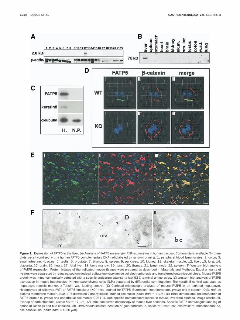
FbspolpehHpFosb
1248 DOEGE ET AL GASTROENTEROLOGY Vol. 130, No. 4
igure 1. Expression of FATP5 in the liver. (A) Analysis of FATP5 messenger RNA expression in human tissues. Commercially available Northernlots were hybridized with a human FATP5 complementary DNA radiolabeled by random priming. 1, peripheral blood lymphocytes; 2, colon; 3,mall intestine; 4, ovary; 5, testis; 6, prostate; 7, thymus; 8, spleen; 9, pancreas; 10, kidney; 11, skeletal muscle; 12, liver; 13, lung; 14,lacenta; 15, brain; 16, heart; 17, fetal liver; 18, bone marrow; 19, tonsil; 20, thymus; 21, lymph node; 22, spleen. (B) Western blot analysisf FATP5 expression. Protein lysates of the indicated mouse tissues were prepared as described in Materials and Methods. Equal amounts of
ysates were separated by reducing sodium dodecyl sulfate/polyacrylamide gel electrophoresis and transferred onto nitrocellulose. Mouse FATP5rotein was immunochemically detected with a specific antiserum against its last 93 C-terminal amino acids. (C) Western blot analysis of FATP5xpression in mouse hepatocytes (H.)/nonparenchymal cells (N.P.) separated by differential centrifugation. The keratin-8 control was used asepatocyte-specific marker. �-Tubulin was loading control. (D) Confocal microscopic analysis of mouse FATP5 in an isolated hepatocyte.epatocytes of wild-type (WT) or FATP5 knockout (KO) mice stained for FATP5 (fluorescein isothiocyanate, green) and �-catenin (Cy3, red) aslasma membrane marker. Blue, 4=,6-diamidino-2-phenylindole–stained cell nuclei (scale bars � 4 �m). (E) Three-dimensional reconstruction ofATP5 protein (I, green) and endothelial cell marker CD31 (II, red) specific immunofluorescence in mouse liver from confocal image stacks (III,verlap of both channels) (scale bar � 17 �m). (F) Immunoelectron microscopy of mouse liver sections. Specific FATP5 immunogold labeling ofpace of Disse (I) and bile canaliculi (II). Arrowheads indicate position of gold particles. D, space of Disse; mv, microvilli; m, mitochondria; bc,
ile canaliculus (scale bars � 0.20 �m).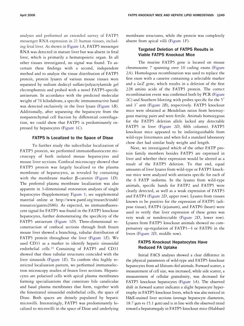
amiRlocmpseawwAntp
FcmFmwTahmrchFcmFueslstcfatDmc
ma
c2fia2r2amgfFkwc
tlraotacakpuvLpl
thmmFstH1
April 2006 FATP5 KNOCKOUT MICE AND HEPATIC LIPID HOMEOSTASIS 1249
nalyses and performed an extended survey of FATP5essenger RNA expression in 21 human tissues, includ-
ng fetal liver. As shown in Figure 1A, FATP5 messengerNA was detected in mature liver but was absent in fetal
iver, which is primarily a hematopoietic organ. In allther tissues investigated, no signal was found. To as-ertain these findings with a second, independentethod and to analyze the tissue distribution of FATP5
rotein, protein lysates of various mouse tissues wereeparated by sodium dodecyl sulfate/polyacrylamide gellectrophoresis and probed with a novel FATP5-specificntiserum. In accordance with the predicted moleculareight of 76 kilodaltons, a specific immunoreactive bandas detected exclusively in the liver lysate (Figure 1B).dditionally, after separating the hepatocyte from theonparenchymal cell fraction by differential centrifuga-ion, we could show that FATP5 is predominantly ex-ressed by hepatocytes (Figure 1C).
FATP5 Is Localized to the Space of Disse
To further study the subcellular localization ofATP5 protein, we performed immunofluorescent mi-roscopy of both isolated mouse hepatocytes andouse liver sections. Confocal microscopy showed that
ATP5 protein was largely localized to the plasmaembrane of hepatocytes, as revealed by costainingith the membrane marker �-catenin (Figure 1D).he preferred plasma membrane localization was alsopparent in 3-dimensional restoration analyses of singleepatocytes (Supplementary Figure 1; see supplementalaterial online at http://www.pamf.org/research/stahl/
esources/gastro2686). As expected, no immunofluores-ent signal for FATP5 was found in the FATP5 knockoutepatocytes, further demonstrating the specificity of theATP5 antiserum (Figure 1D). Three-dimensional re-onstruction of confocal sections through fresh frozenouse liver showed a branching, tubular distribution of
ATP5 protein throughout the liver (Figure 1E). Wesed CD31 as a marker to identify hepatic sinusoidalndothelial cells.39 Costaining of FATP5 and CD31howed that these tubular structures coincided with theiver sinusoids (Figure 1E). To confirm this highly re-tricted localization pattern, we performed immunoelec-ron microscopy studies of frozen liver sections. Hepato-ytes are polarized cells with apical plasma membranesorming specializations that constitute bile canaliculaend basal plasma membranes that form, together withhe fenestrated sinusoidal endothelial cells, the space ofisse. Both spaces are densely populated by hepaticicrovilli. Interestingly, FATP5 was predominantly lo-
alized to microvilli in the space of Disse and underlying t
embrane structures, while the protein was completelybsent from apical villi (Figure 1F).
Targeted Deletion of FATP5 Results inViable FATP5 Knockout Mice
The murine FATP5 gene is located on mousehromosome 7 spanning over 10 coding exons (FigureA). Homologous recombination was used to replace therst exon with a cassette containing a selectable markernd a lacZ gene, which results in a deletion of the first28 amino acids of the FATP5 protein. The correctecombination event was confirmed both by PCR (FigureC) and Southern blotting with probes specific for the 5=nd 3= arm (Figure 2B), respectively. FATP5 knockoutice were obtained at Mendelian ratios from heterozy-
ous mating pairs and were fertile. Animals homozygousor the FATP5 deletion allele lacked any detectableATP5 in liver (Figure 2D, fifth column). FATP5nockout mice appeared to be indistinguishable fromild-type littermates and when fed a standard laboratory
how diet had similar body weight and length.Next, we investigated which of the other FATP pro-
ein family members besides FATP5 are expressed iniver and whether their expression would be altered as aesult of the FATP5 deletion. To that end, equalmounts of liver lysates from wild-type or FATP5 knock-ut mice were analyzed with antisera specific for each ofhe 6 FATP isoforms. In the lysates from wild-typenimals, specific bands for FATP2 and FATP5 werelearly detected, as well as a weak expression of FATP3nd FATP4 (Figure 2D, upper row). Lysates from tissuesnown to be positive for the expression of FATP1 (adi-ose tissue), FATP4 (jejunum), and FATP6 (heart) weresed to verify that liver expression of these genes wasery weak or nondetectable (Figure 2D, lower row).ysates from FATP5 knockout animals showed no com-ensatory up-regulation of FATP1–4 or FATP6 in theivers (Figure 2D, middle row).
FATP5 Knockout Hepatocytes HaveReduced FA Uptake
Initial FACS analyses showed a clear difference inhe physical parameters of wild-type and FATP5 knockoutepatocytes from ad libitum–fed animals. Forward scatter, aeasurement of cell size, was increased, while side scatter, aeasurement of cellular granulosity, was decreased for
ATP5 knockout hepatocytes (Figure 3A). The observedhift in forward scatter indicates a slight hepatocyte hyper-rophy in FATP5 knockout livers, which was also noticed in&E-stained liver sections (average hepatocyte diameters,
8.7 �m vs 15.1 �m) and is in line with the observed trend
oward a hepatomegaly in FATP5 knockout mice (Hubbard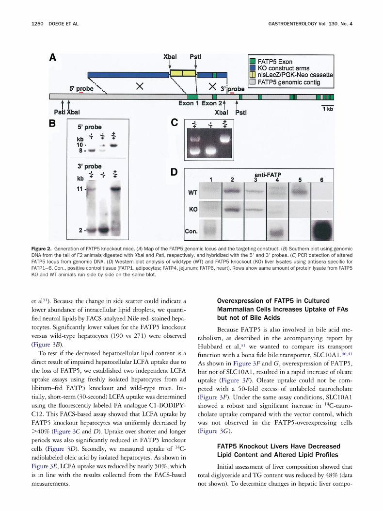
elfitv(
dtultuCF�pcrFim
tHfAbup(scw(
t
FDFFK
1250 DOEGE ET AL GASTROENTEROLOGY Vol. 130, No. 4
t al31). Because the change in side scatter could indicate aower abundance of intracellular lipid droplets, we quanti-ed neutral lipids by FACS-analyzed Nile red–stained hepa-ocytes. Significantly lower values for the FATP5 knockoutersus wild-type hepatocytes (190 vs 271) were observedFigure 3B).
To test if the decreased hepatocellular lipid content is airect result of impaired hepatocellular LCFA uptake due tohe loss of FATP5, we established two independent LCFAptake assays using freshly isolated hepatocytes from adibitum–fed FATP5 knockout and wild-type mice. Ini-ially, short-term (30-second) LCFA uptake was determinedsing the fluorescently labeled FA analogue C1-BODIPY-12. This FACS-based assay showed that LCFA uptake byATP5 knockout hepatocytes was uniformly decreased by40% (Figure 3C and D). Uptake over shorter and longer
eriods was also significantly reduced in FATP5 knockoutells (Figure 3D). Secondly, we measured uptake of 14C-adiolabeled oleic acid by isolated hepatocytes. As shown inigure 3E, LCFA uptake was reduced by nearly 50%, whichs in line with the results collected from the FACS-based
igure 2. Generation of FATP5 knockout mice. (A) Map of the FATP5 gNA from the tail of F2 animals digested with XbaI and PstI, respectivATP5 locus from genomic DNA. (D) Western blot analysis of wild-typATP1–6. Con., positive control tissue (FATP1, adipocytes; FATP4, jejuO and WT animals run side by side on the same blot.
easurements. n
Overexpression of FATP5 in CulturedMammalian Cells Increases Uptake of FAsbut not of Bile Acids
Because FATP5 is also involved in bile acid me-abolism, as described in the accompanying report byubbard et al,31 we wanted to compare its transport
unction with a bona fide bile transporter, SLC10A1.40,41
s shown in Figure 3F and G, overexpression of FATP5,ut not of SLC10A1, resulted in a rapid increase of oleateptake (Figure 3F). Oleate uptake could not be com-eted with a 50-fold excess of unlabeled taurocholateFigure 3F). Under the same assay conditions, SLC10A1howed a robust and significant increase in 14C-tauro-holate uptake compared with the vector control, whichas not observed in the FATP5-overexpressing cells
Figure 3G).
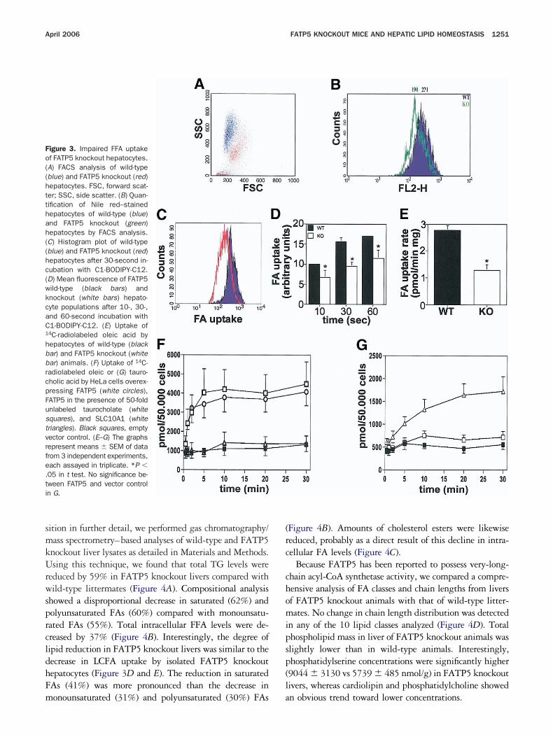
FATP5 Knockout Livers Have DecreasedLipid Content and Altered Lipid Profiles
Initial assessment of liver composition showed thatotal diglyceride and TG content was reduced by 48% (data
ic locus and the targeting construct. (B) Southern blot using genomicnd hybridized with the 5= and 3= probes. (C) PCR detection of alteredT) and FATP5 knockout (KO) liver lysates using antisera specific forFATP6, heart). Rows show same amount of protein lysate from FATP5
enomely, ae (Wnum;
ot shown). To determine changes in hepatic liver compo-
smkUrwsprcldhFm
(rc
chomipsp(l
Fo((htthah((hc(wkcaC1
hbbrcpFustvrfe.ti
April 2006 FATP5 KNOCKOUT MICE AND HEPATIC LIPID HOMEOSTASIS 1251
ition in further detail, we performed gas chromatography/ass spectrometry–based analyses of wild-type and FATP5
nockout liver lysates as detailed in Materials and Methods.sing this technique, we found that total TG levels were
educed by 59% in FATP5 knockout livers compared withild-type littermates (Figure 4A). Compositional analysis
howed a disproportional decrease in saturated (62%) andolyunsaturated FAs (60%) compared with monounsatu-ated FAs (55%). Total intracellular FFA levels were de-reased by 37% (Figure 4B). Interestingly, the degree ofipid reduction in FATP5 knockout livers was similar to theecrease in LCFA uptake by isolated FATP5 knockoutepatocytes (Figure 3D and E). The reduction in saturatedAs (41%) was more pronounced than the decrease in
igure 3. Impaired FFA uptakef FATP5 knockout hepatocytes.A) FACS analysis of wild-typeblue) and FATP5 knockout (red)epatocytes. FSC, forward scat-er; SSC, side scatter. (B) Quan-ification of Nile red–stainedepatocytes of wild-type (blue)nd FATP5 knockout (green)epatocytes by FACS analysis.C) Histogram plot of wild-typeblue) and FATP5 knockout (red)epatocytes after 30-second in-ubation with C1-BODIPY-C12.D) Mean fluorescence of FATP5ild-type (black bars) andnockout (white bars) hepato-yte populations after 10-, 30-,nd 60-second incubation with1-BODIPY-C12. (E) Uptake of
4C-radiolabeled oleic acid byepatocytes of wild-type (blackar) and FATP5 knockout (whitear) animals. (F) Uptake of 14C-adiolabeled oleic or (G) tauro-holic acid by HeLa cells overex-ressing FATP5 (white circles),ATP5 in the presence of 50-foldnlabeled taurocholate (whitequares), and SLC10A1 (whiteriangles). Black squares, emptyector control. (E–G) The graphsepresent means � SEM of datarom 3 independent experiments,ach assayed in triplicate. *P �05 in t test. No significance be-ween FATP5 and vector controln G.
onounsaturated (31%) and polyunsaturated (30%) FAs a
Figure 4B). Amounts of cholesterol esters were likewiseeduced, probably as a direct result of this decline in intra-ellular FA levels (Figure 4C).
Because FATP5 has been reported to possess very-long-hain acyl-CoA synthetase activity, we compared a compre-ensive analysis of FA classes and chain lengths from liversf FATP5 knockout animals with that of wild-type litter-ates. No change in chain length distribution was detected
n any of the 10 lipid classes analyzed (Figure 4D). Totalhospholipid mass in liver of FATP5 knockout animals waslightly lower than in wild-type animals. Interestingly,hosphatidylserine concentrations were significantly higher9044 � 3130 vs 5739 � 485 nmol/g) in FATP5 knockoutivers, whereas cardiolipin and phosphatidylcholine showed
n obvious trend toward lower concentrations.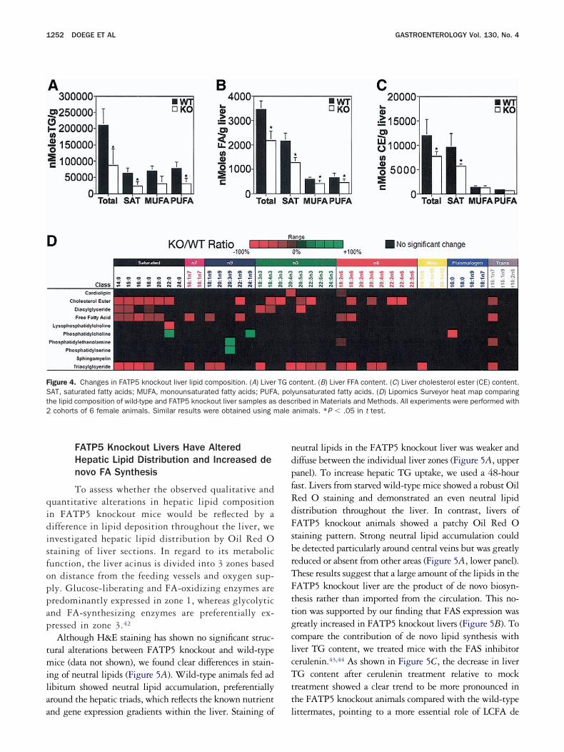
qidisfoppap
tmilaa
ndpfRdFsbrTFttgclcTtt
FSt2 ale
1252 DOEGE ET AL GASTROENTEROLOGY Vol. 130, No. 4
FATP5 Knockout Livers Have AlteredHepatic Lipid Distribution and Increased denovo FA Synthesis
To assess whether the observed qualitative anduantitative alterations in hepatic lipid compositionn FATP5 knockout mice would be reflected by aifference in lipid deposition throughout the liver, wenvestigated hepatic lipid distribution by Oil Red Otaining of liver sections. In regard to its metabolicunction, the liver acinus is divided into 3 zones basedn distance from the feeding vessels and oxygen sup-ly. Glucose-liberating and FA-oxidizing enzymes areredominantly expressed in zone 1, whereas glycolyticnd FA-synthesizing enzymes are preferentially ex-ressed in zone 3.42
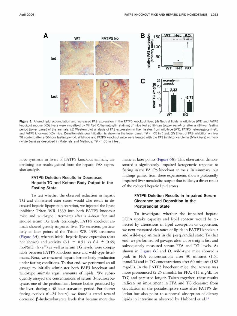
Although H&E staining has shown no significant struc-ural alterations between FATP5 knockout and wild-typeice (data not shown), we found clear differences in stain-
ng of neutral lipids (Figure 5A). Wild-type animals fed adibitum showed neutral lipid accumulation, preferentiallyround the hepatic triads, which reflects the known nutrient
igure 4. Changes in FATP5 knockout liver lipid composition. (A) LiverAT, saturated fatty acids; MUFA, monounsaturated fatty acids; PUFAhe lipid composition of wild-type and FATP5 knockout liver samples as
cohorts of 6 female animals. Similar results were obtained using m
nd gene expression gradients within the liver. Staining of l
eutral lipids in the FATP5 knockout liver was weaker andiffuse between the individual liver zones (Figure 5A, upperanel). To increase hepatic TG uptake, we used a 48-hourast. Livers from starved wild-type mice showed a robust Oiled O staining and demonstrated an even neutral lipidistribution throughout the liver. In contrast, livers ofATP5 knockout animals showed a patchy Oil Red Otaining pattern. Strong neutral lipid accumulation coulde detected particularly around central veins but was greatlyeduced or absent from other areas (Figure 5A, lower panel).hese results suggest that a large amount of the lipids in theATP5 knockout liver are the product of de novo biosyn-hesis rather than imported from the circulation. This no-ion was supported by our finding that FAS expression wasreatly increased in FATP5 knockout livers (Figure 5B). Toompare the contribution of de novo lipid synthesis withiver TG content, we treated mice with the FAS inhibitorerulenin.43,44 As shown in Figure 5C, the decrease in liverG content after cerulenin treatment relative to mock
reatment showed a clear trend to be more pronounced inhe FATP5 knockout animals compared with the wild-type
ontent. (B) Liver FFA content. (C) Liver cholesterol ester (CE) content.yunsaturated fatty acids. (D) Lipomics Surveyor heat map comparingribed in Materials and Methods. All experiments were performed withanimals. *P � .05 in t test.
TG c, poldesc
ittermates, pointing to a more essential role of LCFA de
nds
Tcimsil(nmrmugwqttfd
msffiio
LflwaesspmmmTicl
FkpaT( st.
April 2006 FATP5 KNOCKOUT MICE AND HEPATIC LIPID HOMEOSTASIS 1253
ovo synthesis in livers of FATP5 knockout animals, un-erlining our results gained from the hepatic FAS expres-ion analysis.
FATP5 Deletion Results in DecreasedHepatic TG and Ketone Body Output in theFasting State
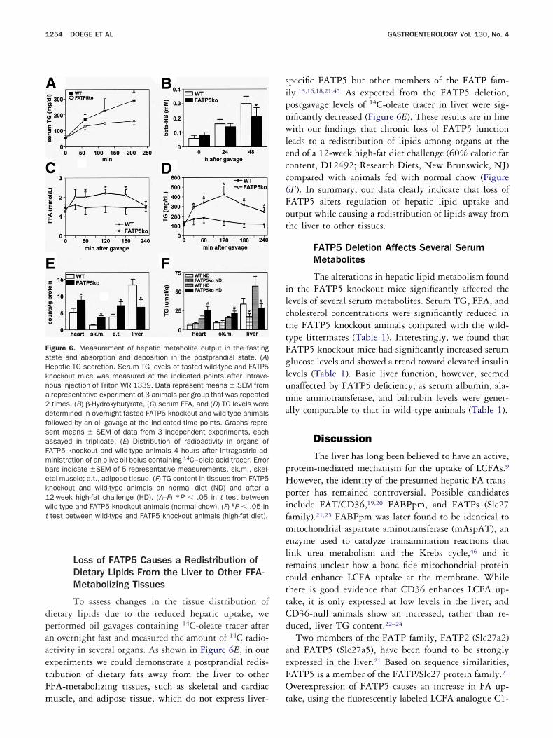
To test whether the observed reduction in hepaticG and cholesterol ester stores would also result in de-reased hepatic lipoprotein secretion, we injected the lipasenhibitor Triton WR 1339 into both FATP5 knockoutice and wild-type littermates after a 4-hour fast and
tudied serum TG levels. Strikingly, FATP5 knockout an-mals showed greatly impaired liver TG secretion, particu-arly at later points of the Triton WR 1339 treatmentFigure 6A), whereas initial hepatic lipase expression (dataot shown) and activity (6.1 � 0.51 vs 6.4 � 0.65)ol/(mL · h · e12) as well as serum TG levels, were compa-
able between FATP5 knockout mice and wild-type litter-ates. Next, we measured hepatic ketone body production
nder fasting conditions. To that end, we performed an oilavage to initially administer both FAP5 knockout andild-type animals equal amounts of lipids. We subse-uently assayed the concentrations of serum �-hydroxybu-yrate, one of the predominant ketone bodies produced byhe liver, during a 48-hour starvation period. For shorterasting periods (0–24 hours), we found a trend toward
igure 5. Altered lipid accumulation and increased FAS expression inockout mouse (KO) livers were visualized by Oil Red O/hematoxylieriod (lower panel) of the animals. (B) Western blot analysis of FASnd FATP5 knockout (KO) mice. Densitometric quantification is shownG content after a 56-hour fasting period. Wild-type and FATP5 knockowhite bars) as described in Materials and Methods. *P � .05 in t te
ecreased �-hydroxybutyrate levels that became more dra- l
atic at later points (Figure 6B). This observation demon-trated a significantly impaired ketogenetic response toasting in the FATP5 knockout animals. In summary, ourndings gained from these experiments show a profoundlympaired liver metabolite output that is likely a direct resultf the reduced hepatic lipid stores.
FATP5 Deletion Results in Impaired SerumClearance and Deposition in thePostprandial State
To investigate whether the impaired hepaticCFA uptake capacity and lipid content would be re-ected by alterations in lipid absorption or deposition,e next measured clearance of lipids in FATP5 knockout
nd wild-type animals in the postprandial state. To thatnd, we performed oil gavages after an overnight fast andubsequently measured serum FFA and TG levels. Ashown in Figure 6C and D, wild-type mice showed aeak in FFA concentrations after 30 minutes (1.51mol/L) and in TG concentrations after 60 minutes (182g/dL). In the FATP5 knockout mice, the increase wasore pronounced (2.25 mmol/L for FFA, 411 mg/dL forG) and persisted longer. Taken together, these results
ndicate an impairment in FFA and TG clearance fromirculation in the postabsorptive state after FATP5 de-etion but also point to a normal absorption of dietary
FATP5 knockout liver. (A) Neutral lipids in wild-type (WT) and FATP5ining of mice fed ad libitum (upper panel) or after a 48-hour fastingssion in liver lysates from wild-type (WT), FATP5 heterozygote (Het),e lower panel. *P � .05 in t test. (C) Effect of FAS inhibition on liverice were treated with the FAS inhibitor cerulenin (black bars) or mock
n then staexprein thut m
ipids in intestine as observed by Hubbard et al.31
dpaaetFm
sipnwlecc6Fot
ilcttFgluna
pHpifmelrcttCd
aeFO
FsHkna2dfsaFmbek1wt
1254 DOEGE ET AL GASTROENTEROLOGY Vol. 130, No. 4
Loss of FATP5 Causes a Redistribution ofDietary Lipids From the Liver to Other FFA-Metabolizing Tissues
To assess changes in the tissue distribution ofietary lipids due to the reduced hepatic uptake, weerformed oil gavages containing 14C-oleate tracer aftern overnight fast and measured the amount of 14C radio-ctivity in several organs. As shown in Figure 6E, in ourxperiments we could demonstrate a postprandial redis-ribution of dietary fats away from the liver to otherFA-metabolizing tissues, such as skeletal and cardiac
igure 6. Measurement of hepatic metabolite output in the fastingtate and absorption and deposition in the postprandial state. (A)epatic TG secretion. Serum TG levels of fasted wild-type and FATP5nockout mice was measured at the indicated points after intrave-ous injection of Triton WR 1339. Data represent means � SEM fromrepresentative experiment of 3 animals per group that was repeatedtimes. (B) �-Hydroxybutyrate, (C) serum FFA, and (D) TG levels were
etermined in overnight-fasted FATP5 knockout and wild-type animalsollowed by an oil gavage at the indicated time points. Graphs repre-ent means � SEM of data from 3 independent experiments, eachssayed in triplicate. (E) Distribution of radioactivity in organs ofATP5 knockout and wild-type animals 4 hours after intragastric ad-inistration of an olive oil bolus containing 14C–oleic acid tracer. Errorars indicate �SEM of 5 representative measurements. sk.m., skel-tal muscle; a.t., adipose tissue. (F) TG content in tissues from FATP5nockout and wild-type animals on normal diet (ND) and after a2-week high-fat challenge (HD). (A–F) *P � .05 in t test betweenild-type and FATP5 knockout animals (normal chow). (F) #P � .05 intest between wild-type and FATP5 knockout animals (high-fat diet).
uscle, and adipose tissue, which do not express liver- t
pecific FATP5 but other members of the FATP fam-ly.13,16,18,21,45 As expected from the FATP5 deletion,ostgavage levels of 14C-oleate tracer in liver were sig-ificantly decreased (Figure 6E). These results are in lineith our findings that chronic loss of FATP5 function
eads to a redistribution of lipids among organs at thend of a 12-week high-fat diet challenge (60% caloric fatontent, D12492; Research Diets, New Brunswick, NJ)ompared with animals fed with normal chow (FigureF). In summary, our data clearly indicate that loss ofATP5 alters regulation of hepatic lipid uptake andutput while causing a redistribution of lipids away fromhe liver to other tissues.
FATP5 Deletion Affects Several SerumMetabolites
The alterations in hepatic lipid metabolism foundn the FATP5 knockout mice significantly affected theevels of several serum metabolites. Serum TG, FFA, andholesterol concentrations were significantly reduced inhe FATP5 knockout animals compared with the wild-ype littermates (Table 1). Interestingly, we found thatATP5 knockout mice had significantly increased serumlucose levels and showed a trend toward elevated insulinevels (Table 1). Basic liver function, however, seemednaffected by FATP5 deficiency, as serum albumin, ala-ine aminotransferase, and bilirubin levels were gener-lly comparable to that in wild-type animals (Table 1).
Discussion
The liver has long been believed to have an active,rotein-mediated mechanism for the uptake of LCFAs.9
owever, the identity of the presumed hepatic FA trans-orter has remained controversial. Possible candidatesnclude FAT/CD36,19,20 FABPpm, and FATPs (Slc27amily).21,25 FABPpm was later found to be identical toitochondrial aspartate aminotransferase (mAspAT), an
nzyme used to catalyze transamination reactions thatink urea metabolism and the Krebs cycle,46 and itemains unclear how a bona fide mitochondrial proteinould enhance LCFA uptake at the membrane. Whilehere is good evidence that CD36 enhances LCFA up-ake, it is only expressed at low levels in the liver, andD36-null animals show an increased, rather than re-uced, liver TG content.22–24
Two members of the FATP family, FATP2 (Slc27a2)nd FATP5 (Slc27a5), have been found to be stronglyxpressed in the liver.21 Based on sequence similarities,ATP5 is a member of the FATP/Slc27 protein family.21
verexpression of FATP5 causes an increase in FA up-
ake, using the fluorescently labeled LCFA analogue C1-
Blwropteritiapcbttjpt
mblmifstp
mL5
tutpwatLFFh
ktsiFmp
siidaFlbaLcFtars
ulabswCIlTFlF
T
BGIFTC�A
AB
Na
April 2006 FATP5 KNOCKOUT MICE AND HEPATIC LIPID HOMEOSTASIS 1255
ODIPY-C12,21 and has also been reported to increaseong-chain and very-long-chain acyl-CoA synthetase28 asell as bile acid/CoA synthetase29,30 activities. Here we
eport that, based on RNA and protein detection meth-ds, FATP5 is indeed a liver-specific protein that isrimarily expressed by hepatocytes where it localizes tohe basal membrane. Immunofluorescent and immuno-lectron localization of FATP5 protein in liver sectionsevealed a preferred localization to the space of Disse, andts expression follows closely the hepatic microvascula-ure. Targeting of FATP5 to the hepatocyte/sinusoidalnterface is congruent with a role in serum FA uptakend the reconjugation of bile acids from the enterohe-atic circulation. While we could show that FATP5learly enhances the uptake of oleate and also acts as aile-CoA ligase (Hubbard et al31), we could demonstratehat it does not function as a general bile transporter likehe SLC10 family members.47 However, because uncon-ugated bile acids can enter cells without a bile trans-orter,48 FATP5 may increase their intracellular concen-ration by metabolic trapping.
Interestingly and similar to FATP5, a targeting toembrane areas juxtaposed to microvasculature has also
een shown for the heart-specific FATP6.37 Our in vivoocalization data are in contrast to earlier reports ofyc-tagged FATP5 protein being predominantly local-
zed to the endoplasmic reticulum of transiently trans-ected COS cells.29 It is important to note that ourtudies were performed with primary tissues and cells,hus eliminating possible artifacts due to the presence ofrotein tags or as a result of overexpression.Loss of FATP5 significantly impacted hepatic lipidetabolism. In line with a postulated role of FATP5 in
CFA uptake, FATP5 knockout hepatocytes showed a
able 1. Body Weights and Serum Parameters of FATP5Knockout and Wild-Type Mice on Normal Chow AdLibitum
ParameterWild-type
miceKnockout
miceP
value
ody wt (g) 29.8 � 2.24 29.6 � 0.65 NSlucose (mg/dL) 92 � 12.8 114 � 11.6 .009
nsulin (ng/mL) 0.76 � 0.15 0.95 � 0.13 .048FA (nmol/L) 1.59 � 0.07 1.39 � 0.03 .0001G (mg/dL) 126 � 12.8 104 � 7.36 .005holesterol (mmol/L) 7.5 � 1.3 4.8 � 1.5 .045-hydroxybuturate (mg/dL) 9.70 � 1.58 7.69 � 2.0 NSlanine aminotransferase(U/L)
33.2 � 2.54 31.8 � 3.09 NS
lbumin (g/dL) 2.2 � 0.44 2.3 � 0.52 NSilirubin (mg/dL) 1.082 � 0.17 0.985 � 0.26 NS
OTE. Parameters are from overnight fasted animals 8–12 weeks ofge (n � 5–6).
0% reduced uptake of both fluorescently and radioac- t
ively labeled LCFAs. It is unlikely that the remainingptake activity is due to passive diffusion because 90% ofhe total hepatic LCFA uptake is due to a saturablerocess.11 Our Western blot analyses showed that bothild-type and FATP5 knockout livers express significant
mounts of FATP2 and, to a lesser degree, FATP3. Weherefore speculate that the FATP5-independent hepaticCFA uptake is largely due to other members of theATP family such as FATP2. A targeted deletion of theATP2 gene in mice has recently been described45;owever, its effect on FA uptake has not been examined.The significantly reduced LCFA uptake by FATP5
nockout hepatocytes provides the first in vivo evidencehat cell surface proteins, such as FATPs, contributeubstantially to this process in the liver. Similarly, othern vivo studies have shown the importance of FATP4 andATP1 for LCFA uptake by the intestine37 and skeletaluscle,49 highlighting the general importance of this
rotein family for FA uptake and metabolism.While our in vitro data clearly show that overexpres-
ion of FATP5 alone results in an increased LCFA uptaken cultured mammalian cells, it is likely that its functions closely associated with other upstream- as well asownstream-located transporters, binding proteins, orcyl-CoA synthetases ensuring efficient LCFA uptake.urther, while FATP5 has been shown to possess very-ong-chain acyl-CoA synthetase activity,28 it remains toe determined whether additional plasma membranecyl-CoA synthetases are required for efficient hepaticCFA uptake. Potential candidates for interactions in-lude CD36,19,20 FABP,19,20 LACS,50 and possiblyATP2.18,45 Although a deeper biochemical and struc-ural understanding of FATP-mediated LCFA uptakewaits further investigation, our data presented in thiseport clearly show that FATP5 is both required andufficient for an efficient hepatic LCFA uptake.
As would be expected from a reduced hepatic LCFAptake, liver FFA, cholesterol ester, diglyceride, and TGevels were all significantly lower in FATP5 knockoutnimals. There were no significant effects on the distri-ution of acyl chain lengths and we did not observe anyigns of perturbed very long-chain fatty acid metabolism,hich would argue that FATP5 very-long-chain acyl-oA synthetase activity plays only a minor role in vivo.
nterestingly, loss of FATP5 protein impacted liver TGevels more than any other FA transport compartment.aken together with the finding that overexpression ofATP1 has been linked to a preferential increase in TGevels,51 this could indicate a predominant channeling ofAs by FATPs toward TG synthesis and storage rather
han mitochondrial import and �-oxidation.
FsFpmuptiibsaa
ukbciasontpieckchpcwipauoaWimuah
hba
Fddtpmihtaamdks
sstlflmukitdFbwfsaed
klcdgTtttaadai
1256 DOEGE ET AL GASTROENTEROLOGY Vol. 130, No. 4
We also observed a disproportionately large effect ofATP5 deletion on saturated FAs compared with monoun-aturated and polyunsaturated FAs. The percentage of 16:0As was decreased in liver TGs and FFAs, whereas theercentage of 18:1n9 was higher in the FATP5 knockoutice. These observations are consistent with a decreased
ptake of dietary FAs and an increased de novo synthesis ofreferentially monounsaturated LCFAs to compensate forhe disturbed LCFA supply from the diet. This hypothesiss supported by the increased concentration of neutral lipidsn FATP5 knockout livers in zone 3 of hepatic lobules andy the increased FAS expression and increased ceruleninusceptibility. Alternatively, FATP5 may have a higherffinity for saturated FAs compared with the postulateddditional liver LCFA transporter(s).
Interestingly, hepatic levels of phospholipids, partic-larly phosphatidylserine, were increased in FATP5nockout livers. Phosphatidylserine is a critical mem-rane phospholipid and can be as much as 10% of theellular phospholipids. It is normally restricted to thenner leaflet of the plasma membrane and participates, inddition to its structural functions, in multiple cellularignaling pathways.52 Additionally, the FA compositionf another phospholipid, phosphatidylcholine, was sig-ificantly different between FATP5 knockout and wild-ype mice. Phosphatidylcholine is the most abundanthospholipid in mammalian cells. Like phosphatidylser-ne, phosphatidylcholine has important structural prop-rties for the cell. Here as well, the monounsaturated FAontent in phosphatidylcholine was increased in thenockout mice while the saturated FA content was de-reased. Similar to the effect of FATP5 deletion onepatic FA composition, this shift was characterizedrimarily by a change in n-9 FAs, that is, the relativeoncentration of 18:1n9 was significantly increased,hich may be due to the increased levels of 18:1n9 FAs
n the liver being shunted into the production of phos-hatidylcholine. Although the direct consequences of thelterations in phospholipid level and composition arenknown, these findings could possibly be linked to thebserved hepatomegaly in the FATP5 knockout animals,s described in our companion report (Hubbard et al31).
hile we cannot exclude the possibility that the decreasen hepatic lipid content is connected to alterations in bileetabolism, it is more likely a result of decreased LCFA
ptake from the circulation because FATP5 knockoutnimals showed no malabsorption of lipids when fed aigh-fat diet (Hubbard et al31).We also observed that the changes in hepatic lipid
omeostasis in FATP5 knockout animals were reflectedy profound alterations in serum metabolites (this report
nd Hubbard et al31). In the FATP5 knockout animals, aFA, TG, and cholesterol levels were significantly re-uced under fasting conditions. This decrease is likely airect result of the reduced liver lipid stores leading tohe observed decrease in hepatic very-low-density li-oprotein output. Interestingly, in FATP5 knockoutice fed on normal chow, we observed significantly
ncreased serum glucose levels and a slight trend towardyperinsulinemia. This finding differs from our observa-ions made on a high-fat diet (Hubbard et al31). Similar,lbeit more pronounced, changes in serum TG, glucose,nd insulin levels were found in the LIRKO mouseodel.53 However, our finding was unexpected because
ecreased hepatocellular FFA levels, as found in FATP5nockout mice, should rather improve hepatic insulinensing and decrease glucose production.54
Further, as a likely result of the depletion of liver lipidtores, we also found severe alterations in metabolite ab-orption and clearance in FATP5 knockout mice. The ke-ogenetic response was severely impaired after FATP5 de-etion during a 48-hour fasting period as assessed by theormation of �-hydroxybutyrate. Conversely, postabsorptiveipid clearance was severely diminished in FATP5 knockoutice, as would be expected from an impaired hepatic FFA
ptake as a result of the FATP5 deletion. While FATP5nockout mouse livers are overall enlarged and show a 25%ncrease in hepatocyte size compared with livers of wild-ype animals, no evidence of fibrosis or other structuralisorders in the FATP5 knockout mouse livers was found.urthermore, serum alanine aminotransferase, albumin, andilirubin levels were similar between FATP5 knockout andild-type mice, again pointing to an unimpaired basic liver
unction. Moreover, in our accompanying report, we couldhow comparable values of the respiratory exchange ratiosnd of expression levels of FA oxidation markers (Hubbardt al31), which as well should exclude a major hepaticysfunction in the FATP5 knockout mouse model.
Taken together, our data describing the FATP5nockout mouse show a significant reduction in bothiver lipid uptake and content. On the other hand, weould demonstrate a decrease in the concentrations ofiverse serum lipids. Besides these findings, our investi-ations point to an unaltered intestinal lipid absorption.o link these findings and to also address the observed
rend toward a hyperglycemic/hyperinsulinemic pheno-ype, we performed oil gavages containing 14C-oleateracer. The results gained from these experiments revealredistribution of dietary fats in the postprandial state
way from the liver toward tissues whose uptake isominated by other members of the FATP family, suchs skeletal and cardiac muscle. This in turn could lead tonsulin desensitization and a decreased glucose uptake
nd utilization by these tissues followed by an elevated
ssFcodtaapoapnalzlFlpcipfi
aFcuslme
1
1
1
1
1
1
1
1
1
1
2
2
2
2
2
2
2
2
April 2006 FATP5 KNOCKOUT MICE AND HEPATIC LIPID HOMEOSTASIS 1257
erum insulin concentration. These findings are also con-istent with our observations that both hearts fromATP5-null mice are enlarged (data not shown) andhronic loss of FATP5 function leads to a redistributionf lipids among organs at the end of a 12-week high-fatiet challenge. Moreover, in line with our hypothesis,hat the partial FFA uptake defect in liver may result inn increased availability of FFAs to other tissues, wouldlso be our observation of an increased thermogenesis,otentially as a consequence of an increased availabilityf FFAs to muscle and brown adipocytes (Hubbard etl31). It is noteworthy that higher insulin levels alsoromote the transcription of SREBP, a key regulator ofutritional homeostasis and insulin action, which inddition could contribute to increased serum glucoseevels due to the stimulation of hepatic glycolytic en-ymes.55,56 Lastly, and linking back to the serum FAevels, the observed higher insulin concentrations in theATP5 knockout mice could result in a suppression ofipolysis in adipocytes leading to a decreased FFA out-ut,57–59 which would also add to a reduced serum lipidoncentration as described previously. Additional stud-es, including euglycemic/hyperinsulinemic clamp ex-eriments, are under way to further investigate thesendings in more detail.
In conclusion, our studies shown here and in theccompanying report by Hubbard et al31 suggest thatATP5 is a protein with multiple activities and canontribute to both bile acid reactivation and hepatic FAptake and lipid accumulation. These findings begin tohed light on the complex interrelationships in hepaticipid biology and should further our understanding ofetabolic disorders, such as nonalcoholic fatty liver dis-
ase, diet-induced obesity, and type 2 diabetes mellitus.
References1. Odaib AA, Shneider BL, Bennett MJ, Pober BR, Reyes-Mugica M,
Friedman AL, Suchy FJ, Rinaldo P. A defect in the transport oflong-chain fatty acids associated with acute liver failure. N EnglJ Med 1998;339:1752–1757.
2. Rinaldo P. Fatty acid transport and mitochondrial oxidation disor-ders. Semin Liver Dis 2001;21:489–500.
3. Masuzaki H, Paterson J, Shinyama H, Morton NM, Mullins JJ,Seckl JR, Flier JS. A transgenic model of visceral obesity and themetabolic syndrome. Science 2001;294:2166–2170.
4. Saltiel AR, Kahn CR. Insulin signalling and the regulation ofglucose and lipid metabolism. Nature 2001;414:799–806.
5. Marchesini G, Brizi M, Bianchi G, Tomassetti S, Bugianesi E,Lenzi M, McCullough AJ, Natale S, Forlani G, Melchionda N.Nonalcoholic fatty liver disease: a feature of the metabolic syn-drome. Diabetes 2001;50:1844–1850.
6. Elsing C, Winn-Borner U, Stremmel W. Confocal analysis of hep-atocellular long-chain fatty acid uptake. Am J Physiol 1995;269:G842–G851.
7. Fitscher B, Klaassen-Schluter C, Stremmel W. Evidence for a
hepatocyte membrane fatty acid transport protein using rat livermRNA expression in Xenopus laevis oocytes. Biochim BiophysActa 1995;1256:47–51.
8. Sorrentino D, Stump DD, Van Ness K, Simard A, Schwab AJ, ZhouSL, Goresky CA, Berk PD. Oleate uptake by isolated hepatocytesand the perfused rat liver is competitively inhibited by palmitate.Am J Physiol 1996;270:G385–G392.
9. Stremmel W. Mechanism of hepatic fatty acid uptake. J Hepatol1989;9:374–382.
0. Stremmel W. Transmembrane transport of fatty acids in theheart. Mol Cell Biochem 1989;88:23–29.
1. Stump DD, Nunes RM, Sorrentino D, Isola LM, Berk PD. Charac-teristics of oleate binding to liver plasma membranes and itsuptake by isolated hepatocytes. J Hepatol 1992;16:304–315.
2. Gore J, Hoinard C. Linolenic acid transport in hamster intestinalcells is carrier-mediated. J Nutr 1993;123:66–73.
3. Stahl A, Hirsch DJ, Gimeno R, Punreddy S, Ge P, Watson N, KotlerM, Tartaglia LA, Lodish HF. Identification of a small intestinalfatty acid transport protein. Mol Cell 1999;4:299–308.
4. Stremmel W. Uptake of fatty acids by jejunal mucosal cells ismediated by a fatty acid binding membrane protein. J Clin Invest1988;82:2001–2010.
5. Sorrentino D, Stump D, Potter BJ, Robinson RB, White R, KiangCL, Berk PD. Oleate uptake by cardiac myocytes is carrier medi-ated and involves a 40-kD plasma membrane fatty acid bindingprotein similar to that in liver, adipose tissue, and gut. J ClinInvest 1988;82:928–935.
6. Schaffer JE, Lodish HF. Expression cloning and characterizationof a novel adipocyte long chain fatty acid transport protein. Cell1994;79:427–436.
7. Abumrad N, Harmon C, Ibrahimi A. Membrane transport of long-chain fatty acids: evidence for a facilitated process. J Lipid Res1998;39:2309–2318.
8. Stahl A, Gimeno RE, Tartaglia LA, Lodish HF. Fatty acid transportproteins: a current view of a growing family. Trends EndocrinolMetab 2001;12:266–273.
9. Abumrad N, Coburn C, Ibrahimi A. Membrane proteins implicatedin long-chain fatty acid uptake by mammalian cells: CD36, FATPand FABPm. Biochim Biophys Acta 1999;1441:4–13.
0. Ibrahimi A, Abumrad NA. Role of CD36 in membrane transport oflong-chain fatty acids. Curr Opin Clin Nutr Metab Care 2002;5:139–145.
1. Hirsch D, Stahl A, Lodish HF. A family of fatty acid transportersconserved from mycobacterium to man. Proc Natl Acad Sci U S A1998;95:8625–8629.
2. Coburn CT, Hajri T, Ibrahimi A, Abumrad NA. Role of CD36 inmembrane transport and utilization of long-chain fatty acids bydifferent tissues. J Mol Neurosci 2001;16:117–121; discussion151–157.
3. Coburn CT, Knapp FF Jr, Febbraio M, Beets AL, Silverstein RL,Abumrad NA. Defective uptake and utilization of long chain fattyacids in muscle and adipose tissues of CD36 knockout mice.J Biol Chem 2000;275:32523–32529.
4. Febbraio M, Abumrad NA, Hajjar DP, Sharma K, Cheng W, PearceSF, Silverstein RL. A null mutation in murine CD36 reveals animportant role in fatty acid and lipoprotein metabolism. J BiolChem 1999;274:19055–19062.
5. Berger J, Truppe C, Neumann H, Forss-Petter S. A novel relativeof the very-long-chain acyl-CoA synthetase and fatty acid trans-porter protein genes with a distinct expression pattern. BiochemBiophys Res Commun 1998;247:255–260.
6. Coe N, Smith A, Frohnert B, Watkins P, Bernlohr D. The fatty acidtransport protein (FATP1) is a very long chain acyl-CoA syn-thetase. J Biol Chem 1999;274:36300–36304.
7. Fitscher BA, Riedel HD, Young KC, Stremmel W. Tissue distribu-tion and cDNA cloning of a human fatty acid transport protein
(hsFATP4). Biochim Biophys Acta 1998;1443:381–385.
2
2
3
3
3
3
3
3
3
3
3
3
4
4
4
4
4
4
4
4
4
4
5
5
5
5
5
5
5
5
5
5
MR
DDtDILa
SchiwNa
1258 DOEGE ET AL GASTROENTEROLOGY Vol. 130, No. 4
8. Steinberg SJ, Kemp S, Braiterman LT, Watkins PA. Role of very-long-chain acyl-coenzyme A synthetase in X-linked adrenoleu-kodystrophy. Ann Neurol 1999;46:409–412.
9. Mihalik SJ, Steinberg SJ, Pei Z, Park J, Kim DG, Heinzer AK,Dacremont G, Wanders RJ, Cuebas DA, Smith KD, Watkins PA.Participation of two members of the very long-chain acyl-CoAsynthetase family in bile acid synthesis and recycling. J BiolChem 2002;277:24771–24779.
0. Steinberg SJ, Mihalik SJ, Kim DG, Cuebas DA, Watkins PA. Thehuman liver-specific homolog of very long-chain acyl-CoA synthetaseis cholate:CoA ligase. J Biol Chem 2000;275:15605–15608.
1. Hubbard B, Doege H, Punreddy S, Wu H, Huang X, Kaushik V, MozellRL, Byrnes JJ, Stricker-Krongrad A, Chou J, Tartaglia LA, Lodish HF,Stahl A, Gimeno RE. Mice deleted for fatty acid transport protein 5have defective bile acid conjugation and are protected from obesity.Gastroenterology 2006;130:1259–1269.
2. Millar JS, Maugeais C, Fuki IV, Rader DJ. Normal production rateof apolipoprotein B in LDL receptor-deficient mice. ArteriosclerThromb Vasc Biol 2002;22:989–994.
3. Nilsson-Ehle P, Schotz MC. A stable, radioactive substrate emulsionfor assay of lipoprotein lipase. J Lipid Res 1976;17:536–541.
4. Folch J, Lees M, Sloane Stanley GH. A simple method for theisolation and purification of total lipides from animal tissues.J Biol Chem 1957;226:497–509.
5. Stremmel W, Berk PD. Hepatocellular influx of [14C]oleate re-flects membrane transport rather than intracellular metabolismor binding. Proc Natl Acad Sci U S A 1986;83:3086–3090.
6. Stremmel W, Strohmeyer G, Berk PD. Hepatocellular uptake ofoleate is energy dependent, sodium linked, and inhibited by anantibody to a hepatocyte plasma membrane fatty acid bindingprotein. Proc Natl Acad Sci U S A 1986;83:3584–3588.
7. Gimeno RE, Ortegon AM, Patel S, Punreddy S, Ge P, Sun Y,Lodish HF, Stahl A. Characterization of a heart-specific fatty acidtransport protein. J Biol Chem 2003;278:16039–16044.
8. Watkins SM, Reifsnyder PR, Pan HJ, German JB, Leiter EH. Lipidmetabolome-wide effects of the PPARgamma agonist rosiglita-zone. J Lipid Res. 2002;43:1809–1817.
9. Frachon S, Gouysse G, Dumortier J, Couvelard A, Nejjari M, MionF, Berger F, Paliard P, Boillot O, Scoazec JY. Endothelial cellmarker expression in dysplastic lesions of the liver: an immuno-histochemical study. J Hepatol 2001;34:850–857.
0. Hagenbuch B, Dawson P. The sodium bile salt cotransport familySLC10. Pflugers Arch 2004;447:566–570.
1. Hagenbuch B, Stieger B, Foguet M, Lubbert H, Meier PJ. Func-tional expression cloning and characterization of the hepatocyteNa/bile acid cotransport system. Proc Natl Acad Sci U S A1991;88:10629–10633.
2. Jungermann K. Role of intralobular compartmentation in hepaticmetabolism. Diabete Metab 1992;18:81–86.
3. Hirsch J. The search for new ways to treat obesity. Proc Natl AcadSci U S A 2002;99:9096–9097.
4. Loftus TM, Jaworsky DE, Frehywot GL, Townsend CA, Ronnett GV,Lane MD, Kuhajda FP. Reduced food intake and body weight inmice treated with fatty acid synthase inhibitors. Science 2000;288:2379–2381.
5. Heinzer AK, Watkins PA, Lu JF, Kemp S, Moser AB, Li YY, MihalikS, Powers JM, Smith KD. A very long-chain acyl-CoA synthetase-deficient mouse and its relevance to X-linked adrenoleukodystro-phy. Hum Mol Genet 2003;12:1145–1154.
6. Berk PD, Wada H, Horio Y, Potter BJ, Sorrentino D, Zhou SL, IsolaLM, Stump D, Kiang CL, Thung S. Plasma membrane fatty acid-binding protein and mitochondrial glutamic-oxaloacetic transam-inase of rat liver are related. Proc Natl Acad Sci U S A 1990;87:3484–3488.
7. Stremmel W. Translocation of fatty acids across the basolateral
rat liver plasma membrane is driven by an active potential-sen- rsitive sodium-dependent transport system. J Biol Chem1987;262:6284–6289.
8. Parks DJ, Blanchard SG, Bledsoe RK, Chandra G, Consler TG,Kliewer SA, Stimmel JB, Willson TM, Zavacki AM, Moore DD,Lehmann JM. Bile acids: natural ligands for an orphan nuclearreceptor. Science 1999;284:1365–1368.
9. Kim JK, Gimeno RE, Higashimori T, Kim HJ, Choi H, Punreddy S,Mozell RL, Tan G, Stricker-Krongrad A, Hirsch DJ, Fillmore JJ, LiuZX, Dong J, Cline G, Stahl A, Lodish HF, Shulman GI. Inactivationof fatty acid transport protein 1 prevents fat-induced insulinresistance in skeletal muscle. J Clin Invest 2004;113:756–763.
0. Suzuki H, Kawarabayasi Y, Kondo J, Abe T, Nishikawa K, Kimura S,Hashimoto T, Yamamoto T. Structure and regulation of rat long-chain acyl-CoA synthetase. J Biol Chem 1990;265:8681–8685.
1. Hatch GM, Smith AJ, Xu FY, Hall AM, Bernlohr DA. FATP1 chan-nels exogenous FA into 1,2,3-triacyl-sn-glycerol and down-regu-lates sphingomyelin and cholesterol metabolism in growing 293cells. J Lipid Res 2002;43:1380–1389.
2. Vance JE. Molecular and cell biology of phosphatidylserine andphosphatidylethanolamine metabolism. Prog Nucleic Acid ResMol Biol 2003;75:69–111.
3. Michael MD, Kulkarni RN, Postic C, Previs SF, Shulman GI,Magnuson MA, Kahn CR. Loss of insulin signaling in hepatocytesleads to severe insulin resistance and progressive hepatic dys-function. Mol Cell 2000;6:87–97.
4. Lam TK, van de Werve G, Giacca A. Free fatty acids increasebasal hepatic glucose production and induce hepatic insulinresistance at different sites. Am J Physiol Endocrinol Metab2003;284:E281–E290.
5. Foretz M, Pacot C, Dugail I, Lemarchand P, Guichard C, Le Liepvre X,Berthelier-Lubrano C, Spiegelman B, Kim JB, Ferre P, Foufelle F.ADD1/SREBP-1c is required in the activation of hepatic lipogenicgene expression by glucose. Mol Cell Biol 1999;19:3760–3768.
6. Kim JB, Spiegelman BM. ADD1/SREBP1 promotes adipocytedifferentiation and gene expression linked to fatty acid metabo-lism. Genes Dev 1996;10:1096–1107.
7. Rebrin K, Steil GM, Getty L, Bergman RN. Free fatty acid as a linkin the regulation of hepatic glucose output by peripheral insulin.Diabetes 1995;44:1038–1045.
8. Rebrin K, Steil GM, Mittelman SD, Bergman RN. Causal linkagebetween insulin suppression of lipolysis and suppression of liverglucose output in dogs. J Clin Invest 1996;98:741–749.
9. Lewis GF, Vranic M, Harley P, Giacca A. Fatty acids mediate theacute extrahepatic effects of insulin on hepatic glucose produc-tion in humans. Diabetes 1997;46:1111–1119.
Received September 12, 2005. Accepted December 14, 2005.Address requests for reprints to: Andreas Stahl, PhD, Palo Altoedical Foundation Research Institute, Ames Building, 795 El Caminoeal, Palo Alto, California 94301. e-mail: [email protected] by a training and feasibility grant from the Stanford
igestive Disease Center (National Institutes of Health grantK56339), a grant from the National Institute of Diabetes and Diges-
ive and Kidney Disease (National Institutes of Health grantK066336-01 to A.S.), and in part by a grant from the National
nstitutes of Health (National Institutes of Health/National Heart,ung, and Blood Institute grant PO1 HL66105 to Dr Harvey F. Lodisht the Whitehead Institute for Biomedical Research).The authors thank Jon Mulholland and Kitty Lee at the Stanford Cell
cience Imaging Facility for expert advice and help with light micros-opy; Ann Strauss, Victoria Fairchild-Huntress, and Dennis Huszar forelp with knockout generation; Dr Harvey F. Lodish for his contribution
n creating FATP5 knockout animals; Dr Sung-Shin Choi for assistanceith lipase assays; BD Bioscience for providing us with controls fororthern blots; the Palo Alto Medical Foundation Research Institutenimal facility staff for their help; and Rosemary Grammer for critical
eading of the manuscript.


![Preliminary Findings on the Use of Targeted Therapy in ... · from diagnosis, despite advances in chemotherapy and use of targeted therapy [3]. Genetic analysis reveals in-volvement](https://img.pdfslide.net/doc/110x75/5f0474847e708231d40e0d6c/preliminary-findings-on-the-use-of-targeted-therapy-in-from-diagnosis-despite.jpg)