Embed Size (px)
Citation preview
Targeted Gold Nanoparticles as Vascular Disrupting Agents during
Radiation Therapy
RI Berbeco1, H Korideck1, S Kunjachan1, R Kumar2, S Sridhar 2, A Detappe1, W Ngwa1 and M Makrigiorgos1
(1) Department of Radiation Oncology, Brigham and Women’s Hospital, Dana-Farber Cancer Institute and Harvard Medical School, Boston, MA
(2) Nanomedicine Science and Technology Center, Northeastern University, Boston, MA
Sept. 16, 2014, S-2Gold nanoparticles as Vascular Disrupting Agents – Ross Berbeco, PhD
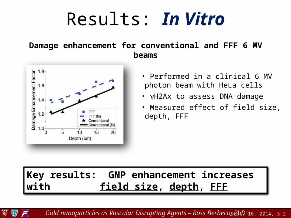
Results: In VitroDamage enhancement for conventional and FFF 6 MV beams
• Performed in a clinical 6 MV photon beam with HeLa cells
• gH2Ax to assess DNA damage
• Measured effect of field size, depth, FFF
Key results: GNP enhancement increases with field size, depth, FFF
Sept. 16, 2014, S-3Gold nanoparticles as Vascular Disrupting Agents – Ross Berbeco, PhD
Results: In Vivo
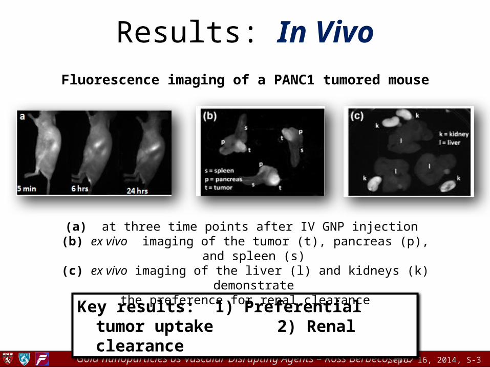
Fluorescence imaging of a PANC1 tumored mouse
Key results: 1) Preferential tumor uptake 2) Renal clearance
(a) at three time points after IV GNP injection (b) ex vivo imaging of the tumor (t), pancreas (p), and spleen (s)
(c) ex vivo imaging of the liver (l) and kidneys (k) demonstrate the preference for renal clearance
Sept. 16, 2014, S-4Gold nanoparticles as Vascular Disrupting Agents – Ross Berbeco, PhD
Results: In Vivo
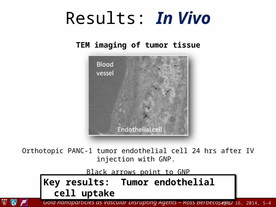
TEM imaging of tumor tissue
Key results: Tumor endothelial cell uptake
Orthotopic PANC-1 tumor endothelial cell 24 hrs after IV injection with GNP.
Black arrows point to GNP
Sept. 16, 2014, S-5Gold nanoparticles as Vascular Disrupting Agents – Ross Berbeco, PhD
Results: In Vivo
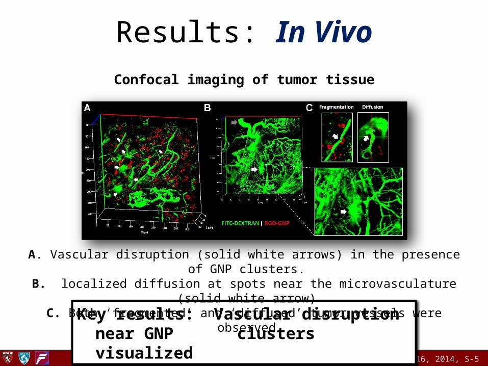
Confocal imaging of tumor tissue
Key results: Vascular disruption near GNP clusters visualized
A. Vascular disruption (solid white arrows) in the presence of GNP clusters. B. localized diffusion at spots near the microvasculature (solid white arrow)
C. Both ‘fragmented’ and ‘diffused’ tumor vessels were observed.
Sept. 16, 2014, S-6Gold nanoparticles as Vascular Disrupting Agents – Ross Berbeco, PhD
Summary/conclusions
• Preliminary results demonstrate that tumor vascular disruption is expected when targeted GNP are combined with radiation
• Justify further in vivo investigation
• Engineering GNP for more efficient targeting, imaging, and therapy
References:Berbeco, R. I., et al. (2011). "Localized dose enhancement to tumor blood vessel endothelial cells via megavoltage x-rays and targeted gold nanoparticles: new
potential for external beam radiotherapy." International Journal of Radiation Oncology Biology Physics 81(1): 270-276.Berbeco, R. I., et al. (2012). "DNA damage enhancement from gold nanoparticles for clinical MV photon beams." Radiation Research 178(6): 604-608.Detappe, A., et al. (2013). "The effect of flattening filter free delivery on endothelial dose enhancement with gold nanoparticles." Medical Physics 40(3): 0317061.Kumar, R., et al. (2013). "Third generation gold nanoparticle platform optimized for radiation therapy." Translational Cancer Research 2(4): 228-239.