Embed Size (px)
Citation preview
International Journal of
Molecular Sciences
Review
Targeting Apolipoprotein E for Alzheimer’s Disease:An Industry Perspective
Georgette L. Suidan and Gayathri Ramaswamy *
Alzheimer’s Disease and Dementia Research Unit, Biogen Inc., Cambridge, MA 02142, USA;[email protected]* Correspondence: [email protected]
Received: 24 March 2019; Accepted: 28 April 2019; Published: 1 May 2019�����������������
Abstract: Apolipoprotein E (apoE), a key lipid transport protein in the brain, is predominantlyproduced by astrocytes. Astrocytes are the most numerous cell type in the brain and are the mainsupport network for neurons. They play a critical role in the synthesis and delivery of cholesterolin the brain. Humans have three common apoE isoforms, apoE2, apoE3 and apoE4, that show astrong genotype effect on the risk and age of onset for sporadic and late onset forms of Alzheimer’sdisease (AD). Carriers of an ε4 allele have an increased risk of developing AD, while those with anε2 allele are protected. Investigations into the contribution of apoE to the development of AD hasyielded conflicting results and there is still much speculation about the role of this protein in disease.Here, we review the opposing hypotheses currently described in the literature and the approachesthat have been considered for targeting apoE as a novel therapeutic strategy for AD. Additionally,we provide our perspective on the rationale for targeting apoE and the challenges that arise withrespect to “drug-ability” of this target.
Keywords: apolipoprotein E; Alzheimer’s disease; astrocytes
1. Introduction
Alzheimer’s disease (AD) is the leading cause of dementia in elderly people. Hallmarks ofthe disease include the deposition of amyloid beta (Aβ) plaques and the presence of neurofibrillarytangles. Thus, strategies that have gained the most traction in the field are predominantly focusedon targeting amyloid and tau proteins [1,2]. However, Alois Alzheimer also reported the presenceof “adipose inclusions” as one of the pathologies in the AD brain, in addition to amyloid and tau,suggesting that AD brains display signs of aberrant lipid metabolism [3,4]. Emerging data clearlyindicate a strong link between AD and lipids. In fact, the brain is the most lipid-rich organ in thebody and—despite accounting for about 2.1% of the total body weight—contains about 25% of totalbody sterols. In the central nervous system (CNS), lipids play a critical role in both structure andfunction [5,6]. For example, unesterified cholesterol plays a specialized role as the major architecturalcomponent of the myelin sheath. In fact, about 70% of total brain cholesterol is present in the myelinsheath. The remaining lipids are present in functionally active pools in neurons and glia. During CNSdevelopment, required cholesterol and lipid pools are obtained exclusively from de novo synthesis.Whether there is any influence of plasma lipids on the brain lipid pool is currently unknown. Given sucha crucial role of lipids in the CNS, maintaining lipid homeostasis in the brain is critical for maintaininga healthy state. It is therefore not surprising that changes in lipid metabolism in the brain can lead toneurodegenerative diseases. However, the most important association was established in 1994 withthe discovery of a profound role for apolipoprotein E (apoE) in AD [7].
Astrocytes are the predominant producers of apoE in the brain [8,9]. In addition to astrocytes,microglia and neurons have also been shown to synthesize apoE [8,10]. ApoE plays an important
Int. J. Mol. Sci. 2019, 20, 2161; doi:10.3390/ijms20092161 www.mdpi.com/journal/ijms
Int. J. Mol. Sci. 2019, 20, 2161 2 of 14
role in lipid transport to neurons [11,12], synaptogenesis [13], cerebrovascular integrity and cerebralblood flow (CBF) [14,15], hippocampal neurogenesis [16], neuroimmune modulation [17] and amyloidclearance [18,19]. The three common human isoforms, apoE2, apoE3 and apoE4, differ from each otherat amino acid positions 112 and 158. ApoE2 has cysteine (Cys) in these two positions, apoE3 hasCys at 112 and arginine (Arg) at 158 and apoE4 has Arg in both positions [20]. These small aminoacid differences in the apoE isoforms elicit a profound genotype-dependent effect on apoE function,such as lipid transport and amyloid clearance, among others [20]. Importantly, these three commonhuman apoE isoforms show a strong genotype effect on the risk and age of onset for sporadic and lateonset forms of Alzheimer’s disease (LOAD), with apoE4 being the strongest known genetic risk factorfor AD.
Despite numerous new genome-wide association studies (GWAS) findings that have been reported,after age, apoE4 is still the strongest known risk factor for AD. While several hypotheses have beenproposed, the exact mechanism by which apoE4 contributes to AD is not known. Consequently, there issignificant debate on whether apoE should be upregulated or downregulated, with reports showingboth protective and pathological roles of apoE4. The goal of this review is to describe the approachesthat have been considered to target apoE as a novel therapeutic strategy for AD.
2. Opposing Hypotheses on the Contribution of ApoE4 to AD
2.1. Loss of Function
ApoE4 has been suggested to be poorly lipidated relative to apoE2 and apoE3. ApoE protein levelsin human cerebrospinal fluid (CSF) are reduced in ε4 carriers compared to ε2 and ε3 by ~30% [21],and the levels of lipid-depleted apoE in ε4 carriers is elevated ~2-fold [22]. Similar results werefound in mice expressing human isoforms of apoE. Utilizing mass spectroscopy and enzyme-linkedimmunosorbent assay (ELISA) methods, it was shown that apoE4-expressing mice have significantlyless apoE in their plasma, brain and CSF compared to apoE2 and apoE3 [23]. Proper lipidation ofastrocytic apoE to form high-density lipoprotein (HDL)-like particles is essential for its stability andproper function; thus, it is hypothesized that poor lipidation of apoE4 leads to reduced functionand downstream pathology [24,25]. Lipidation of apoE in astrocytes is carried out primarily by theadenosine triphosphate (ATP)-binding cassette family member, abca1. Indeed, ATP-binding cassettetransporter a1 (abca1) knock-out mice showed reduced levels of brain apoE and lipidated apoE particlesin their CSF [25]. These mice have also shown reduced dendritic density in the CA1 region of thehippocampus [26] and increased amyloid burden in the brain when crossed with human amyloidprecursor protein (APP)-overexpressing mice [27]. Interestingly, a large scale population study foundthat a loss-of-function variant of abca1 in humans resulted in reduced plasma apoE and increasedrisk of AD, cerebrovascular disease and hemorrhagic stroke, suggesting that this mechanistic axisshould also be considered for improvement of cerebrovascular health and function [28]. In contrast,abca1 overexpression in mice increases lipidation of apoE and enhances amyloid clearance [29].Increasing astrocytic abca1 expression and the resulting increase in apoE lipidation has been shown tohave beneficial effects on axonal damage in a traumatic brain injury mouse model [30,31]. An elegantstudy was recently published utilizing astrocytes derived from human inducible pluripotent stem cells(iPSCs) with either ε3/ε3 or ε4/ε4 genotypes, which provided further evidence in human cells thatsecreted apoE4 is poorly lipidated. Interestingly, astrocytes derived from an ε4/ε4 patient cells did notsupport neuronal survivalas well as ε3/ε3, further supporting the idea that the reduced lipidation stateof apoE4 is tied to neuronal health [32]. Additionally, HDLs have been shown to enhance amyloidclearance through the blood–brain barrier and reduce vascular inflammation, and therefore enhancingapoE lipidation has the potential to be protective to the cerebrovasculature [33,34]. Further supportingdata for the apoE4 loss of function hypothesis are the pathological similarities reported betweenapoE4 and apoE knockout (KO) mice. For example, both strains of mice were shown to have reducedlevels of synaptic proteins PSD-95, GKAP and synaptophysin compared to wild-type and apoE3
Int. J. Mol. Sci. 2019, 20, 2161 3 of 14
mice [15]. The inflammatory response of apoE4 mice was similar to apoE KO when challenged withlipopolysaccharide [17]. Further, work in mice that express human apoE isoforms and five familial ADmutations (termed EFAD) concluded that having apoE4 or no apoE leads to increased age-dependentcognitive dysfunction and reduction in synaptic proteins when compared to mice expressing apoE2or apoE3 [35]. These data collectively support a reduction in function of apoE4 compared to apoE3or apoE2 and suggest that therapies focused on enhancing the lipid-transporting function of apoE4would be beneficial for disease treatment.
2.2. Gain of Toxicity
There are several lines of evidence that support the hypothesis that the apoE4 protein plays a toxicrole in the disease state. Although, the important role of astrocyte-derived apoE under physiologicalconditions is not disputed, the potential for apoE (in particular, apoE4) to take on a determinantal roleunder disease-like conditions has been demonstrated in several animal models. Cerebral ischemiahas been shown to lead to expression of apoE in neurons, a phenomenon not detected in controlrats [36]. Neuronal-expressed apoE4 is subject to cleavage, resulting in the generation of neurotoxicapoE fragments [37,38]. The identity of the enzyme that cleaves neuronal apoE is unknown, but datasupport that it is a chymotrypsin-like serine protease [39,40]. Targeting the cleavage of neuronalapoE is a proposed therapeutic approach for AD [41]. Interestingly, it has been determined thatastrocyte-derived apoE is not subject to cleavage, suggesting that this therapeutic approach would bespecific to neuronal apoE [42]. Further support for this hypothesis was reported in mice expressing thehuman apoE isoforms crossed with mice which develop tau pathology. ApoE KO mice were protected,whereas apoE4 mice had worsened pathology, suggesting that the apoE4 protein in these mice wasexerting a toxic effect [43]. One hypothesis of how apoE4 promotes pathology in AD is through theinitial seeding of Aβ [44]. An apoE-inducible expression mouse model bred with a mouse strainexpressing the human amyloid precursor protein and presenilin with human mutations (APP/PS)was used to demonstrate that increasing expression of astrocytic human apoE4 enhanced amyloiddeposition in mice when expression was turned on prior to initial seeding [45]. These findings werenot present in the apoE3 expressing mice, indicating that apoE4 specifically plays a role in seeding ofhuman Aβ [45]. Other reports suggest that apoE4 exerts its toxic effects through diminished receptorrecycling due to disruption of endocytic transport. Indeed, it has been shown that apoE4 reducesrecycling of critical glutamate receptors (α-amino-3-hydroxy-5-methyl-4-isoxazoleproprionic acid andN-methyl-d-aspartate) and apoE receptors which leads to synaptic dysfunction [46,47]. Others haveshown that treatment of apoE knock-out neurons with recombinant and lipidated apoE4 leads to anaccumulation of insulin receptors in the endosome, which, in turn, reduces mitochondrial respirationand glycolysis [48]. These data supported in vivo work indicating that insulin signaling in the brainwas impaired in aged apoE4 target replacement mice [48]. ApoE4 expressed in GABA-ergic neuronswas shown to result in memory and cognitive deficits in mice [49]. Additionally, astrocyte-mediatedsynaptic pruning has been shown to be allele-dependent, with apoE4 astrocytes demonstrating reducedphagocytic capacity, whereas apoE KO astrocytes were similar to apoE3 and wild-type astrocytes,suggesting a gain-of-toxic function of apoE4 [50]. Together, these data support a detrimental role forapoE4 in disease and suggest that therapeutic intervention should focus on removing or altering thistoxic isoform.
3. Approaches for Targeting ApoE in AD
Although not as mainstream as amyloid and tau approaches, there are approaches for targetingapoE in Alzheimer’s disease being undertaken in academia and industry (Table 1). There are severalunique strategies for targeting apoE and clear indecision about the contribution of apoE4 to ADpathology, as mentioned in the previous sections. Below, we highlight some of the latest findings in thefield, with the exception of gene therapy and mimetic peptide approaches, which are very intriguingand reviewed nicely elsewhere [51].
Int. J. Mol. Sci. 2019, 20, 2161 4 of 14
Table 1. Overview of approaches for targeting apolipoprotein E (apoE) for Alzheimer’s disease (AD).
Approach Rationale Institution/Company Risks/Gaps
Bexarotene (RXR agonist;cancer drug-Targetin).
Increase lipidation ofapoE.
IndianaUniversity/ADDF [19].
Safety issues due to RXR agonistand impaired brain exposure [52].
Abca1 peptide agonist(derived from
carboxy-terminal of apoE).
Increase lipidation ofastrocytic apoE4.
Tel AvivUniversity/ArteryTherapeutics [53].
Unclear mechanism by which thepeptide agonist activates Abca1, a
transmembrane protein.
ApoE4 structurecorrectors.
Convert neuronal apoE4to apoE3-like molecule.
Mitigate neuronaltoxicity caused by apoE4
fragments.
GladstoneInstitute/E-Scape bio
[54,55].
Based on the premise that neuronsexpress apoE. Effect on astrocyticapoE or apoE lipidation unknown.
ApoE antibody.
Target unlipidated apoEassociated with amyloid
plaques. Increaseamyloid clearance.
WashingtonUniversity/Denalitherapeutics [56].
Based on the premise thatunlipidated apoE in associated
with plaques. Amount or origin ofunlipidated apoE in the brain
unknown.
ApoE anti-senseoligonucleotide.
Reduce expression ofapoE4 in the CNS.
WashingtonUniversity/Ionis [57].
Based on the premise that apoE4 istoxic. Safety needs to be assessedas effect of chronic knockdown of
apoE4 is not known.
3.1. Correcting the Structure of ApoE4
Using biophysical and biochemical methods, it has been demonstrated that the presence ofArg at amino acid position 112 in apoE4, instead of Cys 112 as seen in apoE3, results in aninteraction between the amino-terminal low density lipoprotein receptor binding domain andthe carboxy-terminal lipid binding domain via a salt bridge, leading to a property called domaininteraction [58]. Domain interaction has been shown to be responsible for lower levels of secretionof apoE4 from astrocytes relative to apoE3 [59,60]. Additionally, investigators from the GladstoneInstitute have shown that when apoE4 is expressed in neurons under stress, domain interactionis responsible for proteolysis of neuronally expressed apoE4 into neurotoxic fragments [6,39,58].Domain interaction-mediated apoE4 proteolysis was found to result in mitochondrial dysfunction inneurons [41]. Small molecule structure correctors that interfere with this property and convert apoE4to an apoE3-like molecule have been reported to mitigate the neurotoxic effects [54,55]. Importantly,data generated with structure correctors have exclusively focused on the neuron-expressed pool ofapoE, rather than astrocyte-derived. Additional work is necessary to understand the relevance of thisstrategy for astrocyte-derived apoE4 which does not undergo proteolysis [42]. Whether these structurecorrectors would increase lipidation of astrocytic apoE4 is also currently unknown. Furthermore,the feasibility of utilizing a small molecule to correct the structure of a secretory protein that undergoesdynamic lipidation processes remains to be seen. Development of small molecule allosteric modulatorsof apoE4 structure for targeting apoE4-mediated pathology is an interesting strategy and is beingpursued commercially by eScape Bio.
3.2. Increasing ApoE Lipidation via Abca1
Abca1 has been shown to be critical for apoE lipidation [25]. Most known mechanisms regulatingexpression of abca1 are driven by nuclear hormone receptors, such as liver-X-receptor (LXR)andretinoid-X-receptor (RXR) Indeed, treatment of human astrocytes with small molecule agonists of LXRand RXR results in increased abca1 protein levels in the brain and lipidation of CNS apoE [18,19,61,62].Hepatic LXRα activation is a key toxicity associated with these nuclear hormone receptor agonists,resulting in fatty liver and increased plasma triglyceride levels in mice [63]. Recently, pan classI histone deacetylase (HDAC) inhibition was shown to increase astrocytic apoE and abca1 in anLXR-independent manner [64]. However, selectivity issues intrinsic to HDAC inhibitors make this classof molecules unattractive for developing therapeutics. Importantly, several groups have demonstrated
Int. J. Mol. Sci. 2019, 20, 2161 5 of 14
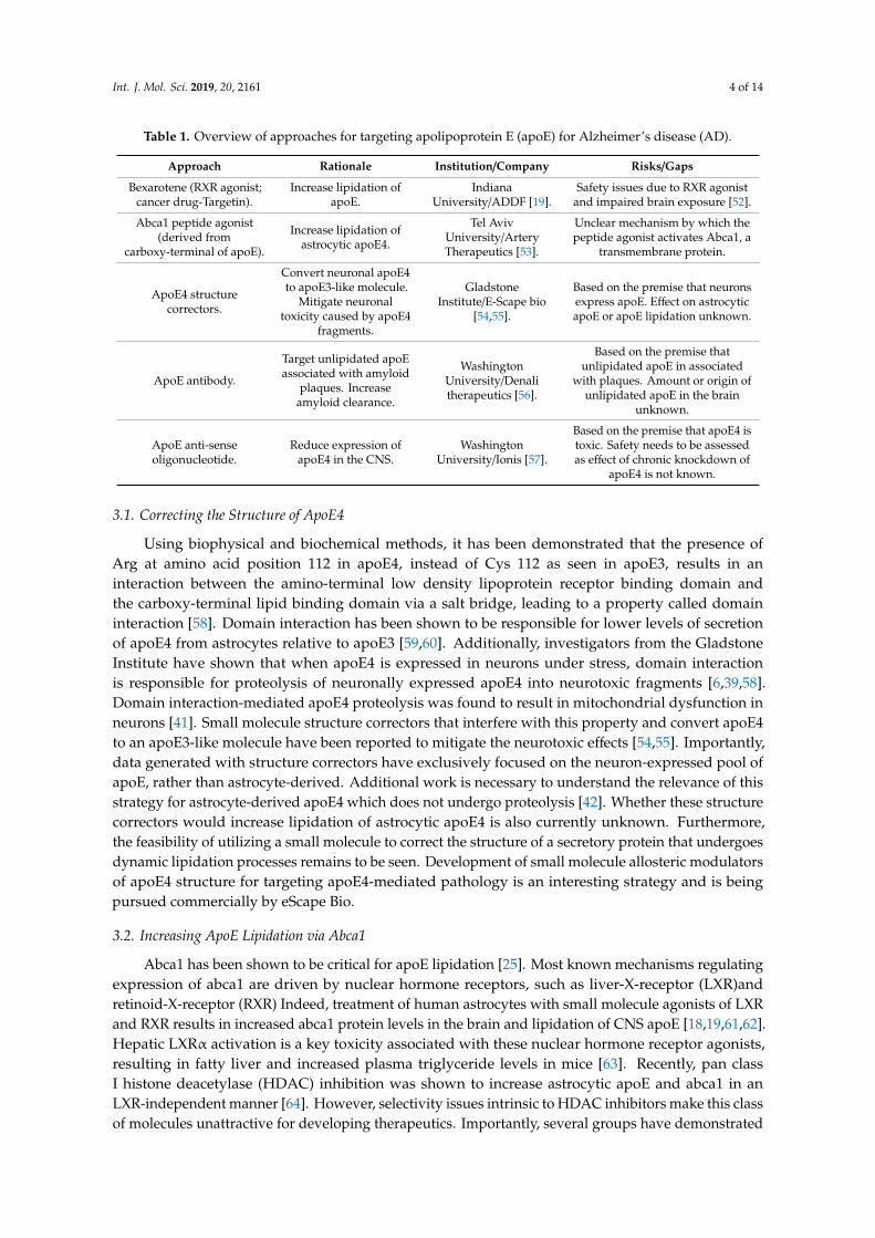
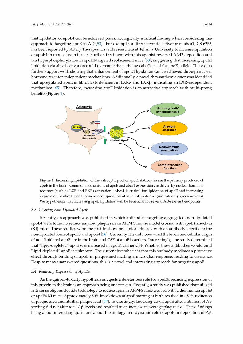
that lipidation of apoE4 can be achieved pharmacologically, a critical finding when considering thisapproach to targeting apoE in AD [53]. For example, a direct peptide activator of abca1, CS-6253,has been reported by Artery Therapeutics and researchers at Tel Aviv University to increase lipidationof apoE4 in mouse brain tissue. Further, treatment with this agonist reversed Aβ42 deposition andtau hyperphosphorylation in apoE4-targeted replacement mice [53], suggesting that increasing apoE4lipidation via abca1 activation could overcome the pathological effects of the apoE4 allele. These datafurther support work showing that enhancement of apoE4 lipidation can be achieved through nuclearhormone receptor-independent mechanisms. Additionally, a novel chrysanthemic ester was identifiedthat upregulated apoE in fibroblasts deficient in LXRα and LXRβ, indicating an LXR-independentmechanism [65]. Therefore, increasing apoE lipidation is an attractive approach with multi-prongbenefits (Figure 1).
Int. J. Mol. Sci. 2018, 19, x FOR PEER REVIEW 5 of 13
inhibitors make this class of molecules unattractive for developing therapeutics. Importantly, several
groups have demonstrated that lipidation of apoE4 can be achieved pharmacologically, a critical
finding when considering this approach to targeting apoE in AD [53]. For example, a direct peptide
activator of abca1, CS-6253, has been reported by Artery Therapeutics and researchers at Tel Aviv
University to increase lipidation of apoE4 in mouse brain tissue. Further, treatment with this agonist
reversed Aβ42 deposition and tau hyperphosphorylation in apoE4-targeted replacement mice [53],
suggesting that increasing apoE4 lipidation via abca1 activation could overcome the pathological
effects of the apoE4 allele. These data further support work showing that enhancement of apoE4
lipidation can be achieved through nuclear hormone receptor-independent mechanisms.
Additionally, a novel chrysanthemic ester was identified that upregulated apoE in fibroblasts
deficient in LXR and LXR indicating an LXR-independent mechanism [65]. Therefore, increasing
apoE lipidation is an attractive approach with multi-prong benefits (Figure 1).
Figure 1. Increasing lipidation of the astrocytic pool of apoE. Astrocytes are the primary producer of
apoE in the brain. Common mechanisms of apoE and abca1 expression are driven by nuclear hormone
receptor (such as LXR and RXR) activation. Abca1 is critical for lipidation of apoE and increasing
expression of abca1 leads to increased lipidation of all apoE isoforms (indicated by green arrows). We
hypothesize that increasing apoE lipidation will be beneficial for several AD-relevant endpoints.
3.3. Clearing Non-Lipidated ApoE
Recently, an approach was published in which antibodies targeting aggregated, non-lipidated
apoE4 were found to reduce amyloid plaques in an APP/PS mouse model crossed with apoE4 knock-
in (KI) mice. These studies were the first to show preclinical efficacy with an antibody specific to the
non-lipidated form of apoE3 and apoE4 [56]. Currently, it is unknown what the levels and cellular
origin of non-lipidated apoE are in the brain and CSF of apoE4 carriers. Interestingly, one study
determined that “lipid-depleted” apoE was increased in apoE4 carrier CSF. Whether these antibodies
would bind “lipid-depleted” apoE is unknown. The current hypothesis is that this antibody mediates
a protective effect through binding of apoE in plaque and inciting a microglial response, leading to
clearance. Despite many unanswered questions, this is a novel and interesting approach for targeting
apoE.
3.4. Reducing Expression of ApoE4
As the gain-of-toxicity hypothesis suggests a deleterious role for apoE4, reducing expression of
this protein in the brain is an approach being undertaken. Recently, a study was published that
utilized anti-sense oligonucleotide technology to reduce apoE in APP/PS mice crossed with either
human apoE3 or apoE4 KI mice. Approximately 50% knockdown of apoE starting at birth resulted
in ~50% reduction of plaque area and fibrillar plaque load [57]. Interestingly, knocking down apoE
Figure 1. Increasing lipidation of the astrocytic pool of apoE. Astrocytes are the primary producer ofapoE in the brain. Common mechanisms of apoE and abca1 expression are driven by nuclear hormonereceptor (such as LXR and RXR) activation. Abca1 is critical for lipidation of apoE and increasingexpression of abca1 leads to increased lipidation of all apoE isoforms (indicated by green arrows).We hypothesize that increasing apoE lipidation will be beneficial for several AD-relevant endpoints.
3.3. Clearing Non-Lipidated ApoE
Recently, an approach was published in which antibodies targeting aggregated, non-lipidatedapoE4 were found to reduce amyloid plaques in an APP/PS mouse model crossed with apoE4 knock-in(KI) mice. These studies were the first to show preclinical efficacy with an antibody specific to thenon-lipidated form of apoE3 and apoE4 [56]. Currently, it is unknown what the levels and cellular originof non-lipidated apoE are in the brain and CSF of apoE4 carriers. Interestingly, one study determinedthat “lipid-depleted” apoE was increased in apoE4 carrier CSF. Whether these antibodies would bind“lipid-depleted” apoE is unknown. The current hypothesis is that this antibody mediates a protectiveeffect through binding of apoE in plaque and inciting a microglial response, leading to clearance.Despite many unanswered questions, this is a novel and interesting approach for targeting apoE.
3.4. Reducing Expression of ApoE4
As the gain-of-toxicity hypothesis suggests a deleterious role for apoE4, reducing expression ofthis protein in the brain is an approach being undertaken. Recently, a study was published that utilizedanti-sense oligonucleotide technology to reduce apoE in APP/PS mice crossed with either human apoE3or apoE4 KI mice. Approximately 50% knockdown of apoE starting at birth resulted in ~50% reductionof plaque area and fibrillar plaque load [57]. Interestingly, knocking down apoE after initiation of Aβseeding did not alter total Aβ levels and resulted in an increase in average plaque size. These findingsbring about interesting questions about the biology and dynamic role of apoE in deposition of Aβ.
Int. J. Mol. Sci. 2019, 20, 2161 6 of 14
They also bring to light the challenge of identifying the best age of intervention for an apoE4-loweringtherapy. Interestingly, a carrier of a mutation which leads to knockdown of apoE protein was reportedin the literature. This was the first apoE null patient to go through extensive cognitive testing and,overall, was largely considered to be normal, suggesting that knockdown of apoE may not be harmfulto the brain [66]. However, it was reported that his memory was below what would be considerednormal for his age and his language performance was also impaired. Data from apoE KO mice indicatethat these mice have age-dependent synaptic loss and cognitive deficits [67,68], however these datacould be confounded by the peripheral hyperlipidemia that results from loss of apoE systemically.Interestingly, mice that were deficient in brain apoE specifically did not display deficits in learningand memory, but still showed synaptic loss, similar to total apoE KO [69]. Whether reducing apoE4expression will have beneficial effects while being safe and tolerable under chronic treatment is acritical question for this approach.
3.5. Our Perspective
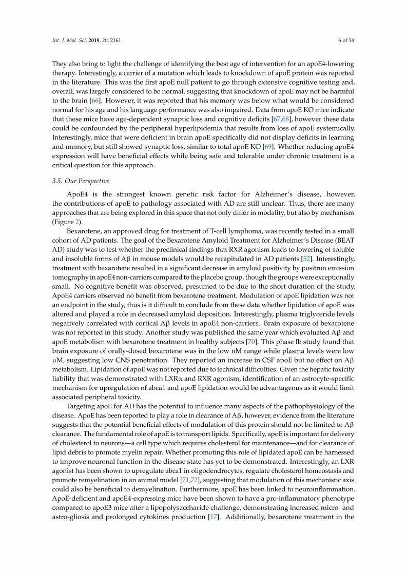
ApoE4 is the strongest known genetic risk factor for Alzheimer’s disease, however,the contributions of apoE to pathology associated with AD are still unclear. Thus, there are manyapproaches that are being explored in this space that not only differ in modality, but also by mechanism(Figure 2).
Bexarotene, an approved drug for treatment of T-cell lymphoma, was recently tested in a smallcohort of AD patients. The goal of the Bexarotene Amyloid Treatment for Alzheimer’s Disease (BEATAD) study was to test whether the preclinical findings that RXR agonism leads to lowering of solubleand insoluble forms of Aβ in mouse models would be recapitulated in AD patients [52]. Interestingly,treatment with bexarotene resulted in a significant decrease in amyloid positivity by positron emissiontomography in apoE4 non-carriers compared to the placebo group, though the groups were exceptionallysmall. No cognitive benefit was observed, presumed to be due to the short duration of the study.ApoE4 carriers observed no benefit from bexarotene treatment. Modulation of apoE lipidation was notan endpoint in the study, thus is it difficult to conclude from these data whether lipidation of apoE wasaltered and played a role in decreased amyloid deposition. Interestingly, plasma triglyceride levelsnegatively correlated with cortical Aβ levels in apoE4 non-carriers. Brain exposure of bexarotenewas not reported in this study. Another study was published the same year which evaluated Aβ andapoE metabolism with bexarotene treatment in healthy subjects [70]. This phase Ib study found thatbrain exposure of orally-dosed bexarotene was in the low nM range while plasma levels were lowµM, suggesting low CNS penetration. They reported an increase in CSF apoE but no effect on Aβmetabolism. Lipidation of apoE was not reported due to technical difficulties. Given the hepatic toxicityliability that was demonstrated with LXRα and RXR agonism, identification of an astrocyte-specificmechanism for upregulation of abca1 and apoE lipidation would be advantageous as it would limitassociated peripheral toxicity.
Targeting apoE for AD has the potential to influence many aspects of the pathophysiology of thedisease. ApoE has been reported to play a role in clearance of Aβ, however, evidence from the literaturesuggests that the potential beneficial effects of modulation of this protein should not be limited to Aβclearance. The fundamental role of apoE is to transport lipids. Specifically, apoE is important for deliveryof cholesterol to neurons—a cell type which requires cholesterol for maintenance—and for clearance oflipid debris to promote myelin repair. Whether promoting this role of lipidated apoE can be harnessedto improve neuronal function in the disease state has yet to be demonstrated. Interestingly, an LXRagonist has been shown to upregulate abca1 in oligodendrocytes, regulate cholesterol homeostasis andpromote remyelination in an animal model [71,72], suggesting that modulation of this mechanistic axiscould also be beneficial to demyelination. Furthermore, apoE has been linked to neuroinflammation.ApoE-deficient and apoE4-expressing mice have been shown to have a pro-inflammatory phenotypecompared to apoE3 mice after a lipopolysaccharide challenge, demonstrating increased micro- andastro-gliosis and prolonged cytokines production [17]. Additionally, bexarotene treatment in the
Int. J. Mol. Sci. 2019, 20, 2161 7 of 14
APP/PS1 mice significantly reduced microgliosis in the hippocampus, suggesting that abca1 and apoElipidation are important for neuroinflammation [73]. ApoE was recently reported to be a “check point”inhibitor of the classical complement cascade in an isoform-dependent manner where apoE4 resultswere similar to apoE KO [74]. ApoE binds to, and is a proposed ligand for, the triggering receptorexpressed on myeloid cells (TREM2) [75,76], a recently identified candidate risk loci for LOAD [77,78].This interaction likely modulates AD pathology, though it is currently unclear what the role of apoE isin TREM2 biology; the data are reviewed nicely elsewhere [79].
Int. J. Mol. Sci. 2018, 19, x FOR PEER REVIEW 7 of 13
cytokines production [17]. Additionally, bexarotene treatment in the APP/PS1 mice significantly
reduced microgliosis in the hippocampus, suggesting that abca1 and apoE lipidation are important
for neuroinflammation [73]. ApoE was recently reported to be a “check point” inhibitor of the classical
complement cascade in an isoform-dependent manner where apoE4 results were similar to apoE KO
[74]. ApoE binds to, and is a proposed ligand for, the triggering receptor expressed on myeloid cells
(TREM2) [75,76], a recently identified candidate risk loci for LOAD [77,78]. This interaction likely
modulates AD pathology, though it is currently unclear what the role of apoE is in TREM2 biology;
the data are reviewed nicely elsewhere [79].
Promote:Lipid delivery [30]
Neuronal growth [26]Neuronal survival [32]
Remyelination [71]Amyloid clearance [18,19]
Neuroimmune modulation [17]Cerebrovascular function [14, 15]
Reduce:Generation of neurotoxic apoE fragments [38, 39]
Tau phosphorylation [42]Mitochondrial dysfunction in neurons [41]
GABA-ergic neuronal dysfunction [49]
Reduce:Tau-mediated neurodegeneration [43]
Aβ seeding [45, 57]Promote:
Reelin signaling [47]Receptor recycling/endocytic transport [46,47]
Mitochondrial dysfunction [48]Insulin signaling [48]
Imp
act
of
stru
ctu
re
on
fu
nct
ion
Figure 2. Multiple hypotheses for targeting apoE and their downstream effects. There are many
reported findings in the literature which support the hypotheses (italicized) aimed at unveiling the
function of apoE in the brain. The approach for targeting apoE in relation to each hypothesis is listed
within the arrow. Cell types involved are astrocytes (red), microglia (purple) and neurons (green). A
lipidated apoE particle is depicted in the center.
Although the primary focus of research on brain-derived apoE has been in the AD field, there
are more data coming out further exploring the effects of the apoE genotype in cognitively normal
people. For instance, there is research on white matter integrity [80] and cerebrovascular reactivity
[81], as well as a host of abnormalities reported in apoE knock-in mice, which are reviewed in detail
[24]. ApoE biology in the brain is extremely complex with many contradictory findings being
reported as to its function and role in the CNS in mice. Therefore, human studies comparing 4
carriers and non-carriers are more likely to provide information on the roles of apoE in health and
disease. Indeed, imaging studies have suggested that 4 carriers have accelerated brain deformations
in the Alzheimer’s disease neuroimaging initiative (ADNI) cohort [82,83]. Interestingly, a recent
study conducted in cognitively normal non-carrier, heterozygotes and homozygous 4 carriers
showed a dose effect on hippocampal morphology, with carriers having increased abnormalities in
Figure 2. Multiple hypotheses for targeting apoE and their downstream effects. There are manyreported findings in the literature which support the hypotheses (italicized) aimed at unveiling thefunction of apoE in the brain. The approach for targeting apoE in relation to each hypothesis is listedwithin the arrow. Cell types involved are astrocytes (red), microglia (purple) and neurons (green).A lipidated apoE particle is depicted in the center.
Although the primary focus of research on brain-derived apoE has been in the AD field, there aremore data coming out further exploring the effects of the apoE genotype in cognitively normal people.For instance, there is research on white matter integrity [80] and cerebrovascular reactivity [81],as well as a host of abnormalities reported in apoE knock-in mice, which are reviewed in detail [24].ApoE biology in the brain is extremely complex with many contradictory findings being reportedas to its function and role in the CNS in mice. Therefore, human studies comparing ε4 carriers andnon-carriers are more likely to provide information on the roles of apoE in health and disease. Indeed,imaging studies have suggested that ε4 carriers have accelerated brain deformations in the Alzheimer’sdisease neuroimaging initiative (ADNI) cohort [82,83]. Interestingly, a recent study conducted incognitively normal non-carrier, heterozygotes and homozygous ε4 carriers showed a dose effect on
Int. J. Mol. Sci. 2019, 20, 2161 8 of 14
hippocampal morphology, with carriers having increased abnormalities in this structure prior to onsetof cognitive decline [84]. The results of these studies suggest that having an ε4 allele is detrimental tomaintenance of brain structures that are impacted by AD.
Another area of AD biology that apoE most likely plays a role in is cerebrovasculardysfunction [85,86]. The brain is a highly vascularized organ and its high energy demand consumes20–25% of the body’s oxygen and glucose stores [87]. Blood flow to the brain is required for proper CNSdevelopment and is necessary for neuronal viability. Vascular disease was shown to strongly correlatewith cognitive dysfunction in AD autopsy samples [88]. Additionally, studies utilizing arterial spinlabeling magnetic resonance imaging to assess CBF have reported in the ADNI cohort that deficitsoccur early in the progression to late onset AD, suggesting that cerebrovascular dysfunction mayplay a causal role in the development of AD [89]. Interestingly, preclinical data suggest a relationshipbetween the apoE genotype and vascular function, however the mechanisms involved are unclear.Cognitively normal carriers of an apoE4 gene demonstrated impaired cerebrovascular reactivity tohypercapnia assessed by blood-oxygen-level-dependent functional magnetic resonance imaging andtranscranial doppler [81,90]. Furthermore, mice expressing human apoE4 have reduced resting CBF [15]and impaired vascular response to neuronal activity (neurovascular coupling) [14]. Blood–brain barrierintegrity is also compromised in apoE KO and apoE4 mice [15,91,92]. Lastly, carrying an apoE4 alleleleads to increased vascular Aβ deposition [93] and homozygotes were reported to have elevatedfibrin(ogen) deposition in Aβ positive vessels [94]. Studies investigating the relationship betweenapoE and cerebrovascular function are needed to increase our understanding of the role of this proteinin vascular dysfunction associated with Alzheimer’s disease.
One clear deficit in the field is the lack of understanding of the relationship between lipidation ofapoE and disease endpoints. Increasing lipidation of apoE is an attractive target for drug discovery asit targets the fundamental apoE function, namely lipid transport. However, the association of apoElipidation status in CSF with markers of AD progression, such as total tau protein, phosphorylated tau,Aβ ratio, and cognitive end points has not been assessed in a large, well-characterized cohort of healthycontrols and AD patients. Total apoE protein levels are informative, and are reduced in apoE4 carriers,but do not directly address the question of lipidation status. One reason for this critical gap in the fieldis the lack of high(er) throughput and quantitative assays to assess protein lipidation, although someadvancements have been made for apolipoprotein A–I [95]. Detection of “lipid-depleted” apoE inthe CSF of a small cohort of patients [22] has also been published and requires centrifugation over apotassium bromide gradient, which is not ideal for screening a large number of samples. Large scalestudies will need to be performed in order to define efficacious dosing for drug discovery programslooking to increase lipidation of apoE.
ApoE biology is highly complex and there are many aspects—such as the cellular origin andlipidation state of apoE—that must be considered when targeting this protein for AD. Additionally,which patient population would most greatly benefit from an apoE-directed therapy and at what stageof the disease it would be most effective are two outstanding questions in the field. In our opinion,addressing these gaps will be critical to the development of effective apoE-directed therapeutics.For example, an apoE lipidation approach may be suitable for all apoE genotypes given the potentialof the mechanism to be restorative. However, the correlation of apoE lipidation state with diseasepathology needs to be determined. Generation of human data in well-characterized patient cohorts willbe critical for addressing this gap. On the other hand, apoE4 reducing approaches may be suitable forapoE4 homozygous patients. Furthermore, work done in apoE KO mice and characterization studiesin a null human do not take into account the compensation that may occur in the complete absenceof this critical protein in the brain, or the effect it may have on CNS lipid metabolism. Additionally,whether perturbation of peripheral lipid homeostasis in the absence of apoE will lead to vasculardysfunction will need to be determined. Thus, it is currently unclear whether reducing apoE expressionin the brain would have a negative outcome with respect to safety. Currently, there are no findings
Int. J. Mol. Sci. 2019, 20, 2161 9 of 14
which suggest that increasing lipidation of endogenous apoE in the brain, including apoE4, would carrya safety risk.
In conclusion, apoE is an attractive and rational target for development of diseasemodifying-therapies for AD. After age, apoE4 is the strongest known risk factor for this disease,and thus targeting apoE could be relevant for those already showing symptoms of the disease.More research is needed in both academia and industry to further uncover the role of this protein inthe brain to answer outstanding questions on how it should be targeted.
Author Contributions: G.L.S., G.R.: Original draft preparation, G.L.S., G.R.: Review and editing.
Acknowledgments: We thank Dominic M. Walsh for review of the manuscript.
Conflicts of Interest: The authors declare no conflict of interest.
References
1. Selkoe, D.J.; Hardy, J. The amyloid hypothesis of Alzheimer’s disease at 25 years. EMBO Mol. Med. 2016, 8,595–608. [CrossRef] [PubMed]
2. Jadhav, S. A walk through tau therapeutic strategies. Acta Neuropathol. Commun. 2019, 7, 22. [CrossRef][PubMed]
3. Foley, P. Lipids in Alzheimer’s disease: A century-old story. Biochim. Biophys. Acta 2010, 1801, 750–753.[CrossRef]
4. Di Paolo, G.; Kim, T.W. Linking lipids to Alzheimer’s disease: Cholesterol and beyond. Nat. Rev. Neurosci.2011, 12, 284–296. [CrossRef]
5. Dietschy, J.M.; Turley, S.D. Thematic review series: Brain Lipids. Cholesterol metabolism in the centralnervous system during early development and in the mature animal. J. Lipid Res. 2004, 45, 1375–1397.[CrossRef] [PubMed]
6. Mahley, R.W. Central Nervous System Lipoproteins: ApoE and Regulation of Cholesterol Metabolism.Arterioscler. Thromb. Vasc. Biol. 2016, 36, 1305–1315. [CrossRef]
7. Leduc, V.; Jasmin-Belanger, S.; Poirier, J. APOE and cholesterol homeostasis in Alzheimer’s disease. Trends Mol.Med. 2010, 16, 469–477. [CrossRef]
8. Zhang, Y.; Chen, K.; Sloan, S.A.; Bennett, M.L.; Scholze, A.R.; O’Keeffe, S.; Phatnani, H.P.; Guarnieri, P.;Caneda, C.; Ruderisch, N.; et al. An RNA-sequencing transcriptome and splicing database of glia, neurons,and vascular cells of the cerebral cortex. J. Neurosci. 2014, 34, 11929–11947. [CrossRef]
9. Zhang, Y.; Sloan, S.A.; Clarke, L.E.; Caneda, C.; Plaza, C.A.; Blumenthal, P.D.; Vogel, H.; Steinberg, G.K.;Edwards, M.S.; Li, G.; et al. Purification and Characterization of Progenitor and Mature Human AstrocytesReveals Transcriptional and Functional Differences with Mouse. Neuron 2016, 89, 37–53. [CrossRef] [PubMed]
10. Huang, Y.; Weisgraber, K.H.; Mucke, L.; Mahley, R.W. Apolipoprotein E: Diversity of cellular origins,structural and biophysical properties, and effects in Alzheimer’s disease. J. Mol. Neurosci. 2004, 23, 189–204.[CrossRef]
11. Nathan, B.P. Differential effects of apolipoproteins E3 and E4 on neuronal growth in vitro. Science 1994, 264,850–852. [CrossRef]
12. Pitas, R.E. Role of apolipoprotein E in modulating neurite outgrowth: Potential effect of intracellularapolipoprotein E. Biochem. Soc. Trans. 1998, 26, 257–262. [CrossRef] [PubMed]
13. Levi, O.; Jongen-Relo, A.L.; Feldon, J.; Roses, A.D.; Michaelson, D.M. ApoE4 impairs hippocampal plasticityisoform-specifically and blocks the environmental stimulation of synaptogenesis and memory. Neurobiol. Dis.2003, 13, 273–282. [CrossRef]
14. Koizumi, K.; Hattori, Y.; Ahn, S.J.; Buendia, I.; Ciacciarelli, A.; Uekawa, K.; Wang, G.; Hiller, A.; Zhao, L.;Voss, H.U.; et al. Apoepsilon4 disrupts neurovascular regulation and undermines white matter integrity andcognitive function. Nat. Commun. 2018, 9, 3816. [CrossRef] [PubMed]
15. Bell, R.D.; Winkler, E.A.; Singh, I.; Sagare, A.P.; Deane, R.; Wu, Z.; Holtzman, D.M.; Betsholtz, C.; Armulik, A.;Sallstrom, J.; et al. Apolipoprotein E controls cerebrovascular integrity via cyclophilin A. Nature 2012, 485,512–516. [CrossRef] [PubMed]
16. Tensaouti, Y.; Stephanz, E.P.; Yu, T.S.; Kernie, S.G. ApoE Regulates the Development of Adult NewbornHippocampal Neurons. eNeuro 2018, 5. [CrossRef]
Int. J. Mol. Sci. 2019, 20, 2161 10 of 14
17. Zhu, Y.; Nwabuisi-Heath, E.; Dumanis, S.B.; Tai, L.M.; Yu, C.; Rebeck, G.W.; LaDu, M.J. APOE genotypealters glial activation and loss of synaptic markers in mice. Glia 2012, 60, 559–569. [CrossRef]
18. Riddell, D.R.; Zhou, H.; Comery, T.A.; Kouranova, E.; Lo, C.F.; Warwick, H.K.; Ring, R.H.; Kirksey, Y.;Aschmies, S.; Xu, J.; et al. The LXR agonist TO901317 selectively lowers hippocampal Abeta42 and improvesmemory in the Tg2576 mouse model of Alzheimer’s disease. Mol. Cell Neurosci. 2007, 34, 621–628. [CrossRef]
19. Cramer, P.E.; Cirrito, J.R.; Wesson, D.W.; Lee, C.Y.; Karlo, J.C.; Zinn, A.E.; Casali, B.T.; Restivo, J.L.;Goebel, W.D.; James, M.J.; et al. ApoE-directed therapeutics rapidly clear beta-amyloid and reverse deficitsin AD mouse models. Science 2012, 335, 1503–1506. [CrossRef]
20. Liu, C.C.; Liu, C.C.; Kanekiyo, T.; Xu, H.; Bu, G. Apolipoprotein E and Alzheimer disease: Risk,mechanisms and therapy. Nat. Rev. Neurol. 2013, 9, 106–118. [CrossRef]
21. Cruchaga, C.; Kauwe, J.S.; Nowotny, P.; Bales, K.; Pickering, E.H.; Mayo, K.; Bertelsen, S.; Hinrichs, A.;Alzheimer’s Disease Neuroimaging, I.; Fagan, A.M.; et al. Cerebrospinal fluid APOE levels:An endophenotype for genetic studies for Alzheimer’s disease. Hum. Mol. Genet. 2012, 21, 4558–4571.[CrossRef] [PubMed]
22. Hanson, A.J.; Bayer-Carter, J.L.; Green, P.S.; Montine, T.J.; Wilkinson, C.W.; Baker, L.D.; Watson, G.S.;Bonner, L.M.; Callaghan, M.; Leverenz, J.B.; et al. Effect of apolipoprotein E genotype and diet onapolipoprotein E lipidation and amyloid peptides: Randomized clinical trial. JAMA Neurol. 2013, 70, 972–980.[CrossRef] [PubMed]
23. Sullivan, P.M.; Han, B.; Liu, F.; Mace, B.E.; Ervin, J.F.; Wu, S.; Koger, D.; Paul, S.; Bales, K.R. Reduced levelsof human apoE4 protein in an animal model of cognitive impairment. Neurobiol. Aging 2011, 32, 791–801.[CrossRef]
24. Rebeck, G.W. The role of APOE on lipid homeostasis and inflammation in normal brains. J. Lipid Res. 2017,58, 1493–1499. [CrossRef] [PubMed]
25. Wahrle, S.E.; Jiang, H.; Parsadanian, M.; Legleiter, J.; Han, X.; Fryer, J.D.; Kowalewski, T.; Holtzman, D.M.ABCA1 is required for normal central nervous system ApoE levels and for lipidation of astrocyte-secretedapoE. J. Biol. Chem. 2004, 279, 40987–40993. [CrossRef] [PubMed]
26. Fitz, N.F.; Carter, A.Y.; Tapias, V.; Castranio, E.L.; Kodali, R.; Lefterov, I.; Koldamova, R. ABCA1 DeficiencyAffects Basal Cognitive Deficits and Dendritic Density in Mice. J. Alzheimer’s Dis. 2017, 56, 1075–1085.[CrossRef] [PubMed]
27. Wahrle, S.E.; Jiang, H.; Parsadanian, M.; Hartman, R.E.; Bales, K.R.; Paul, S.M.; Holtzman, D.M. Deletion ofAbca1 increases Abeta deposition in the PDAPP transgenic mouse model of Alzheimer disease. J. Biol. Chem.2005, 280, 43236–43242. [CrossRef]
28. Nordestgaard, L.T.; Tybjaerg-Hansen, A.; Nordestgaard, B.G.; Frikke-Schmidt, R. Loss-of-function mutationin ABCA1 and risk of Alzheimer’s disease and cerebrovascular disease. Alzheimer’s Dement. 2015, 11,1430–1438. [CrossRef]
29. Wahrle, S.E.; Jiang, H.; Parsadanian, M.; Kim, J.; Li, A.; Knoten, A.; Jain, S.; Hirsch-Reinshagen, V.;Wellington, C.L.; Bales, K.R.; et al. Overexpression of ABCA1 reduces amyloid deposition in the PDAPPmouse model of Alzheimer disease. J. Clin. Investig. 2008, 118, 671–682. [CrossRef]
30. Namjoshi, D.R.; Martin, G.; Donkin, J.; Wilkinson, A.; Stukas, S.; Fan, J.; Carr, M.; Tabarestani, S.; Wuerth, K.;Hancock, R.E.; et al. The liver X receptor agonist GW3965 improves recovery from mild repetitive traumaticbrain injury in mice partly through apolipoprotein E. PLoS ONE 2013, 8, e53529. [CrossRef]
31. Loane, D.J.; Washington, P.M.; Vardanian, L.; Pocivavsek, A.; Hoe, H.S.; Duff, K.E.; Cernak, I.; Rebeck, G.W.;Faden, A.I.; Burns, M.P. Modulation of ABCA1 by an LXR agonist reduces beta-amyloid levels and improvesoutcome after traumatic brain injury. J. Neurotr. 2011, 28, 225–236. [CrossRef]
32. Zhao, J.; Davis, M.D.; Martens, Y.A.; Shinohara, M.; Graff-Radford, N.R.; Younkin, S.G.; Wszolek, Z.K.;Kanekiyo, T.; Bu, G. APOE epsilon4/epsilon4 diminishes neurotrophic function of human iPSC-derivedastrocytes. Hum. Mol. Genet. 2017, 26, 2690–2700. [CrossRef] [PubMed]
33. Robert, J.; Stukas, S.; Button, E.; Cheng, W.H.; Lee, M.; Fan, J.; Wilkinson, A.; Kulic, I.; Wright, S.D.;Wellington, C.L. Reconstituted high-density lipoproteins acutely reduce soluble brain Abeta levels insymptomatic APP/PS1 mice. Biochim. Biophys. Acta 2016, 1862, 1027–1036. [CrossRef]
34. Stukas, S.; Robert, J.; Wellington, C.L. High-density lipoproteins and cerebrovascular integrity in Alzheimer’sdisease. Cell Metab. 2014, 19, 574–591. [CrossRef] [PubMed]
Int. J. Mol. Sci. 2019, 20, 2161 11 of 14
35. Liu, D.S.; Pan, X.D.; Zhang, J.; Shen, H.; Collins, N.C.; Cole, A.M.; Koster, K.P.; Ben Aissa, M.; Dai, X.M.;Zhou, M.; et al. APOE4 enhances age-dependent decline in cognitive function by down-regulating an NMDAreceptor pathway in EFAD-Tg mice. Mol. Neurodegener. 2015, 10, 7. [CrossRef]
36. Horsburgh, K.; Nicoll, J.A. Selective alterations in the cellular distribution of apolipoprotein Eimmunoreactivity following transient cerebral ischaemia in the rat. Neuropathol. Appl. Neurobiol. 1996, 22,342–349. [CrossRef] [PubMed]
37. Xu, Q.; Bernardo, A.; Walker, D.; Kanegawa, T.; Mahley, R.W.; Huang, Y. Profile and regulation ofapolipoprotein E (ApoE) expression in the CNS in mice with targeting of green fluorescent protein gene tothe ApoE locus. J. Neurosci. 2006, 26, 4985–4994. [CrossRef] [PubMed]
38. Huang, Y.; Liu, X.Q.; Wyss-Coray, T.; Brecht, W.J.; Sanan, D.A.; Mahley, R.W. Apolipoprotein E fragmentspresent in Alzheimer’s disease brains induce neurofibrillary tangle-like intracellular inclusions in neurons.Proc. Natl. Acad. Sci. USA 2001, 98, 8838–8843. [CrossRef]
39. Harris, F.M.; Brecht, W.J.; Xu, Q.; Tesseur, I.; Kekonius, L.; Wyss-Coray, T.; Fish, J.D.; Masliah, E.; Hopkins, P.C.;Scearce-Levie, K.; et al. Carboxyl-terminal-truncated apolipoprotein E4 causes Alzheimer’s disease-likeneurodegeneration and behavioral deficits in transgenic mice. Proc. Natl. Acad. Sci. USA 2003, 100,10966–10971. [CrossRef] [PubMed]
40. Mahley, R.W.; Huang, Y.; Weisgraber, K.H. Detrimental effects of apolipoprotein E4: Potential therapeutictargets in Alzheimer’s disease. Curr. Alzheimer Res. 2007, 4, 537–540. [CrossRef] [PubMed]
41. Mahley, R.W.; Weisgraber, K.H.; Huang, Y. Apolipoprotein E4: A causative factor and therapeutic target inneuropathology, including Alzheimer’s disease. Proc. Natl. Acad. Sci. USA 2006, 103, 5644–5651. [CrossRef]
42. Brecht, W.J.; Harris, F.M.; Chang, S.; Tesseur, I.; Yu, G.Q.; Xu, Q.; Dee Fish, J.; Wyss-Coray, T.;Buttini, M.; Mucke, L.; et al. Neuron-specific apolipoprotein e4 proteolysis is associated with increased tauphosphorylation in brains of transgenic mice. J. Neurosci. 2004, 24, 2527–2534. [CrossRef] [PubMed]
43. Shi, Y.; Yamada, K.; Liddelow, S.A.; Smith, S.T.; Zhao, L.; Luo, W.; Tsai, R.M.; Spina, S.; Grinberg, L.T.;Rojas, J.C.; et al. ApoE4 markedly exacerbates tau-mediated neurodegeneration in a mouse model oftauopathy. Nature 2017, 549, 523–527. [CrossRef] [PubMed]
44. Strickland, M.R.; Holtzman, D.M. Dr. Jekyll and Mr. Hyde: ApoE explains opposing effects of neuronalLRP1. J. Clin. Investig. 2019. [CrossRef] [PubMed]
45. Liu, C.C.; Zhao, N.; Fu, Y.; Wang, N.; Linares, C.; Tsai, C.W.; Bu, G. ApoE4 Accelerates Early Seeding ofAmyloid Pathology. Neuron 2017, 96, 1024–1032. [CrossRef] [PubMed]
46. Xian, X.; Pohlkamp, T.; Durakoglugil, M.S.; Wong, C.H.; Beck, J.K.; Lane-Donovan, C.; Plattner, F.; Herz, J.Reversal of ApoE4-induced recycling block as a novel prevention approach for Alzheimer’s disease.eLife 2018, 7. [CrossRef] [PubMed]
47. Chen, Y.; Durakoglugil, M.S.; Xian, X.; Herz, J. ApoE4 reduces glutamate receptor function and synapticplasticity by selectively impairing ApoE receptor recycling. Proc. Natl. Acad. Sci. USA 2010, 107, 12011–12016.[CrossRef]
48. Zhao, N.; Liu, C.C.; Van Ingelgom, A.J.; Martens, Y.A.; Linares, C.; Knight, J.A.; Painter, M.M.; Sullivan, P.M.;Bu, G. Apolipoprotein E4 Impairs Neuronal Insulin Signaling by Trapping Insulin Receptor in the Endosomes.Neuron 2017, 96, 115–129.e5. [CrossRef]
49. Knoferle, J.; Yoon, S.Y.; Walker, D.; Leung, L.; Gillespie, A.K.; Tong, L.M.; Bien-Ly, N.; Huang, Y. ApolipoproteinE4 produced in GABAergic interneurons causes learning and memory deficits in mice. J. Neurosci. 2014, 34,14069–14078. [CrossRef]
50. Chung, W.S.; Verghese, P.B.; Chakraborty, C.; Joung, J.; Hyman, B.T.; Ulrich, J.D.; Holtzman, D.M.; Barres, B.A.Novel allele-dependent role for APOE in controlling the rate of synapse pruning by astrocytes. Proc. Natl.Acad. Sci. USA 2016, 113, 10186–10191. [CrossRef]
51. Yamazaki, Y.; Painter, M.M.; Bu, G.; Kanekiyo, T. Apolipoprotein E as a Therapeutic Target in Alzheimer’sDisease: A Review of Basic Research and Clinical Evidence. CNS Drugs 2016, 30, 773–789. [CrossRef]
52. Cummings, J.L.; Zhong, K.; Kinney, J.W.; Heaney, C.; Moll-Tudla, J.; Joshi, A.; Pontecorvo, M.; Devous, M.;Tang, A.; Bena, J. Double-blind, placebo-controlled, proof-of-concept trial of bexarotene Xin moderateAlzheimer’s disease. Alzheimer’s Res. Ther. 2016, 8, 4. [CrossRef]
53. Boehm-Cagan, A.; Bar, R.; Liraz, O.; Bielicki, J.K.; Johansson, J.O.; Michaelson, D.M. ABCA1 Agonist Reversesthe ApoE4-Driven Cognitive and Brain Pathologies. J. Alzheimer’s Dis. 2016, 54, 1219–1233. [CrossRef][PubMed]
Int. J. Mol. Sci. 2019, 20, 2161 12 of 14
54. Wang, C.; Najm, R.; Xu, Q.; Jeong, D.E.; Walker, D.; Balestra, M.E.; Yoon, S.Y.; Yuan, H.; Li, G.; Miller, Z.A.; et al.Gain of toxic apolipoprotein E4 effects in human iPSC-derived neurons is ameliorated by a small-moleculestructure corrector. Nat. Med. 2018. [CrossRef] [PubMed]
55. Mahley, R.W.; Huang, Y. Small-molecule structure correctors target abnormal protein structure and function:Structure corrector rescue of apolipoprotein E4-associated neuropathology. J. Med. Chem. 2012, 55, 8997–9008.[CrossRef] [PubMed]
56. Liao, F.; Li, A.; Xiong, M.; Bien-Ly, N.; Jiang, H.; Zhang, Y.; Finn, M.B.; Hoyle, R.; Keyser, J.; Lefton, K.B.;et al. Targeting of nonlipidated, aggregated apoE with antibodies inhibits amyloid accumulation. J. Clin.Investig. 2018. [CrossRef]
57. Huynh, T.V.; Liao, F.; Francis, C.M.; Robinson, G.O.; Serrano, J.R.; Jiang, H.; Roh, J.; Finn, M.B.; Sullivan, P.M.;Esparza, T.J.; et al. Age-Dependent Effects of apoE Reduction Using Antisense Oligonucleotides in a Modelof beta-amyloidosis. Neuron 2017, 96, 1013–1023.e4. [CrossRef]
58. Zhong, N.; Weisgraber, K.H. Understanding the basis for the association of apoE4 with Alzheimer’s disease:Opening the door for therapeutic approaches. Curr. Alzheimer Res. 2009, 6, 415–418. [CrossRef]
59. Ramaswamy, G.; Xu, Q.; Huang, Y.; Weisgraber, K.H. Effect of domain interaction on apolipoprotein E levelsin mouse brain. J. Neurosci. 2005, 25, 10658–10663. [CrossRef]
60. Riddell, D.R. Impact of apolipoprotein E (ApoE) polymorphism on brain ApoE levels. J. Neurosci. 2008, 28,11445–11453. [CrossRef]
61. Fitz, N.F.; Castranio, E.L.; Carter, A.Y.; Kodali, R.; Lefterov, I.; Koldamova, R. Improvement of memorydeficits and amyloid-beta clearance in aged APP23 mice treated with a combination of anti-amyloid-betaantibody and LXR agonist. J. Alzheimer’s Dis. 2014, 41, 535–549. [CrossRef] [PubMed]
62. Corona, A.W.; Kodoma, N.; Casali, B.T.; Landreth, G.E. ABCA1 is Necessary for Bexarotene-MediatedClearance of Soluble Amyloid Beta from the Hippocampus of APP/PS1 Mice. J. Neuroimmune Pharmacol.2016, 11, 61–72. [CrossRef]
63. Schultz, J.R.; Tu, H.; Luk, A.; Repa, J.J.; Medina, J.C.; Li, L.; Schwendner, S.; Wang, S.; Thoolen, M.;Mangelsdorf, D.J.; et al. Role of LXRs in control of lipogenesis. Genes Dev. 2000, 14, 2831–2838. [CrossRef][PubMed]
64. Dresselhaus, E.; Duerr, J.M.; Vincent, F.; Sylvain, E.K.; Beyna, M.; Lanyon, L.F.; LaChapelle, E.; Pettersson, M.;Bales, K.R.; Ramaswamy, G. Class I HDAC inhibition is a novel pathway for regulating astrocytic apoEsecretion. PLoS ONE 2018, 13, e0194661. [CrossRef] [PubMed]
65. Fan, J.; Zareyan, S.; Zhao, W.; Shimizu, Y.; Pfeifer, T.A.; Tak, J.H.; Isman, M.B.; Van den Hoven, B.;Duggan, M.E.; Wood, M.W.; et al. Identification of a Chrysanthemic Ester as an Apolipoprotein E Inducer inAstrocytes. PLoS ONE 2016, 11, e0162384. [CrossRef] [PubMed]
66. Mak, A.C.; Pullinger, C.R.; Tang, L.F.; Wong, J.S.; Deo, R.C.; Schwarz, J.M.; Gugliucci, A.; Movsesyan, I.;Ishida, B.Y.; Chu, C.; et al. Effects of the absence of apolipoprotein e on lipoproteins, neurocognitive function,and retinal function. JAMA Neurol. 2014, 71, 1228–1236. [CrossRef] [PubMed]
67. Veinbergs, I.; Mallory, M.; Sagara, Y.; Masliah, E. Vitamin E supplementation prevents spatial learning deficitsand dendritic alterations in aged apolipoprotein E-deficient mice. Eur. J. Neurosci. 2000, 12, 4541–4546.[PubMed]
68. Masliah, E.; Mallory, M.; Ge, N.; Alford, M.; Veinbergs, I.; Roses, A.D. Neurodegeneration in the centralnervous system of apoE-deficient mice. Exp.Neurol. 1995, 136, 107–122. [CrossRef]
69. Lane-Donovan, C.; Wong, W.M.; Durakoglugil, M.S.; Wasser, C.R.; Jiang, S.; Xian, X.; Herz, J.Genetic Restoration of Plasma ApoE Improves Cognition and Partially Restores Synaptic Defects inApoE-Deficient Mice. J. Neurosci. 2016, 36, 10141–10150. [CrossRef]
70. Ghosal, K.; Haag, M.; Verghese, P.B.; West, T.; Veenstra, T.; Braunstein, J.B.; Bateman, R.J.; Holtzman, D.M.;Landreth, G.E. A randomized controlled study to evaluate the effect of bexarotene on amyloid-beta andapolipoprotein E metabolism in healthy subjects. Alzheimer’s Dement. 2016, 2, 110–120.
71. Cantuti-Castelvetri, L.; Fitzner, D.; Bosch-Queralt, M.; Weil, M.T.; Su, M.; Sen, P.; Ruhwedel, T.; Mitkovski, M.;Trendelenburg, G.; Lutjohann, D.; et al. Defective cholesterol clearance limits remyelination in the agedcentral nervous system. Science 2018, 359, 684–688. [CrossRef] [PubMed]
72. Nelissen, K. Liver X receptors regulate cholesterol homeostasis in oligodendrocytes. J. Neurosci. Res. 2012,90, 60–71. [CrossRef]
Int. J. Mol. Sci. 2019, 20, 2161 13 of 14
73. Casali, B.T.; Reed-Geaghan, E.G.; Landreth, G.E. Nuclear receptor agonist-driven modification ofinflammation and amyloid pathology enhances and sustains cognitive improvements in a mouse model ofAlzheimer’s disease. J. Neuroinflamm. 2018, 15, 43. [CrossRef]
74. Yin, C. ApoE attenuates unresolvable inflammation by complex formation with activated C1q. Nat. Med.2019, 25, 496–506. [CrossRef] [PubMed]
75. Bailey, C.C.; DeVaux, L.B.; Farzan, M. The Triggering Receptor Expressed on Myeloid Cells 2 BindsApolipoprotein E. J. Biol. Chem. 2015, 290, 26033–26042. [CrossRef]
76. Atagi, Y. Apolipoprotein E Is a Ligand for Triggering Receptor Expressed on Myeloid Cells 2 (TREM2). J. Biol.Chem. 2015, 290, 26043–26050. [CrossRef]
77. Guerreiro, R.; Wojtas, A.; Bras, J.; Carrasquillo, M.; Rogaeva, E.; Majounie, E.; Cruchaga, C.; Sassi, C.;Kauwe, J.S.; Younkin, S.; et al. TREM2 variants in Alzheimer’s disease. N. Engl. J. Med. 2013, 368, 117–127.[CrossRef] [PubMed]
78. Jonsson, T.; Stefansson, H.; Steinberg, S.; Jonsdottir, I.; Jonsson, P.V.; Snaedal, J.; Bjornsson, S.; Huttenlocher, J.;Levey, A.I.; Lah, J.J.; et al. Variant of TREM2 associated with the risk of Alzheimer’s disease. N. Engl. J. Med.2013, 368, 107–116. [CrossRef] [PubMed]
79. Wolfe, C.M.; Fitz, N.F.; Nam, K.N.; Lefterov, I.; Koldamova, R. The Role of APOE and TREM2 in Alzheimer’sDisease-Current Understanding and Perspectives. Int. J. Mol. Sci. 2018, 20. [CrossRef] [PubMed]
80. Operto, G.; Cacciaglia, R.; Grau-Rivera, O.; Falcon, C.; Brugulat-Serrat, A.; Rodenas, P.; Ramos, R.; Moran, S.;Esteller, M.; Bargallo, N.; et al. White matter microstructure is altered in cognitively normal middle-agedAPOE-epsilon4 homozygotes. Alzheimer’s Res. Ther. 2018, 10, 48. [CrossRef] [PubMed]
81. Suri, S.; Mackay, C.E.; Kelly, M.E.; Germuska, M.; Tunbridge, E.M.; Frisoni, G.B.; Matthews, P.M.; Ebmeier, K.P.;Bulte, D.P.; Filippini, N. Reduced cerebrovascular reactivity in young adults carrying the APOE epsilon4allele. Alzheimer’s Dement. 2015, 11, 648–657. [CrossRef] [PubMed]
82. Shi, J.; Lepore, N.; Gutman, B.A.; Thompson, P.M.; Baxter, L.C.; Caselli, R.J.; Wang, Y.; Alzheimer’s DiseaseNeuroimaging, I. Genetic influence of apolipoprotein E4 genotype on hippocampal morphometry: An N = 725surface-based Alzheimer’s disease neuroimaging initiative study. Hum. Brain Map. 2014, 35, 3903–3918.[CrossRef]
83. Li, B.; Shi, J.; Gutman, B.A.; Baxter, L.C.; Thompson, P.M.; Caselli, R.J.; Wang, Y.; Alzheimer’s DiseaseNeuroimaging, I. Influence of APOE Genotype on Hippocampal Atrophy over Time - An N=1925Surface-Based ADNI Study. PLoS ONE 2016, 11, e0152901. [CrossRef] [PubMed]
84. Dong, Q.; Zhang, W.; Wu, J.; Li, B.; Schron, E.H.; McMahon, T.; Shi, J.; Gutman, B.A.; Chen, K.; Baxter, L.C.;et al. Applying surface-based hippocampal morphometry to study APOE-E4 allele dose effects in cognitivelyunimpaired subjects. NeuroImage Clin. 2019, 22, 101744. [CrossRef]
85. Strickland, S. Blood will out: Vascular contributions to Alzheimer’s disease. J. Clin. Investig. 2018, 128,556–563. [CrossRef] [PubMed]
86. Bell, R.D.; Zlokovic, B.V. Neurovascular mechanisms and blood-brain barrier disorder in Alzheimer’s disease.Acta Neuropathol. 2009, 118, 103–113. [CrossRef]
87. Sweeney, M.D.; Sagare, A.P.; Zlokovic, B.V. Blood-brain barrier breakdown in Alzheimer disease and otherneurodegenerative disorders. Nat. Rev. Neurol. 2018, 14, 133–150. [CrossRef]
88. Serrano-Pozo, A.; Qian, J.; Monsell, S.E.; Frosch, M.P.; Betensky, R.A.; Hyman, B.T. Examination ofthe clinicopathologic continuum of Alzheimer disease in the autopsy cohort of the National AlzheimerCoordinating Center. J. Neuropathol. Exp. Neurol. 2013, 72, 1182–1192. [CrossRef] [PubMed]
89. Iturria-Medina, Y.; Sotero, R.C.; Toussaint, P.J.; Mateos-Perez, J.M.; Evans, A.C.; Alzheimer’s DiseaseNeuroimaging, I. Early role of vascular dysregulation on late-onset Alzheimer’s disease based onmultifactorial data-driven analysis. Nat. Commun. 2016, 7, 11934. [CrossRef] [PubMed]
90. Hajjar, I.; Sorond, F.; Lipsitz, L.A. Apolipoprotein E, carbon dioxide vasoreactivity, and cognition in olderadults: Effect of hypertension. J. Am. Geriatr. Soc. 2015, 63, 276–281. [CrossRef] [PubMed]
91. Hafezi-Moghadam, A.; Thomas, K.L.; Wagner, D.D. ApoE deficiency leads to a progressive age-dependentblood-brain barrier leakage. Am. J. Physiol. 2007, 292, C1256–C1262. [CrossRef] [PubMed]
92. Methia, N.; Andre, P.; Hafezi-Moghadam, A.; Economopoulos, M.; Thomas, K.L.; Wagner, D.D.ApoE deficiency compromises the blood brain barrier especially after injury. Mol. Med. 2001, 7, 810–815.[CrossRef]
Int. J. Mol. Sci. 2019, 20, 2161 14 of 14
93. Peuralinna, T.; Tanskanen, M.; Makela, M.; Polvikoski, T.; Paetau, A.; Kalimo, H.; Sulkava, R.; Hardy, J.;Lai, S.L.; Arepalli, S.; et al. APOE and AbetaPP gene variation in cortical and cerebrovascular amyloid-betapathology and Alzheimer’s disease: A population-based analysis. J. Alzheimer’s Dis. 2011, 26, 377–385.[CrossRef] [PubMed]
94. Hultman, K.; Strickland, S.; Norris, E.H. The APOE varepsilon4/varepsilon4 genotype potentiates vascularfibrin(ogen) deposition in amyloid-laden vessels in the brains of Alzheimer’s disease patients. J. Cereb. BloodFlow Metab. 2013, 33, 1251–1258. [CrossRef] [PubMed]
95. Niedziela-Majka, A.; Lad, L.; Chisholm, J.W.; Lagpacan, L.; Schwartz, K.; Hung, M.; Jin, D.; Fung, W.;Brendza, K.M.; Liu, X.; et al. Lipid-sensing high-throughput ApoA-I assays. J. Biomol. Screen. 2012, 17,1050–1061. [CrossRef] [PubMed]
© 2019 by the authors. Licensee MDPI, Basel, Switzerland. This article is an open accessarticle distributed under the terms and conditions of the Creative Commons Attribution(CC BY) license (http://creativecommons.org/licenses/by/4.0/).


![Targeting Neuroinflammation to Treat Alzheimer’s Disease · 2018-01-12 · gression of neurodegenerative disorders, including AD [48]. Bartzokis et al. [49–52] demonstrated that](https://img.pdfslide.net/doc/110x75/5f7a448a9b3a524e843d1f09/targeting-neuroinflammation-to-treat-alzheimeras-disease-2018-01-12-gression.jpg)