1
Summary 1. Cognitive flexibility requires dynamic routing of information dependent on the current context and goals. 2. Synchronous sub-networks within PFC support rules. These networks organized task-selective neural activity and correlate with behavior. This ability to dynamically bring together task-relevant neural activity may be key to behavioral flexibility. 3. Sub-networks supporting the dominant rule (orientation) show increased alpha synchrony during the competing rule (color), possibly reflecting suppression of the dominant rule. 4. Together, these results suggest a “push-pull” mechanism underlying cognitive competition: beta selects the relevant rule while alpha suppresses alternative rules. The strength of synchrony in rule-selective PFC sub-networks was correlated with behavioral performance. Furthermore, trials with quicker responses showed earlier onset of alpha and beta coherence than trials in which the animals responded slowly (alpha: 166 ms and 104 ms before stimulus onset for fast and slow trials, beta: 46 ms before and 3 ms after stimulus onset). Moreover, the alpha coherence ended later in trials where the animals’ response was slower (12 ms before stimulus onset on fast trials, 51 ms after on slow trials). The correlation between behavioral performance and the degree of synchrony in the rule-selective sub-networks supports our hypothesis that the sub-networks play a role in cognitive flexibility. Sub-Network Synchrony Correlates with Reaction Time Time since Onset of Stimulus (ms) Frequency (Hz) −300 −200 −100 0 100 200 10 20 30 40 50 Slow Reaction Times Average Absolute z-Score of Difference in Coherence 0.5 1.0 1.5 2.0 2.5 −300 −200 −100 0 100 200 10 20 30 40 50 Fast Reaction Times Frequency (Hz) Average Absolute z-Score of Difference in Coherence 0.5 1.0 1.5 2.0 2.5 Time since Onset of Stimulus (ms) Spatial distributions of sub-networks are heavily overlapping... Beta Color Network AS-inf AS-sup PS Beta Orientation Network AS-sup PS AS-inf 1 mm A/P M/L ...but the topological structure is not random Beta Color Network Beta Orientation Network Sub-networks are Extracted from Overlapping Topographies We hypothesize cognitive flexibility relies on the dynamic reorganization of information flow through the brain. Rule-specific synchronous sub-networks may be the mechanism by which such flexibility is achieved: synchrony acts to dynamically modulate the effective connectivity between task-relevant neurons. We found support for this model: PFC local field potentials showed rule-selective changes in synchrony. Two distinct groups of PFC electrode-pairs were more strongly synchronized for either the color or orientation rule at beta-band frequencies (19-40 Hz), around the time of stimulus presentation (when the rule was being followed). These rule-selective synchronous groups of electrodes defined a ‘sub-network’ within PFC for each rule. Interestingly, the orientation sub-networks (defined by stronger beta synchrony during orientation trials) were synchronized at ‘alpha’ (6-16 Hz) frequencies during color rule trials. Since the alpha band is associated with de-selection, it may reflect suppression of the dominant, orientation, ‘beta’ sub-network when following the color rule. Left Left Right Right Red Red Blue Blue Stimulus-Selective Neurons Rule-Selective Neurons Response-Selective Neurons Color Rule Orientation Rule Dynamic Sub-Networks within PFC Support Flexible Rules Time since Onset of Stimulus (ms) 0 10 20 30 40 50 60 70 -200 -100 0 100 200 Stronger for Orientation Rule (N=117) -200 -100 0 100 200 10 20 30 40 50 60 70 0 Stronger for Color Rule (N=90) −3 −2 −1 0 1 2 3 Difference z-Coherence Between Color and Orienation Rules More Coherence during Color Rule More Coherence during Orient. Rule Time since Onset of Stimulus (ms) Frequency (Hz) Synchrony Between Electrodes Changes with Rule Test Stimulus Response Test Stimulus Response Rule Cue Rule Cue z−Score of Phase-locking Value Orientation Rule Trials 0 1 2 10 30 50 70 90 Frequency (Hz) * * ** Synchrony between Stimulus-Selective Neurons and Rule-Selective Local Field Potentials was Task Specific Color-Preferring Local Field Potentials: Orientation-Preferring 0 1 2 10 30 50 70 90 ** ** * * * Frequency (Hz) Color Rule Trials Dynamic Rule-Selective Sub-Networks Synchronize Task-Relevant Information Greater beta-band synchrony between stimulus-selective neurons and the orientation sub-network during the orientation rule. Greater beta-band synchrony between stimulus-selective neurons and the color sub-network during the color rule. Color Rule Orientation Rule Fixation Rule Cue (86-496 ms) Stimulus Response ...CCCCCCOOOOO... Switch Trial (Color to Orientation) Repetition Trial (Orientation to Orientation) M M M M M M M M M M M M L L L L R R R R Color Orientation 80 90 100 Color Orientation Monkey CC Percent Correct Color Orientation Monkey ISA * Switch Repetition *** *** *** * * . Reaction Time (ms) Color to Orientation # of Trials Since Switching Tasks Orientation to Color *** -1 0 1 2 3 4 5 6 7 8 9 10 -1 0 1 2 3 4 5 6 7 8 9 10 196 194 192 190 188 186 184 182 180 196 194 192 190 188 186 184 182 180 Cued Task Switching in Non-Human Primates No Switch Cost Switch Cost Two monkeys were trained on a cued task switching paradigm. The task began with the presentation of a fixation spot at the center of the screen. After acquiring fixation, a cue bordering the screen indicated one of two rules was in effect – either discriminate color or orientation. The monkeys continued to fixate for a brief, randomized, preparatory period until a test stimulus appeared at the center of the screen. Using the cued rule and the relevant feature of the test stimulus, the monkey made a left or right saccade to receive a juice reward. 70% of the trials were incongruent – meaning the test stimulus indicated different responses for the two rules. This ensured consistent application of the rule. Each rule was repeated for at least 20 trials before a probabilistic switch to the other rule. Monkeys show asymmetric switch cost Monkeys were significantly slower when switching away from the orientation rule (into the color rule). In contrast, when switching into the orientation rule, no change in reaction time was observed. This suggests that orientation was the dominant modality, similar to word-reading in the Stroop task. The animals performed both rules well, with no significant difference in behavior between rules. Furthermore, switching rules did not impact the animals’ performance, suggesting they were able to effectively switch rules by slowing their response time. The rule defined the stimulus-response mapping Introduction Cognitive flexibility depends on our ability to quickly and easily switch between tasks. This, in turn, requires dynamic re-mapping of stimulus-response associations based on the current situation. We call these context-dependent, conditional stimulus-response mappings ‘rules.’ Functional imaging and lesion studies have identified the prefrontal cortex (PFC) as important to rule-based task switching in humans. Similarly, electrophysiology studies in non-human primates have found neural correlates for rules in PFC. However, the neural infrastructure supporting cognitive flexibility is unknown. Here we present evidence that different rules are encoded by distinct dynamic, synchronous, sub-networks within prefrontal cortex and that these sub-networks organize the spiking activity of single neurons carrying task-relevant information. Such synchronous sub-networks may provide an ideal mechanism for flexibly associating task-relevant information. Dynamic, synchronous, sub-networks in prefrontal cortex encode stimulus-response rules 599.10/DDD7 Center of Excellence for Learning in Educaon, Science, and Technology Timothy J. Buschman 1,2,3,5 , Eric Denovellis 4,5 , Cinira Diogo 1,3 , Daniel Bullock 4,5 , Earl K. Miller 1,3,5 1 The Picower Inst. for Learning and Memory, 2 The McGovern Inst. for Brain Research, 3 Dept. of Brain and Cognitive Sci., Massachusetts Institute of Technology, Cambridge, MA 4 Cognitive & Neural Systems, Boston Univ., Boston, MA; 5 Ctr. of Excellence for Learning in Education, Sci. and Technol., Boston, MA

Task Subnetworks Sfn 2012

Embed Size (px)

DESCRIPTION

sfn

Citation preview

Summary1. Cognitive flexibility requires dynamic routing of

information dependent on the current context and goals.

2. Synchronous sub-networks within PFC support rules. These networks organized task-selective neural activity and correlate with behavior. This ability to dynamically bring together task-relevant neural activity may be key to behavioral flexibility.

3. Sub-networks supporting the dominant rule (orientation) show increased alpha synchrony during the competing rule (color), possibly reflecting suppression of the dominant rule.

4. Together, these results suggest a “push-pull” mechanism underlying cognitive competition: beta selects the relevant rule while alpha suppresses alternative rules.

The strength of synchrony in rule-selective PFC sub-networks was correlated with behavioral performance. Furthermore, trials with quicker responses showed earlier onset of alpha and beta coherence than trials in which the animals responded slowly (alpha: 166 ms and 104 ms before stimulus onset for fast and slow trials, beta: 46 ms before and 3 ms after stimulus onset). Moreover, the alpha coherence ended later in trials where the animals’ response was slower (12 ms before stimulus onset on fast trials, 51 ms after on slow trials).

The correlation between behavioral performance and the degree of synchrony in the rule-selective sub-networks supports our hypothesis that the sub-networks play a role in cognitive flexibility.

Sub-Network Synchrony Correlates with Reaction Time

Time since Onset of Stimulus (ms)

Freq

uenc

y (H

z)

−300 −200 −100 0 100 200

10

20

30

40

50

Slow Reaction Times

Average A

bsolute z-Score

of Difference in C

oherence

0.5

1.0

1.5

2.0

2.5

−300 −200 −100 0 100 200

10

20

30

40

50 Fast Reaction Times

Freq

uenc

y (H

z)

Average A

bsolute z-Score

of Difference in C

oherence

0.5

1.0

1.5

2.0

2.5

Time since Onset of Stimulus (ms)

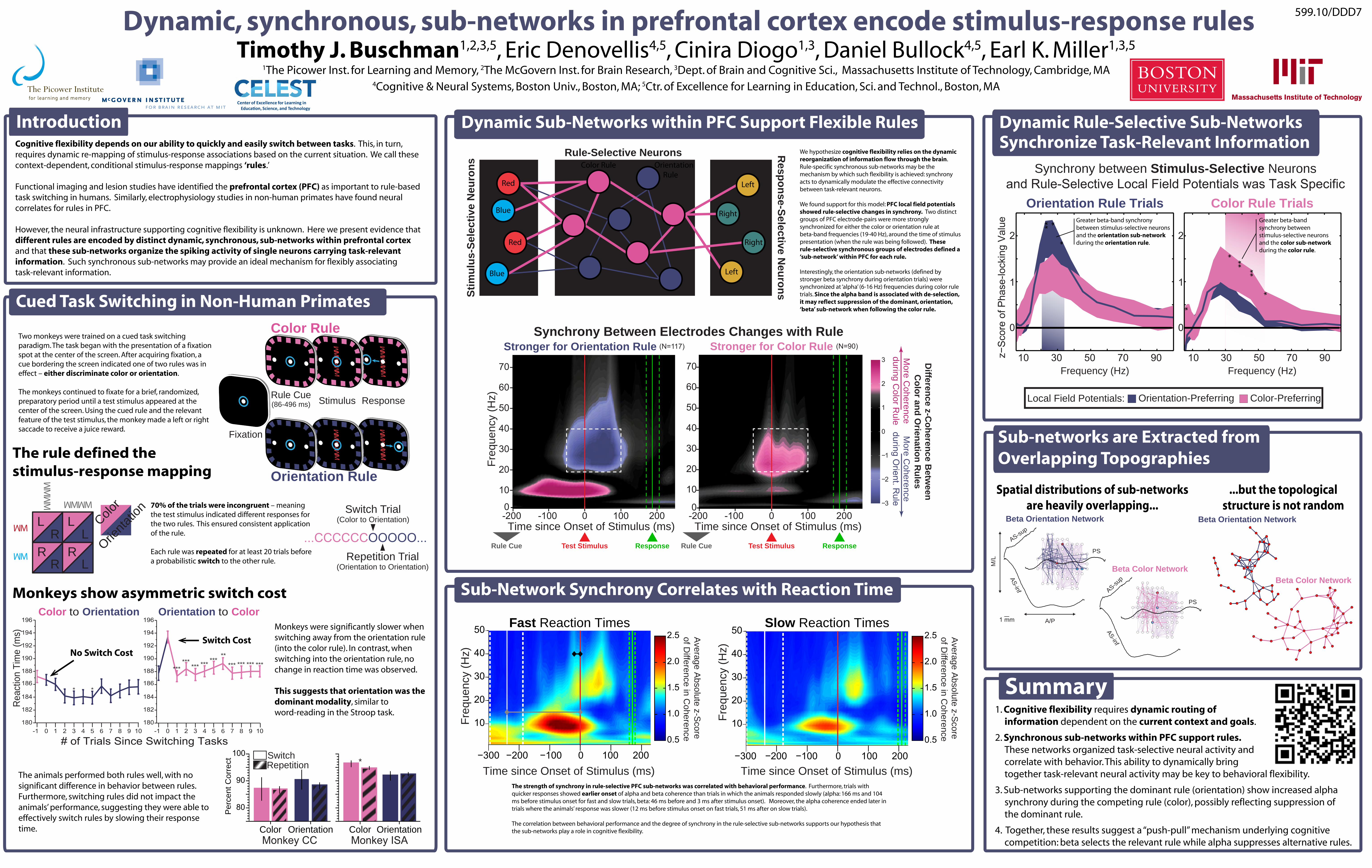
Spatial distributions of sub-networksare heavily overlapping...

Beta Color Network

AS-inf

AS-sup

PS

Beta Orientation Network

AS-sup

PS

AS-inf

1 mm A/P

M/L

...but the topologicalstructure is not random

Beta Color Network

Beta Orientation Network

Sub-networks are Extracted from Overlapping Topographies

We hypothesize cognitive flexibility relies on the dynamic reorganization of information flow through the brain. Rule-specific synchronous sub-networks may be the mechanism by which such flexibility is achieved: synchrony acts to dynamically modulate the effective connectivity between task-relevant neurons.

We found support for this model: PFC local field potentials showed rule-selective changes in synchrony. Two distinct groups of PFC electrode-pairs were more strongly synchronized for either the color or orientation rule at beta-band frequencies (19-40 Hz), around the time of stimulus presentation (when the rule was being followed). These rule-selective synchronous groups of electrodes defined a ‘sub-network’ within PFC for each rule.

Interestingly, the orientation sub-networks (defined by stronger beta synchrony during orientation trials) were synchronized at ‘alpha’ (6-16 Hz) frequencies during color rule trials. Since the alpha band is associated with de-selection, it may reflect suppression of the dominant, orientation, ‘beta’ sub-network when following the color rule.

Left

Left

Right

Right

Red

Red

Blue

Blue

Stim

ulus

-Sel

ectiv

e N

euro

ns

Rule-Selective Neurons Response-Selective N

eurons

Color Rule Orientation Rule

Dynamic Sub-Networks within PFC Support Flexible Rules

Time since Onset of Stimulus (ms)

0

10

20

30

40

50

60

70

-200 -100 0 100 200

Stronger for Orientation Rule (N=117)

-200 -100 0 100 200

10

20

30

40

50

60

70

0

Stronger for Color Rule (N=90)

−3

−2

−1

0

1

2

3 Difference z-C

oherence Betw

eenC

olor and Orienation R

ulesM

ore Coherence

during Color R

uleM

ore Coherence

during Orient. R

ule

Time since Onset of Stimulus (ms)

Freq

uenc

y (H

z)

Synchrony Between Electrodes Changes with Rule

Test Stimulus Response Test Stimulus ResponseRule Cue Rule Cue

z−S

core

of P

hase

-lock

ing

Val

ue

Orientation Rule Trials

0

1

2

10 30 50 70 90Frequency (Hz)

**

**

Synchrony between Stimulus-Selective Neuronsand Rule-Selective Local Field Potentials was Task Specific

Color-PreferringLocal Field Potentials: Orientation-Preferring

0

1

2

10 30 50 70 90

****

*

*

*

Frequency (Hz)

Color Rule Trials

Dynamic Rule-Selective Sub-NetworksSynchronize Task-Relevant Information

Greater beta-band synchrony between stimulus-selective neurons and the orientation sub-network during the orientation rule.

Greater beta-band synchrony between stimulus-selective neurons and the color sub-network during the color rule.

Color Rule

Orientation Rule

Fixation

Rule Cue(86-496 ms) Stimulus Response

...CCCCCCOOOOO...

Switch Trial(Color to Orientation)

Repetition Trial(Orientation to Orientation)

MM

MM

M

M

M

M

M

M

M

M

L LL

LR R

R

R

Color

Orienta

tion

80

90

100

Color OrientationMonkey CC

Per

cent

Cor

rect

Color OrientationMonkey ISA

*SwitchRepetition

***** *** *** ***

*********

***

** .

Reac

tion

Tim

e (m

s)

Color to Orientation

# of Trials Since Switching Tasks

Orientation to Color

***

-1 0 1 2 3 4 5 6 7 8 9 10 -1 0 1 2 3 4 5 6 7 8 9 10

196

194

192

190

188

186

184

182

180

196

194

192

190

188

186

184

182

180

Cued Task Switching in Non-Human Primates

No Switch CostSwitch Cost

Two monkeys were trained on a cued task switching paradigm. The task began with the presentation of a fixation spot at the center of the screen. After acquiring fixation, a cue bordering the screen indicated one of two rules was in effect – either discriminate color or orientation.

The monkeys continued to fixate for a brief, randomized, preparatory period until a test stimulus appeared at the center of the screen. Using the cued rule and the relevant feature of the test stimulus, the monkey made a left or right saccade to receive a juice reward.

70% of the trials were incongruent – meaning the test stimulus indicated different responses for the two rules. This ensured consistent application of the rule.

Each rule was repeated for at least 20 trials before a probabilistic switch to the other rule.

Monkeys show asymmetric switch cost

Monkeys were significantly slower when switching away from the orientation rule (into the color rule). In contrast, when switching into the orientation rule, no change in reaction time was observed.

This suggests that orientation was the dominant modality, similar to word-reading in the Stroop task.

The animals performed both rules well, with no significant difference in behavior between rules. Furthermore, switching rules did not impact the animals’ performance, suggesting they were able to effectively switch rules by slowing their response time.

The rule defined the stimulus-response mapping

IntroductionCognitive flexibility depends on our ability to quickly and easily switch between tasks. This, in turn, requires dynamic re-mapping of stimulus-response associations based on the current situation. We call these context-dependent, conditional stimulus-response mappings ‘rules.’

Functional imaging and lesion studies have identified the prefrontal cortex (PFC) as important to rule-based task switching in humans. Similarly, electrophysiology studies in non-human primates have found neural correlates for rules in PFC.

However, the neural infrastructure supporting cognitive flexibility is unknown. Here we present evidence that different rules are encoded by distinct dynamic, synchronous, sub-networks within prefrontal cortex and that these sub-networks organize the spiking activity of single neurons carrying task-relevant information. Such synchronous sub-networks may provide an ideal mechanism for flexibly associating task-relevant information.

Dynamic, synchronous, sub-networks in prefrontal cortex encode stimulus-response rules 599.10/DDD7

Center of Excellence for Learning in Education, Science, and Technology

Timothy J. Buschman1,2,3,5, Eric Denovellis4,5, Cinira Diogo1,3, Daniel Bullock4,5, Earl K. Miller1,3,5

1The Picower Inst. for Learning and Memory, 2The McGovern Inst. for Brain Research, 3Dept. of Brain and Cognitive Sci., Massachusetts Institute of Technology, Cambridge, MA4Cognitive & Neural Systems, Boston Univ., Boston, MA; 5Ctr. of Excellence for Learning in Education, Sci. and Technol., Boston, MA