Embed Size (px)
Citation preview

Gilbert HabibLa Timone HospitalMarseille - France
TAVI Endocarditis
EuroValves Brussels, 10 March 2016

Vikram– JAMA 2003 ; 290 : 3207
513 patients with complicated IE , 230 (40%) surgical therapy
6 month mortality
IE: a deadly disease!!: native valves

IE: a deadly disease!!: Prosthetic IE
Lalani T– JAMA 2013

Survival after TAVI endocarditis
Amat-Santos IJ et al. Circulation 2015

Vikram– JAMA 2003
513 patients
Native Valve IE
IE: a deadly disease!!
Lalani T– JAMA 2013
1025 patients
Prosthetic Valve IE
Amat-Santos IJ et al.
Circulation 2015
53 patients
TAVI IE

Case 1: TAVI endocarditis
80 year-old man
CHF
TAVI 2 years ago
fever = 38°
BC: staphylococcus coagulase -

CT scan : positive PET CT: negative
Multimodality Imaging

71 year-old man
mitral bioprosthesis 2000
severe Parkinson disease
valve-in-valve MV replacement (transapical) June 2015
october 2015: fever / suspected endocarditis
History of the disease
CHF
systolic murmur 2/6
blood pressure: 100/70 mmHg
arrhythmia (atrial fibrillation)
Clinical examination
Patient 2: Valve-in-valve PVE
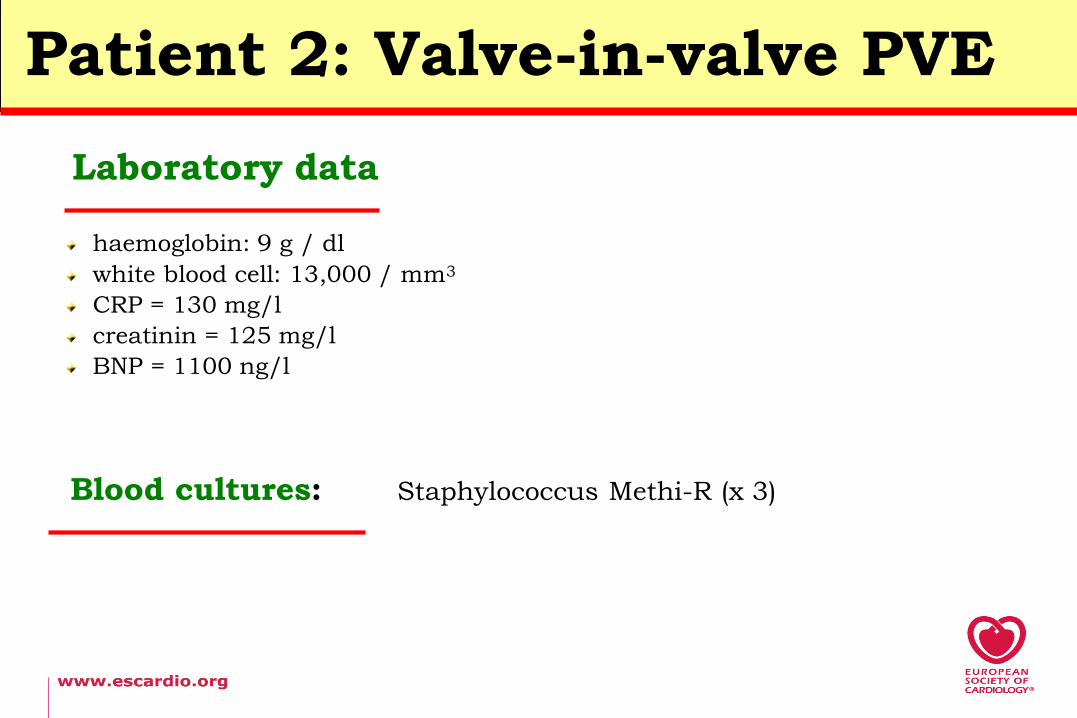
Blood cultures: Staphylococcus Methi-R (x 3)
haemoglobin: 9 g / dl
white blood cell: 13,000 / mm3
CRP = 130 mg/l
creatinin = 125 mg/l
BNP = 1100 ng/l
Laboratory data
Patient 2: Valve-in-valve PVE

TOE October 14th, 2015

TOE October 14th, 2015

TOE October 14th, 2015

Q1: What is your diagnosis?
1. Bioprosthetic Valve-in-valve endocarditis ?
2. Pericardial effusion?
3. LV aneurysm?
4. LV false aneurysm?

cardiac CT scan
Apical false aneurysmMitral annulus
pseudo-aneurysm

18FDG-PET-CT November 4th
Uptake on the apical
LV false aneurysm
Uptake on the
prosthesis

What is your diagnosis?
1. Bioprosthetic Valve-in-valve endocarditis
2. Pericardial effusion
3. LV aneurysm
4. LV false aneurysm

Decision and management
1. Definite IE
2. Initiation of antibiotic therapy
initially: Vancomycin with Gentamycin:
then: Cotrimoxazole with Clindamycin
3. follow-up
repeat TEE
repeat CT scan

Evolution under ATB therapy
October 14th, 2015 October 30th, 2015

Evolution under ATB therapy
October 14th, 2015 October 30th, 2015

Evolution under ATB therapy
October 14th, 2015 October 30th, 2015

Pulsatile false aneurysm

What is your management?
1. Antibiotic therapy alone?
2. Emergency surgery ?
3. Elective surgery?
4. Other?

TAVI Endocarditis
1. incidence
2. prevention
3. diagnosis
4. treatment

• 2572 patients between 2008-2013• 14 centers• Sapien/Corevalve: 40-60%• Median follow-up 1.1 year• 29 IE
Latib A et al. JACC 2014

Amat-Santos IJ et al. Circulation 2015
• 7944 patients between 2007-2014• 21 centers• Sapien/Corevalve: 80-20%• Mean follow-up of 1.1 ± 1.2 year• 53 IE

Olsen NT et al. Circ Cardiovasc int 2015
• 509 patients between 2007-2014• Single-center• Only CoreValve• Median follow-up 1.4 year• 18 IE

TAVI Endocarditis
1. incidence
2. prevention
3. diagnosis
4. treatment
0
20
40
60
80
100
120
140
160
180
200
220
20-24 25-29 30-34 35-39 40-44 45-49 50-54 55-59 60-64 65-69 70-74 75-79 80-84 85-89 90-94 >=95
Inci
den
ce p
er M
illio
n P
op
ula
tio
n
Age,years
Women Men
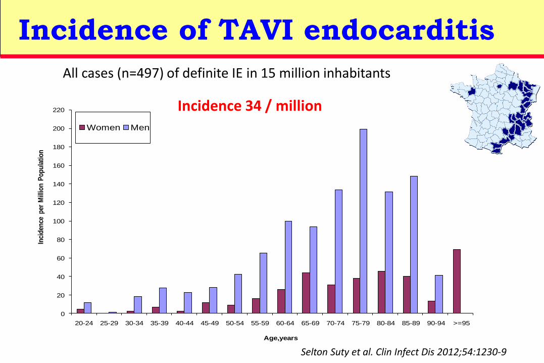
All cases (n=497) of definite IE in 15 million inhabitants
Incidence 34 / million
Selton Suty et al. Clin Infect Dis 2012;54:1230-9
Incidence of TAVI endocarditis

0
20
40
60
80
100
120
140
160
180
200
220
20-24 25-29 30-34 35-39 40-44 45-49 50-54 55-59 60-64 65-69 70-74 75-79 80-84 85-89 90-94 >=95
Inci
den
ce p
er M
illio
n P
op
ula
tio
n
Age,years
Women Men
Incidence 34 / million 194 / million
Selton Suty et al. Clin Infect Dis 2012;54:1230-9
Incidence of TAVI endocarditis

Annual incidence: from 0.4 to 2.1 per 100 pts/year
annual incidence (%) study
0.4 Buellesfeld, JACC 2011
0.6 Généreux, JACC 2012
0.66 Gotzmann, AJC 2014
0.75 PARTNER (2 years FU)
1.1 Latib, JACC 2014
0.7 Amat-Santos, Circulation 2015
2.1 Olsen, Circ Cardiovasc int 2015
Case and cohorts
Specific large studies
T A
V I
Incidence of TAVI endocarditis

Annual incidence: from 0.4 to 2.1 per 100 pts/year
annual incidence (%) study
0.4 Buellesfeld, JACC 2011
0.6 Généreux, JACC 2012
0.66 Gotzmann, AJC 2014
0.75 PARTNER (2 years FU)
1.1 Latib, JACC 2014
0.7 Amat-Santos, Circulation 2015
2.1 Olsen, Circ Cardiovasc int 2015
0.3-1.2 ESC Guidelines 2015
Case and cohorts
Specific large studies
Surgical prosthesis
T A
V I
Surg
ery
Incidence of TAVI endocarditis

TAVI Endocarditis
1. incidence
2. prevention
3. diagnosis
4. treatment
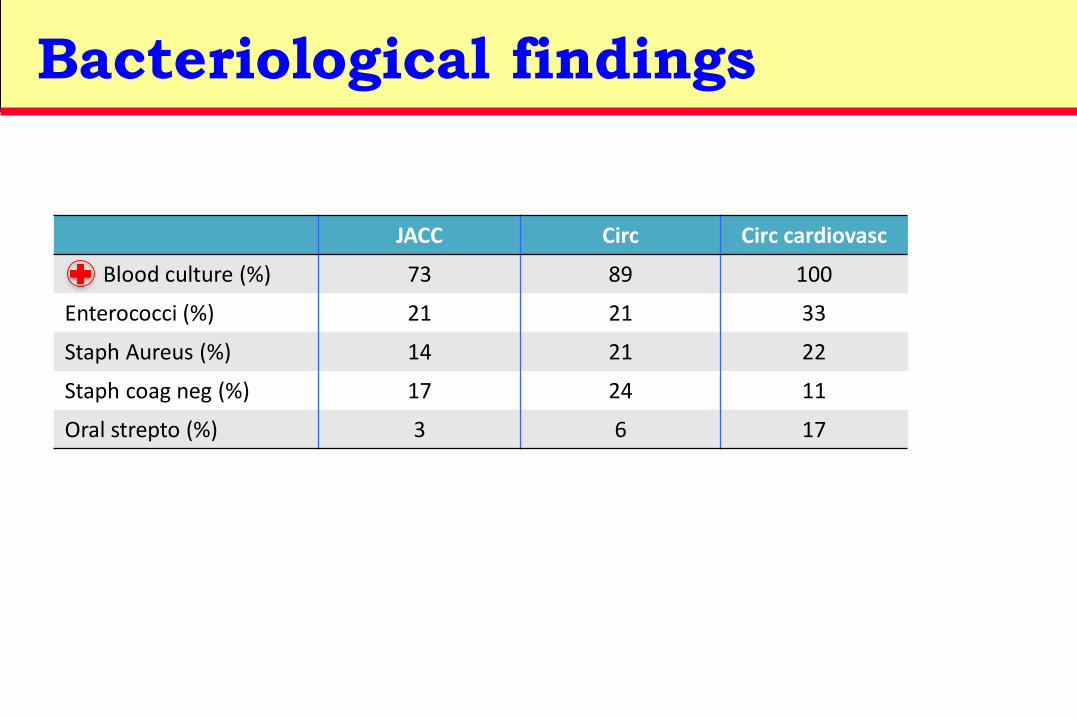
JACC Circ Circ cardiovasc
Blood culture (%) 73 89 100
Enterococci (%) 21 21 33
Staph Aureus (%) 14 21 22
Staph coag neg (%) 17 24 11
Oral strepto (%) 3 6 17
Bacteriological findings

JACC Circ Circ cardiovasc
Blood culture (%) 73 89 100
Enterococci (%) 21 21 33
Staph Aureus (%) 14 21 22
Staph coag neg (%) 17 24 11
Oral strepto (%) 3 6 17
Bacteriological findings

JACC Circ Circ cardiovasc
Blood culture (%) 73 89 100
Enterococci (%) 21 21 33
Staph Aureus (%) 14 21 22
Staph coag neg (%) 17 24 11
Oral strepto (%) 3 6 17
Bacteriological findings

JACC Circ Circ cardiovasc
Blood culture (%) 73 89 100
Enterococci (%) 21 21 33
Staph Aureus (%) 14 21 22
Staph coag neg (%) 17 24 11
Oral strepto (%) 3 6 17
Bacteriological findings

Timing of initial symptoms

Amat-Santos IJ et al. Circulation 2015
Bacteria/microorganisms: 2 enterococci; 4 staph; 3 others
Timing of initial symptoms

Cardiac conditions at highest risk of IE
Recommendations Class Level
Antibiotic prophylaxis should only be considered for patients at highest risk of IE:
1. Patients with a prosthetic valve, including transcatheter valve, or a prosthetic material used for
cardiac valve repair.
2. Patients with previous IE.
3. Patients with congenital heart disease.
a. any cyanotic congenital heart disease
b. congenital heart disease repaired with prosthetic material whether placed surgically or by
percutaneous techniques, up to 6 months after the procedure or lifelong if there remains
residual shunt or valvular regurgitation.
IIa C
Antibiotic prophylaxis is not recommended in other forms of valvular or congenital heart disease. III C

Non-specific prevention measures
41
These measures should ideally be applied to the general population and particularly reinforced in high-risk patients.
• Strict dental and cutaneous hygiene. Dental follow-up should be performed twice a year in high-risk patients and yearly in the others.
• Disinfection of wounds.
• Eradication or decrease of chronic bacterial carriage: skin, urine.
• Curative antibiotics for any focus of bacterial infection.
• No self-medication with antibiotics.
• Strict asepsis control measures for any at-risk procedure.
• Discourage piercing and tattooing.
• Limit the use of infusion catheters and invasive procedure when possible. Favour peripheral over central catheters, and systematic replacement of the peripheral catheter every 3–4 days. Strict adherence to care bundles for central and peripheral cannulae should be performed.

42
Recommendations Class Level
Pre-operative screening of nasal carriage of Staphylococcus aureus is
recommended before elective cardiac surgery in order to treat carriers.I A
Peri-operative prophylaxis is recommended before pacemaker or
implantable cardioverter defibrillator implantation.I B
Elimination of potential sources of dental sepsis is recommended
>2 weeks before implantation of a prosthetic valve or other intracardiac
or intravascular foreign material, except in urgent procedures.
I C
Peri-operative antibiotic prophylaxis should be considered in patients
undergoing surgical or transcatheter implantation of a prosthetic
valve, intravascular prosthetic, or other foreign material.
IIa C
Systematic local treatment without screening for Staphylococcus aureus is not recommended. III C
Habib G et al. Eur Heart Journal 2015
Antibiotic prophylaxis before cardiac or vascular interventions

43
Prophylaxis before TAVI
In the 3 papers: All patients received antibiotic prophylaxis
• JACC: according to institutional practice…
• Circulation: Cephalosporins in 14/21 centers (67%), vancomycin in 6
(28%) and piperacillin/tazobactam in 1 (5%) .
• Circ cardiovasc int: cefuroxime 1.5 g IV pre TAVI, 8 and 16 h after.

In the 3 papers: All patients received antibiotic prophylaxis
• JACC: according to institutional practice…
• Circulation: Cephalosporins in 14/21 centers (67%), vancomycin in 6
(28%) and piperacillin/tazobactam in 1 (5%) .
• Circ cardiovasc int: cefuroxime 1.5 g IV pre TAVI, 8 and 16 h after.
“Cephalosporins are traditionally used,
but this choice could be reconsidered if it is confirmed that
enterococci are important pathogens in very early TAVI-PVE.”
44
Prophylaxis before TAVI

TAVI Endocarditis
1. incidence
2. prevention
3. diagnosis
4. treatment

The Duke echographic criteria
vegetation
Durack DT Am J Med 1994 ; 96 : 200-9
abscess new dehiscence
of prosthetic valve

TOEMorphology
PET CTInflammation /
infection
Cardiac CTPerivalvular lesions
Multimodality imaging in IE

48European Heart Journal (2015) doi:10.1093/eurheartj/ehv319
Clinical suspicion of IE
Modified Duke criteria (Li)
Definite IEPossible/rejected IE but
high suspicionRejected IE
Low suspicion
Nativevalve
Prostheticvalve
1 - Repeat echo
(TTE + TOE)/microbiology
2 - Imaging for embolic events
3 - Cardiac CT
1 - Repeat echo (TTE + TOE)/microbiology
2 - 18F-FDG PET/CT or Leucocytes labeled SPECT/CT
3 - Cardiac CT
4 - Imaging for embolic events
ESC 2015 modified diagnostic criteria
Definite IE Possible IE Rejected IE
ESC 2015 algorithm for diagnosis of IE

TAVI Endocarditis
1. incidence
2. prevention
3. diagnosis
4. treatment

Indications and timing of surgeryIndications for surgery Timing Class Level
1. Heart Failure
Aortic or mitral NVE or PVE with severe acute regurgitation, obstruction or fistula causing refractory pulmonary oedema or cardiogenic shock.
Emergency I B
Aortic or mitral NVE or PVE with severe regurgitation or obstruction causing symptoms of HF or echocardiographic signs of poor haemodynamic tolerance.
Urgent I B
2. Uncontrolled infection
Locally uncontrolled infection (abscess, false aneurysm, fistula, enlarging vegetation). Urgent I B
Infection caused by fungi or multiresistant organisms. Urgent/elective I C
Persisting positive blood cultures despite appropriate antibiotic therapy and adequate control of septic metastatic foci.
Urgent IIa B
PVE caused by staphylococci or non-HACEK Gram negative bacteria. Urgent/elective IIa C
3. Prevention of embolism
Aortic or mitral NVE or PVE with persistent vegetations >10 mm after one or more embolic episode despite appropriate antibiotic therapy.
Urgent I B
Aortic or mitral NVE with vegetations >10 mm, associated with severe valve stenosis or regurgitation, and low operative risk.
Urgent IIa B
Aortic or mitral NVE or PVE with isolated very large vegetations (>30 mm). Urgent IIa B
Aortic or mitral NVE or PVE with isolated large vegetations (>15 mm) and no other indication for surgery.
Urgent IIb C

Evolution under ATB therapy
October 14th, 2015 October 30th, 2015

Decision: transcatheter closure

Amplatzer deployment

Final result
Per procedure TTE November 9, 2015
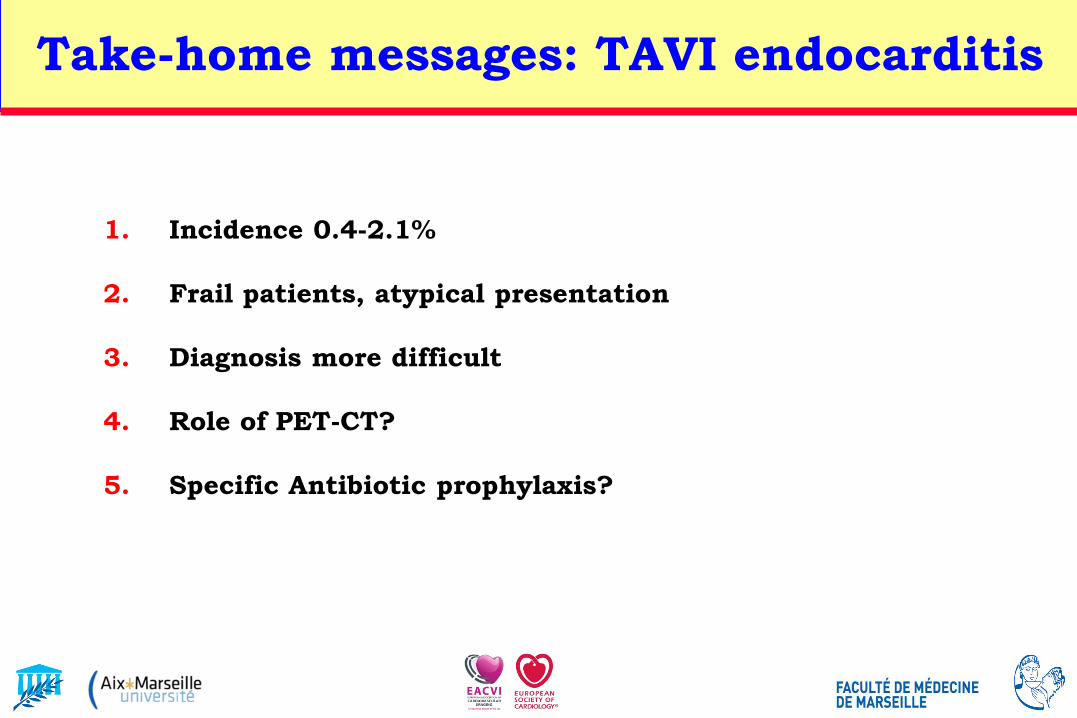
Take-home messages: TAVI endocarditis
1. Incidence 0.4-2.1%
2. Frail patients, atypical presentation
3. Diagnosis more difficult
4. Role of PET-CT?
5. Specific Antibiotic prophylaxis?

ESC Heart House