Embed Size (px)
Citation preview
Cryptogamie, Mycologie, 2016, 37 (4): 509-538© 2016 Adac. Tous droits réservés
doi/10.7872/crym/v37.iss4.2016.509
Taxonomic rearrangement of Anthostomella(Xylariaceae) based on a multigene phylogeny
and morphology
Dinushani A. DARANAGAMAa,b, Erio CAMPORESI e, f, g,Rajesh JEEWON f, Xingzhong LIU a, Marc STADLER d,
Siasamorn LUMYONG h* & Kevin D. HYDE b*
aState Key Laboratory of Mycology, Institute of Microbiology, Chinese Academyof Sciences, No 3 1st West Beichen Road, Chaoyang District Beijing 100101,
People’s Republic of China
bCenter of Excellence in Fungal Research, Mae Fah Luang University,Chiang Rai 57100, Thailand
dHelmholtz-Zentrum für Infektionsforschung GmbH, Department of MicrobialDrugs, Inhoffenstrasse 7, 38124 Braunschweig, Germany
eA.M.B. Gruppo Micologico Forlìvese B Antonio Cicognani, ViaRoma 18,Forlì, Italy
fA.M.B. Circolo Micologico BGiovanni Carini, C.P. 314 Brescia, Italy
gSocietà per gli Studi Naturalistici della Romagna, C.P. 144 Bagnacavallo,RA, Italy
hDepartment of Biology, Faculty of Science, Chiang Mai University,Chiang Mai, 50200 Thailand
iDepartment of Health Sciences, Faculty of Science, University of Mauritius,Reduit, Mauritius, 80837.
Abstract – The genus Anthostomella is heterogeneous and recent DNA based studies haveshown that species are polyphyletic across Xylariaceae. In this study, we present a morphologybased, taxonomic treatment, coupled with a molecular phylogenetic reassessment ofrelationships within Anthostomella. This has resulted in the establishment of two new genera,eight new combinations and three new species among anthostomella-like taxa. Seventeenstrains from 16 anthostomella-like species have been revisited. A re-description ofmorphological characters among these taxa suggests that Anthostomella can be circumscribedbased on immersed ascomata, cylindrical asci with short pedicels and pigmented, equilateralascospores with germ slits, while Anthostomelloides is characterized by oblong-ellipsoidalascospores lacking germ slits. Anthostomella brabeji, A. forlìcesenica, A. leucospermi andA. proteae are transferred to Anthostomelloides. Pseudoanthostomella gen. nov., possessessolitary ascomata, as well as asci with apical rings and is closely related to Neoanthostomella,
* Corresponding author: [email protected] / [email protected]
510 D. A. Daranagama et al.
but can be distinguished based on a combination of characters, such as multiple ascomataand asci completely lacking apical rings. Anthostomella conorum, A. delitescens andA. sepelibilis are reallocated to Pseudoanthostomella with two new species, P. pini-nigraeand P. senecionicola. Neoanthostomella viticola sp. nov. is added to the previously monotypicgenus Neoanthostomella. Alloanthostomella gen. nov is introduced to accommodateAnthostomella rubicola. The only anthostomella-like genus diagnosed with hyaline ascosporesso far, is Alloanthostomella gen. nov. This study represents a comprehensive molecularphylogenetic study based on four independent molecular markers (ITS, 28S rDNA, RPB2and β-tubulin) with a broad taxon sampling across Anthostomella.
Character evolution / Classification / Pyrenomycetes / Sordariomycetes / Taxonomy /Xylariales
IntroductIon
The family Xylariaceae comprises 87 genera (Maharachchikumbura et al.2015; 2016) and over 1300 known species (Stadler et al. 2013). Although manyspecies are characterized by large, conspicuous stromata, others have rudimentarystromata (Daranagama et al. 2016a). Species delimitation and generic recognitioncriteria in Xylariaceae have shifted from morphology (Dennis 1956; Miller 1961;Rogers 1977; Læssøe & Spooner 1994; Ju & Rogers 1996; Daranagama et al.2016b) to DNA based phylogenetic schemes (Sánchez-Ballesteros et al. 2000; Hsiehet al. 2005; Bitzer et al. 2008; Pelaez et al. 2008; Tang et al. 2009; Daranagama etal. 2015a; b; 2016c; d; Senanayake et al. 2015). Most recently chemotaxonomic datahas been used in identification of species and genera (Stadler & Hellwig 2005;Stadler et al. 2008; 2010; 2014a; Daranagama et al. 2014).
Anthostomella Sacc. is a complex genus with approximately 95 species (Lu& Hyde 2000; Daranagama et al. 2015a). The morphological circumscription ofAnthostomella is based on the type species Anthostomella tomicoides Sacc. withimmersed, dark, clypeate ascomata with periphysate, ostiolar canals, 8-spored,cylindrical, unitunicate asci and mostly dark, unicellular ascospores sometimes withsmall cells or appendages at the ends. Daranagama et al. (2015a) confirmed thatAnthostomella is polyphyletic and several anthostomella-like species can beaccommodated into several genera across Xylariaceae. Apart from Anthostomella,there are other genera in Xylariaceae, such as Nemania, Rosellinia, Stilbohypoxylonand Xylaria (Hsieh et al. 2010; Stadler et al. 2013; Daranagama et al. 2015a;b) which may not be monophyletic. Anthostomella species within Xylariaceae arepolyphyletic (Stadler et al. 2013; Daranagama et al. 2015a; Tibpromma et al. 2016).There have been a number of contentious issues in connection with the taxonomyof species that are polyphyletic. In some cases, polyphyletic genera have beentreated as orphans (i. e. Shenoy et al. 2006; Wang et al. 2007). In this study apractical taxonomic treatment for the polyphyletic genus Anthostomella is proposed,which is in concordance with phylogenies recovered from multigene sequenceanalyses. To provide a more robust justification on our taxonomic rearrangement, ithas been necessary to redefine generic concepts based on reexamination of typespecies and micro-morphological variations, which is common taxonomic practicenowadays (Senanayake et al. 2015; Thambugala et al. 2015; Maharachchikumburaet al. 2016). For many years, large number of species has been allocated toAnthostomella, making it more complex and difficult to resolve species relationships.Around 90% of these species had been described only from a morphological
Taxonomic rearrangement of Anthostomella 511
perspective and never been reported with a living culture hence no DNA sequencedata were available to propose a more natural classification. For a meaningfulphylogenetic and morphological reappraisal of Anthostomella, it is essential toobtain fresh collections of Anthostomella species and get axenic cultures to acquireDNA sequence data. In this study we obtained cultures from fresh specimensallowing for additional DNA sequence data to reevaluate the evolutionaryrelationships of anthostomella-like species.
It is important to emphasize the need of reference isolates with accessiblemolecular data, particularly for species identified and described several decades ago.Based on the preliminary phylogenetic analysis on Anthostomella (Daranagamaet al., 2015a) and previous morphological studies (Lu & Hyde 2000; Lee & Crous2003), it is essential to reorganize the taxonomy of Anthostomella species to reflecta more natural classification.
The objectives of this study are as follows: 1) to reassess morphologicalspecies concepts to define species in Anthostomella and anthostomella-like generabased on multi-gene phylogenies; 2) to investigate phylogenetic relationships ofAnthostomella and describe new genera for anthostomella-like species wherenecessary; 3) and evaluate resolving power of various phylogenetic markers foranthostomella-like species.
MAterIAl And MethodS
Sampling, morphological observation and isolation. – Fresh specimenswere obtained from various host plants collected in different provinces of Italy.Morphological examination and microphotography was carried out as described byDaranagama et al. (2014; 2015). Axenic cultures were initiated and maintained inoat agar (OA) medium for observation of asexual morph as described by Daranagamaet al. (2015). Dried materials are deposited in Mae Fah Luang University herbarium(MFLU), Chiang Rai, Thailand and Kunming Institute of Botany (HKAS), Chinaand cultures are deposited at Mae Fah Luang University Culture Collection(MFLUCC), Thailand and the Kunming Institute of Botany Culture Collection(KIBCC), China. Faces of Fungi numbers and Index Fungorum numbers wereobtained as explained in Jayasiri et al. (2015) and Index Fungorum (2016).
DNA extraction, PCR and sequencing. – DNA was extracted and the ITS,LSU, β-TUB and RPB2 genes were amplified following the protocols outlined byDaranagama et al. (2014; 2015a; b) and Thambugala et al. (2015). PCR productswere visualized in 1 % agarose gel electrophoresis, stained with Goldview (GeneshunBiotech, China) with D2000 DNA ladder (Realtimes Biotech, Beijing, China). AllPCR products were purified using the kits according to the manufacture protocolsand DNA sequencing was performed using the same primers as used for PCR in anApplied Biosystem 3730 DNA analyzer at Sinogenomax Company, Beijing, China.
Sequence alignment and phylogenetic analyses. – Raw sequences wereassembled with Contig Express 2003 (Invitrogen, Carlsbad, CA). The assembledconsensus sequences were implemented automatically by Fast Fourier Transformation(MAFFT v7.017) using the E-INS-i algorithm (Katoh & Standley 2013) (http://mafft.cbrc.jp/alignment/server/) and adjusted manually where necessary. Thealignments were edited with an online version of the Gblocks program v. 0.91b9,using parametrical settings for a very low stringency to avoid poorly aligned regions.

512 D. A. Daranagama et al.
Maximum Likelihood (ML) phylogenetic trees were reconstructed usingRAxML 7.4.2 Black Box (Stamatakis 2006; Stamatakis et al. 2008) available in theCIPRES Science Gateway platform (Miller et al. 2010). A combined dataset wasincluded with all default modal parameters in RAxML with ML estimate of 25 persite rate categories. The RAxML software accommodated the GTR model ofnucleotide substitution with the additional options of modeling rate heterogeneity(Γ) and proportion invariable sites (I) Gaps were treated as missing data.
Phylogenetic trees and data files were viewed in MEGA 5 (Tamura et al.2011) and Fig tree v1.4 (Rambaut & Drummond 2008). Maximum Likelihoodbootstrap values (equal to or above 50%) are indicated above or below nodes andconsidered significant if equal or above 70%. The sequences generated in this studyare indicated in bold and deposited in GenBank (Table 1). The alignments andtrees were submitted to TreeBASE (http://purl.org/phylo/treebase/phylows/study/TB2:S19489).
reSultS
Phylogenetic analysesFour different alignments corresponding to each individual gene of ITS,
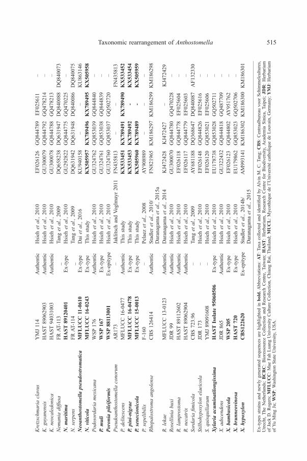
LSU, RPB2 and β-tubulin and a combined alignment of all four genes were analyzed.The ITS alignment consisted of 15 flanks with 314 characters (20% of the original1556 positions). The LSU alignment consisted of 33 flanks with 558 characters(52% of the original 1063 positions). The RPB2 alignment consisted of 22 flankswith 927 characters (70% of the original 1316 positions). The β-tubulin alignmentconsisted of 40 flanks with 858 characters (32% of the original 2631 positions). Thephylogram inferred from the combined analysis of four genes for all isolates ispresented in Fig. 1. Sequence data from 58 isolates, including 17 isolates ofAnthostomella species were used. Sordaria fimicola was used as the outgroup taxon.GenBank accession numbers of the isolates are provided in Table 1.
The Hypoxyloideae (HY) clade is well-supported (90% BS) in the combinedphylogenetic analysis and the analyzed Anthostomella species do not form amonophyletic clade in Xylarioideae (XY). The data show species previouslyidentified as Anthostomella sensu lato to be split across the two subfamilies ofXylariaceae, forming five separate clades (A-E). Anthostomella formosa (similar inmorphology to the type species) is in clade A together with A. obesa, A. pinea andA. helicofissa, which is named herein as Anthostomella sensu stricto. Anthostomellaformosa has close affinities with A. helicofissa and both cluster together with 90%bootstrap support while Anthostomella obesa and A. pinea cluster together with100% bootstrap support (Figs 1 & 2).
Alloanthostomella, Neoanthostomella and Pseudoanthostomella cluster intoa subgroup outside of the two subfamilies Hypoxyloideae and Xylarioideae.Biscogniauxia is suggested without support as the sister clade to Alloanthostomella,Neoanthostomella and Pseudoanthostomella.
A group of anthostomella-like species clusters in a monophyletic clade(Clade B, 73% BS) within Xylarioideae and is introduced in this study as the newgenus Anthostomelloides. Anthostomelloides consists of five species including thetype species, Anthostomelloides krabiensis. Anthostomelloides leucospermi clusterswith An. krabiensis with 100% bootstrap support, while our analyses place An. brabejibasal to the other Anthostomelloides species.

Taxonomic rearrangement of Anthostomella 513
Fig. 1. The best scoring phylogenetic tree based on a combined dataset of ITS, LSU, RPB2 and β-tubulinsequence data produced by maximum likelihood analysis. Bootstrap support values for maximumlikelihood greater than 50% are given. The type strains/specimens are in bold letters and indicated by *.The tree is rooted to Sordaria fimicola. Ex-type strains are in bold.

514 D. A. Daranagama et al.Sp
ecie
sC
ultu
reco
llect
ion/
spec
imen
num
ber
Type
stat
usRe
fere
nce
Gen
Bank
acce
ssio
nnu
mbe
rs
ITS
RPB2
β-tu
bulin
LSU
Amph
irose
llini
afu
shan
ensis
hA
StIs
olat
e91
1112
09Ex
-type
Hsi
ehet
al.,
2010
GU
3394
96GQ848339
GQ495950
–A.
nigr
ospo
rah
ASt
Isol
ate
9109
2308
Ex-ty
peH
sieh
etal
.,20
10G
U32
2457
GQ848340
GQ495951
–An
thoc
anal
issp
arti
MFl
uc
c14
-001
0Ex
-type
Dar
anag
ama
etal
.,20
15a
KP2
9739
4K
P340
522
KP4
0660
5K
P340
536
A.sp
arti
MFl
uc
c14
-055
7Ex
-par
atyp
eD
aran
agam
aet
al.,
2015
aK
P297
395
KP3
4052
3K
P406
606
KP3
4053
7An
nulo
hypo
xylo
nm
orifo
rme
var.
mic
rodi
scum
CB
S123
834
Aut
hent
icTa
nget
al.,
2009
DQ631935
DQ631960
DQ840095
DQ840061
A.ni
tens
MFL
UC
C12
-082
3A
uthe
ntic
Dar
anag
ama
etal
.,20
15a
KJ9
3499
1K
J934
994
KJ9
3499
3K
J934
992
A.st
ygiu
mM
FLU
CC
13-0
826
Aut
hent
icD
aran
agam
aet
al.,
2015
aK
J940
870
KJ9
4086
8K
J940
867
KJ9
4086
9An
thos
tom
ella
form
osa
MFL
UC
C14
-017
0A
uthe
ntic
Dar
anag
ama
etal
.,20
15a
KP2
9740
3K
P340
531
KP4
0661
4K
P340
544
A.helicofissa
MFl
uc
c14
-017
3Ex
-type
Dar
anag
ama
etal
.,20
15a
KP2
9740
6K
P340
534
KP4
0661
7K
P340
547
A.ob
esa
MFl
uc
c14
-017
1Ex
-type
Dar
anag
ama
etal
.,20
15a
KP2
9740
5K
P340
533
KP4
0661
6K
P340
546
A.pi
nea
CB
S12
8205
–C
rous
&G
roen
ewal
d20
10HQ599578
––
–An
thos
tom
ello
ides
brab
eji
CB
S11
0128
–Ja
klits
chan
dVo
glm
ayr
2011
EU55
2098
––
EU55
2098
An.f
orlìc
esen
ica
MFl
uc
c14
-000
7Ex
-type
Dar
anag
ama
etal
.,20
15a
KP2
9739
6K
P340
524
KP4
0660
7K
P340
538
An.k
rabi
ensis
MFl
uc
c15
-067
8Ex
-type
Tibp
rom
ma
etal
.,20
16KX305927
KX305929
–KX305928
An.l
euco
sper
mi
CB
S11
0126
–M
arin
cow
itzet
al.,
2008
EU55
2100
––
–An
.pro
teae
CB
S11
0127
–M
arin
cow
itzet
al.,
2008
EU55
2101
––
–Al
loan
thos
tom
ella
rubi
cola
MFL
UC
C14
-017
5A
uthe
ntic
Dar
anag
ama
etal
.,20
15a
KP2
9740
7K
P340
535
KP4
0661
8K
P340
548
Al.ru
bico
laM
FLU
CC
16-0
479
Aut
hent
icTh
isst
udy
KX
5334
55K
X78
9493
KX
7894
94K
X53
3456
Bisc
ogni
auxi
aca
pnod
esY
MJ
138
Aut
hent
icH
sieh
etal
.,20
10EF
0261
31JX
507779
AY95
1675
–B.
mar
gina
taM
FLU
CC
12-0
740
Aut
hent
icD
aran
agam
aet
al.,
2015
aK
J958
407
KJ9
5840
9K
J958
406
KJ9
5840
8Br
unne
iper
idiu
mgr
acile
ntum
MFl
uc
c14
-001
1Ex
-type
Dar
anag
ama
etal
.,20
15a
KP2
9740
0K
P340
528
KP4
0661
1K
P340
542
B.gr
acile
ntum
MFl
uc
c14
-055
9Ex
-par
atyp
eD
aran
agam
aet
al.,
2015
aK
P297
401
KP3
4052
9K
P406
612
KP3
4054
9D
aldi
nia
conc
entr
ica
CB
S11
3277
Aut
hent
icK
uhne
rtet
al.,
2013
AY61
6683
–K
C97
7274
–D
.dec
ipie
nsC
BS
1228
79A
uthe
ntic
Hsi
ehet
al.,
2005
JX658441
–AY
9516
94–
D.l
ocul
ata
BC
RC
3411
7A
uthe
ntic
Hsi
ehet
al.,
2005
EF02
6145
–AY
9516
98–
Hyp
oxyl
onfe
ndle
riM
FLU
CC
12-0
816
Aut
hent
icD
aran
agam
aet
al.,
2015
aK
M01
7563
KM
0175
66K
M01
7564
KM
0175
65H
.fra
gifo
rme
MU
CL
5126
4A
uthe
ntic
Trie
bele
tal.,
2005
/D
aran
agam
aet
al.,
2015
aK
M18
6294
KM
1862
96K
M18
6301
KM
1862
95
H.l
enor
man
dii
MFL
UC
C13
-012
0A
uthe
ntic
Dar
anag
ama
etal
.,20
15a
KM
0391
35K
M03
9137
KM
0391
38K
M03
9136
H.m
ontic
ulos
umM
FLU
CC
12-0
818
Aut
hent
icD
aran
agam
aet
al.,
2015
aK
M05
2716
KM
0527
19K
M05
2718
KM
0527
17
Tabl
e1.
GB
acce
ssio
nnu
mbe
rsof
the
isol
ates
used
inth
isst
udy
Taxonomic rearrangement of Anthostomella 515K
retz
schm
aria
clav
usY
MJ
114
Aut
hent
icH
sieh
etal
.,20
10EF
0261
26GQ844789
EF02
5611
–K
.guy
anen
sis
HA
ST89
0629
03A
uthe
ntic
Hsi
ehet
al.,
2010
GU
3000
79GQ844792
GQ478214
–K
.neo
cale
doni
caH
AST
9403
1003
Aut
hent
icH
sieh
etal
.,20
10G
U30
0078
GQ844788
GQ478213
–N
eman
iadi
ffusa
FRAT
-113
_Ta
nget
al.,
2009
DQ658238
DQ631947
DQ840088
DQ840073
N.m
ariti
ma
hA
St89
1204
01Ex
-type
Hsi
ehet
al.,
2010
GU
2928
22GQ844775
GQ470225
–N
.ser
pens
FRAT
-114
_Ta
nget
al.,
2009
DQ631942
DQ631948
DQ840086
DQ840075
Neo
anth
osto
mel
laps
eudo
strom
atic
aM
Flu
cc
11-0
610
Ex-ty
peD
aiet
al.,
2016
KU
9401
58–
–K
U86
3146
N.v
itico
laM
Flu
cc
16-0
243
Ex-ty
peTh
isst
udy
KX
5059
57K
X78
9496
KX
7894
95K
X50
5958
Podo
sord
aria
mex
ican
aW
SP17
6A
uthe
ntic
Hsi
ehet
al.,
2010
GU
3247
62GQ853039
GQ844840
–P.
mul
iW
SP16
7Ex
-type
Hsi
ehet
al.,
2010
GU
3247
61GQ853038
GQ844839
–Po
roni
api
leifo
rmis
WSP
8811
3001
Ex-e
pity
peH
sieh
etal
.,20
10G
U32
4760
GQ853037
GQ502720
–Ps
eudo
anth
osto
mel
laco
noru
mA
R17
3–
Jakl
itsch
and
Vogl
may
r20
11FN
4358
13–
–FN
4358
13P.
delit
esce
nsM
FLU
CC
16-0
477
Aut
hent
icTh
isst
udy
KX
5334
51K
X78
9491
KX
7894
90K
X53
3452
P.pi
ni-n
igra
eM
Flu
cc
16-0
478
Ex-ty
peTh
isst
udy
KX
5334
53K
X78
9492
-K
X53
3454
P.se
neci
onic
ola
MFl
uc
c15
-001
3Ex
-type
This
stud
yK
X50
5960
KX
7894
89-
KX
5059
59P.
sepe
libili
sF-
160
–Pe
laez
etal
.,20
08AY
9089
89–
––
Rhop
alos
trom
aan
gole
nse
CB
S12
6414
Aut
hent
icSt
adle
ret
al.,
2010
/D
aran
agam
aet
al.,
2015
aFN
8219
65K
M18
6297
KM
1862
99K
M18
6298
R.le
kae
MFL
UC
C13
-012
3A
uthe
ntic
Dar
anag
ama
etal
.,20
14K
J472
428
KJ4
7242
7–
KJ4
7242
9Ro
selli
nia
buxi
JDR
99A
uthe
ntic
Hsi
ehet
al.,
2010
GU
3000
70GQ844780
GQ470228
–R.
lam
pros
tom
aH
AST
8911
2602
Aut
hent
icH
sieh
etal
.,20
10EF
0261
18GQ844778
EF02
5604
–R.
neca
trix
HA
ST89
0629
04A
uthe
ntic
Hsi
ehet
al.,
2010
EF02
6117
GQ844779
EF02
5603
–So
rdar
iafim
icol
aC
BS
723.
96–
Tang
etal
.,20
09AY
6811
88DQ368647
DQ840087
AF1
3233
0St
ilboh
ypox
ylon
elae
icol
aJD
R17
3–
Hsi
ehet
al.,
2010
EF02
6148
GQ844826
EF02
5616
–S.
quis
quili
arum
YM
J89
0916
08–
Hsi
ehet
al.,
2010
EF02
6120
GQ853021
EF02
5606
–Xy
laria
acum
inat
ilong
issim
ah
ASt
Isol
ate
9506
0506
Ex-ty
peH
sieh
etal
.,20
10EU
1787
38GQ853028
GQ502711
–X.
adsc
ende
nsJD
R86
5A
uthe
ntic
Hsi
ehet
al.,
2010
GU
3224
32GQ844818
GQ487709
–X.
bam
busic
ola
WSP
205
Ex-ty
peH
sieh
etal
.,20
10EF
0261
23GQ844802
AY95
1762
–X.
brun
neov
inos
ah
ASt
720
Ex-ty
peH
sieh
etal
.,20
10EU
1798
62GQ853023
GQ502706
–X.
hypo
xylo
nc
BS1
2262
0Ex
-epi
type
Stad
ler
etal
.,20
14b/
Dar
anag
ama
etal
.,20
15A
M99
3141
KM
1863
02K
M18
6300
KM
1863
01
Ex-ty
pesstrainsandnewly
generatedsequencesarehighlighted
inbo
ld.A
bbre
viat
ions
:At:T
axacollected
andidentifi
edby
Alvin
M.C
.Tang;
cB
S:C
entra
albu
reau
voor
Schi
mm
elcu
lture
s,U
trech
t,Th
eN
ethe
rland
s;B
cr
c:
Bio
ress
ourc
eC
olle
ctio
nan
dR
esea
rch
Cen
tre,T
aiw
an;
hA
St:
Her
bariu
m,
Res
earc
hC
entre
for
Bio
dive
rsity
,Aca
dem
iaSi
nica
,Tai
pei;
Jdr
:H
erba
rium
ofJa
ckD
.Rog
ers;
MFl
uc
c:M
aeFa
hLu
ang
Uni
vers
ityC
ultu
reC
olle
ctio
n,C
hian
gR
ai,T
haila
nd;M
uc
l:M
ycothèquede
l’Université
catholique
deLo
uvain,
Germany;
YM
J:H
erba
rium
ofYu
Min
gJu
;WSP
:Was
hing
ton
Stat
eU
nive
rsity
,USA
.

516 D. A. Daranagama et al.
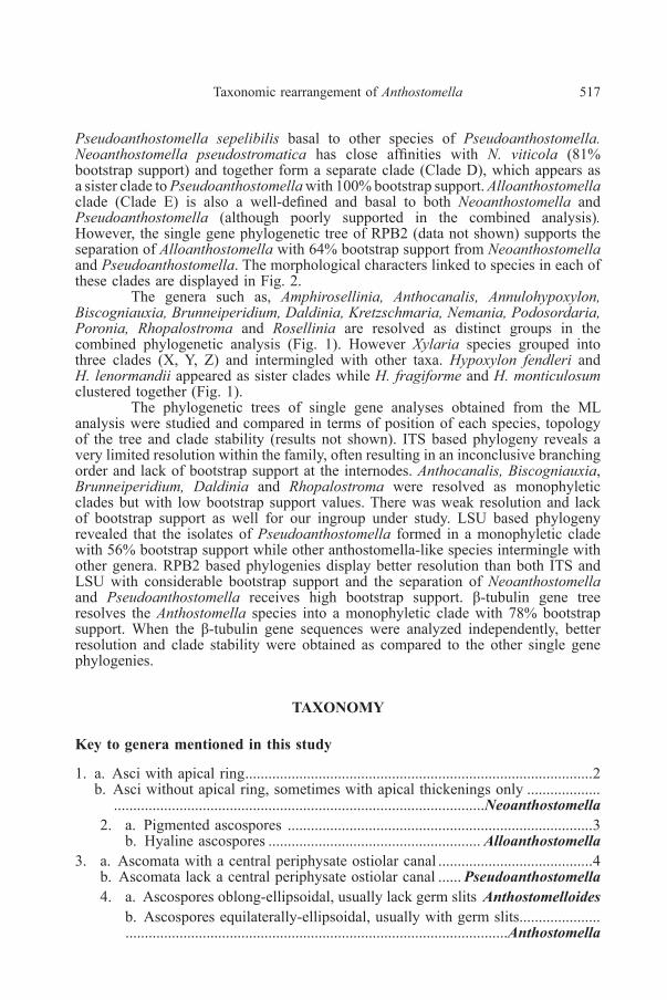
Fig. 2. The best scoring phylogenetic tree based on a combined dataset of ITS, LSU, RPB2 and β-tubulinsequence data produced by Maximum Likelihood analysis. Groups labeled are the same as in Fig. 1.Clades labeled as A-E indicate the different monophyletic subclades of anthostomella-like species.Morphological characters distinguishing each group pertaining to each clade are shown on the right ofthe cladogram with the legend below. Bootstrap support values for maximum likelihood greater than50% are given. The tree is rooted to Sordaria fimicola. Ex-types strains are in bold.
Another group of anthostomella-like species (Clade C, 99% BS) is hereintroduced as the new genus Pseudoanthostomella with five genetically very closespecies, which were previously treated as Anthostomella. Pseudoanthostomellasenecionicola, P. delitescenes and P. conorum cluster in a terminal clade with 81%and 70% bootstrap support. Pseudoanthostomella pini-nigrae branches off fromthem and nested in between P. sepelibilis and the above species. ML analysis places
Taxonomic rearrangement of Anthostomella 517
Pseudoanthostomella sepelibilis basal to other species of Pseudoanthostomella.Neoanthostomella pseudostromatica has close affinities with N. viticola (81%bootstrap support) and together form a separate clade (Clade D), which appears asa sister clade to Pseudoanthostomella with 100% bootstrap support. Alloanthostomellaclade (Clade E) is also a well-defined and basal to both Neoanthostomella andPseudoanthostomella (although poorly supported in the combined analysis).However, the single gene phylogenetic tree of RPB2 (data not shown) supports theseparation of Alloanthostomella with 64% bootstrap support from Neoanthostomellaand Pseudoanthostomella. The morphological characters linked to species in each ofthese clades are displayed in Fig. 2.
The genera such as, Amphirosellinia, Anthocanalis, Annulohypoxylon,Biscogniauxia, Brunneiperidium, Daldinia, Kretzschmaria, Nemania, Podosordaria,Poronia, Rhopalostroma and Rosellinia are resolved as distinct groups in thecombined phylogenetic analysis (Fig. 1). However Xylaria species grouped intothree clades (X, Y, Z) and intermingled with other taxa. Hypoxylon fendleri andH. lenormandii appeared as sister clades while H. fragiforme and H. monticulosumclustered together (Fig. 1).
The phylogenetic trees of single gene analyses obtained from the MLanalysis were studied and compared in terms of position of each species, topologyof the tree and clade stability (results not shown). ITS based phylogeny reveals avery limited resolution within the family, often resulting in an inconclusive branchingorder and lack of bootstrap support at the internodes. Anthocanalis, Biscogniauxia,Brunneiperidium, Daldinia and Rhopalostroma were resolved as monophyleticclades but with low bootstrap support values. There was weak resolution and lackof bootstrap support as well for our ingroup under study. LSU based phylogenyrevealed that the isolates of Pseudoanthostomella formed in a monophyletic cladewith 56% bootstrap support while other anthostomella-like species intermingle withother genera. RPB2 based phylogenies display better resolution than both ITS andLSU with considerable bootstrap support and the separation of Neoanthostomellaand Pseudoanthostomella receives high bootstrap support. β-tubulin gene treeresolves the Anthostomella species into a monophyletic clade with 78% bootstrapsupport. When the β-tubulin gene sequences were analyzed independently, betterresolution and clade stability were obtained as compared to the other single genephylogenies.
tAXonoMY
Key to genera mentioned in this study
1. a. Asci with apical ring..........................................................................................2b. Asci without apical ring, sometimes with apical thickenings only ...................
................................................................................................Neoanthostomella2. a. Pigmented ascospores ...............................................................................3
b. Hyaline ascospores ....................................................... Alloanthostomella3. a. Ascomata with a central periphysate ostiolar canal ........................................4
b. Ascomata lack a central periphysate ostiolar canal ...... Pseudoanthostomella4. a. Ascospores oblong-ellipsoidal, usually lack germ slits Anthostomelloides
b. Ascospores equilaterally-ellipsoidal, usually with germ slits........................................................................................................................Anthostomella

518 D. A. Daranagama et al.
Alloanthostomella Daranagama, Camporesi & K. D. Hyde, gen. nov.Index Fungorum number: IF552371; Facesoffungi number: FoF 02526Etymology: referring to different; yet anthostomella-like species.Saprobic on living branch of Cornus sanguinea L. and on Rubus. Sexual
morph: Ascomata immersed, black, slightly raised conical areas, solitary, sometimesaggregated into clusters, scattered, in cross section globose, with a disc-like, central,black ostiolar dot. Clypeus carbonaceous, comprising intracellular fungal hyphaeand host tissues. Peridium few cell layers, compressed, outwardly comprising thick-walled, light to dark brown cells of textura irregularis, inwardly comprising thin-walled, hyaline cells of textura angularis. Paraphyses few, filamentous, septate. Asci8-spored, unitunicate, cylindrical, short-pedicellate, apically rounded, with a wedge-shaped-cylindrical, J+, apical ring. Ascospores overlapping uniseriate-biseriate,elongate-ellipsoidal, one end tapered and pointed, remain hyaline in asci, bicellularwith rostrate, hyaline, dwarf cell, larger cell hyaline, rarely dark brown, smooth-walled. Asexual morph: Undetermined.
Type species: Alloanthostomella rubicola (Speg. [ex Sacc. & Trotter)Daranagama, Camporesi & K. D. Hyde.
Notes: The monotypic genus Alloanthostomella is introduced toaccommodate Anthostomella rubicola, which is morphologically and phylogeneticallydifferent from other Anthostomella species. Another morphologically reminiscentgenus to Alloanthostomella is Emarcea Duong et al. which shares certain characters,such as ascomata developing beneath a blackened clypeus, papillate ostioles,unitunicate asci with a J+, subapical ring and bicellular, hyaline ascospores lackinggerm slits (Duong et al. 2004). However, the other morphological differencesbetween these two genera (e.g long fusiform ascospores in Emarcea with anobclavate apical cell and a cylindrical basal cell, while Alloanthostomella havedwarf cell and an elongate ellipsoidal, larger cell and further discussed in thetaxonomy section) can be used to differentiate them. In this study, we have beenselective with the ingroup taxa and excluded Emarcea in our analysis since most ofthe available data cannot be authenticated. Along the same line, several xylariaceousspecies had to be excluded from the phylogenetic analyses because of the generationof ambiguous sequence alignment, lack of sequence data from protein gene and highdegree of polytomies encountered in phylograms coupled with low bootstrap support.Previous rDNA based phylogenetic analyses by Duong et al. (2003) indicate thatEmarcea castanopsidicola is closely related to Muscodor species which is nowtreated as genera incertae sedis (Maharachchikumbura et al. 2016).The phylogeneticposition of Emarcea with more taxa, such as Emarcea eucalyptigena, E. rostrisporaand Anthostomella eucalyptorum warrant further investigations.
Alloanthostomella rubicola (Speg.) Daranagama, Camporesi & K. D. Hyde, comb.nov.
Index Fungorum number: IF552372; Facesoffungi number: FoF 00320Fig. 3
Basionym: Entosordaria rubicola Speg., Revta Fac. Agron. Vet. Univ. nac.La Plata, Ser. 2, 6(1): 40 (1910)
≡ Anthostomella rubicola Speg. ex Sacc. & Trotter, Syll. fung. (Abellini)22: 100 (1913)
Saprobic on branch of Cornus sanguinea Sexual morph: Pseudostromatablack, carbonaceous, comprising fungal tissues and epidermal host cells. Ascomata250-300 × 280-320 µm (x̅ = 275 × 310 µm, n = 10), immersed, visible as blackened,

Taxonomic rearrangement of Anthostomella 519
Fig. 3. Alloanthostomella rubicola (MFLU 15-0661). a, b. Pseudostromata on host c, d. Cross sectionof ascoma e. Peridium f-i. Asci j. Paraphyses k-m. Ascospores. Scale bars: a, b = 500 μm, c, d =100 μm, e = 30 μm, f-j = 10 μm, k-m = 10 μm.
520 D. A. Daranagama et al.
slightly raised, conical, stromata-like areas, solitary or aggregated into clusters,scattered, in cross section globose, with a central black ostiolar dot, disc-like, black.Clypeus 60-83 µm diam. at base, 75-80 µm high (x̅ = 68 × 77 µm, n = 10), comprisinghost cells and intracellular fungal hyphae, surrounded by carbonaceous tissues.Peridium 20-30 µm diam. (x̅ = 25.6 µm, n = 10), with a few compressed cell layers,outwardly comprising thick-walled, light-dark brown cells of textura irregularis andinwardly comprising thin-walled, hyaline cells of textura angularis. Paraphyses5-6 µm diam. (x̅ = 5.8 µm, n = 20), few, filamentous, septate. Asci 120-165 × 6-9 µm(x̅ = 157 × 8.4 µm, n = 30), 8-spored, unitunicate, cylindrical, short-pedicellate,apically rounded, with a wedge-shaped to cylindrical, J+ apical ring, 3-5 × 4-5 µm(x̅ = 4 × 4.5 µm, n = 30). Ascospores 22-35 × 5.5-8.7 µm (x̅ = 27 × 6.5 µm,n = 40), overlapping uniseriate to biseriate, ellipsoidal, one end tapered and pointed,in asci always remain hyaline, unicellular with rostrate, hyaline dwarf cell, 4-6 µmlong (x̅ = 5.5 µm, n = 40), larger cell rarely dark brown or mostly hyaline, smooth-walled. Asexual morph: Undetermined.
Culture characteristics: Colonies on Difco OA at 25-27°C reaching 9 cmin 4-5 weeks, at first whitish, felty, azonate, with diffuse grey colour margins, reverseturning citrine (13). Asexual morph was not produced in cultures after 3 months.
Material examined: ITALY, Province of Forlì-Cesena, Trivella di Predappio,on dead branch of Cornus sanguinea (Cornaceae), 26 December 2014, ErioCamporesi, IT 2316 (MFLU 15-0661, reference specimen designated here), livingcultures MFLUCC 16-0479, KUMCC.
Notes: Ascospores of Al. rubicola remain hyaline in the asci. However,rarely mature, brown ascospores occur with a hyaline dwarf cell which is rostrate(Lu & Hyde 2000; Daranagama et al. 2015a). This species has morphologicalsimilarities to Anthostomella appendiculosa (Berk. & Broome) Sacc. However, theascospores in A. appendiculosa are larger and become brown in the asci with acordate dwarf cell and a straight germ slit, while those in Al. rubicola lack a germslit. Attempts to obtain the asexual morph in culture were not successful even after3 months observation. The holotype of A. rubicola collected by Spegazzini, depositedat LPS is from Chile and on Rubus fruticosus L. Due to the differences in the hostand the geographical differences between our collections and the holotype, werefrained from epityification and only designated the fresh collections as referencespecimens.
Anthostomella Sacc., Atti Soc. Veneto-Trent. Sci. Nat., Padova, Sér. 4 4: 84 (1875)Facesoffungi number: FoF 00316Generic description and illustration: See Lu & Hyde (2000) and Daranagama
et al. (2015a).Type species: Anthostomella tomicoides Sacc., Atti Soc. Veneto-Trent. Sci.
Nat., Padova, Sér. 4 4: 101 (1875).Synonymy: Entosordaria tomicoides (Sacc.) Höhn., Sber. Akad. Wiss. Wien,
Math.-naturw. Kl., Abt. 1 129: 166 (1920)= Anthostomella tomicoides var. crassispora Bat., Fischman & Da Matta,
in Batista et al. Atas Inst. Micol. Univ. Recife 4: 224 (1967)Notes: The typification of the genus Anthostomella is long confused as
Saccardo never designated any type material for Anthostomella. Eriksson (1966)designated Anthostomella limitata Sacc. as the lectotype material, the only one withthe original three species with non-appendiculate ascospores. Francis (1975)disagreed to this typification because A. limitata has no true clypeus, which is the

Taxonomic rearrangement of Anthostomella 521
key taxonomic feature of the genus and ascospores have diagonal or spiral germ slitthat is not common to many Anthostomella species. Hence Francis (1975) selectedAnthostomella tomicoides Sacc. as the type of the genus but she was unable to locatethe material and selected Anthostomella italica subsp. affinis Sacc. as the neotype.Lu and Hyde (2000) located the original material of A. tomicoides and lectotypifiedit. Since no living culture of A. tomicoides is available for now, we used A. formosa,which is morphologically similar to the type for stabilization of Anthostomella sensustricto. Anthostomella formosa is similar to A. tomicoides by having darkened,coriaceous ascomata immersed beneath the clypeus, with central, periphysate ostiolarcanal and appendage bearing ascospores with thin mucilaginous sheaths and straightgerm slits (Lu and Hyde 2000).
Anthostomella in a broad sense is currently included in the Xylariaceaewith 85 species (Lu & Hyde 2000; Daranagama et al. 2015a) but this placement stillneeds molecular support and is also not supported in our phylogeny. Furthermore,in our phylogeny the genus sits on a very long branch in between Nemania, Poroniaand Podosordaria and its true affinities may be with other genera Aworld monographof Anthostomella was published by Lu & Hyde (2000). The placement ofAnthostomella sensu stricto was established in Daranagama et al. (2015a). Based onthe molecular data the genus is clearly heterogeneous and true affinities of manyspecies are poorly understood. The genus is characterized by immersed, dark,clypeate ascomata with periphysate ostiolar canals, 8-spored, cylindrical, unitunicateasci and mostly dark, unicellular ascospores, sometimes with small cells orappendages at the ends. The asexual morph of Anthostomella is characterized byconidiophores that are simple, light brown, branched or single and having a few,small, disc-like conidiogenous cells occurring along the denticles of conidiophoresand hyaline, easily dehiscent, ellipsoidal, unicellular conidia (Daranagama et al.2015a). The genus is saprobic and cosmopolitan, distributed in tropics, subtropicsand even temperate regions. The species are distributed on a wide range of hosts andsome species may be host-specific (e. g. A. eucalyptorum).
Anthostomelloides Tibpromma & K.D. Hyde, Turkish Journal of Botany. (2016)40: 2.
Index Fungorum number: IF552117, Facesoffungi number: FoF 02190Saprobic on dead leaves, branches and twigs. Sexual morph: Ascomata
immersed, black, globose, visible as conical, blackened dots, with central periphysateostiolar canal. Clypeus short, margins indistinct, sometimes reduced, comprisingdark, thick-walled, fungal and host tissues, black, globose. Peridium coriaceous,composed of several layers, outwardly comprising brown cells and inwardlycomprising hyaline cells. Hamathecium comprising numerous, hyaline, filamentous,septate, tapering paraphyses. Asci 8-spored, rarely 6-spored, unitunicate, cylindrical,short pedicellate, with a wedge-shaped, J+ apical ring. Ascospores mostly uniseriate,inequilaterally oblong-ellipsoidal, dark brown at maturity, guttulate, with or withouta conspicuous mucilaginous sheath, with or without germ slit, if present germ slitstraight, less than the spore length. Asexual morph: undetermined.
Type species: Anthostomelloides krabiensis Tibpromma & K.D. Hyde,Turkish Journal of Botany. (2016) 40: 2.
Notes:Thisgenuswas introduced recently toaccommodateAnthostomelloideskrabiensis isolated from dead leaves of Pandanus odorifer (Forssk.) Kuntze(Tibpromma et al. 2016). The morphological similarity of Anthostomelloides toother genera such as Anthostomella, Brunneiperidium, Fasciatispora and Nipicolais discussed in Tibpromma et al. (2016). Immersed, globose ascomata with a central,
522 D. A. Daranagama et al.
papillate, periphysate ostiolar canal, short or reduced, black clypeus, asci withwedged-shaped, J+ apical ring and oblong-inequilaterally ellipsoidal ascospores areconsidered as characteristics of this genus. In this study four previously describedAnthostomella species are transferred to Anthostomelloides. Most of theAnthostomelloides species lack germ slits and even when present they are indistinctas in An. krabiensis and An. proteae. The separation of this genus from others ismorphologically and phylogenetically well-supported.
Key to Anthostomelloides species
1. a. Ascospores unicellular......................................................................................2b. Ascospores bicellular ........................................................................A. proteae2. a. Ascospores with mucilaginous sheath.......................................................3
b. Ascospores without mucilaginous sheath .................................................43. a. Ascospores with germ slits .......................................................... A. krabiensis
b. Ascospores without germ slits ................................................. A. forlìcesenica4. a. Ascomata with an intact clypeus........................................ A. leucospermi
b. Ascomata with a reduced clypeus.............................................. A. brabeji
Anthostomelloides brabeji (S.J. Lee & Crous) Daranagama & K. D. Hyde, comb.nov.
Index Fungorum number: IF552373, Facesoffungi number: FoF 02527Basionym: Anthostomella brabeji S.J. Lee & Crous, Mycol. Res. 107(3):
361 (2003)Illustration and description: See Lee & Crous (2003)Notes: According to the description provided (Lee & Crous 2003)
An. brabeji is characterized by perithecioid, immersed ascomata in the host tissue.These ascomata developed under a clypeus visible as blackened, shiny, slightlyraised dots with a central papillate, periphysate ostiolar canal and globose in crosssection. The clypeus of An. brabeji is short and reduced, when compared to otherspecies, but comprises both fungal and host tissues. Asci of An. brabeji containwedged-shaped, amyloidapical ringsanddiffer fromother species inAnthostomelloidesby having elongate ellipsoidal-fusiform, pale brown ascospores (Lee & Crous 2003).
Anthostomelloides forlìcesenica (Daranagama, Camporesi & K.D. Hyde)Daranagama & K. D. Hyde, comb. nov.
Index Fungorum number: IF552374, Facesoffungi number: FoF 02528Basionym: Anthostomella forlìcesenica Daranagama, Camporesi &
K.D. Hyde, Fungal Diversity: 73: 214 (2015)Illustration and description: See Daranagama et al. (2015a), Fungal
Diversity: 73: 214, Fig. 5.Notes: Anthostomelloides forlìcesenica was reported from Italy on Spartium
junceum L. Due to the morphological similarity of this species to An. krabiensis inhaving immersed, globose ascomata, a central periphysate ostiolar canal and ascicontaining wedged-shaped apical rings (Daranagama et al. 2015a), this species istransferred to Anthostomelloides, a move also supported by the phylogenetic analysis.In this study An. forlìcesenica clustered with the type species An. krabiensis. Similar

Taxonomic rearrangement of Anthostomella 523
to An. brabeji, An. forlìcesenica also has a reduced clypeus. Anthostomelloidesforlìcesenica lacks a visible germ slit, while a conspicuous mucilaginous sheath ispresent in An. brabeji (Daranagama et al. 2015a).
Anthostomelloides leucospermi (S.J. Lee & Crous) Daranagama & K. D. Hyde,comb. nov.
Index Fungorum number: IF552375, Facesoffungi number: FoF 02529Basionym: Anthostomella leucospermi S.J. Lee & Crous, Mycol. Res.
107(3): 364 (2003)Illustration and description: See Lee & Crous (2003) Mycol. Res. 107(3):
364. Fig. 2.Notes: This species also exhibits a range of similar characters to the
previously described species of Anthostomelloides, such as immersed, globoseascomata with a central periphysate, papillate ostiolar canal, asci with wedged-shaped apical ring and oblong-ellipsoidal ascospores. Anthostomelloides leucospermiis similar to An. brabeji as both lack ascospore germ-slits and gelatinous sheaths,but differs as the latter has more fusiform ascospores (Lee & Crous 2003).Anthostomelloides leucospermi has a more distinct clypeus than in An. brabeji,where the clypeus is reduced.
Anthostomelloides proteae (S.J. Lee & Crous) Daranagama & K. D. Hyde, comb.nov.
Index Fungorum number: IF552376, Facesoffungi number: FoF 02530Basionym: Anthostomella proteae S.J. Lee & Crous, Mycol. Res. 107(3):
366 (2003)Illustration and description: See Lee & Crous (2003), Mycol. Res. 107(3): 366, Fig. 3.
Notes: Anthostomelloides proteae was reported from South Africa on deadleaves of Protea nitida Mill. (Lee & Crous 2003). Anthostomelloides proteae alsopossess immersed and globose ascomata with a central periphysate ostiolar canal butit lacks a clypeus. According to the illustrations provided by Lee & Crous (2003)the clypeus is reduced and visible as slightly darkened area around the ostiolecomprising subepidermal cells and intracellular fungal hyphae. The ascospores ofAn. proteae are inequilaterally ellipsoidal and bicellular with a small, hyaline, dwarfcell at one end (Lee & Crous 2003). Although Lee & Crous (2003) mentioned thepresence of an indistinct germ slit in ascospore, it is not visible in the illustrationsprovided. However, An. proteae has a conspicuous mucilaginous sheath similar toAn. forlìcesenica and An. krabiensis.
Neoanthostomella D.Q. Dai & K.D. Hyde, Fungal Divers. 10.1007/s13225-016-0367-8 (2016)
Index Fungorum number: IF552041; Facesoffungi number: FoF 02004Saprobic on dead bamboo culms and branches of Vitis vinifera. L. Sexual
morph: Pseudostromata forming blackened, elliptical-irregular-shaped, raised areas,sometimes visible as pustules on the host surface. Ascomata immersed, gregarious,2-5 growing together in a single pseudostroma, in cross section globose-subglobose,dark brown, coriaceous, with a central, periphysate, ostiolate neck. Clypeus black,sometimes elongate, margin indistinct, mixed with dark, thick-walled hyphae inepidermal and subepidermal cells. Peridium comprising several layers of compressed,brown to hyaline cells of textura angularis. Hamathecium comprising dense, long,septate paraphyses intermixed with asci. Asci 8-spored, unitunicate, cylindrical,
524 D. A. Daranagama et al.
short pedicellate, without an apical ring. Ascospores uniseriate-overlapping uniseriate,equilaterally ellipsoidal, with pointed ends, unicellular, brown-olivaceous, guttulate,smooth-walled, with or without mucilaginous sheath, lacking a germ slit. Asexualmorph: Hyphomycetous. Conidiophores macronematous, septate, densely branchedin the upper part, hyaline, smooth, similar to nodulisporium-like branching pattern.Conidiogenous cells with an apical collarette, cylindrical, hyaline, smooth. Conidiahyaline, globose-ellipsoidal, slightly verruculose.
Type species: Neoanthostomella pseudostromatica D.Q. Dai & K.D. Hyde,Fungal Divers. 10.1007/s13225-016-0367-8 (2016)
Notes: Neoanthostomella was introduced by Dai et al. (2016) with a singlespecies N. pseudostromatica, which has morphological affinities with Anthostomellaby having cylindrical, 8-spored asci containing brown ascospores with mucilaginoussheath. However, in N. pseudostromatica the presence of multiple ascomata, whichcluster together in groups of 2-5 within single pseudostroma differ it fromAnthostomella (Lu & Hyde 2000; Daranagama et al. 2015a). According to thedescription provided in Dai et al. (2016) there is a straight germ slit in ascosporesalthough this is not shown in the illustration. We have observed the holotype material(MFLU 15-1190) and noted that the ascospores lack a germ slit. Hence we amendedthe generic description and provided details of the asexual morph.
It is noteworthy to mention herein that our analyses of molecular phylogenydo reveal a close relationship between Pseudoanthostomella and Neoanthostomella(Clade C & D) and hence one may argue that these phenotypically similar generacould be treated as congeneric. Despite our limited taxon sampling here, we considerNeoanthostomella as evolutionary significant and maintain our new genus given thatspecies are differentiated by strictly specific morphological characters (e.g absenceof apical apparatus and presence of central periphysate ostiole) as compared toPseudoanthostomella. Should future studies with an increasing diversity ofNeoanthostomella species reveal an overlap of characters between those two genera,then the possibility of treating those two as congeneric cannot be excluded.
Neoanthostomella viticola Daranagama, Camporesi & K. D. Hyde, sp. nov. Figs 4, 5Index Fungorum number: IF552248; Facesoffungi number: FoF 02392Etymology: Species epithet refers to the host genus Vitis.Holotype: MFLU 15-0691Saprobic on dead branch of Vitis vinifera. L. Sexual morph: Ascomata
160-203 × 180-225 μm (x̅ = 186 µm × 205 µm, n = 10), immersed, visible as black,raised, conical-irregular-shaped areas, coriaceous, clustered, rarely solitary, in crosssection globose, with wide ostiolar neck. Ostiole 82-110 μm diam. at the base × 50-66 μm high (x̅ = 102 µm × 64 µm, n = 10), grey, papillate, with a central periphysateostiolar canal. Clypeus black, thick-walled, margin indistinct, mixed with dark,fungal hyphae in host cell layers. Peridium 34-53 µm wide (x̅ = 41 µm, n = 10),with two cell layers, outwardly comprising thick-walled, compressed, light browncells of textura irregularis and inwardly comprising thick-walled, several layers ofhyaline cells of textura angularis. Paraphyses 2.5-3.4 µm wide at base (x̅ = 3.1 µm,n = 30), slightly longer than the asci, numerous, filamentous, septate. Asci 85-117 ×5-7 µm (x̅ = 91.5 × 6.7 µm, n = 20), 8-spored, unitunicate, cylindrical, longpedicellate, lacking a visible apical ring, sometimes with apical thickenings.Ascospores 5.7-11 × 3.4-4.8 µm (x̅ = 9.2 × 4.1 µm, n = 20) uniseriate-overlappinguniseriate, ellipsoidal, with broad ends, light brown, smooth-walled, lacking a germslit. Asexual morph: Hyphomycetous. Conidiophores 60-80 × 5-8 μm (x̅ = 76 µm× 5.7 µm, n = 40), macronematous, septate, densely branched in the upper part,

Taxonomic rearrangement of Anthostomella 525
Fig. 4. Neoanthostomella viticola (MFLU 15-0691). a, b, c. Appearance of ascomata in host d-f. Crosssection of ascomata g. Peridium h. Clypeus i. Paraphyses j-m. Asci n, o. Ascal apical apex in Melzer’sreagent (Note: lacks an apical ring, apical thickenings visible) p-r.Ascospores. Scale bars: a, c = 500 μm,b = 1000 μm, d, e = 100 μm, f = 30 μm, h-l = 10 μm.
526 D. A. Daranagama et al.
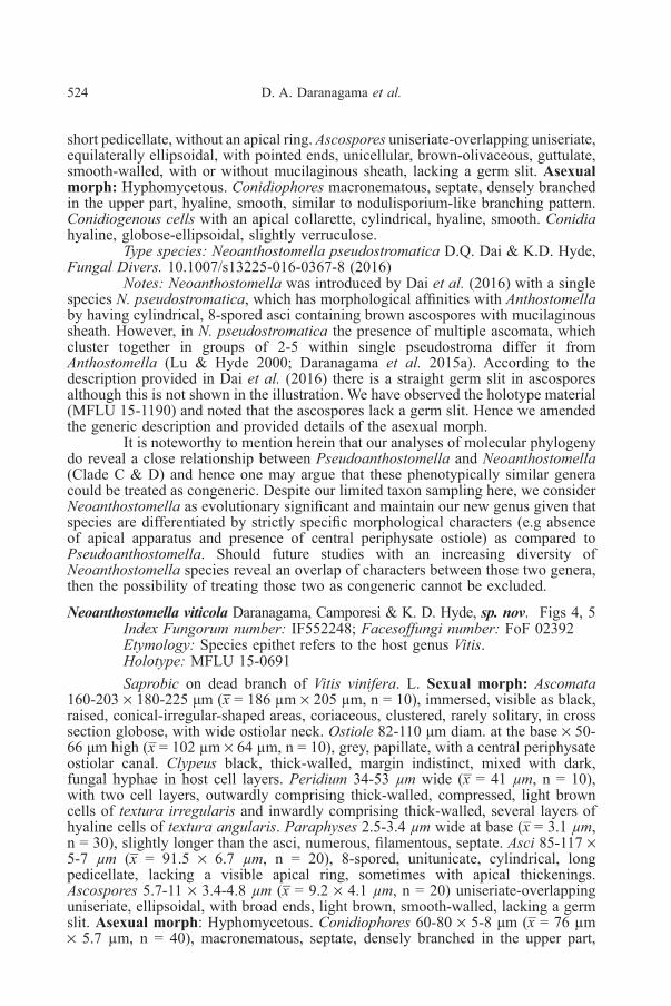
Fig. 5. Asexual morph of Neoanthostomella viticola (MFLUCC 16-0243). Culture on OA; a. Upperview b. Lower view c. Appearance of conidiophores on 3 weeks d. Conidiophores and conidiogenouscells e. Conidia. Scale bars: c = 50 μm, d = 20 μm, e = 10 μm.
hyaline, smooth. Conidiogenous cells 9-12 × 3-6 μm (x̅ = 10.5 µm × 4.5 µm, n = 40),phialidic, with an apical collarette, cylindrical, slightly wider at the base, hyaline,smooth. Conidia 5-8 × 4-6 μm (x̅ = 6.5 µm × 4.3 µm, n = 40), hyaline, globose toellipsoidal, slightly verruculose.
Culture characteristics: Colonies on Difco OA at 25-27oC reaching theedge of 9 cm Petri-dish in 5 weeks, at first whitish, felty, azonate, with fluffymargins; reverse turning light yellow. Production of conidiophores sparse, after3-4 weeks, occurring in the center of the colony.
Material examined: ITALY, Province of Forlì-Cesena, Trivella di Predappio,on dead branch of Vitis vinifera L. (Vitaceae), 31 December 2014, Erio Camporesi,IT 2326 (MFLU 15-0691, holotype), ibid (HKAS 95066, isotype), ex-type livingcultures MFLUCC 16-0243, KUMCC.
Notes: Neoanthostomella viticola is similar to Anthostomella variabilisB.S. Lu & K.D. Hyde in characters such as its smooth-walled, light brown, ellipsoidalascospores and cylindrical, slender asci. However, A. variabilis has deeply immersed

Taxonomic rearrangement of Anthostomella 527
ascomata in the host surface which results in a long, periphysate ostiolar canal (240-400 µm) as observed by Lu & Hyde (2000), which is unique to the species. Incontrast, Neoanthostomella viticola has an ostiolar canal which is wider than itslength. Neoanthostomella viticola is similar to A. limitata Sacc., A. okatina Whittonet al. and A. helicofissa Daranagama et al. However N. viticola differs becauseA. limitata has ascospores with a thin mucilaginous sheath and A. okatina has ascithat lack an apical ring and circular papilla in the ascomata (Lu & Hyde 2000), whileA. helicofissa is characterized by obpyriform ascomata, asci without apical structuresand rather small ascospores with a thin mucilaginous sheath (Daranagama et al.2015a). Neoanthostomella viticola differs from N. pseudostromatica by havingsmaller, pale brown ascospores without a mucilaginous sheath. In addition, N. viticolaproduced a hyphomycete asexual morph in culture, while an asexual morph has notbeen determined in N. pseudostromatica.
Pseudoanthostomella Daranagama, Camporesi & K. D. Hyde, gen. nov.Index Fungorum number: I IF552377; Facesoffungi number: FoF 02531Etymology: Refers to the morphological similarity to Anthostomella.Saprobic on dead leaves, branches, twigs and cones of Pinus. Sexual
morph: Ascomata immersed to semi-immersed, developed beneath the clypeus,blackened, raised, conical-dome-shaped areas, coriaceous or carbonaceous, solitary,rarely aggregated, in cross section mostly subglobose, with reduced clypeus, usuallylacking a periphysate ostiolar canal. Clypeus black, thick-walled, short, comprisingdark fungal hyphae and host epidermal cells. Peridium with two cell layers; outwardlycomprising thick-walled, carbonaceous, compressed, dark brown cells of texturairregularis and inwardly thin-walled, hyaline cells of textura angularis. Paraphysesless than 5 µm wide at base, shorter than the asci, numerous, filamentous, septate.Asci 8-spored, unitunicate, broadly cylindrical-clavate, short-pedicellate, apicallyrounded, with discoid-wedged shaped, J+ apical ring. Ascospores overlappinguniseriate, ellipsoidal, dark brown, smooth-walled, mostly with conspicuousmucilaginous sheath, germ slit on ventral side, straight, extending over the fulllength, rarely absent. Asexual morph: Hyphomycetous; Conidiophores erect, arisingfrom hyphae, complex, septate, dichotomously branched, smooth, hyaline-lightbrown. Conidiogenous cells monoblastic, discrete, terminal on the branches, arisingin clusters of 3-4, denticulate. Conidia hyaline, elongated ellipsoidal-fusiform,sometimes curved at apical end, aseptate, unicellular, smooth.
Type species: Pseudoanthostomella pini-nigrae Daranagama, Camporesi &K. D. Hyde.
Notes: Pseudoanthostomella is erected to accommodate five anthostomella-like species clustered together in a strongly supported monophyletic clade in thephylogenetic analysis. These species share common characters such as blackened,conical to dome shaped, semi-immersed to immersed ascomata, which mostly occuras solitary and rarely aggregated into small groups. The clypeus is usually reduced,but they form thick-walled, dark and short clypei without ostiolar necks. AllPseudoanthostomella species have a peridium comprising two layers of texturairregularis and textura angularis. All the species except P. sepelibilis possessascospores with straight germ slits running the full length of the spore.Pseudoanthostomella has a strong morphological resemblance to the genusAnthostomella but the former does not have a central, periphysate ostiolar canal. Theasci are more clavate than those of Anthostomella and many Anthostomella specieshave ascospores with sigmoid and curved germ slits rather than straight germ slitsas found in Pseudoanthostomella species. Pseudoanthostomella differs from

528 D. A. Daranagama et al.
Neoanthostomella by having asci with J+ apical ring and ascospores with germ slits.It differs from Alloanthostomella by having unicellular, pigmented ascospores withgerm slits. Anthostomelloides species always have immersed ascomata with a centralperiphysate ostiolar canal with more oblong-ellipsoidal ascospores which lack germslits or sometimes have a mucilaginous sheath.
Key to Pseudoanthostomella species
1. a. Ascomata immersed .........................................................................................2b. Ascomata semi-immersed ................................................................................32. a. Asci with J+ apical ring............................................................ P. conorum
b. Asci without apical ring ................................................... P. senecionicola3. a. Ascospores with germ slits ..............................................................................4
b. Ascospores without germ slits ...................................................... P. sepelibilis4. a. Germ slits less than the spore length ....................................P. delitescens
b. Germ slits, full length of spore ............................................ P. pini-nigrae
Pseudoanthostomella conorum (Fuckel) Daranagama, Camporesi & K. D. Hyde,comb. nov.
Index Fungorum number: IF552378; Facesoffungi number: FoF 02532Basionym: Amphisphaeria conorum Fuckel, Jb. nassau. Ver. Naturk. 29-30:
20 (1875) [1877]≡ Anthostomella conorum (Fuckel) Sacc., Syll. fung. (Abellini) 1: 283
(1882)Illustration and Descriptions: See Lu & Hyde (2000), Lee & Crous (2003).
Notes: Similar to other Pseudoanthostomella species, P. conorum has black,conical ascomata, reduced, short clypeus, cylindrical-slightly clavate asci with J+discoid apical ring and ellipsoidal, brown ascospores with straight, full length germslit and surrounded by an even mucilaginous sheath (Lee & Crous 2003). However,P. conorum possesses a central periphysate ostiolar canal though it is not elongateto form an ostiolar neck (Lu & Hyde 2000). According to the description by Lu &Hyde (2000) ascomata in cross section are obpyriform and the otherPseudoanthostomella species have subglobose ascomata.
Pseudoanthostomella delitescens (De Not.) Daranagama, Camporesi & K. D. Hyde,comb. nov. Fig. 6
Index Fungorum number: IF552379; Facesoffungi number: FoF 02389Basionym: Sphaeria delitescens De Not., Monogr. Tuberac. (Milano) 8:
no. 9 (1846)≡ Anthostomella delitescens (De Not.) Sacc., Michelia 1(no. 3): 328 (1878)Saprobic on dead and land cones of Pinus nigra J.F. Arnold. Sexual morph:
Ascomata 340-360 × 285-320 μm (x̅ = 355 × 302 µm, n = 10), semi-immersed,visible as blackened, raised, conical areas, carbonaceous, solitary, rarely clustered,in cross section conical-subglobose, with reduced clypeus. Clypeus 44-55 × 55-73 µm near to the neck (x̅ = 51 × 68 µm, n = 10), dark brown, comprising host cellsand intracellular light brown fungal tissues. Peridium 18-30 µm wide (x̅ = 25 µm,
Taxonomic rearrangement of Anthostomella 529
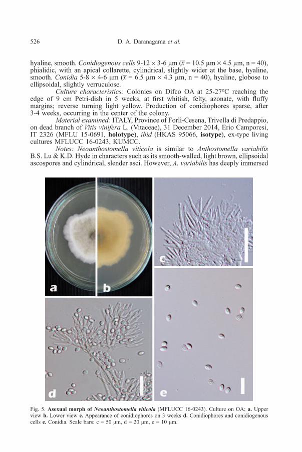
Fig 6. Pseudoanthostomella delitescens (MFLU 15-2660). a, b. Appearance of ascomata on host surfacec. Single ascoma d. Cross section of ascoma e. Reduced clypeus f. Peridium g, h. Immature ascii. Mature ascus j. Discoid, J+ apical ring k. Immature ascospore l, m. Mature ascospores n. Sheatharound ascospore in Indian ink o. Germ slit. Scale bars: a = 1000 µm, b = 500 µm, c = 200 µm,d = 100 µm, e, f = 50 µm, g-i = 20 µm, j = 5 µm, k-o = 10 µm.

530 D. A. Daranagama et al.
n = 10), with two cell layers, outwardly comprising thick-walled, dark brown cellsof textura irregularis and inwardly comprising thin-walled, loosely arranged, hyalinecells of textura angularis. Paraphyses 4-5 µm wide at base (x̅ = 4.4 µm, n = 30),shorter than the asci, numerous, filamentous, septate. Asci 120-145 × 10-15 µm(x̅ = 132 × 13.8 µm, n = 20), 8-spored, unitunicate, cylindrical-clavate, shortpedicellate, apically rounded, with a discoid, J+ apical ring, 1.5-2 µm high × 4-6 µmwide (x̅ = 1.8 × 5.4 µm, n = 20), ascospores appears as if in compartments due tothe presence of mucilaginous sheaths. Ascospores 14-17 × 8-10 µm (x̅ = 15.6 ×9.5 µm, n = 20), uniseriate, broadly ellipsoidal, with pointed ends, dark brown,smooth-walled, with mucilaginous sheath visible in Indian ink, 2.9-3.1 µm thick(x̅ = 3 µm, n = 10), germ slit straight, slightly less than spore length. Asexualmorph: Undetermined.
Material examined: ITALY, Province of Forlì-Cesena, near Passo delleForche – Galeata, on dead and land cone of Pinus nigra (Pinaceae), 16 June 2014,Erio Camporesi, IT 1937 (MFLU 15-2660), ibid (MFLU 15-3277, referencespecimen designated here, HKAS 95062), living cultures MFLUCC 16-0477,KUMCC.
Culture characteristics: Colonies on Difco OA plates at 25-27°C reachingthe edge of 9 cm Petri dish in 4 weeks, at first whitish, felty, azonate, with diffusemargins; reverse turning light yellow after 2-3 weeks.
Notes: Our fresh collection is morphologically similar to the holotypematerial of P. delitescens as described in Lu & Hyde (2000). The holotype was alsocollected from Italy in 1842 on Erica umbellata L. Morphological characters ofP. delitescens described in Lu & Hyde (2000) such as semi-immersed, subgloboseto conical ascomata, appearing as blackened dots, asci with a J+, discoid apical ringand brown ascospores, surrounded by a thin mucilaginous sheath are similar to thosein our collection. However, the colour of ascomata in the holotype is recorded asyellowish brown and our collection has more dark brown to black ascomata whichmay be due to the host differences. Pseudoanthostomella delitescens is differentfrom other Pseudoanthostomella species as it has ascospores with germ slits lessthan the spore length while others have germ slits running the full length of theascospores.
Pseudoanthostomella pini-nigrae Daranagama, Camporesi & K. D. Hyde, sp. nov.Figs 7, 8
Index Fungorum number: IF552250; Facesoffungi number: FoF 02390Etymology: Species epithet refers to the host plant pinus nigra.Holotype: MFLU 15-0652Saprobic on dead and land cones of Pinus nigra J.F.Arnold. Sexual morph:
Ascomata 250-275 × 330-350 μm (x̅ = 263.5 µm × 342 µm, n = 10), semi-immersed,visible as blackened, raised, dome-shaped areas, black, carbonaceous, solitary, incross section globose-subglobose, with flattened top. Clypeus black, thick-walled,short, comprising dark fungal hyphae and host epidermal cells. Peridium 17-25 µmwide (x̅ = 20.5 µm, n = 10), with two cell layers, outwardly comprising thick-walled,carbonaceous, compressed, dark brown cells of textura irregularis and inwardlycomprising thin-walled, hyaline cells of textura angularis. Paraphyses less than5 µm wide at base (x̅ = 4.5 µm, n = 30), slightly shorter than the asci, numerous,filamentous, septate. Asci 90-120 × 11-13.5 µm (x̅ = 106.4 × 12.3 µm, n = 20),8-spored, unitunicate, cylindrical-clavate, short pedicellate, sometimes reduced orabsent, apically rounded, with a discoid-inverted hat-shaped, J+ apical ring, withthin lower ring, 1.5-2.5 µm high × 4.2-5 µm wide (x̅ = 1.7 × 4.7 µm, n = 20).
Taxonomic rearrangement of Anthostomella 531
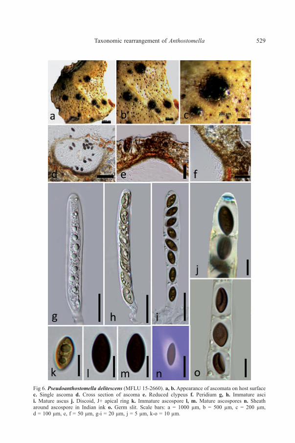
Fig. 7. Pseudoanthostomella pini-nigrae (MFLU 15-3274). a, b. Appearance of ascomata on hostsurface c. Cross section of ascoma d-f. Asci g. Paraphyses h. Apical ring in Melzer’s reagenti, j. Ascospores k. Ascospore showing the germ slit l. Ascospores with a wide band. Scale bars:a-b = 200 μm, c = 100 μm, g = 30 μm, h = 5 μm, d-f & i-l = 20 μm.

532 D. A. Daranagama et al.
Fig. 8. Asexual morph of Pseudoanthostomella pini-nigrae (MFLUCC 16-0478). Culture on OA;a. Upper view b. Lower view c-g. Conidiophores h. Conidiogeneous cells i. Conidia. Scale bars:c-h = 20 μm, i = 10 μm.
Ascospores 10-15 × 8.5-12 µm (x̅ = 14.4 × 10.3 µm, n = 20), overlapping uniseriate,broadly ellipsoidal, dark brown-black, smooth-walled, with conspicuous mucilaginoussheath, 3-3.4 µm thick, germ slit on ventral side of the ascospore, straight, extendingover the full length, sometimes visible as wide horizontal pallid band. Asexualmorph: Hyphomycetous; Conidiophores more than 80 µm long and 2.5-3 µm wide(x̅ = > 80 × 2.7 µm, n = 40), erect, arising from horizontal hyphae, complex, septate,
Taxonomic rearrangement of Anthostomella 533
dichotomously branched, smooth, upper region hyaline, lower region light brown.Conidiogenous cells 10-15 µm long and 2-3 µm diam (x̅ = 13.5 × 2.6 µm, n = 40),monoblastic, discrete, terminal on the branches, arising in clusters of 3-4, denticulate.Conidia 6-8.5 × 2-3.5 µm (x̅ = 7.3 × 2.5 µm, n = 40), hyaline, elongated ellipsoidal-fusiform, sometimes curved at apical end, aseptate, smooth.
Culture characteristics: Colonies on Difco OA plates at 25-27°C reachingthe edge of 9 cm Petri dish in 4 weeks, at first whitish, puffy, azonate, with diffusemargins, developing greyish oily spots in the center of the culture; reverse turningcitrine after 2 weeks. Production of conidiophores rare, starting as greyish spots asthe mycelium becomes melanized.
Material examined: ITALY, Province of Forlì-Cesena, Montecoronaro –Verghereto, on dead and land cones of Pinus nigra (Pinaceae), 30 July 2014, ErioCamporesi, IT 2027 (MFLU 15-0652, holotype), ibid (MFLU 15-3274, HKAS95065, isotypes), ex-type living cultures MFLUCC 16-0478, KUMCC.
Notes: Ascospores of P. pini-nigrae are similar to those of A. nigroannulata(Berk. & M.A. Curtis) Sacc. but in P. pini-nigrae they are smaller, ellipsoidal andsurrounded with a wide mucilaginous sheath. In addition, the asci of P. pini-nigraehave a smaller, discoid apical ring with a thin lower ring, while in A. nigroannulatathe apical ring is wedge-shaped (Lu & Hyde 2000) which also makes it differentfrom other species of Pseudoanthostomella. Ascomata in A. nigroannulata areimmersed, whereas P. pini-nigrae has semi-immersed ascomata appearing as conicalareas on the host surface. Anthostomella nigroannulata has been recorded fromDaemonorops (Arecaceae) and Yucca (Asparagaceae). Pseudoanthostomella pini-nigrae has only been recorded from Pinus nigra (Pinaceae).
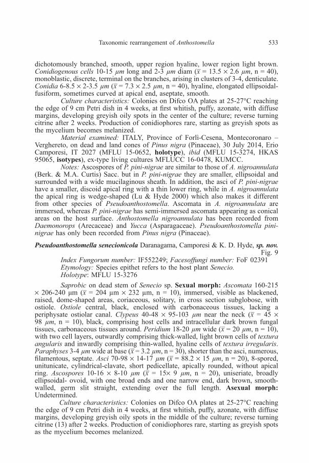
Pseudoanthostomella senecionicola Daranagama, Camporesi & K. D. Hyde, sp. nov.Fig. 9
Index Fungorum number: IF552249; Facesoffungi number: FoF 02391Etymology: Species epithet refers to the host plant Senecio.Holotype: MFLU 15-3276Saprobic on dead stem of Senecio sp. Sexual morph: Ascomata 160-215
× 206-240 μm (x̅ = 204 µm × 232 µm, n = 10), immersed, visible as blackened,raised, dome-shaped areas, coriaceous, solitary, in cross section subglobose, withostiole. Ostiole central, black, enclosed with carbonaceous tissues, lacking aperiphysate ostiolar canal. Clypeus 40-48 × 95-103 µm near the neck (x̅ = 45 ×98 µm, n = 10), black, comprising host cells and intracellular dark brown fungaltissues, carbonaceous tissues around. Peridium 18-20 µm wide (x̅ = 20 µm, n = 10),with two cell layers, outwardly comprising thick-walled, light brown cells of texturaangularis and inwardly comprising thin-walled, hyaline cells of textura irregularis.Paraphyses 3-4 µm wide at base (x̅ = 3.2 µm, n = 30), shorter than the asci, numerous,filamentous, septate. Asci 70-98 × 14-17 µm (x̅ = 88.2 × 15 µm, n = 20), 8-spored,unitunicate, cylindrical-clavate, short pedicellate, apically rounded, without apicalring. Ascospores 10-16 × 8-10 µm (x̅ = 15× 9 µm, n = 20), uniseriate, broadlyellipsoidal- ovoid, with one broad ends and one narrow end, dark brown, smooth-walled, germ slit straight, extending over the full length. Asexual morph:Undetermined.
Culture characteristics: Colonies on Difco OA plates at 25-27°C reachingthe edge of 9 cm Petri dish in 4 weeks, at first whitish, puffy, azonate, with diffusemargins, developing greyish oily spots in the middle of the culture; reverse turningcitrine (13) after 2 weeks. Production of conidiophores rare, starting as greyish spotsas the mycelium becomes melanized.

534 D. A. Daranagama et al.
Fig. 9. Pseudoanthostomella senecionicola (MFLU 15-3276). a. Habitat b. Appearance of ascomata onhost c. Appearance of ascomata on host in side view d. Cross section of an ascoma e. Reduced clypeusf. Peridium g, h. Asci in water i. Paraphyses j. Asci in Melzer’s reagent lack of an apical ringk-n. Ascospores in water o. Ascospore with straight germ slit. Scale bars: a, b = 2000 µm, c = 500 µm,d = 100 µm, e, f = 50 µm, g-o = 10 µm.

Taxonomic rearrangement of Anthostomella 535
Material examined: ITALY, Province of Forlì-Cesena, near Passo delleForche – Galeata, on dead stem of Senecio sp. (Asteraceae), 25 June 2014, ErioCamporesi, IT 1959 (MFLU 15-3276, holotype), ibid (HKAS 95063, isotype) extype living cultures MFLUCC 15-0013, KUMCC.
Notes: Pseudoanthostomella senecionicola is distinct from otherPseudoanthostomella species as the asci lack any visible apical ring. However,species such as Anthostomella sphaeroidea Speg. and A. maderensis Petr. also lackapical ring, but they are thought to be conspecific (Francis 1975, Lu & Hyde 2000).The lack of an apical ring is also characteristic for many species in the stromaticXylariaceae, i.e Hypoxylon notatum Berk. & M.A. Curtis, H. intermedium (Schwein.:Fr.) Y.-M. Ju & J. D. Rogers (Ju and Rogers 1996). Pseudoanthostomellasenecionicola has larger ascospores and unlike A. sphaeroidea, they lack a periphysateostiolar canal. The latter has a very prominent central ostiolar canal (Lu & Hyde2000). Considering the ascospore characters and shape, P. senecionicola is reminiscentto A. vestita Speg. Ascospores of A. vestita however, lack germ slits and have 2-3 µmwide mucilaginous sheaths. In addition A. vestita has globose, larger ascomata witha central periphysate ostiolar canal and longer and narrower asci, with J+ apical ring(Lu & Hyde 2000).
Pseudoanthostomella sepelibilis [(Berk. & M.A. Curtis) Sacc.] Daranagama,Camporesi & K. D. Hyde, comb. nov.
Index Fungorum number: IF552380; Facesoffungi number: FoF 02533Basionym: Sphaeria sepelibilis Berk. & M.A. Curtis, Grevillea 4(no. 32):
146 (1876)≡ Anthostomella sepelibilis (Berk. & M.A. Curtis) Sacc., Syll. fung.
(Abellini) 1: 281 (1882)Illustration and Descriptions: See Lu & Hyde (2000)Notes: Pseudoanthostomella sepelibilis has semi-immersed, brown, dome-
shaped and solitary ascomata, cylindrical-clavate asci with J+ apical ring andellipsoidal ascospores with mucilaginous sheath (Lu & Hyde 2000), which justifiesits position in the genus Pseudoanthostomella. The ascospores of Pseudoanthostomellalack germ slits and they are bicellular, with a large, brown cell and a small, hyalinedwarf cell and a mucilaginous sheath which is slightly thicker at both ends (Lu &Hyde 2000).
Acknowledgements. The authors appreciate the financial support and postgraduatescholarship provided by State Key Laboratory of Mycology, Institute of Microbiology,Chinese Academy of Sciences, Beijing and the Mushroom Research Foundation, Chiang Mai,Thailand. The authors gratefully thank Dr Shaun Pennycook from Landcare ResearchUniversity of Auckland, New Zealand for nomenclature advice on the proposed names.Dr. D. J. Bhat and Dr. Eric Mackenzie are gratefully thanked for their comments on thismanuscript. The University of Mauritius is acknowledged for support and Dr Rajesh Jeewonthanks MFU for the offer of a short term Visiting Professor.
reFerenceS
BITZER J., LÆSSØE T., FOURNIER J., KUMMER V., DECOCK C., TICHY HV., PIEPENBRING M.,PERŠOH D. & STADLER M., 2008— Affinities of Phylacia and the daldinoid Xylariaceae,inferred from chemotypes of cultures and ribosomal DNA sequences. Mycological Research112: 251-270.

536 D. A. Daranagama et al.
CAI L., JEEWON R. & HYDE K. D. 2006 — Phylogenetic investigations of Sordariaceae based onmultiple gene sequences and morphology. Mycological research 110: 137-150.
CROUS P.W. & GROENEWALD J. Z., 2010 — Anthostomella pinea Crous, sp. nov. Persoonia 25:117-159.
DAI D. Q., PHOOKAMSAK R., WIJAYAWARDENE N. N., LI W. J., BHAT D. J., XU J. C.,TAYLOR J. E., HYDE K. D. & CHUKEATIROTE E., Bambusicolous fungi. 2016— FungalDiversity. DOI: 10.1007/s13225-016-0367-8.
DARANAGAMA D. A., LIU X. Z., CHAMUNG S., STADLER M. & HYDE K. D., 2014 —A multigene genealogy reveals the phylogenetic position of Rhopalostroma lekae. Phytotaxa186(4): 177-187.
DARANAGAMA D.A., CAMPORESI E., TIAN Q., LIU X., CHAMYUANG S., STADLER M. &HYDE K.D., 2015a — Anthostomella is polyphyletic comprising several genera inXylariaceae. Fungal Diversity 73: 203-238.
DARANAGAMA D. A., LIU X. Z., CHAMUNG S., STADLER M., BHAKALI A. H. & HYDE K. D.,2015b — Rhopalostroma brevistipitatum sp. nov. from Thailand with an extended genericdescription for Rhopalostroma. Phytotaxa 227: 229-242.
DARANAGAMAD. A., LIU X. Z., CHAMUNG S., STADLER M. & HYDE K. D., 2016a— Do largerXylariaceae make up the most of Xylariomycetidae? Mycosphere 7 (5): 582-601.
DARANAGAMAD. A., LIU X., CHAMYUANG S., STADLER M., BAHKALI A. & HYDE K., 2016b— Towards a natural classification of Sordariomycetes: The genera Frondisphaeria,Immersisphaeria, Lasiobertia, Pulmosphaeria and Yuea (Sordariomycetes incertae sedis).Phytotaxa 258; 153-163.
DARANAGAMAD.A., CAMPORESI E., LIU X. Z., BHATD. J., CHAMYUANG S., BAHKALIA. H.,STADLER M. & HYDE K. D., 2016c — Tristratiperidium microsporum gen. et sp. nov.(Xylariales) on dead leaves of Arundo plinii. Mycological Progress 15; 1-8.
DARANAGAMA D. A., CAMPORESI E., LIU X. Z., CHAMYUANG S., STADLER M., JINGZU S.& HYDE K. D., 2016d — Lopadostoma fagi (Lopadostomataceae) on Fagus sylvatica fromItaly. Studies in Fungi 1; 80-89.
DENNIS R. W. G., 1956 — Further notes on tropical America. Kew Bulletin 1956: 401-404.DUONG L. M., LUMYONG S., HYDE K. D. & JEEWON R., 2004— Emarcea castanopsidicola gen.
et sp. nov. from Thailand, a new xylariaceous taxon based on morphology and DNAsequences. Studies in Mycology 50: 253-260.
FRANCIS S. M., 1975 — Anthostomella Sacc. (Part 1). Mycological Papers 139: 1-97.HSIEH H. M., JU Y. M. & ROGERS J.D., 2005 — Molecular phylogeny of Hypoxylon and closely
related genera. Mycologia 97: 844-865.HSIEH H. M., LIN C. R., FANG M. J., ROGERS J. D., FOURNIER J., LECHAT C. & JU Y. M., 2010
— Phylogenetic status of Xylaria subgenus Pseudoxylaria among taxa of the subfamilyXylarioideae (Xylariaceae) and phylogeny of the taxa involved in the subfamily. MolecularPhylogenetics and Evolution 54: 957-969.
HYDE K. D., UDAYANGAD., MANAMGODAD. S., TEDERSOO L., LARSSON E.,ABARENKOV K.,BERTRAND Y. J. K., OXELMAN B., HARTMANN M., KAUSERUD H., RYBERG M.,KRISTIANSSON E. & NILSSON R. H. 2013 — Incorporating molecular data in fungalsystematics: a guide for aspiring researchers. Current Research in Environmental & AppliedMycology 3: 1-32.
Index Fungorum (2016) http://www.indexfungorum.org/Names/Names.asp. Accessed 26 January 2016.JAKLITSCHW. M. & VOGLMAYR H., 2012— Phylogenetic relationships of five genera of Xylariales
and Rosasphaeria gen. nov.(Hypocreales). Fungal Diversity 52: 75-98.JAYASIRI S.C., HYDE K.D., ARIYAWANSA H.A., BHAT J., BUYCK B., CAI L., DAI Y.C., ABD-
ELSALAM K.A., ERTZ D., HIDAYAT I., JEEWON R., JONES E.B.G., BAHKALI A.H.,KARUNARATHNA S.C., LIU J.K., LUANGSA-ARD J.J., LUMBSCH H.T.,MAHARACHCHIKUMBURA S.S.N., MCKENZIE E.H.C., MONCALVO J.M., GHOBAD-NEJHAD M., NILSSON H., PANG K.A., PEREIRA O.L., PHILLIPS A.J.L., RASPé O.,ROLLINS A.W., ROMERO A.I., ETAYO J., SELÇUK F., STEPHENSON S.L.,SUETRONG S., TAYLOR J.E., TSUI C.K.M., VIZZINI A., ABDEL-WAHAB M.A.,WEN T.C., BOONMEE S., DAI D.Q., DARANAGAMA D.A., DISSANAYAKE A.J.,EKANAYAKA A.H., FRYAR S.C., HONGSANAN S., JAYAWARDENA R.S., LI W.J.,PERERA R.H., PHOOKAMSAK R., DE SILVA N.I., THAMBUGALA K.M., TIAN Q.,WIJAYAWARDENE N.N., ZHAO R.L., ZHAO Q., KANG J.C. & PROMPUTTHA I., 2015— The Faces of Fungi database: fungal names linked with morphology, phylogeny andhuman impacts. Fungal Diversity 74(1):3-18. doi:10.1007/s13225-015-0351-8.
Taxonomic rearrangement of Anthostomella 537
JU Y. M. & ROGERS J.D., 1996 — A revision of the genus Hypoxylon. Mycologia Memoir No. 20.St. Paul (MN): APS Press; 365 pp.
KATOH K. & STANDLEY D. M., 2013 — MAFFT multiple sequence alignment software version 7:improvements in performance and usability. Molecular Biology and Evolution 30: 772-780.
KUHNERT E., FOURNIER J., PERŠOH D., LUANGSA-ARD J. J. D. & STADLER M., 2013— NewHypoxylon species from Martinique and new evidence on the molecular phylogeny ofHypoxylon based on ITS rDNA and β-tubulin data. Fungal Diversity 64: 181-203.
LÆSSØE T. & SPOONER B. M., 1994 — Rosellinia and Astrocystis (Xylariaceae): new species andgenetic concepts. Kew Bulletin 49:1-70.
LEE S. & CROUS P. W., 2003 — New species of Anthostomella on fynbos, with a key to the genus inSouth Africa. Mycological research 107: 360-370.
LU B. S. & HYDE K. D., 2000 — A world monograph of Anthostomella. Fungal Diversity ResearchSeries 4: 1-376.
MAHARACHCHIKUMBURA S. S., HYDE K. D., JONES E. B. G., MCKENZIE E. H. C.,HUANG S. K., ABDEL-WAHAB M. A., DARANAGAMA D. A., DAYARATHNE M.,D’SOUZA M. J., GOONASEKARA I. D., HONGSANAN S., JAYAWARDENA R. S.,KIRK P. M., KONTA S., LIU J. K., LIU Z. Y., NORPHANPHOUN C., PANG K. L.,PERERA R. H., SENANAYAKE I. C., SHANG Q., SHENOY B. D., XIAO Y. P.,BAHKALI A. H., KANG J. C., SOMROTHIPOL S., SUETRONG S., WEN T. C. &XU J. C., 2015 — Towards a natural classification and backbone tree for Sordariomycetes.Fungal Diversity 72: 199-301 DOI 10.1007/s13225-015-0331-z.
MAHARACHCHIKUMBURA S. S. N., HYDE K. D., JONES E. B. G., MCKENZIE E. H. C., BHAT J.,HAWKSWORTH D. L., DAYARATHNE M., HUANG S. K., NORPHANPHOUN C.,SENANAYAKE I. C., PERERA R. H., SHANG Q., XIAO Y., D’SOUZA M. J.,HONGSANAN S., JAYAWARDENA R. S., DARANAGAMA D. A., KONTA S.,GOONASEKARA I. D., ZHUANG W. Y., JEEWON R., PHILLIPS A. L., ABDEL-WAHAB M. A., AL-SADI A. M., BAHKALI A. H., BOONMEE S., BOONYUEN N.,CHEEWANGKOON R., DISSANAYAKE A. J., KANG J., LIU J. K., LIU X. Z., LIU Z. L.,PANG K. L., PHOOKAMSAK R., PROMPUTTHA I., SUETRONG S., WEN T. &WIJAYAWARDENE N. N., 2016 — Families of Sordariomycetes. Fungal Diversity 79:1-317 DOI 10.1007/s13225-016-0369-6.
MARINCOWITZ S., CROUS P. W., GROENEWALD J. Z. & WINGFIELD M. J., 2008— Microfungioccuring on “Proteaceae” in the Fynbos. CBS Fungal Biodiversity Centre.
MILLER J. H., 1961 — A monograph of the world species of Hypoxylon. Athens (GA): University ofGeorgia Press; 158 p.
MILLER M. A., PFEIFFER W. & SCHWARTZ T., 2010— Creating the CIPRES Science Gateway forinference of large phylogenetic trees. In: Proceedings of the Gateway Computing EnvironmentsWorkshop (GCE), 14 Nov. 2010, New Orleans, Louisiana.
PELAEZ F., GONZALEZ V., PLATAS G. & SANCHEZ-BALLESTEROS J., 2008 — Molecularphylogenetic studies within the family Xylariaceae based on ribosomal DNA sequences.Fungal Diversity 31: 111-134.
RAMBAUTA. & DRUMMONDA., 2008— Fig Tree: Tree figure drawing tool, version 1.2. 2. Instituteof Evolutionary Biology, University of Edinburgh.
ROGERS J. D., 1977 — Surface features of the light colored ascospores of some applanate Hypoxylonspecies. Canadian Journal of Botany 55: 2394-2398.
SáNCHEZ-BALLESTEROS J., GONZáLEZ V., SALAZAR O., ACERO J., PORTAL M. A.,JULIÁN M. & RUBIO V., 2000 — Phylogenetic study of Hypoxylon and related generabased on ribosomal ITS sequences. Mycologia 92: 964-977.
SENANAYAKE I. C., MAHARACHCHIKUMBURA S. S. N., HYDE K. D., BHAT J. D.,JONES E. B. G., MCKENZIE E. H. C., DAI D. Q., DARANAGAMA D. A.,DAYARATHNE M. C., GOONASEKARA I. D., KONTA S., LI W. J., SHANG Q. J.,STADLER M., WIJAYAWARDENE N. N., XIAO Y. P., NORPHANPHOUN C., LI Q. R.,LIUX. Z., BAHKALIA. H., KANG J. C.,WANGY.,WENT. C., XU J. C. & CAMPORESI E.,2015 —Towards unraveling relationships in Xylariomycetidae (Sordariomycetes). FungalDiversity 73, 73-144.
SHENOY B. D., JEEWON R., WU W. P., BHAT D. J. & HYDE K. D., 2006 — Ribosomal and RPB2DNA sequence analyses suggest that Sporidesmium and morphologically similar genera arepolyphyletic. Mycological research 110: 916-928.
STADLER M. & HELLWIG V., 2005— Chemotaxonomy of the Xylariaceae and remarkable bioactivecompounds fromXylariales and their associated asexual states. Recent Research Developmentsin Phytochemistry. 9: 41-93.
538 D. A. Daranagama et al.
STADLER M., FOURNIER J., LÆSSØE T., LECHAT C., TICHY H. V. & PIEPENBRING M., 2008— Recognition of hypoxyloid and xylarioid Entonaema species from a comparison ofholomorphic morphology, HPLC profiles, and ribosomal DNA sequences. MycologicalProgress 7: 53-73.
STADLER M., FOURNIER J., GARDT S. & PERŠOH D., 2010 — The phylogenetic position ofRhopalostroma as inferred from a polythetic approach. Persoonia 25: 11-21.
STADLER M., KUHNERT E., PERŠOH D. & FOURNIER J., 2013 — The Xylariaceae as modelexample for a unified nomenclature following the one fungus-one name (1F1N) concept.Mycology: An International Journal on Fungal Biology 4: 5-21.
STADLER M., LÆSSØE T., FOURNIER J., DECOCK C., SCHMIESCHEK B., TICHY H. &PERŠOH D., 2014a — A polyphasic taxonomy of Daldinia (Xylariaceae). Studies inMycology 77: 1-143.
STADLER M., HAWKSWORTH D. L. & FOURNIER J., 2014b— The application of the name Xylariahypoxylon, based on Clavaria hypoxylon of Linnaeus. IMA Fungus 5(1): 57-66.
STAMATAKIS A., 2006 — RAxML-VI-HPC: maximum likelihood-based phylogenetic analyses withthousands of taxa and mixed models. Bioinformatics 22:2688-2690.
STAMATAKIS A., HOOVER P. & ROUGEMONT J., 2008 — A rapid bootstrap algorithm for theRAxML web servers. Systematic Biology 57: 758-771.
TAMURA K., PETERSON D., PETERSON N., STECHER G., NEI M. & KUMAR S., 2011 —MEGA5: molecular evolutionary genetics analysis using maximum likelihood, evolutionarydistance, and maximum parsimony methods. Molecular Biology and Evolution 28: 2731-2739.
TANGA. M. C., JEEWON R. & HYDE K. D., 2009—A re-evaluation of the evolutionary relationshipswithin the Xylariaceae based on ribosomal and protein-coding gene sequences. FungalDiversity 34:155-153.
THAMBUGALAK.M.,HYDEK.D., TANAKAK.,TIANQ.,WANASINGHED.N.,ARIYAWANSAH.,JAYASIRI S. C., BOONMEE S., CAMPORESI E., HASHIMOTO A., HIRAYAMA K.,SCHUMACHER R. K., PROMPUTTHA I. & LIU Z. Y., 2015 — Towards a naturalclassification and backbone tree for Lophiostomataceae, Floricolaceae and Amorosiaceaefam.nov. Fungal Diversity 74: 199-266.
TIBPROMMA S., DARANAGAMA D. A., BOONMEE S., PROMPUTTHA I., NONTACHAI-YAPOOM S. & HYDE K. D., 2016 — Paraanthostomella krabiensis gen. et sp. nov.(Xylariaceae) from Pandanus odorifer (Pandanaceae). Turkish Journal of Botany. (In press).
TRIEBEL D., PERSOH D., WOLLWEBER H. & STADLER M., 2005 — Phylogenetic relationshipsamong Daldinia, Entonaema and Hypoxylon as inferred from ITS nrDNA sequences. NovaHedwigia 80: 25-43.
WANG H. K., APTROOT A., CROUS P. W., HYDE K. D. & JEEWON R., 2007 — The polyphyleticnature of Pleosporales: an example from Massariosphaeria based on rDNA and RBP2 genephylogenies. Mycological research 111: 1268-1276.