Embed Size (px)
Citation preview
CLINICAL MICROBIOLOGY REVIEWS, OCt. 1994, p. 479-504 Vol. 7, No. 40893-8512/94/$04.00+0Copyright C) 1994, American Society for Microbiology
Taxonomy, Biology, and Clinical Aspects of Fusarium SpeciestPAUL E. NELSON,1* M. CECILIA DIGNANI,2 AND ELIAS J. ANAISSIE2*
Fusarium Research Center, Department of Plant Pathology, The Pennsylvania State University, University Park,Pennsylvania 16802,1 and Department of Medical Specialties, Section of Infectious Diseases, The University of Texas
M. D. Anderson Cancer Center, Houston, Texas 770302
INTRODUCTION .............................................ITQrP'%D'V d'% LYTCADYYTAX Q'VQrVVlLArFlr7H.
.480140.1L131'JtIUr rUk''.A IU N 'I N IIUli LVA11................................................................................. 481
Splitters................................ 481Wollenweber and Reinking................................ 481Gerlach and Nirenberg................................ 481Raillo and Bilai................482Joade................................482
Lumpers................................ 482Snyder and Hansen.482.................................Messiaen and Cassini ................................ 483Matuo................................ 483
Moderates................................ 483Gordon................................ 483Booth................................ 483Nelson, Toussoun, and Marasas................................ 483
TAXONOMY .......................................................................................................
General Characteristics of Fusarium Species .............................................
Primary Characters Used To Separate Species in Fusarium Taxonomyws__LS_.~M + s%
.484
.484AQAiviorUpoUigyonrU lu.. ..a.ruturXuuIa.54
Microconidia..............................484Microconidiophores ............................. 484Chlamydospores.484
Secondary Characters Useful in Separating Species in Fusarium Taxonomy.PROBLEMS IN WORKING WITH FUSARIUM SPECIES.
Transfer Methods ................................................................
CultureMedia.Cultural Mutation................................................................
TOXIGENICITY OF FUSARIUM SPECIES............................................................
Mycotoxins and Mycotoxicoses ............................................................... .
Human Diseases Associated with Toxigenic Fusarium Species.Alimentary toxicaleukia.
(i) First stage................................................................
(ii) Secondstage.(iii) Thirdstage.
Urov or Kashin-Beck disease.Akakabi-byo (scabby grain intoxication).
Animal Diseases Associated with Toxigenic Fusarium Species.Hemorrhagicsyndrome.Estrogenicsyndrome.Feed refusal and emetic syndromes.Fescuefoot.Degnala disease...................................................................................................Moldy sweet potato toxicosis (atypical interstitial pneumonia)...................
r.....................484.484.484AQR
.................................................487.487.487.487.487.488A400
.488
.488
.488
.488
.488
..488
.489
..489
..489AxQ
* Corresponding authors. Mailing address for Paul E. Nelson:Fusarium Research Center, Department of Plant Pathology, ThePennsylvania State University, University Park, PA 16802. Phone:(814) 865-9773. Fax: (814) 863-7217.
Mailing address for Elias J. Anaissie: Department of MedicalSpecialties, Section of Infectious Diseases, The University of Texas M.D. Anderson Cancer Center, Box 47, 1515 Holcombe Blvd., Houston,TX 77030.
t Contribution 1990 from the Fusarium Research Center, Depart-ment of Plant Pathology, The Pennsylvania State University, Univer-sity Park.
479
AQi
.................................................
I...............................................
................................................
..407............................................
,
on April 29, 2020 by guest
http://cmr.asm
.org/D
ownloaded from
480 NELSON ET AL.
AeQ
monilhjorme..........49
Fusarins........490Fumonisins........490
MYCOTOXICOSES ASSOCIATED WITH THE GROWTH OF F. MONILIFORME ON CORN .................490Equine LEM ..................................................................................... 490PPE ..................................................................................... 490Experimental Liver Cancer ..................................................................................... 491Esophageal Cancer ..................................................................................... 491Distribution of Fusarium Species That Produce Fumonisins ...........................................................................492
INFECTIONS CAUSED BY FUSARIUM SPECIES ..................................................................................... 492Introduction.492_
PATHOGENESIS OF INFECTION......................Experimental Infections......................................Ocular infection...............................................Brain infection..................................................Disseminated infection.....................................
Determinants of Virulence of Fusarium spp....Fusarium mycotoxins.......................................Enzyme production .................... .
Adherence to prosthetic material...................Host Response to Infection..................................
CLINICAL SPECTRUM AND MANAGEMENTForeign-Body-Associated Fusarial Infection.....
Keratitis in contact lens wearers....................Peritonitis following CAPD .................... .
Catheter-associated fungemia.........................Single Organ Invasion..........................................Fusarium keratitis.............................................nn,vohnamvl>ns: c
..493A02
.....................................................................................................49..493..494AifA....................................................................................................494
..495
..495
..495
..495
..495
..495
..495AO6
.'.....................-............... -...............................................Y.v
Si
JnyI InIumycI Usis ............................................................................................................................................... U4vSkin infections ............................................. 496Otitis..............................................496Bone and joint infections............................................. 496Invasive intranasal infection ............................................. 496Brain abscess............................................. 496Pneumonia..............................................496lisseminated Multiorgan Infection............................................. 496Fnidtlminanov A97Pathogenesis.......................498Clinical picture............................................. 498Outcome ............................................ 498uisceptibility of Fusarium Species to AntifungalAgents.498_
100qAQO
INTRODUCTION
Fusarium species have been important for many years asplant pathogens causing diseases such as crown rot, headblight, and scab on cereal grains; vascular wilts on a wide rangeof horticultural crops; root rots; cankers; and other diseasessuch as pokkah-boeng on sugarcane and bakanae disease ofrice (28). In the last 20 years, Fusarium species have beenstudied extensively because the mycotoxins they produce canbe a threat to animal and human health (124). Mycotoxins are
secondary metabolites produced by fungi that are associatedwith a variety of animal diseases and some human healthproblems (123). More recently, Fusarium species have becomeimportant as pathogens of human patients with compromisedimmune systems (6, 8).Fusarium species are widely distributed in soil and on
subterranean and aerial plant parts, plant debris, and otherorganic substrates (28, 32, 79, 255). They are common in
tropical and temperate regions and are also found in desert,alpine, and arctic areas, where harsh climatic conditions pre-vail (28, 32, 35, 59, 77, 78, 80, 92, 98, 110, 117, 162-164, 242,251, 255). Many Fusarium species are abundant in fertilecultivated and rangeland soils but are relatively uncommon inforest soils (32, 35, 38, 92, 117, 214, 226). Fusarium species areoften regarded as soilborne fungi because of their abundancein soil and their frequent association with plant roots, as eitherparasites or saprophytes. However, many have active or passivemeans of dispersal in the atmosphere and are common colo-nizers of aerial plant parts, where they may result in diseases ofconsiderable economic importance (27, 33, 59, 60, 89, 113, 118,150, 165, 204). Some of these airborne Fusarium species areencountered rarely in isolations of cultures from soil or roots.The widespread distribution of Fusarium species may beattributed to the ability of these fungi to grow on a widerange of substrates and their efficient mechanisms for dispersal(32).
F. MONILIFORME..................Mycotoxins Produced by F.
x,. ... a .
D
CONCLUSION..........................................................................RFFRRFNCR-
Moniliformin........................................................................................................................................................490
...................................................................................................I
...................................................................................................I
......................................................................................................d#Yu
AG4
CLIN. MICROBIOL. REV.
on April 29, 2020 by guest
http://cmr.asm
.org/D
ownloaded from
FUSARIUM SPECIES 481
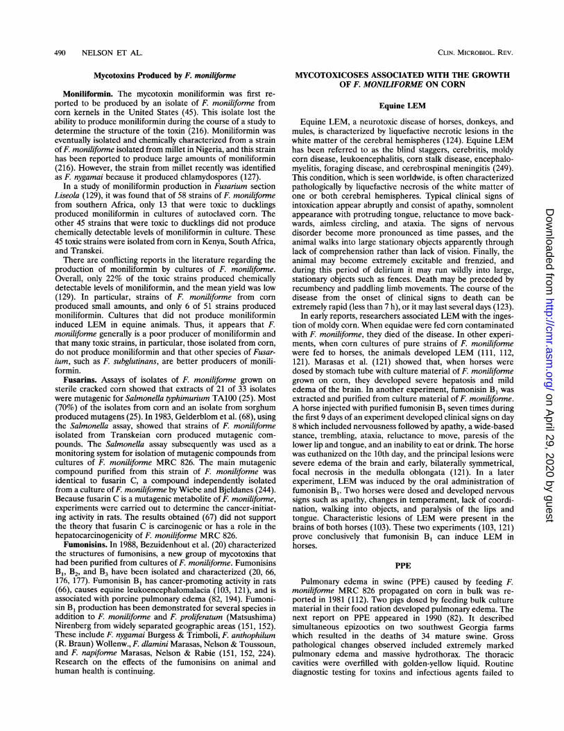
HISTORY OF FUSARIUM SYSTEMATICSThe taxonomy of Fusarium species has always been a
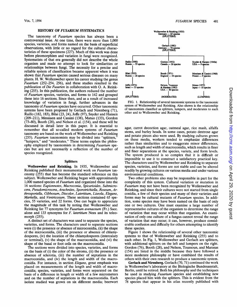
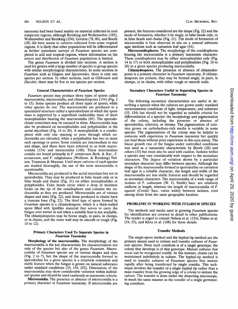
controversial issue. At one time, there were more than 1,000species, varieties, and forms named on the basis of superficialobservations, with little or no regard for the cultural charac-teristics of these specimens (227). Much of this work was donebefore pleomorphism and variation in fungi were recognized.Systematists of that era generally did not describe the wholeorganism and made no attempt to look for similarities orrelationships between fungi. The necessity for a precise andreliable system of classification became apparent when it wasshown that Fusarium species caused serious diseases on manyplants. H. W. Wollenweber spent his career studying the genusFusarium (252-254, 256), and these studies resulted in thepublication of Die Fusarien in collaboration with 0. A. Reink-ing (255). In this publication, the authors reduced the numberof Fusarium species, varieties, and forms to 142 and groupedthese into 16 sections. Since then, and as a result of increasedknowledge of variation in fungi, further advances in thetaxonomy of Fusarium species have occurred. Other taxonomicsystems have been proposed by Gerlach and Nirenberg (71),Raillo (182, 183), Bilai (23, 24), Joffe (97), Snyder and Hansen(209-211), Messiaen and Cassini (138), Matuo (133), Gordon(73-80), Booth (28), and Nelson et al. (154), and these will bediscussed in detail later in this paper. It is essential toremember that all so-called modem systems of Fusaiumtaxonomy are based on the work of Wollenweber and Reinking(255). Fusarium taxonomists may be divided into "splitters,""lumpers," and "moderates." These terms explain the philos-ophy employed by taxonomists in determining Fusarium spe-cies but are not necessarily a reflection of the number ofspecies recognized.
SplittersWollenweber and Reinking. In 1935, Wollenweber and
Reinking published their monumental work on Fusarium tax-onomy (255) that has become the standard reference on thissubject. Wollenweber and Reinking began with approximately1,000 named species of Fusarium and organized these into the16 sections: Eupionnotes, Macroconia, Spicarioides, Submicro-cera, Pseudomicrocera, Arachnites, Sporotrichiella, Roseum, Ar-throsporiella, Gibbosum, Discolor, Lateritium, Liseola, Elegans,Martiella, and Ventricosum. These sections contained 65 spe-cies, 55 varieties, and 22 forms. One can begin to appreciatethe magnitude of this task by noting that Wollenweber andReinking list 77 synonyms for Fusarium avenaceum (Fr.) Sacc.alone and 133 synonyms for F. lateritium Nees and its teleo-morph (255).A distinct set of characters was used to separate the species,
varieties, and forms. The characters used to separate sectionswere (i) the presence or absence of microconidia, (ii) the shapeof the microconidia, (iii) the presence or absence of chlamy-dospores, (iv) the location of the chlamydospores (intercalaryor terminal), (v) the shape of the macroconidia, and (vi) theshape of the basal or foot cells on the macroconidia.The sections were divided into species, varieties, and forms
on the basis of (i) the color of the stroma, (ii) the presence orabsence of sclerotia, (iii) the number of septations in themacroconidia, and (iv) the length and width of the macro-conidia. For instance, in section Elegans, great emphasis wasplaced on measurements of the length and width of macro-conidia; species, varieties, and forms were separated on thebasis of a difference in length or width of a few micrometersand on the number of septations in the macroconidia. Eachisolate studied was grown on six different media; beerwort
Wollenweber & Reinking (1935)Gennanyc65
Ge (1982)Germany-78 species
(S I)
Railo (1950)Russia-55 species
Bilai (1 )Russia -26 species
Joffe (1974)Isael-33 species
(SPPrS)
Gordon (1952)Canada- 26 species
Booth (J971)England- 44 species
Nelson, Toussoun& Massas (1983)USA-30 species
(MODLRThS)
Snyder& Hansen (1940s)U A _
Messiaen & cassi (1968)France -9 species
Mato(1971)Japan- 10 species
(LUl1RS)FIG. 1. Relationship of several taxonomic systems to the taxonomic
system of Wollenweber and Reinking. Also shown is the relationshipof taxonomists classified as splitters, lumpers, and moderates to eachother and to Wollenweber and Reinking.
agar, carrot decoction agar, oatmeal agar, rice mash, alfalfastems, and barley heads. In some cases, potato dextrose agarand potato pieces also were used. By studying cultures grownon these media, workers tended to emphasize differencesrather than similarities and to exaggerate minor differences,such as length and width of macroconidia, which results in finerand finer separations at the species, variety, and form levels.The system produced is so complex that it is difficult orimpossible to use it to construct a satisfactory practical key.The characters used by Wollenweber and Reinking to separatespecies, varieties, and forms are not stable and can be alteredreadily by growing cultures on various media and under variousenvironmental conditions.Two other problems also may be responsible in part for the
complexity of this system. Cultural variation or mutation inFusanium may not have been recognized by Wollenweber andReinking, and since their cultures were not started from singleconidia, a few of their species and many of their varieties andforms may be cultural mutants of Fusarium species. In addi-tion, some species may have been named on the basis of onlyone or two cultures. One must examine a large number ofrepresentative cultures of the organism to determine the rangeof variation that may occur within that organism. An exami-nation of only one culture of a fungus cannot reveal the rangeof variation that may occur; it can, however, produce consid-erable confusion and difficulty for others attempting to identifythese species.
Figure 1 shows the relationship of several other taxonomicsystems to that of Wollenweber and Reinking (255) and toeach other. In Fig. 1, Wollenweber and Gerlach are splitters,with additional splitters on the left and lumpers on the right.Gordon (76), Booth (28), and Nelson, Toussoun, and Marasas(154) are listed in the middle because they have followed amore moderate philosophy or have combined the results ofothers with their own research to produce a taxonomic system.
Gerlach and Nirenberg. Gerlach (70, 71) continued the workin Wollenweber's laboratory at the Biologische Bundesanstalt,Berlin, until he retired. Both his philosophy and the techniqueshe used in studying Fusarium species and establishing newspecies placed him with the splitters. This is evident from the78 species that appear in his atlas recently published with
VOL. 7, 1994
on April 29, 2020 by guest
http://cmr.asm
.org/D
ownloaded from
482 NELSON ET AL.
Nirenberg (71), a well-illustrated work that uses excellentphotographs and line drawings to supplement Wollenweber'soriginal drawings included in part. Gerlach and Nirenberggrew cultures on the eight different media used by Wollenwe-ber and Reinking and under conditions that accentuate differ-ences. They concentrated on these differences rather than on
similarities, with the result that a slight difference in gross
culture morphology may have been the basis for designating a
new species or variety. New species were established on thebasis of a single culture and, in some cases, on a single mutantculture. This philosophy led to a complex taxonomic systemthat is difficult to use for the same reasons that Wollenweberand Reinking's system is difficult to use.
Raillo and Bilai. The systems of Raillo (182, 183) and Bilai(23, 24) are not as well understood as other systems (Fig. 1).Raillo studied morphological characters useful in taxonomyand concluded the following: the form of the apical cell was theguiding character in species determination; the incurvature ofconidia, length of the apical cell, number of septa, and width ofconidia were the characters used in separating subspecies andvarieties; and cultural characters such as pigment, presence ofsclerotia, and mode of spore formation were useful in separat-ing forms only. She also studied variability in Fusarium byinitiating cultures from single conidia and found that the formof the apical cell and the incurvature of conidia remainedconstant in cultures developed from single conidia; the numberof septa was constant in isolates within a single-conidiumculture; the length and width of conidia varied considerably inseparate isolates within a single-conidium culture; the numberof sclerotia varied greatly in separate isolates within a single-conidium culture; and the mode of spore formation (pion-notes, pseudopionnotes, and sporodochia) varied in separateisolates within a single-conidium culture.
Bilai (23, 24) recognized the importance of cultural variationor mutation in Fusarium taxonomy. She did a critical analysisof several characters used in Fusarium taxonomy by studyingexperimental variability of individual isolates and establishingthe range of variation for some species. In addition, she studiedexperimental morphogenesis in single-conidium isolates inculture, paying particular attention to the effects of tempera-ture, moisture, length of growth period, and composition of themedium, as well as to the method of germination and aging ofconidia. Her results showed that the range of variability was
greater than that accepted in the description of many speciesand often included the features of the whole section. On thebasis of these results, she revised the taxonomy of the genus to
include only nine sections, 26 species, and 29 varieties. Some ofher changes, such as combining section Liseola with sectionElegans and combining section Gibbosum with section Dis-color, are difficult to understand. This system may have beenused in Russia, but it has not been accepted and used in otherparts of the world.
Joffe. Joffe (97) began working on Fusarium in the late 1940sin Russia and later immigrated to Israel and continued hiswork. He examined a large number of isolates of Fusarium
from soil, wilting or decaying plants, and seed. These isolateswere collected in the warm, semiarid climate of Israel and the
cold climate of Russia. Other isolates were received fromresearch institutes in several countries. His philosophy and
approach to Fusarium taxonomy is similar to that of Wollen-
weber and Reinking (255) and Gerlach (70, 71). In fact, his
so-called modern system appears to be simply a restatement of
Wollenweber and Reinking's sections and species with theaddition of some names by Gerlach. He included 13 sections,33 species, and 14 varieties.
Lumpers
Snyder and Hansen. Snyder and Hansen (209-211) areconsidered the ultimate lumpers. In the 1930s, W. C. Snyderwent to Berlin and spent a year working with Wollenweber.When Snyder returned to Berkeley, he began an extensiveresearch program on the biology and taxonomy of Fusariumspecies in cooperation with H. N. Hansen, who pioneered theuse of single-conidium cultures. In the 1940s, they publishedtheir results of studies on the taxonomy of Fusarium species inthree papers (209-211). In essence, they made nine species outof Wollenweber and Reinking's 16 sections. Snyder and Han-sen's system is based primarily on the morphology of themacroconidia and an extensive study of the general nature andvariability of Fusarium species. The basis for their work was anextensive single-conidium analysis of cultures of Fusariumspecies under identical conditions of substrate and otherenvironmental conditions. These studies revealed a great rangeof variability in conidial length, width, and septation, in kindsand intensities of pigments produced, and in the presence orabsence of sporodochia and sclerotia among the subcultures ofthe same original single-conidial culture. Working with sectionElegans, they found that progeny of a single parent may beplaced in different species and even in different subsections.This was an indication that the characters used for identifica-tion to species level by Wollenweber and Reinking were toonarrow.
Snyder and Hansen's work with F. oxysporum Schlecht.emend. Snyd. & Hans. (section Elegans) is the basis for theirsystem (209). This work illustrated the importance of culturalvariation in taxonomy and is generally accepted. Their workwith F. solani (Mart.) Sacc. emend Snyd. & Hans., which is alsogenerally accepted, showed that the variations are inheritable(209). The remaining work, including the lumping of severalsections into one species, is not generally accepted (211). Thelumping of Wollenweber and Reinking's sections Arthrospori-ella, Discolor, Gibbosum, and Roseum into F. roseum Link hascaused a great deal of confusion and controversy. The reduc-tion in recognized species eliminated the convenience ofnaming certain fungi that previously had been known byspecies names. The members of these sections that werepathogenic on cereals were further distinguished by the formaspecialis name cerealis. Later, Snyder and his colleagues pro-posed the adoption of the nonbotanical name, cultivar, forcertain infraspecies populations differing in conidial morphol-ogy (213). They state, "The cultivar provides a means ofinformally naming plants. It has nothing to do with taxonomyor classification, and therefore is entirely independent of thebotanical variety which implies relationship and position in ascheme of plant classification. These two systems of namingserve different purposes and may supplement one another, butneither takes the place of the other." Following these propos-als, if one had a pathogenic strain of F. graminearum Schwabe,it would be written F. roseum f. sp. cerealis 'Graminearum.' Ifthe strain was not pathogenic, the name would be F. roseum.There is a fundamental flaw in the cultivar concept because
Snyder et al. (213) considered it an informal device. Conse-quently, they proposed only a few cultivars that they did notdescribe and thus left no formal guidelines for future workers.Later, Nash and Snyder (146) and Snyder and Nash (214)named additional cultivars without descriptions. In short, theconcept of cultivars was never completely explained or final-ized.On the basis of continued study and usage, it has been
concluded that the concept of a single species, F. roseum asproposed by Snyder and Hansen (211), cannot be justified and
CLIN. MICROBIOL. REV.
on April 29, 2020 by guest
http://cmr.asm
.org/D
ownloaded from
FUSARIUM SPECIES 483
should be abandoned. The reasons for this conclusion are asfollows. (i) The reduction of all -species in sections Roseum,Arthrosporiella, Gibbosum, and Discolor was an oversimplifica-tion based on insufficient cultural studies and largely anextrapolation from earlier work on sections Elegans, Martiella,and Ventricosum (209, 210). (ii) There are no substantialmorphological characters common to all populations includedin F. roseum by Snyder and Hansen. A few characters arecommon to most populations, but taxonomically these are ofsecondary importance. Most populations are reported to formchlamydospores, but their formation is erratic in culture.Within populations, the formation of microconidia andchlamydospores can be highly variable even under standardconditions. Thus, there is no sound biological reason forplacing these populations within a single species on the basis ofmorphological characteristics (154). (iii) The species in Fusar-ium sections Roseum, Arthrosporiella, Gibbosum, and Discolorare distinct and can be recognized and separated (154, 255).(iv) The designation f. sp. cerealis to denote pathogenicity tocereals is not valid as shown by Tammen (223), and hissuggestion to use f. sp. cerealis simply to designate pathogen-esis is confusing and unnecessary. (v) The use of trinomials andquadrinomials as names is unnecessary, cumbersome, andconfusing. (vi) The use of the name F. roseum f. sp. cerealis andcultivar names has caused confusion and misunderstanding inregard to the correct identification of fungi in these sectionsand, for mycologists, plant pathologists, mycotoxicologists, andothers working with these species, has reduced the value ofpublications in which the name F. roseum is used (125).
Messiaen and Cassini. Messiaen and Cassini (138) followedSnyder and Hansen's system, but they used botanical varietiesinstead of cultivars at the subspecies level in F. roseum. Theyprovided descriptions for each variety, and a key was providedfor the entire system.
Matuo. Matuo (133) also followed the Snyder and Hansensystem and provided a key to the entire system. Matuo andKobayashi (134) reported that Hypocrea splendens Phil. &Plowr. produced a conidial state that they named F. spendens.However, further work showed that this was most likely aNectria hyperparasite (29). Matuo was also in favor of lumpingF. lateritium and F. roseum, but this concept has received verylittle support.
Moderates
Gordon. Gordon worked in Canada from the 1930s to the1960s with Fusarium species isolated from cereal seed, varioushost plants, and soil from both temperate and tropical geo-graphic areas (73-80). His taxonomic system was a compro-mise between that of Wollenweber and Reinking (255) andSnyder and Hansen (209-211) but is more closely allied to thatof Wollenweber and Reinking.
Booth. Booth (28) modified Gordon's system, added infor-mation from his studies, and expanded the information onperfect states (29). A major contribution was information onconidiophores and conidiogenous cells useful in the taxonomyof Fusarium species. He showed the value of the presence ofpolyphialides versus monophialides in separating sections andspecies. The length and shape of the microconidiophores alsowere shown to be reliable characters in separating F. oxyspo-rum, F. solani, and F. moniliforne Sheldon. Booth made a realeffort to bridge the gap between the taxonomic mycologists andplant pathologists and other groups that work with theseorganisms.
Nelson, Toussoun, and Marasas. The philosophy of Nelson,Toussoun, and Marasas is set forth in two publications (148,
154). In these publications, they point out that there is nosingle taxonomic system in use today that is completely satis-factory for the identification of all Fusarium species. Thecontinued proliferation of "new" or "modern" systems for thetaxonomy of Fusarium species does not solve the problem.New or modern systems for the taxonomy of Fusarium speciesthat ignore the collective wisdom and errors of past researchare likely to be counterproductive to the development of abetter practical taxonomic treatment of the genus (148). Onthis basis, these workers (154) selected what they consideredthe best parts of several systems and combined them withresults of their own research to develop a compromise systemin which utility for practical identification was emphasized. Thesystem included F. oxysporum and F. solani, as described bySnyder and Hansen (209, 210), and information on conidio-phores, especially that on microconidiophores, as described byBooth (28). The sections of Wollenweber and Reinking (255)containing toxigenic species as well as some other sectionswere retained. However, the number of species was reduced,and varieties and forms were combined with the appropriatespecies. In their opinion, these changes were justified becausemany of the varieties and forms may have been culturalvariants or mutants. The publication of Nelson et al. (154) isillustrated with photographs of macroconidia, microconidia,conidiophores, and chlamydospores produced on carnationleaf agar (54). Fusarium species grow well on this medium,produce sporodochia readily, and produce uniform conidia oftypical morphology suitable for observation and identificationof Fusarium species. Their book is cross-referenced to thetaxonomic systems of Wollenweber and Reinking (255),Gerlach and Nirenberg (71), Booth (28), Joffe (97), Snyder andHansen (209-211), and Messiaen and Cassini (138), and theindex lists all known species names from these systems. If thespecies name is not known, synoptic keys are provided foridentification of sections and species.
TAXONOMY
Many species, populations within species, and unidentifiedpopulations in the genus Fusarium exhibit a remarkable degreeof variation with respect to morphological, cultural, and phys-iological characteristics. This capacity for variation may ex-plain, in part, the ability of Fusarium species to colonizediverse ecological niches in most geographic areas of theworld. However, variation has led to considerable difficulties inthe development of a stable and widely accepted taxonomicsystem for the genus. The proliferation in the number ofspecies described prior to 1900 can be attributed in part tovariability in many Fusarium populations as well as to inade-quate criteria for delimiting taxa (227, 255).During the last decade, mycologists and plant pathologists
have reached a reasonable degree of consensus on the taxon-omy of Fusarium species. The basic approach proposed andillustrated by Nelson et al. (154) and Burgess et al. (33) hasbeen accepted by many workers and is based largely on DieFusarien (255). Since 1982, several new species have beenrecognized (34, 37, 126, 128, 153) and some species have beenemended (36) or transferred to another genus [e.g., F. stoveriBooth to Michrodochium stoveri (Booth) Samuels and Hallett(200)]. It is not surprising that additional populations ofFusarium species distinctive enough to be recognized as newtaxa have been identified, as it is only in the last 20 years thatintensive and extensive surveys of Fusarium populations asso-ciated with various crops and soils in the hot semiarid andsubtropical regions of the world have been completed (34, 35,120). Prior to the completion of these surveys, Fusarium
VOL. 7, 1994
on April 29, 2020 by guest
http://cmr.asm
.org/D
ownloaded from
484 NELSON ET AL.
taxonomy had been based mainly on material collected in cooltemperate regions, although Reinking and Wollenweber (185),Wollenweber and Reinking (254), Gordon (78, 80), and Booth(28) did have access to cultures collected from some tropicalregions. It is likely that other populations will be differentiatedas further systematic surveys of Fusarium species are com-pleted in arid and tropical regions, where information on thenature and distribution of Fusarium populations is limited.The genus Fusarium is divided into sections. A section is
used for genera with a large number of species to group specieswith similar morphological characteristics. In some sections inFusarium such as Elegans and Spicarioides, there is only onespecies per section. In other sections, such as Gibbosum andDiscolor, there may be five to ten species per section.
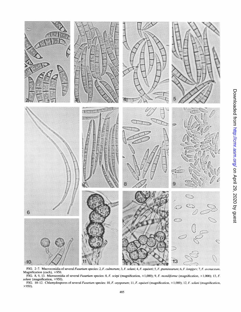
General Characteristics of Fusarium SpeciesFusarium species may produce three types of spores called
macroconidia, microconidia, and chlamydospores (154) (Fig. 2to 13). Some species produce all three types of spores, whileother species do not. The macroconidia are produced in aspecialized structure called a sporodochium in which the sporemass is supported by a superficial cushionlike mass of shortmonophialides bearing the macroconidia (85). The sporodo-chium sometimes may be encased in slime. Macroconidia mayalso be produced on monophialides and polyphialides in theaerial mycelium (Fig. 14 to 20). A monophialide is a condio-phore with only one opening or pore through which en-doconidia are extruded, while a polyphialide has two or moresuch openings or pores. Some conidia are intermediate in sizeand shape, and these have been referred to as both macro-conidia (154) and mesoconidia (168). These intermediateconidia are found primarily in F. semitectum Berk. & Rav., F.avenaceum, and F. subglutinans (Wollenw. & Reinking) Nel-son, Toussoun & Marasas. Until more cultures of each speciesare studied thoroughly, the use of the term mesoconidia isquestionable.
Microconidia are produced in the aerial mycelium but not insporodochia. They may be produced in false heads only or infalse heads and chains (Fig. 21) on either monophialides orpolyphialides. False heads occur when a drop of moistureforms on the tip of the conidiophore and contains the en-doconidia as they are produced. Microconidia are of variousshapes and sizes (Fig. 22), and those produced in chains havea truncate base (Fig. 22). The third type of spore formed byFusarium species is a chlamydospore, which is a thick-walledspore filled with lipidlike material that serves to carry thefungus over winter in soil when a suitable host is not available.The chlamydospores may be borne singly, in pairs, in clumps,or in chains, and the outer wall may be smooth or rough (Fig.10 to 12).
Primary Characters Used To Separate Species inFusarium Taxonomy
Morphology of the macroconidia. The morphology of themacroconidia is the key characteristic for characterization notonly of the species but also of the genus Fusarium. Macro-conidia of Fusarium species are of various shapes and sizes(Fig. 2 to 7), but the shape of the macroconidia formed insporodochia for a given species is a relatively consistent andstable feature when the fungus is grown on natural substratesunder standard conditions (33, 154, 252). Dimensions of themacroconidia may show considerable variation within individ-ual species and should be used cautiously as taxonomic criteria.
Microconidia. The presence or absence of microconidia is aprimary character in Fusarium taxonomy. If microconidia are
present, the features considered are the shape (Fig. 22) and themode of formation, whether it be singly, in false heads only, orin false heads and chains (Fig. 21). The mode of formation ofmicroconidia is best observed in situ on a natural substrateagar medium such as carnation leaf agar (54).
Microconidiophores. The morphology of the conidiophoresbearing the microconidia is a primary taxonomic character.These conidiophores may be either monophialides only (Fig.14 to 17) or both monophialides and polyphialides (Fig. 18 to20) in a given species producing microconidia.
Chlamydospores. The presence or absence of chlamydos-pores is a primary character in Fusanium taxonomy. If chlamy-dospores are present, they may be formed singly, in pairs, inclumps, or in chains, with either rough or smooth walls.
Secondary Characters Useful in Separating Species inFusarium Taxonomy
The following secondary characteristics are useful in de-scribing a species when the cultures are grown under standardenvironmental conditions of light, temperature, and substrate(33, 154) but should not be regarded as suitable criteria fordifferentiation of a species: the morphology and pigmentationof the colony, including the presence or absence ofsporodochia, sclerotia, or stroma. The pigmentation of colo-nies grown on carbohydrate-rich media is variable in somespecies. The pigmentation of the colony may be helpful tosomeone with experience in Fusanium taxonomy but quicklycan lead those without prior experience in this area astray. Thelinear growth rate of the fungus under controlled conditionswas used as a taxonomic characteristic by Booth (28) andothers (33) but must also be used with caution. Isolates withina species may vary considerably with respect to the secondarycharacters. The degree of variation shown by a particularsecondary character may differ between species. Although theshape of the macroconidia formed in sporodochia on carnationleaf agar is a reliable character, the length and width of themacroconidia are less stable features and should be regardedas secondary characters. The macroconidia of a wide range ofisolates of F. culmorum (W. G. Smith) Sacc. are relativelyuniform in length, whereas the length of macroconidia of F.equiseti (Corda) Sacc. varies widely between isolates, evenamong those from the same geographic location.
PROBLEMS IN WORKING WITH FUSARIUM SPECIES
The methods and media used in growing Fusarium speciesfor identification are covered in detail in other publications.The reader is urged to consult Nelson et al. (154), Fisher et al.(54, 55), and Klotz et al. (108) for these details.
Transfer Methods
The single-spore method and the hyphal-tip method are theprimary means used to initiate and transfer cultures of Fusar-ium species. Since each conidium is of a single genotype, thecolony that develops is of that genotype. Mutant cultures thatoccur can be recognized readily. In this manner, clones can bemaintained indefinitely in culture. The hyphal-tip method isused to transfer cultures of Fusarium species that mutaterapidly after being transferred by single conidia. This tech-nique involves the transfer of a single hyphal tip rather than amass transfer from the growing edge of a colony to initiate theculture. The transfer is done under the dissecting microscope,in much the same manner as the transfer of a single germinat-ing conidium.
CLIN. MICROBIOL. REV.
on April 29, 2020 by guest
http://cmr.asm
.org/D
ownloaded from
~~~I
I~4.J½-> - ,' J-.4 7 5)l
K
C
, N.
K,-( ( I; jg
A
-- (, . ¾,K. ,.<¼
6 t.
11~~~~~'~ .,._
/NJ
I ,.
",_
10 .riiQq 6FIG. 2-7. Macroconidia of several Fusarium species: 2, F. culmorum; 3, F. solani; 4, F. equiseti; 5, F graminearum; 6, F longipes; 7, F. avenaceumn.
Magnification (each), X950.FIG. 8, 9, 13. Microconidia of several Fusarium species: 8, F scirpi (magnification, x 1,000); 9, F moniliforme (magnification, x 1,000); 13, F
solani (magnification, x950).FIG. 10-12. Chlamydospores of several Fusarium species: 10, F oxysporum; 11, F equiseti (magnification, x 1,000); 12, F. solani (magnification,
x950).
485
I */
I
A.... .."
C-,
on April 29, 2020 by guest
http://cmr.asm
.org/D
ownloaded from
486 NELSON ET AL.
I.)
b
e.14 15 16 17
.,.i.
9, ~~~..W
iW
r.(.
'F
19
-1 a
20 21
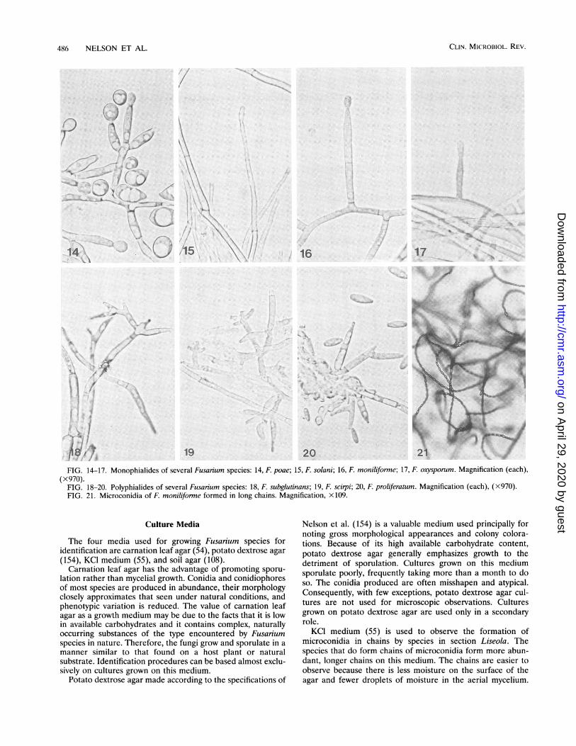
FIG. 14-17. Monophialides of several Fusarium species: 14, F. poae; 15, F. solani; 16, F. moniliforme; 17, F. oxysporum. Magnification (each),(x970).
FIG. 18-20. Polyphialides of several Fusarium species: 18, F. subglutinans; 19, F. scirpi; 20, F. proliferatum. Magnification (each), (X970).FIG. 21. Microconidia of F. moniliforme formed in long chains. Magnification, X109.
Culture Media
The four media used for growing Fusarium species foridentification are carnation leaf agar (54), potato dextrose agar(154), KCl medium (55), and soil agar (108).
Carnation leaf agar has the advantage of promoting sporu-lation rather than mycelial growth. Conidia and conidiophoresof most species are produced in abundance, their morphologyclosely approximates that seen under natural conditions, andphenotypic variation is reduced. The value of carnation leafagar as a growth medium may be due to the facts that it is lowin available carbohydrates and it contains complex, naturallyoccurring substances of the type encountered by Fusariumspecies in nature. Therefore, the fungi grow and sporulate in amanner similar to that found on a host plant or naturalsubstrate. Identification procedures can be based almost exclu-sively on cultures grown on this medium.
Potato dextrose agar made according to the specifications of
Nelson et al. (154) is a valuable medium used principally fornoting gross morphological appearances and colony colora-tions. Because of its high available carbohydrate content,potato dextrose agar generally emphasizes growth to thedetriment of sporulation. Cultures grown on this mediumsporulate poorly, frequently taking more than a month to doso. The conidia produced are often misshapen and atypical.Consequently, with few exceptions, potato dextrose agar cul-tures are not used for microscopic observations. Culturesgrown on potato dextrose agar are used only in a secondaryrole.KCI medium (55) is used to observe the formation of
microconidia in chains by species in section Liseola. Thespecies that do form chains of microconidia form more abun-dant, longer chains on this medium. The chains are easier toobserve because there is less moisture on the surface of theagar and fewer droplets of moisture in the aerial mycelium.
CLIN. MICROBIOL. REV.
II
il
i
on April 29, 2020 by guest
http://cmr.asm
.org/D
ownloaded from
FUSARIUM SPECIES 487
a b c d
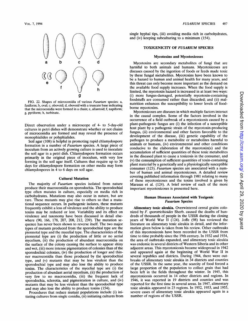
FIG. 22. Shapes of microconidia of various Fusanum species: a,
fusiform; b, oval; c, obovoid; d, obovoid with a truncate base indicatingthat the microconidia were formed in a chain; e, allantoid; f, napiform;g, pyriform; h, turbinate.
Direct observation under a microscope of 4- to 5-day-oldcultures in petri dishes will demonstrate whether or not chainsof microconidia are formed and may reveal the presence ofmonophialides or polyphialides.
Soil agar (108) is helpful in promoting rapid chlamydosporeformation in a number of Fusarium species. A large piece ofinoculum from an actively growing culture is used to inoculatethe soil agar in a petri dish. Chlamydospore formation occurs
primarily in the original piece of inoculum, with very fewforming in the soil agar itself. Cultures that require up to 30days for chlamydospore formation on other media may formchlamydospores in 4 to 6 days on soil agar.
Cultural MutationThe majority of Fusarium species isolated from nature
produce their macroconidia on sporodochia. The sporodochialtype often mutates in culture, especially on media rich incarbohydrates. Mutations may also occur in nature, but arerare. These mutants may give rise to others so that a muta-tional sequence occurs. In pathogenic isolates, these mutantsfrequently exhibit a loss of virulence and the ability to producetoxins may be reduced or lost. Variability and its effect on
virulence and taxonomy have been discussed in detail else-where (90, 166, 178, 207, 208, 212, 239). The mutation se-
quence has never been shown to reverse itself. The two majortypes of mutants produced from the sporodochial type are thepionnotal type and the mycelial type. The characteristics of thepionnotal type are (i) the production of little or no aerialmycelium, (ii) the production of abundant macroconidia on
the surface of the colony causing the surface to appear shinyand wet, (iii) more intense pigmentation of colonies than of thesporodochial colonies, (iv) the production of longer and thin-ner macroconidia than those produced by the sporodochialtype, and (v) mutants that may be less virulent than thesporodochial type and may also lose the ability to producetoxins. The characteristics of the mycelial type are (i) theproduction of abundant aerial mycelium, (ii) the production ofvery few to no macroconidia, (iii) the frequent lack ofsporodochia, sclerotia, and pigmentation in culture, and (iv)mutants that may be less virulent than the sporodochial typeand may also lose the ability to produce toxins (154).
Procedures that reduce mutant populations include (i) ini-tiating cultures from single conidia, (ii) initiating cultures from
single hyphal tips, (iii) avoiding media rich in carbohydrates,and (iv) keeping subculturing to a minimum (154).
TOXIGENICITY OF FUSARIUM SPECIES
Mycotoxins and Mycotoxicoses
Mycotoxins are secondary metabolites of fungi that areharmful to both animals and humans. Mycotoxicoses arediseases caused by the ingestion of foods or feeds made toxicby these fungal metabolites. Mycotoxins have been known tobe a hazard to human and animal health for many years, andthis threat can only become more important as the demand onthe available food supply increases. When the food supply islimited, the mycotoxin hazard is increased in at least two ways:(i) more fungus-damaged, potentially mycotoxin-containingfoodstuffs are consumed rather than discarded; and (ii) mal-nutrition enhances the susceptibility to lower levels of food-borne mycotoxins.
Mycotoxicoses are diseases in which multiple factors interactin the causal complex. Some of the factors involved in theoccurrence of a field outbreak of a mycotoxicosis caused by aplant-pathogenic fungus are (i) the infection of a susceptiblehost plant by a pathogenic strain of the mycotoxin-producingfungus, (ii) environmental and other factors favorable to thedevelopment of the disease, (iii) genetic capability of thepathogen to produce a metabolite or metabolites harmful toanimals or humans, (iv) environmental and other conditionsconducive to the elaboration of the mycotoxin(s) and theaccumulation of sufficient quantities of these toxic metabolitesin the diseased plant to cause a toxicosis in the consumer, and(v) the consumption of sufficient quantities of toxin-containingplant material by a genetically and a physiologically susceptibleconsumer (123). Fusarium species are associated with a num-ber of human and animal mycotoxicoses. A detailed reviewcovering published information through 1981 relating to manyof these mycotoxicoses and the toxins involved is given byMarasas et al. (124). A brief review of each of the moreimportant mycotoxicoses is presented here.
Human Diseases Associated with ToxigenicFusarium Species
Alimentary toxic aleukia. Overwintered cereal grains colo-nized by F. sporotrichioides Sherb. caused the deaths of hun-dreds of thousands of people in the USSR during the closingyears of World War II (124). Joffe (98) has reviewed theliterature on alimentary toxic aleukia in detail, and the infor-mation given below is taken from his review. Other outbreaksof this mycotoxicosis have been recorded in the USSR fromtime to time probably since the 19th century. In 1932 and 1934,the area of outbreaks expanded, and alimentary toxic aleukiawas endemic in several districts of Western Siberia and in otheradjacent areas. This mycotoxicosis became widespread in 1942and appeared again at the beginning of World War II inseveral republics and districts. During 1944, there were out-breaks of alimentary toxic aleukia in 34 districts and countiesof the USSR. In the same year, the scarcity of food forced alarge proportion of the population to collect grain that hadbeen left in the fields throughout the winter. In 1945, thismycotoxicosis occurred in 14 other districts and regions. In1946, it was reported in 19 districts and counties and wasreported for the first time in several areas. In 1947, alimentarytoxic aleukia appeared in 23 regions. In 1952, 1953, and 1955,severe cases of alimentary toxic aleukia appeared again in anumber of regions of the USSR.
VOL. 7, 1994
on April 29, 2020 by guest
http://cmr.asm
.org/D
ownloaded from
488 NELSON ET AL.
This mycotoxicosis occurred in families that had gatheredgrain from fields after the snow had melted in the spring. Thedisease usually appeared after at least 2 kg of food preparedfrom toxic overwintered grain had been consumed. Lesions inthe hematopoietic system were the result of a toxic substanceaccumulating in the body and usually appeared 2 to 3 weeksafter the consumption of toxic grain. Prosomillet (Panicummiliaceum L.) and wheat (Triticum aestivum L.) were found tobe the most toxic grains. People consuming a balanced dietwere less susceptible to the toxin than people who weresuffering from malnutrition. Grain harvested during springthaws was toxic, while grain harvested during autumn or winterbefore the snow melted was either nontoxic or slightly toxic(93).
Overwintered cereal grains colonized by F. sporotrichioidesand F. poae (Peck) Wollenw. are the cause of alimentary toxicaleukia (98, 124). The toxin produced by these organisms iscalled T-2 toxin and was first isolated and characterized byBamburg (14) and Bamberg et al. (15).
Clinically, alimentary toxic aleukia is usually divided intothree stages. If the disease is diagnosed during the first stage,or during the transition from the second to the third stage,early hospitalization and treatment save lives. However, if thedisease is not detected until the third stage, the patient'scondition is usually critical, and death results.
(i) First stage. The symptoms of the first stage appear ashort time after ingestion of toxic grain and include primarychanges in the mouth cavity and gastrointestinal tract. Thepatient feels a burning sensation in the mouth, tongue, throat,palate, esophagus, and stomach since the toxin affects themucous membranes. Inflammation of the gastric and intestinalmucosa results in vomiting, diarrhea, and abdominal pain. Inmost cases, excessive salivation, headache, dizziness, weakness,fatigue, and tachycardia occur, and in some cases fever andsweating occur. The first stage may last for 3 to 9 days.
(ii) Second stage. The second stage is often called the latentstage because the patient feels well and is capable of normalactivity. The main features are disturbances in the hematopoi-etic system characterized by a progressive leukopenia, with agranulocytopenia and a relative lymphocytosis. In addition,there are anemia and a decrease in the platelet count, as wellas a lowering of the patient's resistance to bacterial infections.This stage usually lasts for 3 to 4 weeks but may be as long as8 weeks.
(iii) Third stage. The first visible sign of the third stage is theappearance of petechial hemorrhages on the skin of the trunk,in the axillary and inguinal areas, on the lateral surfaces ofarms and thighs, on the chest, and, in serious cases, on the faceand head. Necrotic changes soon appear in the throat, causingdifficulty and pain on swallowing. Secondary bacterial infec-tions often occur in these necrotic sites, and suppression of thepatient's immune system and the infection may result in deathof the patient.Urov or Kashin-Beck disease. Urov or Kashin-Beck disease
is a chronic disabling, deforming, dystrophic osteoarthosisinvolving the peripheral joints and spine that occurs endemi-cally among the Cossacks in the valley of the Urov River ineastern Siberia as well as in North Korea and northern China(124). The disease begins slowly and often asymptomatically inchildren of preschool or school age. In the early stages,patients experience pain in some of their joints and the jointsbecome thickened. The disease then develops slowly andchronically and is manifested as a shortening of the long bones,thickening and subsequent deformity of the joints, flexorcontractures, and muscular atrophy.
Climatic peculiarities that occurred in areas where this
disease was epidemic were marked temperature changes dur-ing the day and the occurrence of the major portion of therainfall during late summer or early fall when cereals werematuring and grain harvest was in progress. These climaticfactors were conducive to a high level of infection by Fusariumspecies in harvested grains. Considerable experimental evi-dence has been produced in the former USSR that Urovdisease is caused by certain strains of F. poae, but the myco-toxin(s) involved has not been positively identified and theetiology of the disease has not been resolved (124).Akakabi-byo (scabby grain intoxication). In Japan, sporadic
epidemics of akakabi-byo (red mold disease or scab) of wheat,barley, oats, rye, and rice caused by F. graminearum can affectmore than one-third of the national production of thesecereals. Infected cereal grain is frequently associated withoutbreaks of human mycotoxicosis characterized by anorexia,nausea, vomiting, headache, abdominal pain, diarrhea, chills,giddiness, and convulsions. Isolates of F. graminearum fromscabby cereal grains from Japan are known to produce thetrichothecenes deoxynivalenol, nivalenol, fusarenon-X, diace-toxyscirpenol, neosolaniol, and T-2 toxin in culture. An isolateof F. sporotrichioides from scabby wheat from Japan producednivalenol, fusarenon-X, diacetylnivalenol, and T-2 toxin inculture. Scabby cereals in Japan are known to be contaminatedwith deoxynivalenol and nivalenol and may be infected by bothF. graminearum and F. sporotrichioides. It is possible thatsynergistic interactions between deoxynivalenol and other tri-cothecenes may be involved in the human mycotoxicosiscaused by the consumption of scabby cereal grains in Japan.
Evidence exists that the consumption of cereals infected byF. graminearum has resulted in cases of a human mycotoxicosischaracterized by emesis in Japan, Korea, and the formerUSSR. Although deoxynivalenol and nivalenol are known tooccur naturally in scabby cereal grains in Japan, these twotrichothecenes have not been directly implicated in cases ofhuman mycotoxicosis. Thus, it is not known if deoxynivalenoland/or nivalenol is responsible for the clinical signs of scabbygrain intoxication in humans or if other factors also areinvolved (124).
Animal Diseases Associated with Toxigenic Fusarium Species
Hemorrhagic syndrome. Outbreaks of a hemorrhagic syn-drome characterized by bloody diarrhea, necrotic oral lesions,hemorrhagic gastroenteritis, and extensive hemorrhages inmany organs occur sporadically in animals such as cattle, pigs,and poultry in the north central United States and elsewhere.The disease is associated with the ingestion of moldy cereals,particularly corn, and some of the most toxic Fusarium specieshave been isolated from these feeds.
F. sporotrichioides and F. poae are the fungi most oftenassociated with such feeds. These weakly pathogenic fungiinfect the host in the field, develop saprophytically after thedeath of the host, and produce mycotoxins during overwinter-ing in the field and/or during storage of the harvested host, allof which render the diseased plant toxic when consumed.The hemorrhagic syndrome in farm animals and alimentary
toxic aleukia in humans are closely related, if not identical,syndromes. Both are caused by trichothecene mycotoxins, suchas T-2 toxin and diacetoxyscirpenol, produced primarily by F.sporotrichioides (123, 124).
Estrogenic syndrome. In many countries, sporadic fieldoutbreaks of hyperestrogenism in animals, particularly pigs,are caused by the consumption of cereals, particularly corn andbarley, infected by F. graminearum and contaminated with theestrogenic metabolite, zearalenone. Pigs are the most sensitive
CLIN. MICROBIOL. REV.
on April 29, 2020 by guest
http://cmr.asm
.org/D
ownloaded from
FUSARIUM SPECIES 489
animals, and primarily the genitals and reproductive organs areinvolved. In prepuberal gilts, the vulva becomes swollen,hyperemic, and edematous, the mammary glands are swollen,and in severe cases, there may be vaginal and rectal prolapse.True estrus is not commonly observed, but breeding sows mayshow prolonged estrus cycles. Young males may undergo afeminizing effect, with enlargement of the mammary glands,atrophy of the testes, and swelling of the prepuce. In matureboars, a marked decrease in libido may occur. Infertility,reduced litter size, and weak piglets are also manifestations ofthe estrogenic syndrome.
Feed refusal and emetic syndromes. Sporadic field out-breaks of feed refusal by pigs, sometimes associated withvomiting, are caused by the infection of cereals, particularlycorn and barley, by F. graminearum in the midwestern UnitedStates, Japan, and elsewhere. The reduced palatability of thescabby grain is reflected in decreased weight gains and slowergrowth rates of pigs and is associated with nausea and emesisin animals forced by starvation to eat the grain.
It has been established that the mycotoxin deoxynivalenoloccurs in cereals infected by F. graminearum and is associatedwith field outbreaks of feed refusal and emesis in animals,particularly pigs. Deoxynivalenol is found at levels capable ofinducing the characteristic clinical signs of these syndromesunder experimental conditions. However, there is reason tobelieve that deoxynivalenol alone is not responsible for all ofthe feed refusal and emetic activity ofF. graminearum-infectedcereals and that other factors may also be involved (123, 124).
Fescue foot. Winter pastures of tall fescue (Festuca arundi-nacea Schreb.) in the United States, Australia, and NewZealand are associated with sporadic outbreaks of a disease incattle known as fescue foot. This disease is characterized bylameness, loss of weight, arched back, elevated body temper-ature, and dry gangrene involving the hind feet, tail tip, andears, with sloughing of the most distal parts of these extremi-ties. Although the clinical signs of fescue foot are very remi-niscent of the gangrenous form of ergot poisoning, ergotsclerotia apparently are not involved in the disease. The typicalclinical signs of fescue foot have been reproduced experimen-tally in cattle with ethanol extracts of toxic fescue hay.
Several Fusarium species have been isolated from toxic hay.One strain isolated was identified as F. sporotrichioides, and itproduces several mycotoxins in culture. However, since thesemycotoxins have not been shown to occur naturally in toxicfescue hay, the role of F. sporotrichioides in fescue foot of cattleis unknown at present (123).Degnala disease. Degnala disease occurs during winter in
buffaloes and cattle fed almost exclusively on rice straw inlow-lying, waterlogged, rice-growing areas of Pakistan andIndia. The disease is characterized by edematous swelling ofthe legs and necrosis, gangrene, and sloughing of the extrem-ities. The characteristic clinical signs were reproduced inbuffalo calves after feeding them rice straw from farms wherefield outbreaks had occurred. Later, the disease was repro-duced experimentally in buffalo calves fed cultures of an isolateof F. equiseti on rice straw. The organism had been isolatedfrom toxic rice straw, but the mycotoxins produced by thisstrain have not been identified. In addition to F. equiseti, somecultures of F. semitectum also have been isolated from toxicrice straw. At present, it is not certain whether this disease iscaused by F. equiseti or F. semitectum or both. Since the toxinsproduced by these strains have not been identified, the etiologyof this disease is not known at present (123).Moldy sweet potato toxicosis (atypical interstitial pneumo-
nia). Field outbreaks of a fatal respiratory disease of cattle inJapan and the United States have been attributed to the
ingestion of moldy sweet potatoes (Ipomoea batateas L.) forseveral years. This toxicosis is caused by four lung-toxicfuranoterpenoides present in sweet potato tubers infected byF. solani. Affected cattle exhibit severe respiratory distress, arapid respiratory rate, typical extension of the head and neckassociated with dyspnea, and frothy exudate around the mouthbefore death. The disease has been reproduced experimentallyin cattle with sweet potatoes artificially inoculated with F.solani isolated from moldy sweet potatoes associated with afield outbreak of the disease in Georgia. Although it isuncertain at present whether F. solani is involved in etiology ofthe bovine atypical interstitial pneumonia and whether or notthe pulmonary toxins present in moldy sweet potatoes arespecific degradation products of F. solani, the fact remains thatmoldy sweet potatoes cause field outbreaks of respiratorydisease in cattle. Moreover, these potent lung toxins have beenfound naturally in sweet potatoes offered for sale in supermar-kets in the United States, and they are not destroyed by normalcooking procedures. It is evident that the consumption ofmoldy sweet potatoes is potentially dangerous to human andanimal health (123, 124).
F. MONILIFORME
Currently, F. moniliforme is the Fusarium species receivingmost of the attention from research workers studying myco-toxins. F. moniliforne is one of the most prevalent fungiassociated with basic human and animal dietary staples such ascorn (124, 149). This fungus has been suspected of beinginvolved in human and animal diseases since its originaldescription in 1904 (203). In the early 1900s, widespread fieldoutbreaks of a disease in animals associated with the ingestionof moldy corn occurred in the United States (173). Peters (173)reported that the hooves of cattle and horses sloughed, pigsshed their bristles, chickens lost their feathers, some animalsdeveloped convulsions, and a high percentage of affectedanimals died. F. moniliforme was the fungus most commonlyassociated with moldy corn and was implicated as the cause ofthe disease "moldy corn toxicosis" (173). In some areas of theworld, F. moniliforme has been associated with high rates ofhuman esophageal cancer. In southern Africa, the highest rateof human esophageal cancer occurs in the southwestern dis-tricts of the Transkei, where corn is the main dietary staple(131). Several strains of F. monilifonne isolated from cornproduced in these districts have been found to be acutely toxicto ducklings (111). When culture material of these isolatesgrown on autoclaved corn was fed to experimental animals, thelesions induced included cirrhosis and nodular hyperplasia ofthe liver and intraventricular cardiac thrombosis in rats, leu-koencephalomalacia and toxic hepatosis in horses, pulmonaryedema in pigs, nephrosis and hepatosis in sheep, and acutecongestive heart failure in baboons (111, 112). When corn-based feed that was naturally contaminated with F. monili-forme and was associated with outbreaks of equine leukoen-cephalomalacia (LEM) was fed to rats, multiple hepaticnodules and pale depressed hepatic areas resulted. Histologi-cal examination revealed multiple hepatic neoplastic nodulesand large areas of adenofibrosis and cholangiocarcinomas(248). Marasas et al. (122) found that isolate MRC 826 of F.moniliforme grown on autoclaved corn and fed at a dietarylevel of 4% for 286 days and 2% for the rest of the experimentwas hepatocarcinogenic. This material caused hepatocellularcarcinoma in 80% and ductular carcinoma of the liver in 63%of the rats that survived for more than 450 days.
VOL. 7, 1994
on April 29, 2020 by guest
http://cmr.asm
.org/D
ownloaded from
490 NELSON ET AL.
Mycotoxins Produced by F. moniliforme
Moniliformin. The mycotoxin moniliformin was first re-ported to be produced by an isolate of F. moniliforme fromcorn kernels in the United States (45). This isolate lost theability to produce moniliformin during the course of a study todetermine the structure of the toxin (216). Moniliformin waseventually isolated and chemically characterized from a strainofF. moniliforme isolated from millet in Nigeria, and this strainhas been reported to produce large amounts of moniliformin(216). However, the strain from millet recently was identifiedas F. nygamai because it produced chlamydospores (127).
In a study of moniliformin production in Fusarium sectionLiseola (129), it was found that of 58 strains of F. moniliformefrom southern Africa, only 13 that were toxic to ducklingsproduced moniliformin in cultures of autoclaved corn. Theother 45 strains that were toxic to ducklings did not producechemically detectable levels of moniliformin in culture. These45 toxic strains were isolated from corn in Kenya, South Africa,and Transkei.There are conflicting reports in the literature regarding the
production of moniliformin by cultures of F. moniliforme.Overall, only 22% of the toxic strains produced chemicallydetectable levels of moniliformin, and the mean yield was low(129). In particular, strains of F. moniliforme from cornproduced small amounts, and only 6 of 51 strains producedmoniliformin. Cultures that did not produce moniliformininduced LEM in equine animals. Thus, it appears that F.moniliforme generally is a poor producer of moniliformin andthat many toxic strains, in particular, those isolated from corn,do not produce moniliformin and that other species of Fusar-ium, such as F. subglutinans, are better producers of monili-formin.
Fusarins. Assays of isolates of F. moniliforme grown onsterile cracked corn showed that extracts of 21 of 33 isolateswere mutagenic for Salmonella typhimurium TA100 (25). Most(70%) of the isolates from corn and an isolate from sorghumproduced mutagens (25). In 1983, Gelderblom et al. (68), usingthe Salmonella assay, showed that strains of F. moniliformeisolated from Transkeian corn produced mutagenic com-pounds. The Salmonella assay subsequently was used as amonitoring system for isolation of mutagenic compounds fromcultures of F. moniliforme MRC 826. The main mutageniccompound purified from this strain of F. monilifonne wasidentical to fusarin C, a compound independently isolatedfrom a culture ofF. moniliforme by Wiebe and Bjeldanes (244).Because fusarin C is a mutagenic metabolite of F. moniliforme,experiments were carried out to determine the cancer-initiat-ing activity in rats. The results obtained (67) did not supportthe theory that fusarin C is carcinogenic or has a role in thehepatocarcinogenicity of F. moniliforme MRC 826.
Fumonisins. In 1988, Bezuidenhout et al. (20) characterizedthe structures of fumonisins, a new group of mycotoxins thathad been purified from cultures of F. moniliforme. FumonisinsBl, B2, and B3 have been isolated and characterized (20, 66,176, 177). Fumonisin B1 has cancer-promoting activity in rats(66), causes equine leukoencephalomalacia (103, 121), and isassociated with porcine pulmonary edema (82, 194). Fumoni-sin B1 production has been demonstrated for several species inaddition to F. moniliforme and F. proliferatum (Matsushima)Nirenberg from widely separated geographic areas (151, 152).These include F. nygamai Burgess & Trimboli, F. anthophilum(R. Braun) Wollenw., F. dlamini Marasas, Nelson & Toussoun,and F. napiforme Marasas, Nelson & Rabie (151, 152, 224).Research on the effects of the fumonisins on animal andhuman health is continuing.
MYCOTOXICOSES ASSOCIATED WITH THE GROWTHOF F. MONILIFORME ON CORN
Equine LEM
Equine LEM, a neurotoxic disease of horses, donkeys, andmules, is characterized by liquefactive necrotic lesions in thewhite matter of the cerebral hemispheres (124). Equine LEMhas been referred to as the blind staggers, cerebritis, moldycorn disease, leukoencephalitis, corn stalk disease, encephalo-myelitis, foraging disease, and cerebrospinal meningitis (249).This condition, which is seen worldwide, is often characterizedpathologically by liquefactive necrosis of the white matter ofone or both cerebral hemispheres. Typical clinical signs ofintoxication appear abruptly and consist of apathy, somnolentappearance with protruding tongue, reluctance to move back-wards, aimless circling, and ataxia. The signs of nervousdisorder become more pronounced as time passes, and theanimal walks into large stationary objects apparently throughlack of comprehension rather than lack of vision. Finally, theanimal may become extremely excitable and frenzied, andduring this period of delirium it may run wildly into large,stationary objects such as fences. Death may be preceded byrecumbency and paddling limb movements. The course of thedisease from the onset of clinical signs to death can beextremely rapid (less than 7 h), or it may last several days (123).
In early reports, researchers associated LEM with the inges-tion of moldy corn. When equidae were fed corn contaminatedwith F. moniliforne, they died of the disease. In other experi-ments, when corn cultures of pure strains of F. moniliformewere fed to horses, the animals developed LEM (111, 112,121). Marasas et al. (121) showed that, when horses weredosed by stomach tube with culture material of F. moniliformegrown on corn, they developed severe hepatosis and mildedema of the brain. In another experiment, fumonisin B1 wasextracted and purified from culture material of F. moniliforme.A horse injected with purified fumonisin B1 seven times duringthe first 9 days of an experiment developed clinical signs on day8 which included nervousness followed by apathy, a wide-basedstance, trembling, ataxia, reluctance to move, paresis of thelower lip and tongue, and an inability to eat or drink. The horsewas euthanized on the 10th day, and the principal lesions weresevere edema of the brain and early, bilaterally symmetrical,focal necrosis in the medulla oblongata (121). In a laterexperiment, LEM was induced by the oral administration offumonisin Bl. Two horses were dosed and developed nervoussigns such as apathy, changes in temperament, lack of coordi-nation, walking into objects, and paralysis of the lips andtongue. Characteristic lesions of LEM were present in thebrains of both horses (103). These two experiments (103, 121)prove conclusively that fumonisin B1 can induce LEM inhorses.
PPE
Pulmonary edema in swine (PPE) caused by feeding F.moniliforme MRC 826 propagated on corn in bulk was re-ported in 1981 (112). Two pigs dosed by feeding bulk culturematerial in their food ration developed pulmonary edema. Thenext report on PPE appeared in 1990 (82). It describedsimultaneous epizootics on two southwest Georgia farmswhich resulted in the deaths of 34 mature swine. Grosspathological changes observed included extremely markedpulmonary edema and massive hydrothorax. The thoraciccavities were overfilled with golden-yellow liquid. Routinediagnostic testing for toxins and infectious agents failed to
CLIN. MICROBIOL. REV.
on April 29, 2020 by guest
http://cmr.asm
.org/D
ownloaded from
FUSARIUM SPECIES 491
establish an etiology, and these epizootics appeared to repre-sent an unrecognized disease problem (82).To investigate this disease, feed consisting of corn screenings
from the 1989 crop common to both farms where this problemoccurred were used (82). Deaths began about 5 days after thescreenings were first fed and ceased 24 h after screenings wereremoved as the feed source. On day 7 of the feeding study, oneanimal was found dead and a severely dyspneic animal waseuthanized on the same day. At necropsy, both animalsexhibited marked pulmonary edema and hydrothorax previ-ously seen in field cases. Corn screenings were cultured, and F.moniliforme was recovered from samples from both farms.Preliminary data showed that the concentration of fumonisinB1 ranged from 105 to 155 ,ug/g in the two feed samples (82).Additional tests with pure fumonisin B1 and fumonisin B2 weredone on pigs. One pig was injected daily with fumonisin B1 anddied on day 5. This pig developed pulmonary edema andexhibited lesions similar to those observed in field and otherexperimental cases (82).During the 1989 corn harvest season, numerous outbreaks of
PPE were reported and were generally confined to the centralUnited States (194). In almost all cases, feed containing cornand/or corn screenings from the 1989 harvest was implicated asthe causative factor. Because of the 1981 report of a PPE-likesyndrome (112), feed samples were collected for mycologicalevaluation and chemical analyses. Five feed samples associatedwith PPE cases, primarily corn and/or corn screenings, wereobtained from farms in southeastern Iowa. F. moniliforme wasisolated from all samples, and F. proliferatum was isolated fromone sample. The isolates of F. moniliforme from feed producedfumonisin B1 in amounts ranging from 900 to 2,350 ,ug/g andfumonisin B2 in amounts ranging from 120 to 350 ,ug/g whengrown in corn culture. The single isolate of F. proliferatumproduced 1,670 ,ug of fumonisin B1 and 150 ,ug of fumonisin B2per g when grown in corn culture. These data (194) and thedata of Harrison et al. (82) indicate that fumonisin B1 isprobably the cause of PPE.Ross et al. (194) also examined feed samples obtained
during the 1989 corn harvest season. A total of nine feedsamples were obtained from farms in southeastern Iowa: twowere associated with a case of equine LEM, five were associ-ated with cases of PPE, and two samples were not associatedwith animal health problems. All samples were primarily cornand/or corn screenings. F. moniliforme was isolated from allnine samples, and F. proliferatum was isolated from one LEMsample, one PPE sample, and one nonproblem sample. All ofthe isolates of F. moniliforme and F. proliferatum producedfumonisins in corn cultures in amounts ranging from 960 to2,350 ,ug/g, suggesting that the potential exists for fumonisincontamination in any feed containing these two species.
Experimental Liver Cancer
Kriek et al. (111) isolated 21 strains of F. moniliforme (F.verticillioides) from crops of corn in South Africa and theTranskei. These strains did not produce moniliformin, and themajority were toxic to ducklings. Acute mortality was commonin ducklings fed corn culture material of these isolates. In ratsfed this material, the mean time to death was at least 24 dayseven with the most toxic isolate. Cirrhosis and nodular hyper-plasia of the liver and acute and proliferative endocardiallesions with concurrent intraventricular thrombosis were en-countered frequently (111). In a later study (122), culturematerial on corn of F. moniliforne MRC 826 was fed to rats ona lifelong basis. At a dietary level of 8%, culture material washepatotoxic and caused 100% mortality. Hepatic lesions in rats
that died were characterized by cirrhosis, nodular hyperplasia,and bile duct proliferation. At lower dietary levels, culturematerial was hepatocarcinogenic and caused hepatocellularcarcinoma and ductular carcinoma of the liver. No hepatocel-lular or ductular carcinomas occurred in the control animals.Hepatocellular carcinomas in the experimental rats invariablydeveloped in severely cirrhotic livers showing nodular hyper-plasia. Adenofibrosis developed concurrently with hepatocel-lular carcinoma (122).
In other experiments (155, 248), corn that was being fedduring an epizootic of LEM was obtained from a farm in which9 of 15 horses died. Clinical and neuropathological lesionswere consistent with the diagnosis of LEM. The locally growncorn, which was not treated with fungicide, was ground finelyand fed unsupplemented to rats. The predominant speciesrecovered from this corn was F. moniliforme. Analysis of thefeed for aflatoxins at the <0.9-ppb level was negative. Controlanimals euthanized and evaluated on day 176 were free ofsignificant gross lesions. Gross lesions in all test rats necropsiedfrom 123 to 176 days postfeeding were confined to the liver andconsisted of multiple hepatic nodules and pale depressedhepatic areas. Histological examination revealed multiple he-patic neoplastic nodules and large areas of adenofibrosis andcholangiocarcinomas. This study was the first to report on thehepatocarcinogenicity of a sample of equine feed infested withF. moniliforme (155, 248).
Gelderblom et al. (65) fed a corn-based diet containing 50,ug of partially pure (not less than 90%) fumonisin B1 per g,isolated from culture material of F. moniliforme MRC 826, toa group of 25 rats over a period of 26 months. A control groupof 25 rats received the same diet without fumonisin Bl. Theliver was the main target organ in fumonisin B1-treated rats,and the hepatic pathological changes were identical to thosepreviously reported in rats fed culture material of F. monili-forme MRC 826. All fumonisin BI-treated rats that died orwere killed from 18 months onward suffered from a micro- andmacronodular cirrhosis and had large expansile nodules ofcholangiofibrosis at the hilus of the liver. Ten of 15 fumonisinBl-treated rats (66%) that were killed and/or died between 18and 26 months developed primary hepatocellular carcinoma,and metastases to the heart, lungs, or kidneys were present in4 of the rats. No neoplastic changes were observed in any of thecontrol rats. Chronic interstitial nephritis was present in thekidneys of fumonisin B1-treated rats killed after 26 months. Noneoplastic changes were observed in the esophagus, heart, orforestomach of fumonisin B1-treated rats, which was contraryto previous findings when culture material of the fungus wasfed to rats (122). It was concluded that fumonisin B1 isresponsible for the hepatocarcinogenic and the hepatotoxic,but not all of the other toxic, effects of culture material of F.moniliforme MRC 826 in rats (65).
Esophageal Cancer
In Africa, the highest human esophageal cancer rate occursin the southwestern districts of the Transkei, while the rate inthe northeastern region of the Transkei is relatively low (124,130, 131). Corn is the main dietary staple in both areas. In acomparative study of the mycoflora of home-grown cornproduced in the two areas, the most striking and consistentdifference was the significantly high incidence of F. moniliformein corn produced in the area with a high rate of cancer (122,130, 222). Esophageal carcinoma as it occurs in nature has notbeen induced in animals with cultures of F. moniliforme, andconsequently there is no experimental proof of a causativerelationship.
VOL. 7, 1994
on April 29, 2020 by guest
http://cmr.asm
.org/D
ownloaded from
492 NELSON ET AL.
In China, F. moniliforne is one of the fungi most frequentlyassociated with foodstuffs in Lin Xian county in Henan Prov-ince, which is one of the highest-risk areas for esophagealcancer in the world (122). According to Yang (258), cornmealinoculated with isolates of F. moniliforme from Lin Xiancounty has been found to induce tumors in several differentorgans in rats (122). Although carcinoma of the esophagus asit occurs in nature was not observed, the F. moniliforme-infected cornmeal induced epithelial hyperplasia, precancer-ous changes, and papillomas of the esophagus and stomach ofrats and mice (122).At present, it is not possible to postulate a causative role for
F. moniliforme in the etiology of human esophageal cancerbecause there is no direct experimental evidence of a cause-and-effect relationship. However, the finding that the incidenceof F. moniliforme in corn is correlated with the esophagealcancer rate in Transkei (131) and the frequent association ofF.moniliforme with foodstuffs in areas of China (258) with higherrates merit further investigation of the possible role of F.moniliforme in the etiology of human esophageal cancer (122).
Distribution of Fusarium Species That Produce Fumonisins
Much of the early work on production of fumonisins wasdone with F. moniliforme MRC 826. However, recent testinghas shown that other strains of F. moniliforme as well as otherFusarium species also produce these mycotoxins. Thiel et al.(224) tested F. decemcellulare Brick, F. sporotrichioides, F.poae, F. tricinctum (Corda) Sacc., F. avenaceum, F. semitectum,F. camptoceras Wollenw. & Reinking, F. equiseti, F. acumina-tum Ell. & Ev., F. scirpi Lambotte & Fautr., F. longipesWollenw. & Reinking, F. sambucinum Fuckel, F. graminearum,F. reticulatum Mont., F. compactum (Wollenw.) Gordon, F.lateritium, F. moniliforme, F. proliferatum, F. subglutinans, F.anthophilum, F. oxysporum, F. nygamai, and F. napiforme. Ofthe species tested, only F. moniliforme, F. proliferatum, and F.nygamai produced fumonisins. In another study, Nelson et al.(151) tested strains of F. moniliforme from various substratesand geographic areas for production of fumonisin B1. Theytested strains from corn-based feed from the United States;from millet and sorghum in Nigeria and Zimbabwe; from cornkernels from Nepal; from sorghum stalks and grain, corn,sugarcane, and soil from Australia; from good-quality corn foruse in poultry rations in the United States; and from corn silksfrom Iowa and strains associated with mycotic keratitis andvarious types of cancer in humans from the United States andCanada. They considered those strains that produced up to 50ppm of fumonisin B1 to be low producers, those producing 50to 500 ppm to be intermediate producers, and those producing>500 ppm to be high producers. Most of the strains fromcorn-based feed associated with LEM were high producers (16of 20), whereas fewer strains from millet and sorghum werehigh producers (4 of 15) and a number of strains did notproduce the compound. Only one strain from corn kernelsfrom Nepal was a high producer (1 of 10), most strains fromgood-quality corn for poultry rations were high producers (7 of8), and the strains from corn silks were mainly high producers(8 of 9). All strains from sorghum stalks and grain, corn,sugarcane, and soil from Australia were nonproducers (10 of10). Several of the strains from humans (9 of 13) were highproducers.
In another study, Nelson et al. (152) tested species otherthan F. moniliforne, including F. proliferatum, F. subglutinans,F. anthophilum, F. beomiforme Nelson, Toussoun & Burgess, F.dlamini, F. napiforme, and F. nygamai. They found that 17 of 31strains of F. proliferatum were high producers, 1 of 17 strains of
F. anthophilum was a high producer (and 2 of 17 strains werelow producers), 5 of 9 strains of F. dlamini were low tointermediate producers, 5 of 31 strains of F. napiforme werelow to intermediate producers, and 6 of 27 strains of F.nygamai were high producers. F. subglutinans and F. beomi-forme did not produce detectable amounts of fumonisin B1.These results indicate that the potential for production offumonisins in natural substrates and agricultural commoditiesexists in strains from a variety of substrates and geographicareas. Of the species tested, F. moniliforme, F. proliferatum, F.nygamai, and F. napiforme are probably the most importantproducers of fumonisin B1 because of their association withfood grains such as corn, millet, and sorghum and with animalmycotoxicoses such as equine LEM and PPE.
INFECTIONS CAUSED BY FUSARIUM SPECIES
Introduction
As described earlier, Fusarium species are soilborne fungithat are distributed worldwide and are known to be plant,animal, and human pathogens. Fusarium spp. can affect hu-mans by producing either mycotoxicosis or invasive disease.For example, alimentary toxic aleukia is a mycotoxicosis thatcaused the death of hundreds of thousands of people in Russiaat the close of World War II (124). This disease affects thehematopoietic system and develops after the ingestion ofoverwintered cereal grains colonized with F. sporotrichioidesand F. poae (96, 124). The toxin produced by these organismsis called T-2 toxin. Invasive fusariosis in humans requires twofactors for development: exposure to the pathogen and somedegree of local or systemic impairment of the host defenses.Fusarium spp. have been recovered in different areas of all
five continents (28, 32). Although they are often regarded assoilborne, wind and rain could play an important role in thedissemination of these fungi. Circumstantial evidence of winddispersion of Fusarium spores as far as 400 km has beenreported (165). A survey of airborne fungi carried out in theUnited States indicated that Fusarium spp. were more fre-quently present in air samples than were Aspergillus spp. (39).Wind dispersal may explain the isolation of Fusarium spp.
from 17% of throat specimens of 27 nonhospitalized healthyadults, primarily medical school students (44). The presence ofhigh levels of antibodies to extracellular polysaccharides ofmolds in the sera of 125 healthy subjects indicates that healthyhumans are frequently in contact with the fungal extracellularpolysaccharides, presumably by inhalation (157). Fusariumspp. have also been isolated from the flora of the conjunctivalsac (11) with an incidence that correlated with the number ofairborne fungi in the environment.
Disseminated invasive fusariosis was first reported in 1973(42). Since then, several other cases and a few small series ofsystemic fusarial hyalohyphomycosis in immunosuppressedpatients have been reported (1, 6, 8, 26, 63, 106, 132, 137, 140,144, 145, 147, 161, 169, 187, 189, 219, 230, 232, 234, 243, 263,265), with a significant increase being noted recently. Localizedor systemic fusarial infections have been reported from allaround the world (Fig. 23). Over the last 10 years, theFusarium Research Center at The Pennsylvania State Univer-sity, University Park, has received 208 cultures representing 11different Fusarium species isolated from patients with fusarialhyalohyphomycosis.The following will focus on the experimental infection,
pathogenesis, clinical manifestations, treatment, and outcomeof infections caused by Fusarium spp.
CLIN. MICROBIOL. REV.
on April 29, 2020 by guest
http://cmr.asm
.org/D
ownloaded from
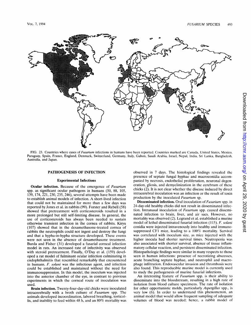
FUSARIUM SPECIES 493
FIG. 23. Countries where cases of Fusarium infections in humans have been reported. Countries marked are Canada, United States, Mexico,Paraguay, Spain, France, England, Denmark, Switzerland, Germany, Italy, Gabon, Saudi Arabia, Israel, Nepal, India, Sri Lanka, Bangladesh,Australia, and Japan.
PATHOGENESIS OF INFECTION
Experimental Infections
Ocular infection. Because of the emergence of Fusariumspp. as significant ocular pathogens in humans (50, 88, 105,139, 174, 221, 230, 235, 246), several attempts have been madeto establish animal models of infection. A short-lived infectionthat could not be maintained for more than a few days wasreported by Jones et al. in rabbits (99). Forster and Rebell (58)showed that pretreatment with corticosteroids resulted in amore prolonged but still self-limiting disease. In general, theuse of corticosteroids has always been needed to sustainotherwise transient infection in the cornea of rabbits. Kiryu(107) showed that in the dexamethasone-treated cornea ofrabbits the neutrophils could not ingest and destroy the fungiand that a hypha-in-hypha structure developed. These eventswere not seen in the absence of dexamethasone treatment.Burda and Fisher (31) developed a fusarial corneal infectionmodel in rats. An increased rate of infectivity was observedwith steroid pretreatment. Finally, O'Day et al. (159) devel-oped a rat model of fulminant ocular infection culminating inendophthalmitis that resembled remarkably that encounteredin humans. F. solani was the infectious agent, and infectioncould be established and maintained without the need forimmunosuppression. In this model, the inoculum was injectedinto the anterior chamber of the eye, in contrast to previousexperiments in which the corneal route of inoculation wasused.
Brain infection. Twenty-four-day-old chicks were inoculatedintracerebrally with a broth culture of Fusarium spp. Theanimals developed incoordination, labored breathing, torticol-lis, and inability to feed within 48 h, and an 80% mortality was
observed in 7 days. The histological findings revealed thepresence of septate fungal hyphae and macroconidia accom-panied by necrosis, endothelial proliferation, neuronal degen-eration, gliosis, and demyelinization in the cerebrum of thesechicks (2). It is not clear whether the disease induced by directintracerebral inoculation was an infection or the result of toxinproduction by the inoculated Fusarium sp.
Disseminated infection. Oral inoculation of Fusarium spp. in24-day-old healthy chicks did not result in disseminated infec-tion. Intranasal inoculation of Fusarium spp. caused dissemi-nated infection to brain, liver, and air sacs. However, nomortality was observed (2). Legrand et al. established a murinemodel of lethal disseminated fusarial infection (115). F. solaniconidia were injected intravenously into healthy and immuno-suppressed CF1 mice, leading to a 100% mortality. Survivalwas correlated with inoculum size, as mice injected with thehigher inocula had shorter survival times. Neutropenia wasalso associated with shorter survival, absence of tissue inflam-matory cellular reaction, and persistent disseminated infection.Histopathologic findings were similar in many respects to thoseseen in human infections: presence of necrotizing abscesses,acute branching septate hyphae, and neutrophil and macro-phage infiltration. Endovascular invasion and thrombosis werealso found. This reproducible murine model is currently usedto study the pathogenesis of murine fusarial infections.An interesting feature of Flusarium spp. is their ability to
disseminate into the bloodstream, resulting in a high rate ofisolation from blood culture specimens. The rate of isolationfor other opportunistic molds, particularly Aspergilluls spp., isvery low (6). In order to understand this phenomenon, ananimal model that would allow frequent sampling of adequatevolumes of blood was needed; hence, a rabbit model of
VOL. 7, 1994
w9
on April 29, 2020 by guest
http://cmr.asm
.org/D
ownloaded from
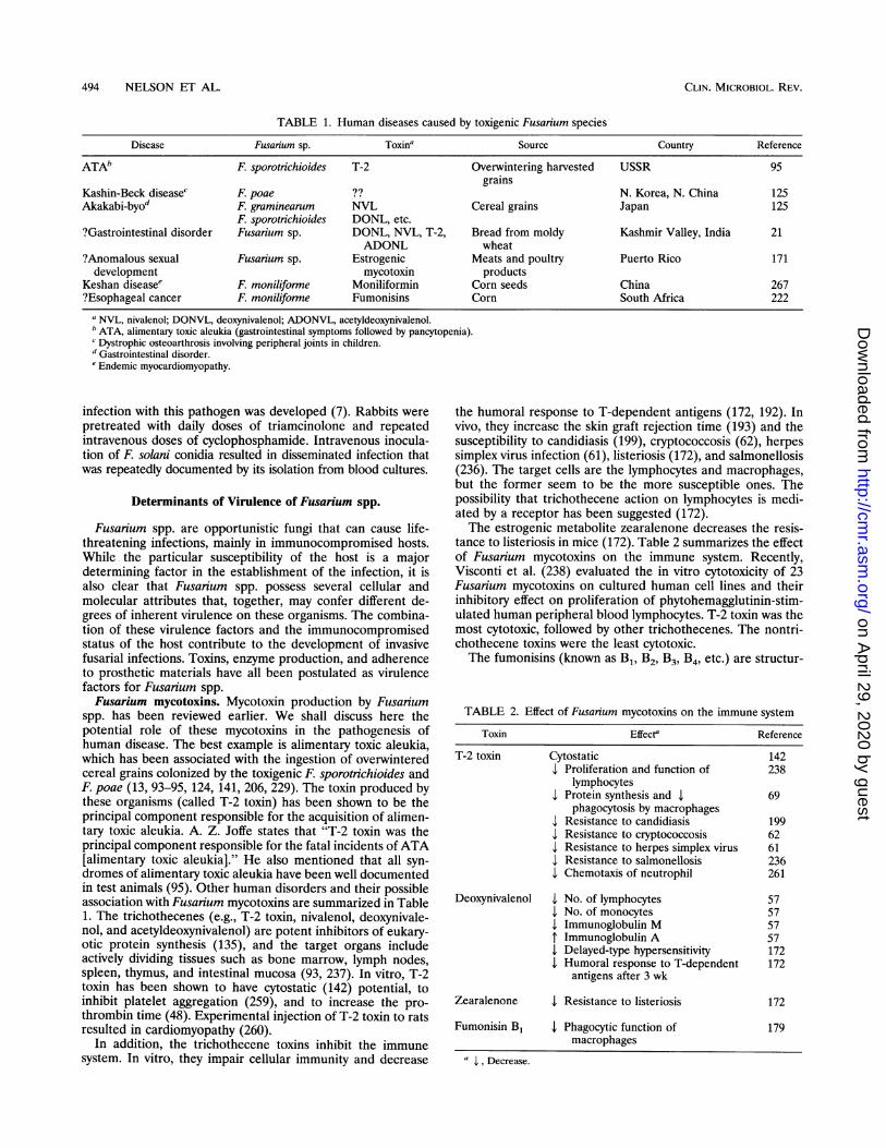
494 NELSON ET AL.
TABLE 1. Human diseases caused by toxigenic Fusarium speciesDisease Fusarium sp. Toxina Source Country Reference
ATAb F. sporotnchioides T-2 Overwintering harvested USSR 95grains
Kashin-Beck diseasec F. poae ?? N. Korea, N. China 125Akakabi-byod F. graminearum NVL Cereal grains Japan 125
F. sporotnichioides DONL, etc.?Gastrointestinal disorder Fusanum sp. DONL, NVL, T-2, Bread from moldy Kashmir Valley, India 21
ADONL wheat?Anomalous sexual Fusarium sp. Estrogenic Meats and poultry Puerto Rico 171development mycotoxin products
Keshan diseasee F. moniliforme Moniliformin Corn seeds China 267?Esophageal cancer F. moniliforme Fumonisins Corn South Africa 222
a NVL, nivalenol; DONVL, deoxynivalenol; ADONVL, acetyldeoxynivalenol.b ATA, alimentary toxic aleukia (gastrointestinal symptoms followed by pancytopenia).c Dystrophic osteoarthrosis involving peripheral joints in children.d Gastrointestinal disorder.eEndemic myocardiomyopathy.
infection with this pathogen was developed (7). Rabbits werepretreated with daily doses of triamcinolone and repeatedintravenous doses of cyclophosphamide. Intravenous inocula-tion of F. solani conidia resulted in disseminated infection thatwas repeatedly documented by its isolation from blood cultures.
Determinants of Virulence of Fusarium spp.
Fusarium spp. are opportunistic fungi that can cause life-threatening infections, mainly in immunocompromised hosts.While the particular susceptibility of the host is a majordetermining factor in the establishment of the infection, it isalso clear that Fusarium spp. possess several cellular andmolecular attributes that, together, may confer different de-grees of inherent virulence on these organisms. The combina-tion of these virulence factors and the immunocompromisedstatus of the host contribute to the development of invasivefusarial infections. Toxins, enzyme production, and adherenceto prosthetic materials have all been postulated as virulencefactors for Fusarium spp.Fusarium mycotoxins. Mycotoxin production by Fusarium
spp. has been reviewed earlier. We shall discuss here thepotential role of these mycotoxins in the pathogenesis ofhuman disease. The best example is alimentary toxic aleukia,which has been associated with the ingestion of overwinteredcereal grains colonized by the toxigenic F. sporotrichioides andF. poae (13, 93-95, 124, 141, 206, 229). The toxin produced bythese organisms (called T-2 toxin) has been shown to be theprincipal component responsible for the acquisition of alimen-tary toxic aleukia. A. Z. Joffe states that "T-2 toxin was theprincipal component responsible for the fatal incidents of ATA[alimentary toxic aleukia]." He also mentioned that all syn-dromes of alimentary toxic aleukia have been well documentedin test animals (95). Other human disorders and their possibleassociation with Fusarium mycotoxins are summarized in Table1. The trichothecenes (e.g., T-2 toxin, nivalenol, deoxynivale-nol, and acetyldeoxynivalenol) are potent inhibitors of eukary-otic protein synthesis (135), and the target organs includeactively dividing tissues such as bone marrow, lymph nodes,spleen, thymus, and intestinal mucosa (93, 237). In vitro, T-2toxin has been shown to have cytostatic (142) potential, toinhibit platelet aggregation (259), and to increase the pro-
thrombin time (48). Experimental injection of T-2 toxin to ratsresulted in cardiomyopathy (260).
In addition, the trichothecene toxins inhibit the immunesystem. In vitro, they impair cellular immunity and decrease
the humoral response to T-dependent antigens (172, 192). Invivo, they increase the skin graft rejection time (193) and thesusceptibility to candidiasis (199), cryptococcosis (62), herpessimplex virus infection (61), listeriosis (172), and salmonellosis(236). The target cells are the lymphocytes and macrophages,but the former seem to be the more susceptible ones. Thepossibility that trichothecene action on lymphocytes is medi-ated by a receptor has been suggested (172).The estrogenic metabolite zearalenone decreases the resis-
tance to listeriosis in mice (172). Table 2 summarizes the effectof Fusarium mycotoxins on the immune system. Recently,Visconti et al. (238) evaluated the in vitro cytotoxicity of 23Fusarium mycotoxins on cultured human cell lines and theirinhibitory effect on proliferation of phytohemagglutinin-stim-ulated human peripheral blood lymphocytes. T-2 toxin was themost cytotoxic, followed by other trichothecenes. The nontri-chothecene toxins were the least cytotoxic.The fumonisins (known as B1, B2, B3, B4, etc.) are structur-
TABLE 2. Effect of Fusanum mycotoxins on the immune system
Toxin Effecte Reference
T-2 toxin Cytostatic 142a Proliferation and function of 238
lymphocytes4 Protein synthesis and 4 69
phagocytosis by macrophages4 Resistance to candidiasis 199v Resistance to cryptococcosis 624 Resistance to herpes simplex virus 614 Resistance to salmonellosis 2364 Chemotaxis of neutrophil 261
Deoxynivalenol 4 No. of lymphocytes 574 No. of monocytes 57
IImmunoglobulin M 574 Immunoglobulin A 574 Delayed-type hypersensitivity 1724 Humoral response to T-dependent 172
antigens after 3 wk
Zearalenone 4. Resistance to listeriosis 172
Fumonisin B1 4 Phagocytic function of 179macrophages
a I, Decrease.
CLIN. MICROBIOL. REV.
on April 29, 2020 by guest
http://cmr.asm
.org/D
ownloaded from
FUSARIUM SPECIES 495
ally similar to the long-chain (sphingoid) base backbones ofsphingolipids and are known to inhibit sphingolipid synthesis(262). This event seems to be the first step in the pathogenesisof cellular toxicity, which may result in brain, pulmonary, andliver damage in animals and cause equine LEM (240), livercancer in rats (156), and PPE (83). Fumonisins were shown tocause significant reduction in the phagocytic potential ofchicken peritoneal macrophages after 4 h of in vitro exposureto the toxin (179).Fusarium toxins could also play an important role in the
pathogenesis of skin infections. Dermal toxicity of Fusariumtoxins T-2, diacetoxyscirpenol, fusarenon, and butenolide wereevaluated individually and in combination on the shaved skinof guinea pigs. Erythema and induration were observed on skinpatches treated with the toxins. These findings correlatedhistologically with the thickening of stratum malpighii, mild tomoderate degeneration of fibrocytes, and cellular infiltration (22).
All Fusarium mycotoxin effects seem to be dose dependentand reversible (83, 238, 250, 262). This raises the possibilitythat toxin production in humans with fusarial infections mayplay a role in the establishment and persistence of fusarialinfections, particularly in patients who are already immuno-compromised. It is important to note, however, that mycotoxinproduction by Fusarium species causing invasive hyalohypho-mycosis in human tissue has not been demonstrated.Enzyme production. Definitive evidence linking enzyme
production to the virulence of Fusarium species is lacking. Invitro production of proteases by F. solani and of collagenasesby F. moniliforme has been documented (100). Whether theseenzymes play a role in the pathogenesis of human infectionsremains to be determined. On the basis of electron microscopyfindings, Kiryu (107) suggested that these enzymes might havea role in the pathogenesis of fusarial keratitis.Adherence to prosthetic material. The ability of Fusarium
species to adhere to silastic catheters has been reported.McNeely et al. (136) were the first to demonstrate by electronmicroscopy that Fusarium species invaded the silastic catheterwall of continuous ambulatory peritoneal dialysis (CAPD)catheters and occluded the catheter. Kerr et al. (104) reportedon a patient with Fusarium peritonitis who was undergoingCAPD. Electron micrographs showed that fungi were adherentto but had not invaded the silastic catheter wall. At ourinstitution, we observed by electron microscopy the invasionand destruction of the wall of a silastic intravenous catheter byF. oxysporum (180). This catheter belonged to a patient withcatheter-related fusarial fungemia.
Contact lenses (especially soft contact lenses with high watercontent) can harbor Fusarium spp. (196). Approximately 14%of soft contact lenses in asymptomatic lens wearers are colo-nized with fungus (175). Laboratory studies have confirmed theability of fungi to adhere, penetrate, and proliferate within theinterior matrix of the contact lens (205, 257). Therefore,Fusarium spp. are able to colonize and sometimes invadeforeign bodies such as contact lenses and silastic catheters, butthe ability to colonize host superficial and organ surfaces hasnot been thoroughly studied. A few reports have mentionedsaprophytic colonization of skin ulcers (114), burn scars (18),and a traumatic wound (158). It is conceivable, however, thatthe ability to colonize tissues could be a major virulence factorfor Fusarium spp.
Host Response to Infection
The interaction of the immune system with F. solani wasexamined in our laboratory. We concluded that granulocytesand macrophages play essential roles in the immune defense
against fusariosis. Macrophages inhibit germination of conidiaand growth of hyphae, while granulocytes inhibit only hyphalgrowth (9).
CLINICAL SPECTRUM AND MANAGEMENT
Foreign-Body-Associated Fusarial Infection
Keratitis in contact lens wearers. Fungi are not part of thenormal ocular flora (247), even during contact lens wear (40),but they frequently contaminate contact lens paraphernalia orthe lens itself (49, 205). Fusarium spp. have been cultured fromsoft contact lenses during use (205). It has been postulated thatafter an initial period of attachment fungal products maydegrade hydrophilic polymers and hence, allow fungal invasionand multiplication within the interior matrix of the lens (205).Fungal keratitis may occur in 4 to 27% of contact lens wearersdepending on the type of lenses (245), and Fusarium spp. havebeen reported in several cases (102, 218, 245).The most frequently noted predisposing factor for keratitis
was improper lens care, which led to contamination of contactlens paraphernalia (245). Aerosolization of conidia, particu-larly in windy conditions, may also lead to contamination of thelens during use. An additional risk factor includes the presenceof a pathologic corneal condition such as a herpes simplexkeratitis, accompanied by a chronic epithelial defect, andconcomitant local corticosteroid and antibacterial medication(245). Removal of the contaminated lens in addition to topicaltreatment with natamycin should be part of the therapeuticregimen. Prevention of contact lens-related keratitis includesavoidance of moldy environments and of nonsterile or reusablecleaning solutions. In addition, handwashing before lens ma-nipulation and frequent cleaning and sterilization of the lensparaphernalia are also recommended (245).
Peritonitis following CAPD. Seven patients with fusarialperitonitis following CAPD were reported from five differentinstitutions (41, 104, 136, 189, 264). The clinical presentationwas insidious, with fever, abdominal pain, and decreasingdrainage from the peritoneal catheter. The peritoneal fluid wascloudy in most of these patients. The outcome was uniformlygood after removal of the catheter alone or in combinationwith antifungal therapy. One patient had persistently positiveFusarium cultures of her dialysate fluid for almost a year andprogressive occlusion of the cannula lumen by the "fungalvegetation" but no clinical symptoms of peritonitis (104).Fungi growing in the lumens of the catheters were eitherplugging the pores of the catheter or invading it.
Catheter-associated fungemia. Catheter-associated funge-mia caused by F. chlamydosporum was reported in a patientwith lymphocytic lymphoma (106). We have also recently caredfor a patient with catheter-related fusarial infection (180).Electron microscope studies of the central venous catheterrevealed plugging of the catheter with masses of fungal hyphaeand also showed invasion and destruction of the catheter wall.
All patients with peritoneal or central venous catheter-associated fusarial infections recovered uneventfully when thecatheter was removed. In cases of bloodstream infections,antifungal chemotherapy was also instituted. Of note is that allof these patients either had a normal peripheral neutrophilcount or had suffered only transient episodes of neutropenia.While these infections cannot be considered localized, theoverall good outcome can be attributed to the lack of tissueinvasion and the presence of a potential removable focus ofinfection.
Given the absence of reports of fusarial infections associatedwith devices made of hard materials (titanium and platinum)
VOL. 7, 1994
on April 29, 2020 by guest
http://cmr.asm
.org/D
ownloaded from
496 NELSON ET AL.
such as artificial heart valves or hip prostheses, and despite thevery large number of patients with such implants, the size orthe adherence properties of Fusarium spp. may not allow themto attach to prostheses made of such hard materials.
Single Organ Invasion
Fusarium keratitis. Fusarium spp. (especially F. solani) arethe most frequent cause of fungal keratitis in the United States(47, 101). Predisposing factors include corneal trauma due toimplantation of vegetable or soil matter in patients engaging inoutdoor activities (139, 235), preexisting allergic conjunctivitis(230), the use of hydrophilic contact lenses (205) for prolongedperiods, and possibly the topical use of corticosteroids andantibacterial eye drop medications (230, 245). The clinicopath-ological features and diagnosis of fusarial keratitis are similarin many regards to those of fungal keratitis caused by otherorganisms. Topical natamycin is the treatment of choice givenits excellent antifusarial activity in vitro (186, 195), its cornealpenetration (160), and its safety profile (191). Silver sulfadia-zine has been shown to be active against fusarial keratitis (230).One case of F. moniliforme keratitis was successfully treatedwith cyclopiroxolamine (50). However, fusarial keratitis isrelatively resistant to treatment with azoles when comparedwith the early and fast response of keratitis caused byAspergil-lus spp. (56, 184, 225). If therapy is delayed, fusarial keratitismay progress to endophthalmitis (174). Fusarial endoph-thalmitis may result from infection initiated from exogenous orendogenous sources. Exogenous sources include extension offungal corneal ulcer (which can precede the development ofendophthalmitis by 2 weeks to 5 months) and surgical ornonsurgical trauma (174). Endogenous infection may resultfrom hematogenous spread of the fungus from another site ofinfection in the body (116). Rapid and accurate diagnosis ofendophthalmitis is essential if vision is to be salvaged, but inmost reported cases patients have had a poor visual outcome(174).
Onychomycosis. Rush-Munro et al. (197) reported on sev-eral cases of onychomycosis caused by F. oxysporum whichtended to cause highly characteristic milky lesions on greattoenails. The earliest sign is a white spot at the base of the nailor an extension proximally of the free border. With moreadvanced infection, the area of nail affected increases, occa-sionally in a patchy fashion but mostly as a regular extension upor down the nail. There may be onychogryposis, and in a fewpatients, the whole toenail becomes opaque, distinctly milkywhite, and thickened at the free border. Ultimately, destruc-tion of part or the entire nail may ensue. While most infectionsare asymptomatic, swelling and tenderness at the base of thenail may occur. Some patients may seek advice for cosmeticreasons. In the reported series, F. oxysponum was more fre-quently encountered than other filamentous fungi. It has beenpostulated that Fusarium spp. invade great toenails after soilcontamination, particularly in people who tend to walk in opensandals or barefooted. Trauma to the great toenail and accu-mulation of debris in the lateral folds will introduce theorganism. Similar findings were noted in a recent series byVelez and Diaz (233). F. oxysporum penetrates and invades thekeratinous part of the nail plate and the hair shaft (241).Although onychomycosis by Fusarium spp. usually behaves as alocalized infection in immunocompetent individuals, it couldalso represent the portal of entry leading to disseminatedfusarial infection in immunocompromised patients (72) (Fig.24). A spreading cellulitis of the great toe is usually present.Treatment includes removal of the infected keratin and topicalor systemic antimycotic therapy (197).
Skin infections. Fusarium spp. have a propensity to invadeskin structures both directly and through the bloodstream.Following initial colonization, localized cutaneous infectionsdevelop in the presence of certain predisposing factors such asexcessive moisture (52, 53), burn (18), trauma (228), andimmunosuppression (19, 52, 263). Several types of clinicallesions have been noted on the skin, including granulomas (19,215), ulcers (114), necrosis (18), pustules (46), vesicles (228),painful nodules (263), mycetomas (17, 86, 188), and pannicu-litis (170).An apparently healthy, 16-year-old Sri Lankan girl devel-
oped extensive subcutaneous granulomatous lesions due to F.oxysporum (12). These lesions had started almost at infancyand had failed to respond to topical therapy. A combination ofketoconazole and fluorocytosine resulted in marked improve-ment. The most recent reported case of mycetoma respondedto long-term administration of ketoconazole (17). During a10-year period, Fusanum and Aspergillus spp. were the mostcommon etiologies of fungal burn wound infections (18).
Otitis. One case of external otitis caused by Fusarium spp.was reported among a series of 83 cases of mycotic externalotitis in Central Africa. Washing of the ear followed by topicaleconazole and amphotericin B was effective in eradicating theinfection (109).Bone and joint infections. The first reported case of Fusar-
ium osteomyelitis is that of a 7-year-old boy who fell andpunctured his knee with a thorn (30). Three weeks later, thediagnosis of fusarial osteomyelitis of the tibia was made. Thepatient recovered completely after surgical drainage and intra-venous amphotericin B therapy. Two other cases of Fusariumosteomyelitis were reported in two healthy patients: followingsurgery in one (167) and trauma in the other (158). Both werereceiving broad-spectrum antibacterial agents at the time ofthe infection. Successful outcome was achieved after surgicaldebridement and local administration of amphotericin B. Inone of these cases (158), a concurrent case of wound coloni-zation by Fusanium spp. was reported in another patient in thesame ward and nosocomial spread was suggested. Septicarthritis caused by Fusanium spp. has been reported in twopatients (81, 91). The infection developed after trauma andresponded to surgical intervention and amphotericin B.
Invasive intranasal infection. Invasive intranasal F. oxyspo-rum infection occurred in a 58-year-old diabetic woman withmetastatic sigmoid adenocarcinoma. The clinical presentationwas suggestive of rhinocerebral zygomycosis. Biopsy and cul-ture of the internasal scar led to the diagnosis (231).
Brain abscess. A patient with chronic mononucleosislikeillness associated with Epstein-Barr virus developed a fatalfusarial brain abscess (217).Pneumonia. A 5-year-old boy with dysmyelopoietic syn-
drome developed F. moniliforne pneumonia after receivinginduction chemotherapy. He was probably exposed to conidiaof Fusarium spp. while harvesting wheat (266). The patientresponded to a total dose of 47 mg of amphotericin B per kgand right lower lobe lobectomy; several other cases of lunginvolvement were reported in the setting of disseminatedfusariosis (26, 243, 265).
Disseminated Multiorgan Infection
Disseminated infections have occurred mainly in cancerpatients undergoing intensive chemotherapy or bone marrowtransplantation. Since the first description of disseminatedfusarial infection in an immunocompromised host in 1973 (42),a significant increase in the incidence of disseminated fusarialinfections has been recognized in humans.
CLIN. MICROBIOL. REV.
on April 29, 2020 by guest
http://cmr.asm
.org/D
ownloaded from
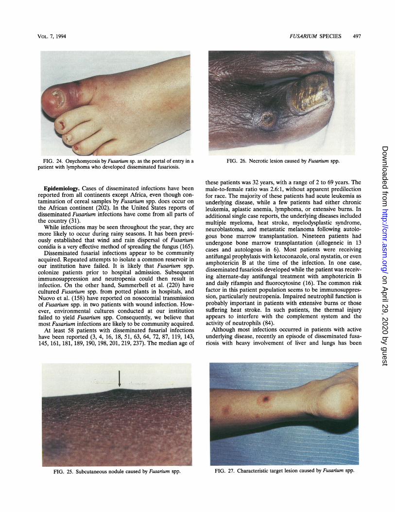
FUSARIUM SPECIES 497
FIG. 24. Onychomycosis by Fusarium sp. as the portal of entry in apatient with lymphoma who developed disseminated fusariosis.
Epidemiology. Cases of disseminated infections have beenreported from all continents except Africa, even though con-tamination of cereal samples by Fusarium spp. does occur onthe African continent (202). In the United States reports ofdisseminated Fusarium infections have come from all parts ofthe country (31).While infections may be seen throughout the year, they are
more likely to occur during rainy seasons. It has been previ-ously established that wind and rain dispersal of Fusariumconidia is a very effective method of spreading the fungus (165).
Disseminated fusarial infections appear to be communityacquired. Repeated attempts to isolate a common reservoir inour institution have failed. It is likely that Fusarium spp.colonize patients prior to hospital admission. Subsequentimmunosuppression and neutropenia could then result ininfection. On the other hand, Summerbell et al. (220) havecultured Fusarium spp. from potted plants in hospitals, andNuovo et al. (158) have reported on nosocomial transmissionof Fusarium spp. in two patients with wound infection. How-ever, environmental cultures conducted at our institutionfailed to yield Fusarium spp. Consequently, we believe thatmost Fusanum infections are likely to be community acquired.At least 58 patients with disseminated fusarial infections
have been reported (3, 4, 16, 18, 51, 63, 64, 72, 87, 119, 143,145, 161, 181, 189, 190, 198, 201, 219, 237). The median age of
FIG. 26. Necrotic lesion caused by Fusarium spp.
these patients was 32 years, with a range of 2 to 69 years. Themale-to-female ratio was 2.6:1, without apparent predilectionfor race. The majority of these patients had acute leukemia asunderlying disease, while a few patients had either chronicleukemia, aplastic anemia, lymphoma, or extensive burns. Inadditional single case reports, the underlying diseases includedmultiple myeloma, heat stroke, myelodysplastic syndrome,neuroblastoma, and metastatic melanoma following autolo-gous bone marrow transplantation. Nineteen patients hadundergone bone marrow transplantation (allogeneic in 13cases and autologous in 6). Most patients were receivingantifungal prophylaxis with ketoconazole, oral nystatin, or evenamphotericin B at the time of the infection. In one case,disseminated fusariosis developed while the patient was receiv-ing alternate-day antifungal treatment with amphotericin Band daily rifampin and fluorocytosine (16). The common riskfactor in this patient population seems to be immunosuppres-sion, particularly neutropenia. Impaired neutrophil function isprobably important in patients with extensive burns or thosesuffering heat stroke. In such patients, the thermal injuryappears to interfere with the complement system and theactivity of neutrophils (84).Although most infections occurred in patients with active
underlying disease, recently an episode of disseminated fusa-riosis with heavy involvement of liver and lungs has been
I
FIG. 27. Characteristic target lesion caused by Fusanum spp.
VOL. 7, 1994
4
FIG. 25. Subcutaneous nodule caused by Fusayium spp.
on April 29, 2020 by guest
http://cmr.asm
.org/D
ownloaded from
498 NELSON ET AL.
reported in an apparent healthy farmer, although no immuno-logical studies were done (119).The species of Fusarium implicated in disease include F.
solani in almost half of the cases, followed by F. oxysporum andF. moniliforme. Several other species have also caused infec-tion.
Pathogenesis. Two modes of acquisition of the infectionseem likely: (i) respiratory, following inhalation of conidia asevidenced by the sinopulmonary involvement in several pa-tients (63); and (ii) cutaneous, as in cases following cellulitis ofthe toe or finger, in catheter-related infections, and in patientswith burns (72, 106, 243). The prevalence of Fusarium spp. ingrain foods and the involvement of the gastrointestinal tract insome cases raise the possibility, though unlikely, of a gastro-intestinal route of infection (7, 119). Thus far, Fusarium spp.are the only opportunistic molds that can be easily recoveredfrom the bloodstream (7, 137). Usually, patients with positiveblood cultures tend to have concomitant skin lesions. It hasbeen speculated that the toxins produced by Fusarium spp. mayenhance the breakdown of tissues and thus facilitate entry offusaria into the systemic circulation (137). It is also possiblethat the growth of Fusarium spp. may not be inhibited by bloodcomponents in cultures as are Aspergillus spp. (137). These andother hypotheses are being examined in a rabbit model ofsystemic fusarial infection in which the organism can beregularly isolated from the bloodstream (7).
Histopathologically, fusarial infections may mimic any num-ber of other mycoses exhibiting moniliaceous, septate, branch-ing or nonbranching hyphae. Because of these morphologicsimilarities, identification of the fungus obtained from culturesis required to establish a fusarial etiology (6). Immunohisto-logical staining has been successfully used to diagnose tissueinvasion (161). Like Aspergillus spp., Fusarium spp. have apropensity for vascular invasion, resulting in thrombosis andtissue necrosis.
Clinical picture. The disseminated infection by Fusariumspp. usually presents as persistent fever unresponsive to broad-spectrum antibiotics in a profoundly neutropenic cancer pa-tient (6). The infection can involve almost any organ. Sinusitisand/or rhinocerebral infection, endophthalmitis, or pyomyosi-tis may be the presenting problem. In the proper setting ofimmunosuppression, several findings should raise the index ofsuspicion for Fusarium infection. These include a preceding orconcomitant toe or finger cellulitis, the presence of cutaneousor subcutaneous lesions, and the growth of a mold from ablood culture. Three types of cutaneous lesions can be ob-served: multiple erythematous subcutaneous nodules (Fig. 25),which are very suggestive of Fusarium infection in this setting;painful erythematous maculas and papules with progressivecentral infarction (Fig. 26) similar to the ecthyma-gangreno-sum-like lesions observed with disseminated aspergillosis; andtarget lesions consisting of ecthyma-gangrenosum-like lesionssurrounded by a thin rim of erythema (Fig. 27). The targetlesion is only occasionally observed, but it has not beenassociated with other opportunistic mycoses and could thus bea useful diagnostic clue. Extensive cellulitis of the face or of theextremities with or without fascitis may also occur.The high (60%) rate of isolation of Fusarium spp. from the
bloodstream is in sharp contrast to the rare (<5%) isolation ofAspergillus spp. or other opportunistic molds in similar clinicalconditions. Similarly, more than two-thirds of patients withdisseminated Fusarium infection have skin lesions comparedwith the rare occurrence (<10%) of such lesions in dissemi-nated aspergillosis (6, 137).Upon recovery from myelosuppression, the infection may
either resolve completely or become chronic and localized to
sinuses, lungs, eye, brain, joint, or muscle (237), with thepotential for relapse and dissemination upon reinstitution ofcytotoxic chemotherapy (137). A small subset of patients withhlematogenous infection but without organ involvement hasbeen observed (6). These patients have had a successfuloutcome when antifungal therapy was promptly instituted andthe central venous catheter was removed. Whether responsewas related to the short duration of neutropenia or to thetherapeutic measures taken remains to be determined.Outcome. The status of the host and the extent of the
infection are the most important factors predicting the out-come of Fusarium infections. Among 58 episodes of dissemi-nated fusarial infection that developed in 53 patients (3, 4, 16,51, 63, 64, 72, 87, 143, 145, 189, 190, 201, 219, 237), 17 had asuccessful outcome. The improvement in 16 of these patientswas associated with the recovery of the bone marrow function.Only one neutropenic patient appeared to have cleared theinfection while still neutropenic following chemotherapy andautologous bone marrow transplant. The infection had devel-oped while the patient had been receiving alternate-day anti-fungal treatment with amphotericin B and daily rifampin andfluorocytosine. Response was achieved after 6 weeks of am-photericin B, rifampin, granulocyte-macrophage colony-stimu-lating factor, and granulocyte transfusions while the neutrophilcount remained below 0.25 x 10⁹ per liter (16). Hence,successful outcome is related to the control of the underlyingdisease and recovery from neutropenia. Age, sex, dose, andduration of intravenous amphotericin B, presence or absenceof positive blood cultures or skin lesions, and the Fusarium spp.involved are all factors that appear to be significantly lessrelated to outcome than host immunity. Surgical resection ofinfected tissue, when possible, may be an important compo-nent of therapy. Limited experimental and clinical experiencesuggests a therapeutic benefit from the antifungal triazole,SCH39304, which is no longer available (10). Hence, optimaltherapy remains to be determined. Given the overall grimprognosis for patients with disseminated infection, effectivepreventive measures are urgently needed. Infection controlpolicies aimed at reducing the number of airborne fungalconidia in the hospital setting should be properly implemented.Prophylaxis with novel antifungal agents that have good activ-ity against Fusarium spp. and shortening the duration ofneutropenia with colony-stimulating factors (43) may alsoprove useful for preventing these devastating infections. Fusar-ium onychomycosis should be treated aggressively, includingnail removal in those patients with hematologic malignanciesundergoing treatment with cytotoxic chemotherapy with orwithout bone marrow transplantation. This approach coulddecrease the risk of developing disseminated Fusarium infec-tions during the periods of neutropenia.
Susceptibility of Fusarium Species to Antifungal Agents
In vitro, the inhibitory activity by miconazole, itraconazole,and fluorocytosine for Fusarium isolates is poor (186). Ampho-tericin B and natamycin are the most active agents. However,in vitro susceptibility or resistance to these antifungal agentsmay not predict the clinical outcome of Fusarium infection. Inour model of murine systemic Fusarium infection, amphoteri-cin B at a dose of 1 mg/kg/day was unable to alter an ultimatelyfatal outcome when the treatment was commenced 1 h afterinoculation of a clinical isolate of F. solani (5).
CLIN. MICROBIOL. REV.
on April 29, 2020 by guest
http://cmr.asm
.org/D
ownloaded from
FUSARIUM SPECIES 499
CONCLUSION
The two most important taxonomic systems for Fusaniumspecies are those of Wollenweber and Reinking and Snyderand Hansen. Other systems that have been used are comparedwith these two. At present, one must rely on morphologicalcharacters for taxonomy, but this should change in the next 5 to10 years as molecular techniques are applied to the taxonomyof this important genus. Problems such as cultural mutationare discussed along with the difficulties presented by thisphenomenon. Several Fusarium species are toxigenic, and themycotoxins and mycotoxicoses produced by these fungi presenta potential health problem. Many of the toxins produced bythese fungi are not yet characterized chemically. Toxins pro-duced by F. moniliforne are discussed separately because agroup of mycotoxins produced by this fungus are found readilyin corn and are carcinogenic in rats. They pose a potentialthreat to human health.Human infections by Fusarium species can be superficial or
limited to single organs in otherwise healthy patients. Suchinfections are rare and tend to respond well to therapy. Bycontrast, disseminated fusarial hyalohyphomycosis hasemerged as a significant and usually fatal infection in theimmunocompromised host. Successful outcome is determinedby the degree of immunosuppression and the extent of theinfection. These infections may be clinically suspected on thebasis of constellation of clinical and laboratory findings, whichshould lead to prompt therapy, probably with one of the newerantifungal agents. Perhaps the use of such agents or the use ofcolony-stimulating factors may improve the outcome of thisdevastating infection. However, until new approaches fortreatment develop, effective preventive measures are urgentlyneeded.
REFERENCES1. Abramowsky, C. R., D. Quinn, W. D. Bradford, and N. F. Conant.
1974. Systemic infection by Fusarium in a burned child. J.Pedriatr. 84:561-564.
2. Adetosoye, A. I., and D. F. Adene. 1978. The response of chickensto experimental infection with Fusanium spp. Bull. Anim. HealthProd. Afr. 26:198-200.
3. Agamanolis, D. P., D. K. Kalwinsky, C. E. Krill, Jr., S. Dasu, B.Halasa, and P. G. Galloway. Fusarium meningoencephalitis in achild with acute leukemia. Neuropediatrics 22:110-112.
4. Alvarez-Franco, M., M. Reyes-Mujica, and A. S. Paller. 1992.Cutaneous Fusarium infection in an adolescent with acute leu-kemia. Pediatr. Dermatol. 9:62-65.
5. Anaissie, E., R. Hachem, C. Legrand, P. Legenne, P. Nelson, andG. Bodey. 1992. Lack of activity of amphotericin B in systemicmurine fusarial infection. J. Infect. Dis. 165:1155-1157.
6. Anaissie, E., H. Kantarjian, P. Jones, B. Barlogie, M. Luna, G.Lopez-Berenstein, and G. Bodey. 1986. Fusarium, a newly recog-nized fungal pathogen in immunosuppressed patients. Cancer57:2141-2145.
7. Anaissie, E., C. Legrand, R. Hachem, P. Nelson, and G. Bodey.1990. Recovery of Fusarium spp. from the bloodstream using arabbit model of systemic fusariosis, abstr. F-28, p. 413. Abstr.90th Annu. Meet. Am. Soc. Microbiol. 1990. American Societyfor Microbiology, Washington, D.C.
8. Anaissie, E., P. Nelson, M. Beremand, D. Kontoyannis, and M.Rinaldi. 1992. Fusarium caused hyalohyphomycosis, an overview.Curr. Top. Med. Mycol. 4:231-249.
9. Anaissie, E. J., S. C. Bhalla, S. E. Vartivarian, J. H. Rex, E.Gunter, P. Bhatnagar, and K. Mehta. Unpublished data.
10. Anaissie, E. J., D. P. Kantoyiannis, S. Vartivarian, H. M.Kantarjian, S. O'Brien, S. A. Giralt, S. Anderson, C. Karl, R. E.Champlin, and G. Bodey. 1993. Effectiveness of an oral triazolefor opportunistic mold infections in patients with cancer: experi-ence with SCH 39304. Clin. Infect. Dis. 17:1022-1031.
11. Ando, N., and K. Takatori. 1982. Fungal flora of the conjunctival
sac. Am. J. Ophthalmol. 94:67-74.12. Attapatta, M. C., and C. Anandakrishnan. 1986. Extensive
subcutaneous hyalohyphomycosis caused by Fusarium oxysporum.J. Med. Vet. Mycol. 24:105-111.
13. Austwick, P. K C. 1984. Fusarium infections in man and animals,p. 129-140. In M. 0. Moss and E. Smith (ed.), The appliedmycology of Fusarium. Cambridge University Press, Cambridge.
14. Bamberg, J. R. 1968. Mycotoxins of the trichothecene familyproduced by cereal molds. Ph.D. thesis. University of Wisconsin,Madison.
15. Bamberg, J. R., W. F. 0. Marasas, N. V. Riggs, E. B. Smalley, andF. M. Strong. 1968. Toxic spiro-epoxy compounds from fusariaand other Hyphomycetes. Biotechnol. Bioeng. 10:445-455.
16. Barrios, N. J., D. V. Kirkpatrick, A. Murciano, K. Stine, R. B.Van Dyke, and J. Humbert. 1990. Successful treatment of dis-seminated Fusarium infection in an immunocompromised child.Am. J. Pediatr. Hematol. Oncol. 12:319-324.
17. Baudraz-Rosselet, F., M. Monod, L. Borradori, J. M. Ginalsky,B. Vion, C. Boccard, and E. Frenk. 1992. Mycetoma of the footdue to Fusarium sp. treated with oral ketoconazole. Dermatology184:303-305.
18. Becker, W. K., W. G. Cioffi, A. T. McManus, S. H. Kim, W. F.McManus, A. D. Mason, and B. A. Pruitt, Jr. 1991. Fungal burninfection. A 10-year experience. Arch. Surg. 126:44-48.
19. Benjamin, R. P., J. L. Callaway, N. F. Conant, and N. Durham.1970. Facial granuloma associated with Fusarium infection. Arch.Dermatol. 101:598-600.
20. Bezuidenhout, S. C., W. C. A. Gelderblom, C. P. Gorst-Allman,R. M. Horak, W. F. 0. Marasas, G. Spiteller, and R. Vleggaar.1988. Structure elucidation of fumonisins, mycotoxins fromFusarium moniliforme. J. Chem. Soc. Chem. Commun. 1988:743-745.
21. Bhat, R. V., S. R. Beedu, Y. Ramakrishna, and K. L. Munshi.1989. Outbreak of trichothecene mycotoxicosis associated withconsumption of mould-damaged wheat production in KashmirValley, India. Lancet i:35-37.
22. Bhavanishankar, T. N., H. P. Ramesh, and T. Shantha. 1988.Dermal toxicity of Fusarium toxins in combinations. Arch. Toxi-col. 61:241-244.
23. Bilai, V. I. 1955. The fusaria (biology and systematics). AkademiaNaukwe Ukrainskii, SSR, Kiev.
24. Bilai, V. L. 1970. Experimental morphogenesis in the fungi of thegenus Fusarium and their taxonomy. Ann. Acad. Sci. Fenn. Ser.A IV Biol. 168:7-18.
25. Bjeldanes, L. F., and S. V. Thomson. 1979. Mutagenic activity ofFusarium moniliforme isolates in the Salmonella typhimuriumassay. Appl. Environ. Microbiol. 37:1118-1121.
26. Blazar, B. R., D. D. Hurd, D. C. Snover, J. W. Alexander, andP. B. McGlove. 1984. Invasive Fusarium infections in bonemarrow transplant recipients. Am. J. Med. 77:645-651.
27. Bolkan, H. A., J. C. Dianese, and F. P. Cupertino. 1979. Pineap-ple flowers as principal infections sites for Fusarium moniliformevar. subglutinans. Plant Dis. Rep. 63:655-657.
28. Booth, C. 1971. The Genus Fusarium, p. 237. CommonwealthMycology Institute, Kew, Surrey, England.
29. Booth, C. 1981. Perfect states (teleomorphs) of Fusanium species,p. 446-452. In P. E. Nelson, T. A. Toussoun, and R. J. Cook (ed.),Fusarium: diseases, biology, and taxonomy. Pennsylvania StateUniversity Press, University Park.
30. Bourguinon, R. L., A. F. Walsh, J. C. Flynn, C. Baro, and E.Spinos. 1976. Fusanium species osteomyelitis-a case report. J.Bone Joint Surg. Am. Vol. 58:722-723.
31. Burda, C. D., and E. Fisher. 1959. The use of cortisone inestablishing experimental fungal keratitis in rats. Am. J. Ophthal-mol. 48:330-335.
32. Burgess, L. W. 1981. General ecology of the fusaria, p. 225-235.In P. E. Nelson, T. A. Toussoun, and R. J. Cook (ed.), Fusanium:diseases, biology, and taxonomy. Pennsylvania State UniversityPress, University Park.
33. Burgess, L. W., C. M. Liddell, and B. A. Summerell. 1988.Laboratory manual for Fusarium research, 2nd ed. University ofSydney, Sydney, Australia.
34. Burgess, L. W., P. E. Nelson, and T. A. Toussoun. 1982. Charac-
VOL. 7, 1994
on April 29, 2020 by guest
http://cmr.asm
.org/D
ownloaded from
500 NELSON ET AL.
terization, geographic distribution and ecology of Fusariumcrookwellense sp. nov. Trans. Br. Mycol. Soc. 79:497-505.
35. Burgess, L. W., P. E. Nelson, T. A. Toussoun, and G. A. Forbes.1988. Distribution of Fusarium species in sections Roseum,Arthrosporiella, Gibbosum, and Discolor recovered from grass-land, pasture, and pine nursery soils of eastern Australia. Myco-logia 80:815-824.
36. Burgess, L. W., P. E. Nelson, T. A. Toussoun, and W. F. 0.Marasas. 1985. Fusanum scirpi: emended description and noteson geographic distribution. Mycologia 77:212-218.
37. Burgess, L. W., and D. Trimboli. 1986. Characterization anddistribution of Fusarium nygamai, sp. nov. Mycologia 78:223-229.
38. Burgess, L. W., A. H. Wearing, and T. A. Toussoun. 1975. Surveysof Fusaria associated with crown rot of wheat in eastern Austra-lia. Aust. J. Agric. Res. 26:791-799.
39. Caplin, I., and D. L. Unger. 1983. Molds on the southernCalifornia deserts. Ann. Allergy 50:260-263.
40. Charles, A. M., M. Callender, and T. Grosvenor. 1973. Efficacy ofchemical asepticizing system for soft contact lenses. Am. J.Ophthalmol. 50:777.
41. Chiaradia, V., D. Schinella, L. Pascoli, and G. F. Santini. 1990.Fusarium peritonitis in peritoneal dialysis: report of two cases.Microbiologica 13:77-78.
42. Cho, C. T., T. S. Vats, J. T. Lowman, J. W. Brandsberg, and F. E.Tosh. 1973. Fusarium solani infection during treatment for acuteleukemia. J. Pediatr. 83:1028-1031.
43. Clark, S. C. 1987. The human hematopoietic colony-stimulatingfactors. Science 236:1229-1237.
44. Cohen, R., F. J. Roth, E. Delgado, D. G. Ahearn, and M. H.Kalser. 1969. Fungal flora of the normal human small and largeintestine. N. Engl. J. Med. 280:638-641.
45. Cole, R. J., J. W. Kirksey, H. G. Cutler, B. L. Doupnik, and J. C.Peckham. 1973. Toxin from Fusarium moniliforne: effects onplants and animals. Science 179:1324-1326.
46. Collins, M. S., and M. G. Rinaldi. 1977. Cutaneous infection inman by Fusarium moniliforme. Sabouraudia 15:151-160.
47. DeVoe, A. G., and M. Silva-Hutner. 1972. Fungal infections of theeye, p. 208-240. In D. Locatcher-Khorazo and B. C. Seegal (ed.),Microbiology of the eye. C. V. Mosby, St. Louis.
48. Doerr, J. A., W. E. Huff, H. T. Tung, R. D. Wyatt, and P. B.Hamilton. 1974. A survey of T-2 toxin, ochratoxin and aflatoxinfor their effects on the coagulation of blood in young broilerchickens. Poult. Sci. 53:1728-1734.
49. Donzis, P. B., B. J. Mondino, B. A. Weissman, and D. A.Bruckner. 1987. Microbial contamination of contact lens caresystems. Am. J. Ophthalmol. 104:325.
50. Duran, J. A., A. Malvar, and M. Pereiro. 1989. Fusarium monili-forme keratitis. Acta Ophthalmol. 67:710-713.
51. El-Ani, A. 1990. Disseminated infection caused by Fusanumsolani in a patient with aplastic anemia. N. Y. State J. Med.90:609-610.
52. English, M. 1968. Invasion of the skin by filamentous non-dermatophyte fungi. Br. J. Dermatol. 80:282-286.
53. English, M. P., R. J. Smith, and R. R. M. Harman. 1971. Thefungal flora of ulcerated legs. Br. J. Dermatol. 84:567-581.
54. Fisher, N. L., L. W. Burgess, T. A. Toussoun, and P. E. Nelson.1982. Carnation leaves as a substrate and for preserving Fusariumspecies. Phytopathology 72:151-153.
55. Fisher, N. L., W. F. 0. Marasas, and T. A. Toussoun. 1983.Taxonomic importance of microconidial chains in Fusariumsection Liseola and effects of water potential on their formation.Mycologia 75:693-698.
56. Fitzsimons, R., and A. L. Peters. 1986. Miconazole and ketocon-azole as a satisfactory first-line treatment for keratomycosis. Am.J. Ophthalmol. 101:605-608.
57. Forsell, J. H., M. F. Witt, J. H. Tai, R. Jensen, and J. J. Pestka.1986. Effects of 8-week exposure of the B6C3F1 mouse to dietarydeoxynivalenol (vomitoxin) and zearalenone. Food Chem. Toxi-col. 24:213.
58. Forster, R. K., and G. Rebell. 1975. Animal model of Fusariumsolani keratitis. Am. J. Ophthalmol. 79:510-515.
59. Francis, R. G., and L. W. Burgess. 1975. Surveys of fusaria andother fungi associated with stalk rot of maize in eastern Australia.
Aust. J. Agric. Res. 26:801-807.60. Francis, R. G., and L. W. Burgess. 1977. Characteristics of two
populations of Fusarium roseum 'Graminearum' in eastern Aus-tralia. Trans. Br. Mycol. Soc. 68:421-427.
61. Friend, S. C. E., L. A. Babiuk, and H. B. Schiefer. 1983. The effectof dietary T-2 toxin on the immunological function and herpessimplex reactivation in Swiss mice. Toxic. Appl. Pharmacol.69:234.
62. Fromentin, H., S. Salazar-Mejicanos, and F. Mariat. 1981.Experimental cryptococcosis in mice treated with diacetoxyscir-penol, a mycotoxin of Fusarium. Sabouraudia 19:311.
63. Gamis, A. S., T. Gudnason, G. S. Giebink, and N. K. Ramsay.1991. Disseminated infection with Fusarium in recipients of bonemarrow transplantation. Rev. Infect. Dis. 13:1077-1088.
64. Garcia, J., and W. Schell. 1990. Fatal dissemination and resolu-tion of Fusarium solani infection in cancer patients followingchemotherapy, p. 410, abstr. F-13. Abstr. 90th Annu. Meet. Am.Soc. Microbiol. 1990. American Society for Microbiology, Wash-ington, D.C.
65. Gelderblom, W. C., N. P. Kriek, W. F. 0. Marasas, and P. G.Thiel. 1991. Toxicity and carcinogenicity of the Fusarium monili-forme metabolite, fumonisin B1 in rats. Carcinogenesis 12:1247-1251.
66. Gelderblom, W. C. A., K. Jaskiewicz, W. F. 0. Marasas, P. G.Thiel, R. M. Horak, R. Vleggaar, and N. P. J. Krielk 1988.Fumonisins: novel mycotoxins with cancer-promoting activityproduced by Fusarium moniliforme. Appl. Environ. Microbiol.54:1806-1811.
67. Gelderblom, W. C. A., P. G. Thiel, K. Jaskiewicz, and W. F. 0.Marasas. 1985. Investigations on the carcinogenicity of fusarinC-a mutagenic metabolite of Fusanium moniliforme. Carcino-genesis 7:1899-1901.
68. Gelderblom, W. C. A., P. G. Thiel, K. J. van der Merwe, W. F. 0.Marasas, and H. S. C. Spies. 1983. A mutagen produced byFusarium moniliforme. Toxicon 21:467-473.
69. Gerberick, G. F., and W. G. Scorenson. 1983. Toxicity of T-2toxin, a Fusarium mycotoxin, to alveolar macrophages in vitro.Environ. Res. 32:269.
70. Gerlach, W. 1981. The present concept of Fusanium classification,p. 413-426. In P. E. Nelson, T. A. Toussoun, and R. J. Cook (ed.),Fusarium: diseases, biology, and taxonomy. Pennsylvania StateUniversity Press, University Park.
71. Gerlach, W., and H. Nirenberg. 1982. The genus Fusarium-apictorial atlas. Mitt. Biol. Bundesanst. Land Forstwirtsch. Berl.Dahlem 209:1-406.
72. Girmenia, C., W. Arcese, A. Micozzi, P. Martino, P. Bianco, andG. Morace. 1992. Onychomycosis as a possible origin of dissem-inated Fusarium solani infection in a patient with severe aplasticanemia. Clin. Infect. Dis. 14:1167. (Letter.)
73. Gordon, W. L. 1944. The occurrence of Fusarium species inCanada. I. Species of Fusarium isolated from farm samples ofcereal seed in Manitoba. Can. J. Res. Sect. C Bot. Sci. 22:282-286.
74. Gordon, W. L. 1952. The occurrence of Fusarium species inCanada. II. Prevalence and taxonomy of Fusarium species incereal seed. Can. J. Bot. 30:209-251.
75. Gordon, W. L. 1954. The occurrence of Fusarium species inCanada. III. Taxonomy of Fusarium species in the seed ofvegetable, forage, and miscellaneous crops. Can. J. Bot. 32:576-590.
76. Gordon, W. L. 1954. The occurrence of Fusarium species inCanada. IV. Taxonomy and prevalence ofFusarium species in thesoil of cereal plots. Can. J. Bot. 32:622-629.
77. Gordon, W. L. 1956. The occurrence of Fusarium species inCanada. V. Taxonomy and geographic distribution of Fusariumspecies in soil. Can. J. Bot. 34:833-846.
78. Gordon, W. L. 1956. The taxonomy and habitats of the Fusariumspecies in Trinidad, B. W. I. Can. J. Bot. 34:847-864.
79. Gordon, W. L. 1959. The occurrence of Fusarium species inCanada. VI. Taxonomy and geographic distribution of Fusariumspecies on plants, insects, and fungi. Can. J. Bot. 37:257-290.
80. Gordon, W. L. 1960. The taxonomy and habitats of Fusariumspecies from tropical and temperate regions. Can. J. Bot. 38:643-658.
CLIN. MICROBIOL. REV.
on April 29, 2020 by guest
http://cmr.asm
.org/D
ownloaded from
FUSARIUM SPECIES 501
81. Gradon, J., A. Lerman, and L. Lutwick. 1990. Septic arthritis dueto Fusarium moniliforme. Rev. Infect. Dis. 12:716-717. (Letter.)
82. Harrison, L. R, B. M. Colvin, J. T. Greene, L. E. Newman, andJ. R Cole. 1990. Pulmonary edema and hydrothorax in swineproduced by fumonisin B,, a toxic metabolite of Fusariummoniliforme. J. Vet. Diagn. Invest. 2:217-221.
83. Hasheck, W. M., G. Motelin, D. K. Ness, K. S. Harlin, W. F. Hall,R F. Vesonder, R E. Peterson, and V. R Beasley. 1992.Characterization of fumonisin toxicity in orally and intravenouslydosed swine. Mycopathologia 117:83-96.
84. Hatherhill, J. R, G. 0. Till, L. H. Bruner, and P. A. Ward. 1986.Thermal injury, intravascular hemolysis, and toxic oxygen prod-ucts. J. Clin. Invest. 78:629-936.
85. Hawksworth, D. L, B. C. Sutton, and G. C. Ainsworth. 1983.Ainsworth and Bisby's dictionary of the fungi, 7th ed. Common-wealth Mycology Institute, Kew, Surrey, England.
86. Hay, R J., and D. W. R MacKenzie. 1983. Mycetoma (madurafoot) in the United Kingdom-a survey of 44 cases. Clin. Exp.Dermatol. 8:553-562.
87. Helm, T. N., D. L. Longworth, G. S. Hall, B. J. Bolwell, B.Fernandez, and K. Tomecki. 1990. Case report and review ofresolved fusariosis. J. Am. Acad. Dermatol. 23:393-398.
88. Hemo, I., J. Peer, and I. Polacheclk 1989. Fusanum oxysporumkeratitis. Ophthalmologica 198:3-7.
89. Horst, R K., P. E. Nelson, and T. A. Toussoun. 1970. Aerobiologyof Fusarium spp. associated with stem rot of Dianthus caryophyl-lus. Phytopathology 60:1296.
90. Hwang, S.-W. 1948. Variability and perithecium production of ahomothallic form of the fungus, Hypomyces solani. Farlowia3:315-326.
91. Jakle, C., J. Leek, D. Olson, and D. L. Robbins. 1983. Septicarthritis due to Fusarium solani. J. Rheumatol. 10:151-153.
92. Jeschke, N., P. E. Nelson, and W. F. 0. Marasas. 1990. Fusariumspecies isolated from soil samples collected at different altitudesin the Transkei, southern Africa. Mycologia 82:727-733.
93. Joffe, A. 1978. Fusarium poae and Fusarium sporotrichioides asprincipal causal agents of alimentary toxic aleukia. In T. D.Wyllie and L. G. Morehouse (ed.), Mycotoxic fungi, mycotoxins,mycotoxicoses: an encyclopedia handbook. Marcel Dekker, NewYork.
94. Jofe, A. A. 1971. Alimentary toxic aleukia, p. 139. In S. Kadis, A.Ciegler, and S. J. Ajl (ed.), Microbial toxins, vol. 7. AcademicPress, Inc., New York.
95. Joffe, A. Z. 1983. Environmental conditions conducive to Fusar-ium toxin formation causing serious outbreaks in animals andman. Vet. Res. Commun. 7:187-193.
96. Joffe, A. Z. 1986. Fusarium species: their biology and toxicology,p. 225-298. John Wiley & Sons, New York.
97. Joffe, A. Z. 1974. A modern system of Fusarium taxonomy.Mycopathol. Mycol. Appl. 53:201-228.
98. Joffe, A. Z., and J. Palti. 1967. Fusanum equiseti (Cda.) Sacc. inIsrael. Isr. J. Bot. 16:1-18.
99. Jones, B. R, D. B. Jones, and Y. Clayton. 1969. Corneal andintraocular infection due to Fusarium solani. Trans. Ophthalmol.Soc. U. K. 89:757-779.
100. Jones, D. B. 1978. Pathogenesis of bacterial and fungal keratitis.Trans. Ophthalmol. Soc. U. K. 98:367-371.
101. Jones, D. B., R. K. Forster, and G. Rebell. 1972. Fusarium solanikeratitis treated with natamycin (pimaricin): 18 consecutivecases. Arch. Ophthalmol. 88:147.
102. Jones, D. B., R. Sexton, and G. Rebell. 1970. Mycotic keratitis insouth Florida. A review of 39 cases. Trans. Ophthalmol. Soc.U. K. 84:781-797.
103. Kellerman, T. S., W. F. 0. Marasas, P. G. Thiel, W. C. A.Gelderblom, M. Cawood, and J. A. W. Coetzer. 1990. Leukoen-cephalomalacia in two horses induced by oral dosing of fumonisinB1. Onderstepoort J. Vet. Res. 57:269-275.
104. Kerr, C. M., J. R Perfect, P. C. Craven, J. H. Jorgensen, D. J.Drutz, J. D. Shelburne, H. A. Gallis, and R A. Gutman. 1983.Fungal peritonitis in patients in continuous ambulatory perito-neal dialysis. Ann. Intern. Med. 99:334-336.
105. Khairallah, S. H., K. A. Byrne, and K. F. Tabbara. 1992. Fungalkeratitis in Saudi Arabia. Doc. Ophthalmol. 79:269-276.
106. Kiehn, T. E., P. E. Nelson, E. M. Bernard, F. F. Edwards, B.Koziner, and D. Armstrong. 1985. Catheter-associated fungemiacaused by Fusarium chlamydosporum in a patient with lympho-cytic lymphoma. J. Clin. Microbiol. 21:501-504.
107. Kiryu, H. 1991. Invasion and survival of Fusarium solani in thedexamethasone-treated cornea of rabbits. J. Med. Vet. Mycol.29:395-406.
108. Klotz, L. V., P. E. Nelson, and T. A. Toussoun. 1988. A mediumfor enhancement of chlamydospore formation in Fusanium spe-cies. Mycologia 80:108-109.
109. Kombila, M., M. Gomez de Diaz, C. de Bievre, G. Crepet, J. C.Debrie, E. Belembaogo, and D. Richard-Lenoble. 1989. Fungalotitis in Libreville. Study of 83 cases. Bull. Soc. Pathol. Exot.82:201-207.
110. Kommedahl, T., C. E. Windels, and D. S. Lang. 1975. Compari-son of Fusarium populations in grasslands of Minnesota andIceland. Mycologia 67:38-44.
111. Kriek, N. P. J., W. F. 0. Marasas, and P. G. Thiel. 1981. Hepato-and cardiotoxicity of Fusarium verticillioides (F. moniliforme)isolates from southern African maize. Food Cosmet. Toxicol.19:447-456.
112. Kriek, N. P. J., T. S. Kellerman, and W. F. 0. Marasas. 1981. Acomparative study of the toxicity of Fusarium verticillioides (=F.moniliforme) to horses, primates, pigs, sheep, and rats. Onder-stepoort J. Vet. Res. 48:129-131.
113. Kuhlman, E. G., L. D. Dwinell, P. E. Nelson, and C. Booth. 1978.Characterization of the Fusarium causing pitch canker of south-ern pines. Mycologia 70:1131-1143.
114. Landau, M., A. Srebrnik, R Wolf, E. Bashi, and S. Brenner.1992. Systemic ketoconazole treatment for Fusarium leg ulcers.Int. J. Dermatol. 31:511-512.
115. Legrand, C., E. Anaissie, R. Hashem, P. Nelson, G. Bodey, and J.Ro. 1991. Experimental fusarial hyalophyphomycosis in a murinemodel. J. Infect. Dis. 164:944-948.
116. Lieberman, T. W., A. P. Ferry, and E. J. Bottone. 1979. Fusariumsolani endophthalmitis without primary corneal involvement.Am. J. Ophthalmol. 88:764-767.
117. Lim, G., and C. H. Chew. 1970. Fusarium in Singapore soils. PlantSoil 33:673-677.
118. Lukezic, F. L., and W. J. Kaiser. 1966. Aerobiology of Fusanumroseum 'Gibbosum' associated with crown rot of boxed bananas.Phytopathology 56:545-548.
119. Madhavan, M., C. Ratnakar, A. J. Veliath, R Kanungo, S.Robinson Smile, and B. Sanjay. 1992. Primary disseminatedfusarial infection. Postgrad. Med. J. 68:143-144.
120. Marasas, W. F. O., L. W. Burgess, R Y. Anelich, S. C. Lam-precht, and D. J. van Schalkwyk. 1987. Survey of Fusaniumspecies associated with plant debris in South African soils. S. Afr.J. Bot. 54:63-71.
121. Marasas, W. F. O., T. S. Kellerman, W. C. A. Gelderblom,J. A. W. Coetzer, P. G. Thiel, and J. J. van der Lugt. 1988.Leukoencephalomalacia in a horse induced by fumonisin B1isolated from Fusarium moniliforme. Onderstepoort J. Vet. Res.55:197-203.
122. Marasas, W. F. O., N. P. J. Kriek, J. E. Fincham, and S. J. vanRensburg. 1984. Primary liver cancer and oesophogeal basal cellhyperplasia in rats caused by Fusarium moniliforme. Int. J.Cancer 34:383-387.
123. Marasas, W. F. O., and P. E. Nelson. 1987. Mycotoxicology:introduction to the mycology, plant pathology, chemistry, toxicol-ogy, and pathology of naturally occurring mycotoxicoses inanimals and man. Pennsylvania State University Press, UniversityPark.
124. Marasas, W. F. O., P. E. Nelson, and T. A. Toussoun. 1984.Toxigenic Fusarium species: identity and mycotoxicology. Penn-sylvania State University Press, University Park.
125. Marasas, W. F. O., P. E. Nelson, and T. A. Toussoun. 1985.Taxonomy of toxigenic fusaria, p. 3-14. In J. Lacy (ed.), Tricho-thecenes and other mycotoxins. John Wiley & Sons, New York.
126. Marasas, W. F. O., P. E. Nelson, and T. A. Toussoun. 1985.Fusarium dlamini, a new species from southern Africa. Mycologia77:971-975.
127. Marasas, W. F. O., P. E. Nelson, and T. A. Toussoun. 1988.
VOL. 7, 1994
on April 29, 2020 by guest
http://cmr.asm
.org/D
ownloaded from
502 NELSON ET AL.
Reclassification of two important moniliformin-producing strainsof Fusarium, NRRL 6022 and NRRL 6322. Mycologia 80:407-410.
128. Marasas, W. F. O., C. J. Rabie, A. Lubben, P. E. Nelson, T. A.Toussoun, and P. S. van Wyk. 1987. Fusarium napiforme, a newspecies from millet and sorghum in southern Africa. Mycologia79:910-914.
129. Marasas, W. F. O., P. G. Thiel, C. J. Rabie, P. E. Nelson, andT. A. Toussoun. 1986. Moniliformin production in Fusariumsection Liseola. Mycologia 78:242-247.
130. Marasas, W. F. O., S. J. van Rensburg, and C. J. Mirocha. 1979.Incidence of Fusarium species and the mycotoxins, deoxynivale-nol and zearalenone, in corn produced in esophageal cancerareas in Transkei. J. Agric. Food Chem. 27:1108-1112.
131. Marasas, W. F. O., F. C. Wehner, S. J. van Rensburg, and D. J.van Schalkwyk. 1981. Mycoflora of corn produced in humanesophageal cancer areas in Transkei, southern Africa. Phytopa-thology 71:792-796.
132. Matsuda, T., and T. Matsumoto. 1986. Disseminated hyalohypho-mycosis in a leukemic patient. Arch. Dermatol. 122:1171-1175.
133. Matuo, T. 1972. Taxonomic studies of phytopathogenic fusaria inJapan. Rev. Plant Prot. Res. 5:34-45.
134. Matuo, T., and T. Kobayashi. 1960. A new Fusarium, the conidialstate ofHypocrea splendens Phil. & Plowr. Trans. Mycol. Soc. Jpn.2:13-15.
135. McLaughlin, C. S., M. H. Vaughan, I. M. Campbell, C. M. Wie,M. E. Starford, and B. S. Hansen. 1977. Inhibition of proteinsynthesis by trichothecenes, p. 262. In J. V. Rodricks, C. W.Hesseltine, and M. A. Mehlman (ed.), Mycotoxins in human andanimal health. Proceedings of a conference on mycotoxins inhuman and animal health, University of Maryland, College Park.Pathotox Publishers, Park Forest South, Ill.
136. McNeely, D. J., S. I. Vas, N. Dombros, and D. G. Oreopoulus.1981. Fusarium peritonitis: an uncommon complication of con-tinuous ambulatory peritoneal dialysis. Peritoneal Dial. Bull.1:94-96.
137. Merz, W. G., J. E. Karp, M. Hoagland, M. Jett-Coheen, J. M.Junkins, and A. F. Hood. 1988. Diagnosis and successful treat-ment of fusariosis in the immunocompromised host. J. Infect.Dis. 158:1046-1055.
138. Messiaen, C. M., and R. Cassini. 1968. Recherches sur lesfusarioses. IV. La systematique des Fusarium. Ann. Epiphyt.(Paris) 19:387-454.
139. Mino de Gaspar, H., G. Zoulek, M. E. Paredes, R. Alborno, D.Medina, M. Centurion de Morinigo, M. Ortiz de Fresco, and F.Aguero. 1991. Mycotic keratitis in Paraguay. Mycoses 34:251-254.
140. Minor, R., M. Pfaller, R. Gingrish, and L. Burns. 1989. Dissem-inated Fusanium infections in patients following bone marrowtransplantation. Bone Marrow Transplant. 4:653-658.
141. Mirocha, C. J. 1984. Mycotoxicosis associated with Fusarium, p.141-155. In M. 0. Moss and E. Smith (ed.), The appliedmycology of Fusarium. Cambridge University Press, Cambridge.
142. Morel-Chany, E., P. Burtin, G. Trincal, and C. Frayssinet. 1980.Cytostatic effects of T2 toxin on cultured human neoplastic cellsof intestinal origin. Bull. Cancer (Paris) 67:149-154.
143. Mowbray, D., A. S. Paller, P. E. Nelson, and R. L. Kaplan. 1988.Disseminated Fusarium solani infection with cutaneous nodulesin a bone marrow transplant patient. Int. J. Dermatol. 27:698-701.
144. Mutton, K. J., T. J. Lucas, and J. L. Harkness. 1980. Dissemi-nated Fusarium infection. Med. J. Aust. 2:624-625.
145. Nadler, J. 1990. Disseminated fusarial infection. Rev. Infect. Dis.12:162. (Letter.)
146. Nash, S. M., and W. C. Snyder. 1965. Quantitative and qualitativecomparisons of Fusarium populations in cultivated fields andnoncultivated parent soils. Can. J. Bot. 43:939-945.
147. Nati, R., F. Dreyfus, J. Langre, F. Brunet, P. Robin, C. Bordier,and J. Dupouy-Camet. 1988. Localizations cutanees d'une septi-cemie a Fusarium oxysporum survenant chez un patient en aplasiemedullaire. Ann. Dermatol. Venereol. 115:337-338.
148. Nelson, P. E. 1991. History of Fusarium systematics. Phytopathol-ogy 81:1045-1048.
149. Nelson, P. E. 1992. Taxonomy and biology of Fusarium monili-forme. Mycopathologia 117:29-36.
150. Nelson, P. E., B. W. Pennypacker, T. A. Toussoun, and R. K.Horst. 1975. Fusarium stub dieback of carnation. Phytopathology65:575-581.
151. Nelson, P. E., R. D. Plattner, D. D. Shackelford, and A. E.Desjardins. 1991. Production of fumonisins by Fusarium monili-forme strains from various substrates and geographic areas. Appl.Environ. Microbiol. 57:2410-2412.
152. Nelson, P. E., R. D. Plattner, D. D. Shackelford, and A. E.Desjardins. 1992. Fumonisin B, production by Fusarium speciesother than F. moniliforme in section Liseola and by some relatedspecies. Appl. Environ. Microbiol. 58:984-989.
153. Nelson, P. E., T. A. Toussoun, and L. W. Burgess. 1987. Character-ization of Fusanum beomiforme sp. nov. Mycologia 79:884-889.
154. Nelson, P. E., T. A. Toussoun, and W. F. 0. Marasas. 1983.Fusarium species: an illustrated manual for identification. Penn-sylvania State University Press, University Park.
155. Nelson, P. E., and T. M. Wilson. 1986. Hepatocarcinogenicity ofcorn screenings naturally contaminated with Fusarium monili-forme, p. 535-544. In P. S. Steyn and R. Vleggaar (ed.), Myco-toxins and phycotoxins. Elsevier Science Publishers B. V., Am-sterdam.
156. Norred, W. P., E. Wang, H. Yoo, R. T. Riley, and A. H. Merrill, Jr.1992. In vitro toxicology of fumonisins and the mechanisticimplications. Mycopathologia 117:73-78.
157. Notermans, S., J. Dufrenne, L. M. Wijnands, and W. B. Engel.1988. Human serum antibodies to extracellular polysaccharides(EPS) of moulds. J. Med. Mycol. 26:41-48.
158. Nuovo, M. A., J. E. Simmonds, M. S. Chacho, and J. C.McKitrick. 1988. Fusarium solani osteomyelitis with probablenosocomial spread. Am. J. Clin. Pathol. 90:738-741.
159. O'Day, D. M., P. L. Akrabawi, L. D. Richmond, B. R. Jones, andY. Clayton. 1979. An animal model of Fusarium solani endoph-thalmitis. Br. J. Ophthalmol. 63:277-280.
160. O'Day, D. M., W. S. Head, R. D. Robinson, and J. A. Clanton.1986. Corneal penetration of topical amphotericin B and nata-mycin. Curr. Eye Res. 5:877-882.
161. Okuda, C., M. Ito, Y. Sato, K. Oka, and M. Hotchi. 1987.Disseminated cutaneous Fusarium infection with vascular inva-sion in a leukemic patient. J. Med. Vet. Mycol. 25:177-186.
162. Onyike, N. B. N., and P. E. Nelson. 1992. Fusarium speciesassociated with sorthum grain from Nigeria, Lesotho, and Zim-babwe. Mycologia 84:452-459.
163. Onyike, N. B. N., and P. E. Nelson. 1993. The distribution ofFusarium species in soils planted to millet and sorghum inLesotho, Nigeria, and Zimbabwe. Mycopathologia 121:105-114.
164. Onyike, N. B. N., P. E. Nelson, and W. F. 0. Marasas. 1991.Fusarium species associated with millet grain from Nigeria,Lesotho, and Zimbabwe. Mycologia 83:708-712.
165. Ooka, J. J., and T. Kommedahl. 1977. Wind and rain dispersal ofFusarium moniliforme in corn fields. Phytopathology 67:1023-1026.
166. Oswald, J. W. 1949. Cultural variation, taxonomy and pathoge-nicity of Fusarium species associated with cereal root rots.Phytopathology 39:359-376.
167. Page, J. C., C. Friedlander, and G. L. Dockery. 1982. Postoper-ative Fusarium osteomyelitis. J. Foot Surg. 21:174-176.
168. Pascoe, I. G. 1990. Fusarium morphology. I. Identification andcharacterization of a third conidial type, the mesoconidium.Mycotaxon 37:121-160.
169. Patoux-Pibouin, M., A. Couatarmanach, F. Le Gall, C. Bergeron,C. De-Bievre, C. Guiguen, and J. Chevrant-Breton. 1992. Fusar-ium solani fusariosis in a leukemic adolescent. Ann. Dermatol.Venereol. 119:377-380.
170. Patterson, J. W., P. C. Brown, and A. H. Broecker. 1989.Infection-induced panniculitis. J. Cutan. Pathol. 16:183-193.
171. Perez Comas, A., C. A. Saenz de Rodriguez, and F. Sanchez Lugo.1991. Abnormalities of sexual development in Puerto Rico: statusreport. Bol. Assoc. Med. P. R. 83:306-309.
172. Pestka, J. J., J. H. Tai, M. F. Witt, D. E. Dixon, and J. H. Forsell.1987. Suppression of immune response in the B6C3F1 mouseafter dietary exposure to the Fusarium mycotoxins deoxynivalc-nol (vomitoxin) and zearalenone. Food Chem. Toxicol. 25:297-304.
CLIN. MICROBIOL. REV.
on April 29, 2020 by guest
http://cmr.asm
.org/D
ownloaded from
FUSARIUM SPECIES 503
173. Peters, A. T. 1904. A fungus disease in corn. Agric. Exp. Stn.Nebr. Annu. Rep. 17:13-22.
174. Pflugfelder, S. C., H. W. Flynn, T. A. Zwickey, R. K. Forster, A.Tsiligianni, W. W. Culberston, and S. Mandelbaum. 1988. Exog-enous fungal endophthalmitis. Ophthalmology 95:19-30.
175. Pitts, R. E., and J. H. Krachmer. 1979. Evaluation of soft contactlens disinfection in the home environment. Arch. Ophthalmol.97:470.
176. Plattner, R. D., W. P. Norred, C. W. Bacon, K. A. Voss, R.Peterson, D. D. Shackelford, and D. Weisleder. 1990. A methodof detection of fumonisins in corn samples associated with fieldcases of equine leukoencephalomalacia. Mycologia 82:698-702.
177. Plattner, R. D., and D. D. Shackelford. 1992. Biosynthesis oflabeled fumonisins in liquid cultures of Fusarium moniliforme.Mycopathologia 117:17-22.
178. Prasad, N. 1949. Variability of the cucurbit root-rot fungus,Fusarium (Hypomyces) solani f. cucurbitae. Phytopathology 39:133-141.
179. Qureshi, M. A., and W. M. Hagler, Jr. 1992. Effect of fumonisinB, exposure on chicken macrophage functions in vitro. Poult. Sci.71:104-112.
180. Raad, I., R. Hachem, and E. Anaissie. Unpublished data.181. Raicu, M. D. 1983. Infectie diseminata cu Fusarium sp. Rev. Ig
Bacteriol. Virusol. Parazitol. Epidemiol. 28:69-74.182. Raillo, A. 1935. Diagnostic estimation of morphological and
cultural characters of species in the genus Fusarium. Bull. PlantProt. II Leningrad (Phytopathol.) 7:1-100.
183. Raillo, A. 1950. Briby roda Fusarium. State Publ. Moskva. Gosizd-vo selk-hoz. lit-ry.
184. Rajasekaran, J., P. A. Thomas, C. M. Kalavathy, P. C. Joseph,and D. J. Abraham. 1987. Itraconazole therapy for fungal kera-titis. Indian J. Ophthalmol. 35:157-160.
185. Reinking, 0. A., and H. W. Wollenweber. 1927. Tropical fusaria.Philipp. J. Sci. 32:103-253.
186. Reuben, A., E. Anaissie, P. E. Nelson, R. Hashem, C. Legrand,D. H. Ho, and G. P. Bodey. 1989. Antifungal susceptibility of 44isolates of Fusariumn species determined by using a broth microdi-lution method. Antimicrob. Agents Chemother. 33:1647-1649.
187. Richarson, S. E., R. M. Bannatyne, R. M. Summerbell, J.Milliken, R. Gold, and S. S. Weitzman. 1988. Disseminatedfusarial infection in the immunocompromised host. Rev. Infect.Dis. 10:1171-1181.
188. Rippon, J. W. 1988. Mycetoma, p. 80-118. In J. W. Rippon (ed.),Medical mycology: the pathogenic fungi and the pathogenicactinomycetes, 3rd ed. W. B. Saunders, Philadelphia.
189. Rippon, J. W., R. A. Larson, D. M. Rosenthal, and J. Clayman.1988. Disseminated cutaneous and peritoneal hyalohyphomyco-sis caused by Fusarium species. Three cases and review of theliterature. Mycopathologia 101:105-111.
190. Robertson, M. J., M. A. Socinski, R. J. Soiffer, R. W. Finberg, C.Wilson, K. C. Anderson, L. Bosserman, D. N. Sang, I. F. Salkin,and J. Ritz. 1991. Successful treatment of disseminated Fusariuminfection after autologous bone marrow transplantation for acutemyeloid leukemia. Bone Marrow Transplant. 8:143-145.
191. Rokicka Milewska, R., D. Derulska, D. Lipnicki, A. SkrobowskaWozniak, and A. Moszcenska. 1989. The use of 2.5% natamycinas orally administered drops, in the treatment of fungal infectionsof the oral cavity in children with chronic blood diseases. J. Int.Med. Res. 17:82-86.
192. Rosenstein, Y., R. R. Kretschmer, and C. Lafarge-Frayssinet.1981. Effect of Fusarium toxins, T-2 toxin and diacetoxyscirpenolon murine T-independent immune responses. Immunology 44:555.
193. Rosenstein, Y., C. Lafarge-Frayssinet, G. Lespinats, F. Loisillier,P. Lafont, and C. Frayssinet. 1979. Immunosuppressive activityof Fusarium toxins: effects on antibody synthesis and skin graftsof crude extracts, T2-toxin and diacetoxyscirpenol. Immunology36:111-117.
194. Ross, P. F., P. E. Nelson, J. L. Richard, G. D. Osweiler, L. G.Rice, R. D. Plattner, and T. M. Wilson. 1990. Production offumonisins by Fusarium moniliforme and Fusarium proliferatumisolates associated with equine leukoencephalomalacia and apulmonary edema syndrome in swine. Appl. Environ. Microbiol.56:3225-3226.
195. Rotowa, N. A., H. J. Shadomy, and S. Shadomy. 1990. In vitroactivities of polyene and imidazole antifungal agents againstunusual opportunistic fungal pathogens. Mycoses 33:203-211.
196. Ruben, M. 1966. Preliminary observations of soft (hydrophilic)contact lenses. Proc. R. Soc. Med. 59:531.
197. Rush-Munro, F. M., H. Black, and J. M. Dingley. 1971. Onych-omycosis caused by Fuisarium oxysporum. Aust. J. Dermatol.12:18-29.
198. Rutten, A., D. Hantschke, and M. Goos. 1990. Kutane Manifes-tation einer Pilzepsis. Hautartz 41:392-393.
199. Salazar, S., H. Fromentin, and F. Mariat. 1981. Effects ofdiacetoxyscirpenol on experimental candidiasis of mice. C. R.Acad. Sci. Paris 290:877.
200. Samuels, G. J., and S. C. Hallett. 1983. Michrodochium stoveriand Monographella stoveri, new combinations for Fusarium stoveriand Micronectriella stoveri. Trans. Br. Mycol. Soc. 81:473-483.
201. Schneller, F. R., S. C. Gulati, I. B. Cunningham, R. J. Oreilly,H. J. Schmitt, and B. D. Clarkson. 1990. Fusarium infections inpatients with hematologic malignancies. Leukemia Res. 14:961-966.
202. Sharman, M., J. Gilbert, and J. Chelkowsky. 1991. A survey ofthe occurrence of the mycotoxin moniliformin in cereal samplesfrom sources worldwide. Food Addit. Contam. 8:459-466.
203. Sheldon, J. L. 1904. A corn mold (Fusarium moniliforme n. sp.).Agric. Exp. Stn. Nebr. Annu. Rep. 17:23-43.
204. Siddiqi, M. A., and D. C. M. Corbett. 1963. Coffee bark diseasesin Nyasaland. I. Pathogenicity, description and identity of thecausal organism. Trans. Br. Mycol. Soc. 46:91-101.
205. Simmons, R. B., J. R. Buffington, M. Ward, L. A. Wilson, andD. G. Ahearn. 1986. Morphology and ultrastructure of fungi inextended-wear soft contact lenses. J. Clin. Microbiol. 24:21.
206. Smith, J. E., and M. 0. Moss. 1985. Mycotoxins: formation,analysis and significance, p. 73-82. John Wiley & Sons, London.
207. Snyder, W. C. 1940. White perithecia and the taxonomy ofHypomyces ipomoeae. Mycologia 32:646-648.
208. Snyder, W. C., and H. N. Hansen. 1939. The importance ofvariation in the taxonomy of fungi. Proc. 6th Pacific Sci. Congr.4:749-752.
209. Snyder, W. C., and H. N. Hansen. 1940. The species concept inFusarium. Am. J. Bot. 27:64-67.
210. Snyder, W. C., and H. N. Hansen. 1941. The species concept inFusarium with reference to section Martiella. Am. J. Bot. 27:738-742.
211. Snyder, W. C., and H. N. Hansen. 1945. The species concept inFusarium with reference to Discolor and other sections. Am. J.Bot. 32:657-666.
212. Snyder, W. C., and H. N. Hansen. 1954. Variation and speciationin the genus Fusarium. Ann. N. Y. Acad. Sci. 60:16-23.
213. Snyder, W. C., H. N. Hansen, and J. W. Oswald. 1957. Cultivarsof the fungus Fusarium. J. Madras Univ. Sect. B 27:185-192.
214. Snyder, W. C., and S. M. Nash. 1968. Relative incidence ofFusarium pathogens of cereals in rotation plots at Rothamsted.Trans. Br. Mycol. Soc. 51:417-425.
215. Soo-Pin, O., C. Tein-Tai, H. Tse-Hsun, C. Hong-Sing, and H.Hsu-Yung. 1987. Granuloma annulare-like skin lesion due toFusarium roseum therapy with ketoconazole. Arch. Dermatol.123:167-168. (Letter.)
216. Springer, J. P., J. Clardy, R. J. Cole, J. W. Kirksey, R. K. Hill,R. M. Carlson, and J. L. Isidor. 1974. Structure and synthesis ofmoniliforme, a novel cyclobutane microbial toxin. J. Am. Chem.Soc. 96:2267-2268.
217. Steinberg, G. K., R. H. Britt, D. R. Enzymann, J. L. Finlay, andA. M. Arvin. 1983. Fusarium brain abscess. J. Neurosurg. 58:598-608.
218. Strelow, S. A., H. D. Kent, R. C. Eagle, Jr., and E. J. Cohen. 1992.A case of soft contact lens related Fusarium solani keratitis.Contact Lens Assoc. Ophthalmol. J. 18:125-127.
219. Sturm, A. W., W. Grave, and W. S. Kwee. 1989. DisseminatedFusarilm oxysporum infection in a patient with heatstroke.Lancet i:968.
220. Summerbell, R. C., S. Krajden, and J. Kane. 1989. Potted plantsin hospitals as reservoirs of pathogenic fungi. Mycopathologia106: 13-22.
VOL. 7, 1994
on April 29, 2020 by guest
http://cmr.asm
.org/D
ownloaded from
504 NELSON ET AL.
221. Sundaram, B. M., S. Badrinath, and S. Subramanian. 1989.Studies on mycotic keratitis. Mycoses 32:568-572.
222. Sydenham, E. W., P. G. Thiel, W. F. 0. Marasas, G. S. Shephard,D. J. van Schalkwyk, and K. R. Koch. 1990. Natural occurrenceof some Fusarium mycotoxins in corn from low and high esoph-ageal cancer prevalence areas of the Transkei, Southern Africa. J.Agric. Food Chem. 38:1900-1903.
223. Tammen, J. 1958. Pathogenicity of Fusarium roseum to carnationand to wheat. Phytopathology 48:423-426.
224. Thiel, P. G., W. F. 0. Marasas, E. W. Sydenham, G. S. Shephard,W. C. A. Gelderblom, and J. J. Nieuwenhuis. 1991. Survey offumonisin production by Fusarium species. Appl. Environ. Mi-crobiol. 57:1089-1093.
225. Torres, M. A., J. Mohamed, H. Cavazos-Adame, and L. A.Martinez. 1985. Topical ketoconazole for fungal keratitis. Am. J.Ophthalmol. 100:293-298.
226. Toussoun, T. A. 1975. Fusarium-suppressive soils, p. 145-151. InG. W. Bruehl (ed.), Biology and control of soilborne plantpathogens. American Phytopathology Society, St. Paul, Minn.
227. Toussoun, T. A., and P. E. Nelson. 1975. Variation and speciationin the fusaria. Annu. Rev. Phytopathol. 13:71-82.
228. Uchiyama, N., Y. Shindo, H. Mikoshiba, and H. Saito. 1987.Cutaneous infection caused by Fusanum solani and Fusariumoxysporum. J. Dermatol. 14:471-475.
229. Ueno, Y. 1977. Trichothecenes: overview address, p. 189-207. InJ. V. Rodricks, C. V. Hesseltine, and M. A. Melzman (ed.),Mycotoxins in human and animal health. Pathotox, Park ForestSouth, Ill.
230. Vajpayee, R. B., S. K Gupta, U. Bareja, and K. Kishore. 1990.Ocular atopy and mycotic keratitis. Ann. Ophthalmol. 22:369-372.
231. Valenstein, P., and W. A. Schell. 1986. Primary intranasal Fusar-ium infection. Potential for confusion with rhinocerebral zygo-mycosis. Arch. Pathol. Lab. Med. 110:751-754.
232. Veglia, K. S., V. J. Marks, and P. A. Danville. 1987. Fusarium asa pathogen: a case report of Fusarium sepsis and review of theliterature. J. Am. Acad. Dermatol. 116:260-263.
233. Velez, H., and F. Diaz. 1985. Onychomycosis due to saprophyticfungi. Report of 25 cases. Mycopathologia 91:87-92.
234. Venditti, M., A. Micozzi, G. Gentile, L. Polonelli, G. Morace, P.Bianco, G. Awisati, G. Papa, and P. Martino. 1988. InvasiveFusanium solani infections in patients with acute leukemia. Rev.Infect. Dis. 10:653-660.
235. Venugopal, P. L., T. L. Venugopal, A. Gomathi, E. S. Ra-makrishna, and S. Ilavarasi. 1989. Mycotic keratitis in Madras.Indian J. Pathol. Microbiol. 32:190-197.
236. Vidal, D., and S. Mavet. 1989. In vitro and in vivo toxicity ofT2-toxin, a Fusarium mycotoxin, to mouse peritoneal macro-phages. Infect. Immun. 57:2260-2264.
237. Viscoli, C., E. Castagnola, C. Moroni, A. Garaventa, G. Manno,and C. Savioli. 1990. Infection with Fusarium species in twochildren with neuroblastoma. Eur. J. Clin. Microbiol. Infect. Dis.9:773-776.
238. Visconti, A., F. Minervini, G. Lucivero, and V. Gambatesa. 1991.Cytotoxic and immunotoxic effects of Fusarium mycotoxins usinga rapid colorimetric bioassay. Mycopathologia 113:181-186.
239. Waite, B. H., and R. H. Stover. 1960. Studies on Fusanum wilt ofbananas. VI. Variability and the cultivar concept in Fusariumoxysporum f. cubense. Can. J. Bot. 38:985-994.
240. Wang, E., W. P. Norred, C. W. Bacon, R. T. Riley, and A. H.Merrill, Jr. 1991. Inhibition of sphingolipids biosynthesis byfumonisins. Implications for diseases associated with Fusariummoniliforme. J. Biol. Chem. 266:14,486-14,490.
241. Watanabe, S., and K Nabekura. 1964. Onychomycosis caused byFusarium oxysporum. Jpn. J. Med. Mycol. 5:229. (In Japanese.)
242. Wearing, A. H., and L. W. Burgess. 1977. Distribution of Fusar-ium roseum 'Graminearum' Group I and its mode of survival ineastern Australian wheat belt soils. Trans. Br. Mycol. Soc.69:429-442.
243. Wheeler, M. S., M. R. McGinnis, W. A. Schell, and D. H. Walker.1981. Fusarium infection in burned patients. Am. J. Clin. Pathol.75:304-311.
244. Wiebe, L. A., and L. F. Bjeldanes. 1981. Fusarin C, a mutagenfrom Fusarium moniliforme grown on corn. J. Food Sci. 46:1424-1426.
245. Wilhelmus, K. R., N. M. Robinson, R. A. Font, M. B. Hamill, andD. B. Jones. 1988. Fungal keratitis in contact lens wearers. Am. J.Ophthalmol. 106:708-714.
246. Williams, G., F. Billson, R. Husain, S. A. Howlader, N. Islam, andK. McClellan. 1987. Microbiological diagnosis of suppurativekeratitis Bangladesh. Br. J. Ophthalmol. 71:315-321.
247. Wilson, L. A., D. G. Ahearn, D. B. Jones, and R. R. Sexton. 1969.Fungi from the outer normal eye. Am. J. Ophthalmol. 67:52.
248. Wilson, T. M., P. E. Nelson, and C. R. Knepp. 1985. Hepaticneoplastic nodules, adenofibrosis, and cholangiocarcinomas inmale Fisher 344 rats fed corn naturally contaminated withFusarium moniliforme. Carcinogenesis 6:1155-1160.
249. Wilson, T. M., P. E. Nelson, T. B. Ryan, C. D. Rouse, C. W.Pittman, T. P. Neal, M. L. Porterfield, and G. K. Saunders. 1985.Linking leukoencephalomalacia to commercial horse rations.Vet. Med. 80:63-69.
250. Wilson, T. M., P. F. Ross, D. L. Owens, L. G. Rice, S. A. Green,S. J. Jenkins, and H. A. Nelson. 1992. Experimental reproductionof ELEM. A study to determine the minimum toxic dose inponies. Mycopathologia 117:115-120.
251. Windels, C. E., and T. Kommedahl. 1974. Population differencesin indigenous Fusarium species by corn culture of prairie soil.Am. J. Bot. 61:141-145.
252. Wollenweber, H. W. 1913. Studies on the Fusarium problem.Phytopathology 3:24-29.
253. Wollenweber, H. W. 1914. Identification of species of Fusariumoccurring on the sweet potato. J. Agric. Res. 2:251-286.
254. Wollenweber, H. W., and 0. A. Reinking. 1925. Aliquot Fusariatropicalia nova vel revisa. Phytopathology 15:155-169.
255. Wollenweber, H. W., and 0. A. Reinking. 1935. Die Fusarien, ihreBeschreibung, Schadwirkung und Bekampfung. Paul Parey, Ber-lin.
256. Wollenweber, H. W., C. D. Sherbakoff, 0. A. Reinking, H.Johann, and A. A. Bailey. 1925. Fundamentals for taxonomicstudies of Fusarium. J. Agric. Res. 30:833-843.
257. Yamaguchi, T., A. Hubbard, A. Fukushima, T. Kimura, and H. E.Kaufman. 1984. Fungus growth on soft contact lenses withdifferent water contents. Contact Lens Assoc. Ophthalmol. J.10:166.
258. Yang, C. S. 1980. Research on esophageal cancer in China: areview. Cancer Res. 40:2633-2644.
259. Yarom, R., R. More, A. Eldor, and B. Yagen. 1984. The effect ofT-2 toxin on human platelets. Toxicol. Appl. Pharmacol. 73:210-217.
260. Yarom, R., R. More, Y. Sherman, and B. Yagen. 1983. T-2toxin-induced pathology in the hearts of rats. Br. J. Exp. Pathol.64:570-577.
261. Yarom, R., Y. Sheruan, R. More, I. Ginsburg, R Borinski, and B.Yagen. 1984. T-2 toxin effect on bacterial infection and leucocytefunctions. Toxicol. Appl. Pharmacol. 75:60-68.
262. Yoo, H. S., W. P. Norred, E. Wang, A. H. Merrill, Jr., and R. T.Riley. 1992. Fumonisin inhibition of the novo sphingolipid bio-synthesis and cytotoxicity are correlated in LLC-PKi cells. Toxi-col. Appl. Pharmacol. 114:9-15.
263. Young, C. N., and A. M. Meyers. 1979. Opportunistic fungalinfections by Fusarium oxysporum in a renal transplant patient.Sabouraudia 17:219-223.
264. Young, J. B., I. H. Ahmed-Jushuf, A. M. Brownjohn, F. M.Parsons, S. J. Foulkes, and E. G. Evans. 1984. Opportunisticperitonitis in continuous ambulatory peritoneal dialysis. Clin.Nephrol. 22:268-269.
265. Young, N. A., K. J. Kwon-Chung, T. T. Kubota, A. E. Jennings,and R. I. Fisher. 1978. Disseminated infection by Fusaniummoniliforme during treatment of malignant lymphoma. J. Clin.Microbiol. 7:589-594.
266. Zach, T. L., R. G. Penn, D. J. Gnarra, and J. L. Bever. 1987.Fusarium moniliforme pneumonia. Nebr. Med. J. 1987:p. 6-9.
267. Zhang, H., and J. L. Li. 1989. Study on toxicological mechanismof moniliformin. Acta Microbiol. Sin. 29:93-100.
CLIN. MICROBIOL. REV.
on April 29, 2020 by guest
http://cmr.asm
.org/D
ownloaded from


![ThePrograminMuonandNeutrino Physics: SuperBeams ... · 1.1.FeasibilityStudies suchafacility[5]and,morerecently,initiatedastudytocomparethephysicsreachofa NeutrinoFactorywiththatofconventionalneutrinobeams[6](https://img.pdfslide.net/doc/110x75/5ff0c2af8540c36b5543be8b/theprograminmuonandneutrino-physics-superbeams-11feasibilitystudies-suchafacility5andmorerecentlyinitiatedastudytocomparethephysicsreachofa.jpg)