Embed Size (px)
Citation preview
Reprinted from Rickettsiology: Current Issues and PerspectivesVolume 590 of the Annals of the New York Academy of Sciences0 -- June 26, 1990
tCV, Chura~ic ioof the Origin of DNA-N4 Replication of the Coxiella burnetii
Chromosome
SHU-YIN CHEN,. ' c TIMOTHY A. HOOVER, '
HERBERT A. THOMPSONa AND JIM C. WILLIAM f;'aDepartment of Microbiology and Immunology
West Virginia University Health Sciences CenterMorgantown, West Virginia 26506
bUnited States Army Medical Research Instituteof Infectious Diseases
Department of Intracellular PathogensBacteriology Division
Fort DetrickFrederick, Maryland 21701-5011
dOffice of the Director of Intramural Research Programs
National Institute of Allergy and Infectious DiseasesNational Institutes of HealthBethesda, Maryland 20892
INTRODUCTION
Replication of the bacterial chromosome initiates at a unique site, the origin ofreplication (oriC), and proceeds bidirectionally.' DNA replication in bacteria iscontrolled at the level of initiation. Initiation of chromosome replication in theEscherichia coli cell cycle occurs when a certain cell mass per chromosomalorigin of replication is obtained. 2 When a cell contains several origins, they are allinitiated simultaneously.3 The initiation of replication at E. coli oriC is a compli-cated event, which has been elucidated through construction of minichromo-somes (plasmids which contain oriC as their only replication origin) and studies ofminichromosome replication in an in vitro replication system. The chromosomereplication origins of six species of gram-negative bacteria have been cloned andsequenced. 4- 2 Included are members of the family enterobacteriaceae and ofthose distantly related to the enterobacteriaceae. The oriCs from organisms otherthan E. coli can function as origins in E. coli and use E. coli initiation factors. Incomparing the nucleotide sequences of these bacterial oriCs, a consensus struc-ture was found, which included multiple GATC sequences, DnaA binding sites.and AT-rich direct repeat regions. 12.' 3
The minimal sequence necessary for autoromous repli ttion of E. coli oriC is245 bp.' 4 However, DNA sequences adjacent to the right of this 245-bp minimalregion are required for proper function of the origin in vivo. This sequence en-codes a 16-kilodalton (kDa) MioC (modulation of initiation at oriC) protein ofunknown function, 5 a 17-kDa AsnC protein, an activator of the asnA gene,' 6 . 7
'Address correspondence to Dr. Shu-Yin Chen, Department of Microbiology and Immu-nology, West Virginia University Health Sciences Center, Morgantown, West Virginia26506.
-- _491
This documecnt has been appoe .d~~:.ihc~tcn c -jd sole; its
492 ANNALS NEW YORK ACADEMY OF SCIENCES
and the "incompatibility regions," incB and incC, which cover the mioC pro-moter and most of the asnC gene.' 8".9 Transcription from the mioC promoter,negatively regulated by the DnaA protein in vitro2° and in vivo,2 is essential forproper origin function. Deletion of the nucleotide sequence covering the mioCpromoter leads to decreased minichromosome copy number and increased segre-gational instability. 8 ,22,23
Studies on minichromosomes have enabled us to characterize the replicationproperties of the oriC and to obtain information on the mechanisms of control ofchromosome replication. Studies on E. coli minichromosomes containing oriChave indicated that (i) initiation of replication (on E. coli minichromosomes) re-quires a number of the same gene products as on the chromosome, including RNApolymerase, gyrase, primase, DnaA, DnaB, and DnaC proteins; 24-29 (ii) initiationof replication (in both systems) does not require the polA gene product, whereascolEl-type replicons do;14,30 (iii) minichromosomes are present in higher copynumbers than the chromosome and are not maintained stably in the host;8' 21' 31' 32
and (iv) minichromosome replication frequency is governed by the same mecha-nism that controls chromosome replication.33
DNA replication of Coxiella burnetii, the obligate intraphagolysosomal bacte-rium, is not understood. Previous studies have shown that the microorganismreplicated its chromosome and plasmid DNA independently after removal fromeukaryotic host cells.3 3a The origin of the C. burnetii chromosome has been clonedby construction of the minichromosome using an ori-search plasmid (Chen et al.;manuscript in preparation). Three minichromosomes, pSYC1, pSYC2, andpSYC3, were obtained by this method. A putative origin was found in theseminichromosomes, which was in a 5.8-kb EcoR I fragment and functioned as areplication origin in E. coli (Chen et al.; manuscript in preparation). As the micro-organism is a gram-negative-like bacterium in many aspects, 34- 37 similarities be-tween the replication origins of C. burnetii and those of other gram-negativebacteria were expected. In the current study, we determined the homology of theorigin of C. burnetii and those of other gram-negative bacteria by Southern hy-bridization analysis and determined some physiogenetic properties of the C.burnetii minichromosome in E. coli.
MATERIALS AND METHODS
Bacterial Strains, Plasmids and Growth Conditions
The bacterial strains and plasmids used are listed in TABLE I. Bacteria weregrown in L-broth38 or on L agar plates at 37°C. Antibiotic-resistant cells wereselected by addition of 50 p g/ml kanamycin (Calbiochem Corp., San Diego, CA),or 50 /g/ml ampicillin (Boehringer Mannheim Biochemicals, Indianapolis, IN) tothe growth medium.
Preparation of Chromosomal and Plasmid DNAs
C. burnetii Nine Mile strain phase I clone 7 (CB9MIC7)3 9 was cultured and iso-lated as described previously. 40 C. burnetii chromosomal DNA was extracted by athermolysin-SDS procedure." Chromosomal DNAs of the gram-negative bacterialisted in TABLE I were isolated by a procedure described by Takeda et al.,8 except
CHEN el al.: C. bunmetii ORIGIN OF DNA REPLICATION 493
that 0. 1% SDS was used as a substitute for Sarkosyl. DNAs were then purified viacesium chioride-ethidium bromide gradient centrifugation.
Plasmid DNAs were isolated in small scale by an alkaline-lysis procedure 42
and in large scale by a cleared lysate procedure413 followed by cesium chloride-etbidium bromide equilibrium centrifugation.
Enzymes
Restriction endonucleases Acc 1, Ava 1, BamH 1, Bgl II, Cla 1, EcoR I,EcoR V, Hinc 11, Hind III, Hpa I, Kpn I, Nci 1, Pst 1, Puu 1, Pvu 11, SalI, Sma 1,
TABLE 1. Bacterial Strains and Plasmids
Designation Genotypea and Phenotypeb SourceE. coli strain
JZ279 F- recA56 hsdR lacY galK2 J. Zyskind 5
galT22 metBl trpR55 supE44supFS8
JZ294 F- polAl argH hsdR rpsL thyA36 J. Zyskind3
DH5aF' F' endAl hsdR17 (rj, mk) supE44 BRL,thi-l A- recAl gyrA relAlA(IacZYA-argF) U 1694680dIacZAM15
Salmonella typhimurium Wild type WVUdKiebsiella pneumoniae Wild type WVUdEnterobacter aero genes Wild type WVUdPlasmids
pML2I ori(col) Kinr J. Zyskind"piZI0l ori(col) ori(Eco) Apr J. Zyskind 5
pEMBL8 +)ori(col) ori(FI) Lac* Apr L. Dented'pSYCI ori(Cbu) Kmir This lab,pSYC2 ori(Cbu) Ki This lab,pSYC3 ori(Cbu) Kin' This lab,
aAbbreviations used are those of Bachmann and Low.'7
b Abbreviations for drug resistance: Ap, ampicillin; Kmn, kanamycin. The type of originof DNA replication carried by plasmids is shown as col, colE I replicon; Eco, E. coli oriC:Fl, phage Fl replicon; Cbu, C. burnetli oriC.
cBRL, Bethesda Research Laboratories, Gaithersburg, MD.d WVU, Department of Microbiology and Immunology, West Virginia University.IDetails of the construction of these three C. burnei minichromosoines will be pub-
lished elsewhere.
Sau3A I, Xba 1, Xho I (Bethesda Research Laboratories, Gaithersburg, MD) wereused as recommended by the supplier. T4 DNA ligase (New England Biolabs,Inc., Beverly, MA), alkaline phosphatase (Boehringer Mannheim Biochemicals),and DNA polymerase I (Klenow fragment, Boehringer Mannheim Biochemicals)were used under conditions recommended by the suppliers.
DNA Labeling
Restriction endonuclease DNA fragments were labeled by the following meth-ods: (i) oligo-labeling, 4 ,4 (ii) nick translation,50 and (iii) 3'-end labeling. 5' Oligo-
494 ANNALS NEW YORK ACADEMY OF SCIENCES
labeling was used to label DNA with [a-32P]dCTP; labeling was performed asrecommended by the supplier (Multiprime DNA Labeling System; AmershamCorp., Arlington Heights, IL). A nick-translation kit (Bethesda Research Labora-tories) was used to label DNA with biotin- I l-dUTP (Bethesda Research Labora-tories). The reaction was carried out as recommended by the manufacturer. Thebiotin-labeled DNAs were separated from unincorporated nucleotides by ethanolprecipitation. The 3' end of the 5.8-kb EcoR I C. burnetii origin fragment waslabeled by using the Klenow fragment of E. coli DNA polymerase I. The DNAfragment was labeled with [a-32P]dATP in a 25-1.d filling-in reaction mixture con-taining I l.g of sample DNA; I MM [a-32P]dATP (sp. act., 5000 gCi/mmole);20 pM dCTP, dGTP, and dTP; and I unit of the Klenow fragment of DNApolymerase I. The reaction mixture was incubated at room temperature for 30min. The 32p-labeled DNA was separated from the unincorporated 132 PjdATPusing a chromatography column (nick column; Phamacia LKB Biotechnology,Piscataway, NJ) and was precipitated with ethanol. The 3'-end-labeled DNAwas then used for restriction analysis.
DNA-DNA Hybridization
DNA-DNA hybridizations using 32P-labeled DNA probes were performed bythe standard procedure.5 2 Alternatively, the biotin-labeled DNAs were used asprobes, and hybridizations were carried out at 42°C in the presence of formamide.Prehybridization and hybridization were performed in 50% (prehybridization) or45% (hybridization) formamide, 5% dextran sulfate, and 100 to 200 /g/ml biotiny-lated probe at 42°C for 16-20 h as recommended by the manufacturer (BethesdaResearch Laboratories). The nitrocellulose filters were washed under high- orlow-stringency conditions. The hybridized biotinylated probes were detected asrecommended by the supplier (BluGene kit, Bethesda Research Laboratories).
Relative Copy Number Determination
E. coil JZ2795 containing plasmids pSYCI, pSYC2, pSYC3 (see TABLE I) orcontaining pML2114 was inoculated from an overnight colony into a selectivemedium (L-broth containing 50 /g/ml kanamycin) and incubated in a 37"C shakerto a density of 300 Klett units. Plasmid DNAs were extracted from 1.5 ml ofculture by the alkaline-lysis procedure. A 5.19-kb DNA fragment was used asinternal reference. This reference DNA was added to the cultures to a concentra-tion of 0.133 g/mIl before extraction. Plasmid DNAs were suspended in 10 Ml of10 mM Tris-HCl (pH 8.0), 1 mM EDTA buffer containing 100 Mg/ml RNAse. Onefifth of the volume of each DNA extract was used for endonuclease digestion andagarose gel electrophoresis to determine the relative amount of these plasmidDNAs.
Test for Plasmid Stability
E. coli JZ279 containing plasmid pSYCI or pML21 was inoculated from anovernight colony on selective agar into a selective medium. The cells were thenincubated at 370C with constant shaking overnight. The overnight culture wasdiluted with fresh, prewarmed, non-selective medium and incubated in a shaker at

CHEN et al.: C. burnedi ORIGIN OF DNA REPLICATION 495
370C. At intervals (every two generations), the samples of the cultures werediluted with fresh L-broth and plated on selective and non-selective agar plates.To obtain exponentially growing cells for at least 20 generations, the cultureswere diluted at suitable intervals (every two generations) with fresh, prewarmed,non-selective medium. The fraction of plasmid-containing colonies were deter-mined by measuring the bacterial number on non-selective and selective media.
RESULTS
Determination of Homology between E. coli oriC and the C. burnetii Origin
Southern hybridization was used to determine whether C. burnetii chromo-somal DNA contains a region homologous to E. coli oriC (FIG. 1). A 300-bp DNAfragment derived from pJZ10145 by double digestion with restriction endonu-cleases Ava I and Hind III was used as a biotin-labeled probe. This DNA fragmentcontains E. coli oriC sequences at positions -45 to +245.6.7 The minimum regionof the E. coli oriC is located between positions +22 to +267 of the origin se-quence. Chromosomal DNAs from various gram-negative bacteria were used asreference controls. Sequences of the DNA in the oriCs of these bacteria .10 were80% (or more) homologous to E. coli oriC.II Strong hybridization occurred to theorigin fragment of these bacterial chromosomal DNAs (FIG. IB): the 19.4-kbEcoR I fragment of Salmonella typhimurium DNA (lane 4),9 the 17.5-kb Sal Ifragment of Enterobacter aerogenes DNA (lane 5),5 the 10.2-kb Sal I fragment ofKlebsiella pneumoniae DNA (lane 6),' and the 9.4-kb EcoR I fragment of E. coliDNA (lane 7).7 Very little hybridization to a 5-6-kb fragment of C. burnetii chro-mosomal DNA occurred (lane 3). No hybridization of the oriC to other bacterialchromosomal DNAs was observed when the same type of analyses was per-formed under high stringency conditions (not shown). The result indicated thatthe C. burnetii chromosome contained a 5-6-kb EcoR I fragment that had lessthan 80% homology to the E. coli oriC.
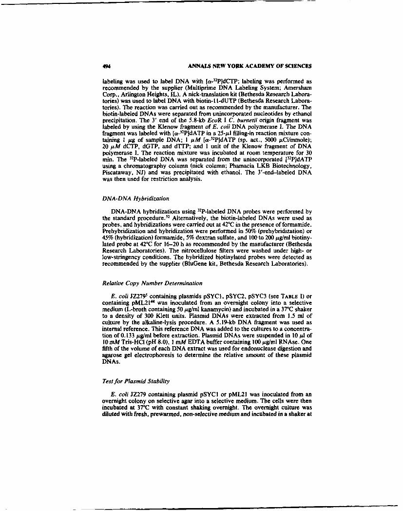
DNA homology of the origin region of the C. burnetii chromosome to those ofother gram-negative bacterial chromosomes was also determined by Southernblot (FIG. 2). The 5.8-kb EcoR I C. burnetii origin fragment, derived from pSYC I(Chen et al., manuscript in preparation), was used as a 32P-labeled probe (FIG. 2).Hybridization analysis was carried out under low stringency conditions, whichallowed 25% mismatches. The probe hybridized only to C. burnetii DNA of thecorresponding size (FIG. 2B). No hybridization to other bacterial DNAs wasobserved.
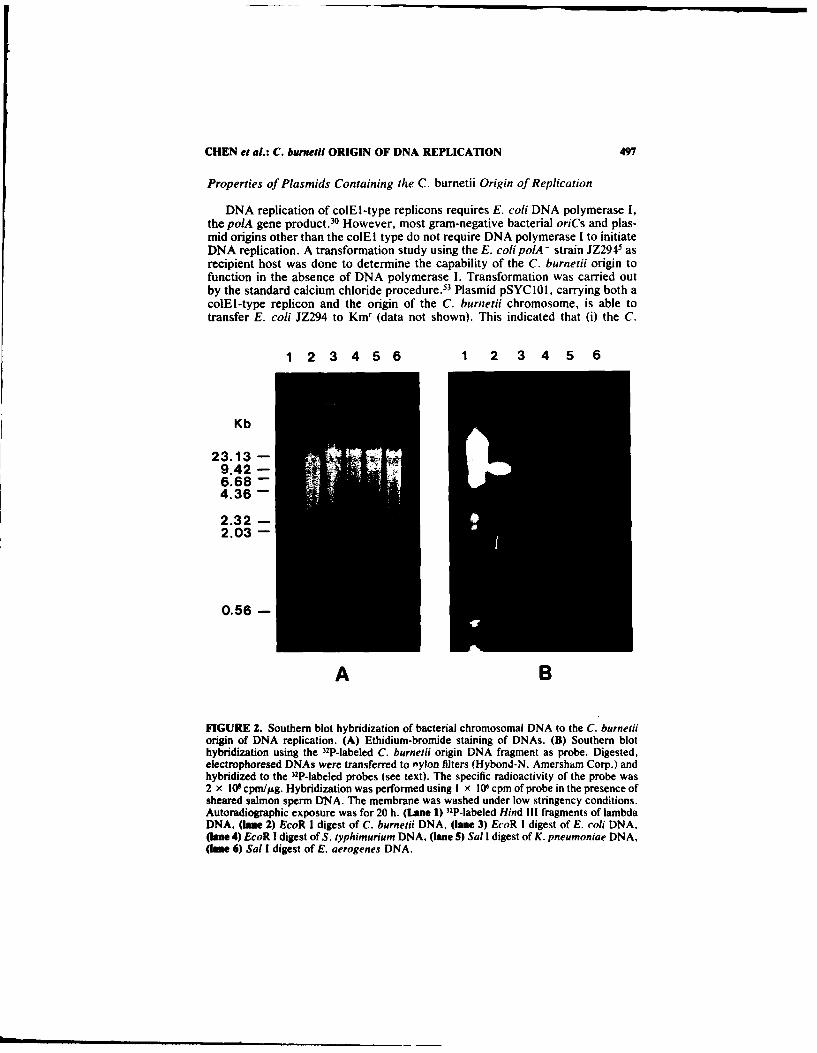
Restriction Map of the 5.8-kb C. burnetii Origin Fragment
Minichromosomes containing the C. burnetii origin as the only replicationorigin are not stably maintained in E. coli hosts (see below). Plasmid pSYC101,which has a higher copy number and which can be maintained stably in the host,was therefore constructed for the restriction map analysis. The 5.8-kb originfragment obtained from EcoR I digestion of plasmid pSYCI was ligated to anEcoR I-treated vector, pEMBL8(+). E. coli DH5aF' cells were transformedand used as the source of the 5.8-kb C. burnetii origin fragment. The restrictionmap (FIG. 3) of the C. burnetii origin fragment was deduced from molecularweights of restriction fragments obtained from gel electrophoresis of single, dou-

496 ANNALS NEW YORK ACADEMY OF SCIENCES
1 2 3 4 5 6 7 1 2 3 4 5 6 7
Kb
23.139.426.68
4.36
2.322.03
0.56
A BFIGURE 1. Southern blot hybridization of bacterial chromosomal DNAs to the E. coli oriC.(A) Ethidium-bromide staining of DNAs. (B) Southern blot hybridization using E. coli oriCas probe. Plasmid pSYC I and chromosomal DNA from C. burnetii, E. coli, S. typhimurium,E. aerogenes and K. pneumoniae were digested with appropriate restriction enzymes. Therestriction fragments were separated on a 0.9% agarose gel, transferred to a nitrocellulosefilter, and hybridized to the biotin-labeled E. coli oriC probe (see text) as described inMATERIALS AND METHODS. The filter was washed under low stringency conditions. Thehybridized biotinylated probes were detected by a BluGene technique as described in MATE-RIALS AND METHODS. (LaUe 1) biotinylated Hind III fragments of lambda DNA, (lane 2)EcoR I digest of plasmid pSYCI, (lane 3) EcoR I digest of C. burnetii Nine Mile I clone 7chromosomal DNA, (lane 4) EcoR I digest of S. typhimurium DNA, (lane 5) Sal I digest ofE. aerogenes DNA, (lane 6) Sal I digest of K. pneumoniae DNA, (lane 7) EcoR I digest of E.coli DNA. Arrowhead, 5-6-kb fragment of C. burnetii chromosomal DNA shows littlehybridization with the oriC probe.
ble, or partial digests of the DNA fragments with 18 restriction endonucleases (seebelow). 3'-end-labeled DNA was used in the partial digestion analysis. The fol-lowing restriction endonucleases which recognize hexamers were used to mapthis DNA fragment: Acc I, Ava I, BamH I, Bgl II, Cla I, EcoR I, EcoR V, Hinc II,Hind III, Hpa I, Kpn I, Pst I, Pvu I, Pvu II, Sal I, Sma 1, Xba I, and Xho I. Thosenot appearing on the map (FiG. 3) have no recognition sites on this DNAfragment.
CHEN et al.: C. burnetii ORIGIN OF DNA REPLICATION 497
Properties of Plasmids Containing the C. burnetii Origin of Replication
DNA replication of colEI-type replicons requires E. coli DNA polymerase I,the polA gene product.30 However, most gram-negative bacterial oriCs and plas-mid origins other than the colEl type do not require DNA polymerase I to initiateDNA replication. A transformation study using the E. coli polA - strain JZ294 5 asrecipient host was done to determine the capability of the C. burnetii origin tofunction in the absence of DNA polymerase I. Transformation was carried outby the standard calcium chloride procedure.5 3 Plasmid pSYCIOI, carrying both acolEl-type replicon and the origin of the C. burnetii chromosome, is able totransfer E. coli JZ294 to Kmr (data not shown). This indicated that (i) the C.
1 2 3 4 5 6 1 2 3 4 5 6
Kb
23.139.42 -
6.68 -4.36 -
2.32 -2.03 -
0.56 -
A B
FIGURE 2. Southern blot hybridization of bacterial chromosomal DNA to the C. burnetiiorigin of DNA replication. (A) Ethidium-bromide staining of DNAs. (B) Southern blothybridization using the 32P-labeled C. burnetii origin DNA fragment as probe. Digested,electrophoresed DNAs were transferred to nylon filters (Hybond-N, Amersham Corp.) andhybridized to the 32P-labeled probes (see text). The specific radioactivity of the probe was2 x 109 cpm//sg. Hybridization was performed using I x 106 cpm of probe in the presence ofsheared salmon sperm DNA. The membrane was washed under low stringency conditions.Autoradiographic exposure was for 20 h. (Lane 1) 32P-labeled Hind Ill fragments of lambdaDNA, (lane 2) EcoR I digest of C. burnetii DNA, (lane 3) EcoR I digest of E. coli DNA,(lane 4) EcoR I digest of S. typhimurium DNA, (lane 5) Sal I digest of K. pneumoniae DNA,(lane 6) Sal I digest of E. aerogenes DNA.
491 ANNALS NEW YORK ACADEMY OF SCIENCES
E
0 d uC ~ 0
2 3 4 5 KI4~ bI J , II
-ri CLl -0 2- --
0 0
IW I
FIGURE 3. Restriction endonuclease recognition sites in the origin region of the C. burne-tii chromosome.
burnetii origin was used for initiation of plasmid pSYCI01 replication in thepolA- strain and (ii) like most gram-negative bacterial oriCs, C. burnetii oriCdoes not require DNA polymerase I to initiate replication. The polA-indepen-dence of the C. burnetii origin made it possible to determine the minimal oriCregion by an ori-test.14
The relative copy numbers of the minichromosomes containing a C. burnetiiorigin in E. coil JZ279 cells were determined by comparison of the density of DNAbands to those of pML21 in an ethidium bromide-stained gel (FIG. 4). Theseplasmids all contain a 6.8-kb kanamycin fragment, while all of the minichromo-somes contain the 5.8-kb C. burnetii origin fragment. The marker DNA was usedas internal reference control, which appeared equally in all of the samples. Theamounts of plasmids pSYC 1, pSYC2, and pSYC3 are about one-half or less of thatof pML21.
The stability of C. burnetii minichromosomes in E. coli was determined bymeasuring the percentage of kanamycin-resistant cells after incubation in non-selective medium. Plasmid pSYC I was lost rapidly during cell growth when selec-tive pressure was removed (FIG. 5). However, plasmid pML21 remained rela-tively stable under the same test conditions. Statistically, the variation of thepercentage of kmr cells containing pML21 is not significant (not shown). Percent-ages of knr cells in the culture containing the C. burneti minichromosomesdropped from 50 to almost 0 during growth in the non-selective medium for 20generations, an interval of about 40 h (FIG. 5). About 40% of the overnight culturein selective medium consisted of plasmid-free cells (generation 0). This data sug-gested that the C. burnetii minichromosome was unstable in the E. coil cells,regardless of whether the selective pressure was present or not.
CHEN et al.: C. burnetii ORIGIN OF DNA REPLICATION 499
DISCUSSION
Hybridization analysis and the deduced restriction map of the C. burnetiiorigin suggested that the C. burnetii origin is not similar to the E. coli origin ofreplication, at least at the level of the primary structure. The nucleotide se-quences of the oriC of E. coli and those of the reference bacteria used in this studyare highly conserved (80-84% homology)." Strong hybridizations occurred be-tween the E. coli oriC probe and the reference bacterial origin fragments under
1 2 345
Kb
J 23.13 -
9.42 -
6.68 - ,: Marker DNA4.36 -
~2.32_
2.03 -
0.56 -
FIGURE 4. Gel electrophoretic determination of relative copy numbers of the Km' plas-mids containing the C. burnetii origin in E. coli JZ279. Cells were inoculated from anovernight colony in selective agar plates and were cultured in selective medium at 37°C to adensity of 300 Klett units. After addition of 0.2 /sg of the 5.19-kb DNA fragment as aninternal reference, plasmid DNAs of each culture were isolated from 1.5 ml of culture. Therelative amount of plasmid DNA was estimated by agarose gel electrophoresis. DNA wasdissolved in 10 Ml of 10 mM Tris-HCI, I mM EDTA buffer (pH 8.0) containing 100 /Ag/mlRNase. 2 gI of each preparation was used for EcoR I digestion and agarose gel electrophore-sis. (Lane 1) size marker: Hind Ill fragment of lambda DNA. (lane 2) pML21, (lane 3)pSYCI, (lane 4) pSYC2, (lane 5) pSYC3.
500 ANNALS NEW YORK ACADEMY OF SCIENCES
low stringency conditions (FIG. 1). However, hybridization between E. coli oriCand the C. burneti origin fragment was much weaker. It is concluded that theC. burneii oriC is less than 80% homologous to the E. coli oriC.
When the 5.8-kb C. burnetii origin fragment was used as probe, hybridizationoccurred only to the C. burnetii chromosomal DNA fragment with the corre-
100 •
E
* 50
0
I I I I I I I I I I I
0 2 4 6 8 10 12 14 16 18 20
GENERATIONS
FIGURE 5. Stability of plasmid pSYCI in E. coli JZ279. E. coli JZ279 containing thepSYCI (0) or pML21 (U) was grown exponentially for 20 generations in non-selectivemedium as described in MATERIALS AND METHODS, and the samples were taken at suitableintervals. Percentages of plasmid-containing cells (kanamycin resistant, KmR) were deter-mined by direct counting of cells plated on selective and non-selective plates.
sponding length (5.8 kb: FIG. 2). The result was different from that in FIGURE 1:no hybridization was observed between E. coli and C. burnetii DNAs. This dis-crepancy might be due to the differences of both systems in the sensitivity of theprobes, the method to detect the labeled probes, and the conditions of hybridiza-tion and post-hybridization wash. In any case, it demonstrates that there is very
CHEN et al.: C. burnei ORIGIN OF DNA REPLICATION 501
little DNA sequence homology between the origins of the C. burnetii chromosomeand those of gram-negative bacterial DNA. The result also suggested that theregions adjacent to the C. burnetii oriC are not similar to those of E. co/i or othertested gram-negative bacteria. The uniqueness of the oriC, and of its adjacentregions in the C. burnetii chromosome, is also implied by its restriction pattern.Restriction recognition sites, such as BamH I, Bgl 11, and Pst I, which appear oneor more times in the tested gram-negative bacteria origins,4 6. 9
,10 were not found in
the C. burnetii origin fragment. Moreover, gene organization near the origin re-gion of those gram-negative bacteria are similar. 91.
.,54 The organization of open
reading frames (ORFs) near the origin of the chromosome of the gram-positivebacteria Bacillus subtilis is similar to that at a location about 40 kb away from theoriC of the E. co/i chromosome (rpmH-dnaA-dnaN-recF-gyrB) 5
1, 6 The gene or-
ganization near the B. subtilis origin is believed to represent a primordial repliconfrom which the chromosome of both gram-positive and gram-negative bacteriahave evolved. Further studies are needed to reveal the gene organization near theC. burnetii oriC and the evolutionary implications for the microorganism.
The properties of instability and high copy number are present in minichromo-somes carrying E. co/i oriC or other gram-negative bacterial origins. -9 -
18 .21 31 .32
Although the exact copy number of the C. burnetii minichromosome was notdetermined, comparison of its relative copy number to that of pML21 suggeststhat the C. burneiji minichromosome has only about one-half the copy number ofpML21, which suggests that (i) C. burneiii minichromosomes have lower copynumbers than the colEl-type plasmids in E. coli but have a higher copy numberthan that of the E. co/i chromosome or (ii) these minichromosomes are not stablymaintained or segregated in the E. co/i host. The decrease of the amount ofplasmid DNA in non-selectively growth culture and the high proportion of plas-mid-free cells in the growth culture (FIG. 5) reflected the instability of the plasmid.Moreover, the growth-curve studies indicate that cultures of E. co/i JZ279 carry-ing the pSYCI plasmid exhibited longer generation times than that carrying thepML21 plasmid (data not shown). This may be again due to the low percentage ofthe kmr cells in the whole population. E. coli minichromosomes initiate theirreplication in synchrony with the host chromosome.3 3 Like the chromosome,minichromosomes initiate, on the average, at the same cell mass per origin; 2 andat a low growth rate the minichromosome was lost at a higher frequency. 2" Thehigh loss-frequencies of minichromosomes might be an indication of (i) slightincompatibility, (ii) unstable segregation, or (iii) the presence of a high copy lethal(HCL) region. The incompatibility properties of E. co/i minichromosomes havebeen shown to be in the mioC promoter, which was negatively regulated by DnaAprotein and required methylation of the GATC site within the promoter sequencefor full activity. 19 However, the E. co/i minichromosomes can be stabilized by thesop (stability of plasmid) genes from plasmid F,2 ' whose only function is proposedto be partitioning of plasmids at cell division. This fact indicates that the instabil-ity of E. coli minichromosome might be largely due to occasional segregationfailure rather than to competition with the chromosome for replication factors.Another interpretation of the instability of the minichromosome is that it indicatesthe presence of a gene product which is detrimental to the host cell when presentin higher than normal amounts.' 9 Like E. co/i minichromosomes, the instability ofC. burneiii minichromosomes in E. co/i hosts might be largely due to the lack of apartition system at cell division. The presence of a HCL region is less likely in the5.8-kb EcoR I fragment, since pSYC 101 is present in higher copy number in the E.coli host (data not shown).
502 ANNALS NEW YORK ACADEMY OF SCIENCES
REFERENCES
1. MASTERS, M. & P. BRODA. 1971. Nature New Biol. 232: 137-140.2. DONACHIE. W. D. 1968. Nature 219: 1077-1079.3. SKARSTAD, K., E. BOYE & H. B. STEEN. 1986. EMBO J. 5: 1711-1717.4. CLEARY. J. M., D. W. SMITH, N. E. HARDING & J. W. ZYSKIND. 1982. 1. Bacteriol.
150: 1467-1471.5. HARDING, N. E., J. M. CLEARY, D. W. SMITH, J. J. MICHoN, W. S. A. BRUSILOW &
J. W. ZYSKIND. 1982. J. Bacteriol. 152: 983-993.6. MEIJER, M., E. BECK, F. G. HANSEN, H. E. N. BERGMANS, W. MESSER, K. VON
MEYENBURG & H. SCHALLER. 1979. Proc. Natl. Acad. Sci. USA 76: 580-584.7. SUGIM~OTO, K., A. OKA. H. SUGISAKI. M. TAKANAMI, A. NISHIMURA, S. YASUDA &
Y. HIROTA. 1979. Proc. Natl. Acad. Sci. USA 76: 575-579.8. TAKEDA, Y., N. E. HARDING, D. W. SMITH & J. W. ZYSKIND. 1982. Nucleic Acids
Res. 10: 2639-2650.9. ZYSKINO, J. W.. L. T. DEEN & D. W. SMITH. 1979. Proc. Nat]. Acad. Sci. USA 76:
3097-3101.10. ZYSKIND. J. W. & D. W. SMITH. 1980. Proc. Natl. Acad. Sci. USA 77: 2460-2464.11. ZYSKIND, J. W., J. M. CLEARY, W. S. A. BRUSILOW. N. E. HARDING & D. W.
SMITH. 1983. Proc. Natl. Acad. Sci. USA 80: 1164-1168.12. YASUDA, S. & Y. HiROTA. 1977. Proc. Nai. Acad. Sci. USA 74: 5458-5462.13. ZYSKIND, J. W. & D. W. SMITH. 1986. Cell 46: 489-490.14. OKA, A., K. SUGIMOTO, M. TAKARAMI & Y. HIROTA. 1980. Mol. Gen. Genet. 178: 9-
20.15. HANSEN, F. G., S. KoFOED, K. VON MEYENBURG & T. ATLUNG. 1981. ICN-UCLA
Symp. Mol. Cell Biol. 22: 37-55.16. Kb4LLING, R., C. KOCHERER, M. A. SCHAUZU, H. LaTHER & W. MESSER. 1987.
UCLA Symp. Mol. Cell Biol. New Ser. 47: 429-439.17. KOiLLING, R. & H. LOTHER. 1985. J. Bacteriol. 164: 3 10-315.18. STUITJE, A. R. & M. MEIJER. 1983. Nucleic Acids Res. 11: 5775-5791.19. YAMAGUCHI, K. M., YAMAGUCHI & J.-l. TomIZAWA. 1982. Proc. Natl. Acad. Sci.
USA 19: 5347-5351.20. LOTHER, H.. R. KOLLING, C. KOCHERER & 'V. SCHAUZU. 1985. EMBO J. 4: 555-
560.21. L0BNER-OLESEN, A., T. ATLUNG & K. V. RA~smUSSEN. 1987. J. Bacterial. 169: 2835-
2842.22. STUITJE, A. R., N. DE WIND, J. C. VAN DER SPEK, T. H. PoRs & M. MEIJER. 1986.
Nucleic Acids Res. 11: 5775-579L.23. TANAKA, M. & S. HiRAGA. 1985. Mol. Gen. Genet. 200: 2 1-26.24. FULLER, R. S., J. M. KAGUNI & A. KoRNHERG. 1981. Proc. Nai. Acad. Sci. USA
78: 7370-7374.25. FULLER, R. S. & A. KORNBERG. 1983. Proc. Nai. Acad. Sci. USA 80: 5817-5821.26. KAGUNI, J. M. & A. KORNHERG. 1984. Cell 38: 183-190.27. OGAWA, T., T. A. BAKER, A. VAN DER ENDE & A. KORNBERG. 1985. Proc. Nai.
Acad. Sci. USA 82: 3562-3566.28. FUNNELL. B. E., T. A. BAKER & A. KORNOERG. 1986. J. Bacterial. 261: 5616-5626.29. FUNNELL, B. ':, T. A. BAKER & A. KORNBERG. 1987. J. Biol. Chem. 262: 10327-
10334.30. KINGSBURY, 0. T. & D. R. HELINSKI. 1973. J. Bacteriol. 114: 1116-1124.31. MESSER, W., H. E. N. BERGMANS, M. MEIJER, J. E. WOMACK, F. G. HANSEN &
K. VON MEYENBURG. 1978. Mol. Gen. Genet. 162: 269-275.32. VON MEYENBURG, K., F. G. HANSEN. E. RiISE, H. E. N. BEROMANS, M. MEIJER &
W. MESSER. 1979, Cold Spring Harbor Symp. Quant. Biol. 43: 121-128.33. LEONARD, A. C. & C. E. HELMSTETTER. 1986. Proc. Natl. Acad. Sci. USA 33: 5 101 -
5105.33a. CHEN, S.-Y., M. VODKIN. H. A. THOMPSON & J. C. WILLIAMS. 1990. J. Gen.
Microbial. 136: 89-96.
CHEN et a/.: C. bawnei ORIGIN OF DNA REPLICATION 503
34. AMANO, K.-I. & 1. C. WILLIAMS. 1984. J. Bacteriol. 160: 989-993.35. AMANO, K.-I. & J. C. WILLIAMS. 1984. J. Bacteriol. 160: 994-1002.36. BURTON, P. R., J. STUECKEMANN & D. PARETsKy. 1975. J. Bacteriol. 122: 316-324.37. JERRELS, T. R., D. J. HINRICHS & L. P. MALLAVIA. 1974. Can. J. Microbiol. 20:
1465-1470.38. LENNOX, E. S. 1955. Virology 1: 190-206,39. WILLIAMS, J. C., M. C. PEACOCK & T. F. MCCAtJL. 198 1. Infect. Immun. 32: 840-
851.40. ZUERNER, R. L. & H. A. THOMPSON. 1983. J1. Bacteriol. 156: 186-19).41. SAMUEL, J. E., M. E. FRAZIER, M. L. KAHN, L. S. THOMASHOW & L. P. MALLAVIA.
1983. Infect. Immun. 41: 488-493.42. MANIATIS, T., E. F. FRITSCH & J. SAMBROOK. 1982. In Molecular Cloning, a Labora-
tory Manual. pp. 368-369. Cold Spring Harbor Laboratory. Cold Spring Harbor,N.Y.
43. KATZ, L., D. T. KINGSBURY & D. R. HEINSKI. 1973. J. Bacteniol. 114: 577-591.44. HERSHFIELD, V., H. W. BOYER, L. CHOW & D. R. HEuNSKIc. 1976. J. Bacteriol.
126: 447-453.45. SMITH, E. W., A. M. GARLAND, G. HERMEAN. R. E. ENNS, T. A. BAKER & J. W.
ZYSKIND. 1985. EMBO J1. 4: 1319-1324.46. DENTE, L., G. CESARENI & R. CORTESE. 1983. Nucleic Acids Res. 11: 1645-1655.47. BACHMANN, B. J. & K. B. Low. 1980. Microbiol. Rev. 44: 1-56.48. FEINBERG, A. P. & B. VOGELSTEIN. 1983. Anal. Biochem. 132: 6-13.49. FEINBERG, A. P. & B. VOGELSTEIN. 1984. Anal. Biochem. 137(Addendum): 266-267.50. RIGBY, T. W. J., M. DIECK(MANN, C. RHODES & P. BERG. 1977. J. Mol. Biol. 113:
237-25 1.51. DROUIN, J. 1980. J. Mol. Biol. 140: 15-34.52. SOUTHERN, E. M. 1975. J. Mol. Biol. 98: 503-5 17.53. LEDERBERG, E. M. & S. N. COHEN. 1974. J. Bacteriol. 119: 1072-1074.54. VON MEYENBURG, K., F. G. HANSEN, L. D. NIELSEN & E. RiISE. 1978. Mol. Gen.
Genet. 160: 287-295.55. MORIYA, S., N. OGASAWARA & H. YOSHIKAWA. 1985. Nucleic Acids Res. 13: 225 1-
2265.56. VON MEYENBURG, K. & F. G. HANSEN. 1988. In Escheric/iia coi and Salmonella
typhimurium: Cellular and molecular biology. J. L. Ingraham, K. B. Low, B. Maga-sanik, F. C. Neidhardt, M. Schaechter & J. E. Umbarger, Ed!..: 1555-1577. Ameri-can Society for Microbiology. Washington, D.C.
J1