Embed Size (px)
Citation preview

Teaching Module V
Contrast-Induced Nephropathy – CIN

Contrast-Induced Nephropathy – CIN This slide deck was prepared by
Professor S K Morcos
Consultant Radiologist
University of Sheffield
Sheffield Teaching Hospitals
NHS Trust

Contents
• Pathophysiology
• Risk factors
• Clinical picture
• Incidence
• Outcome
• Prevention
• Importance of the dose of CM
• Areas of confusion
Contrast-induced nephrotoxicity (CIN)

The kidney is the main route of
elimination of CM
• Increase RVR
• Decrease GFR
• Diuresis
• Natriuresis
• Enzymuria
• Structural changes [Osmotic nephrosis]
It represents the normal response
of the kidney to CM exposure
Modulation of production ofintrarenal vasoactive mediators
↑ Endothelin (vasoconstriction)↑ Adenosine (vasoconstriction)↓ NO (vasodilatation) ↓ Prostacycline (vasodilatation)

CM Normal kidneys, no risk factors
No clinical problem
Risk factorsRenal impairment + DM
Dehydration
Congestive heart failure
Age over 70 years old
Administration of nephrotoxic drugs
Dose and type of CM

Contrast-media-induced nephrotoxicity (CIN)
Definition
It implies impairment in renal function:
an increase in serum creatinine by more
than 25% or 0.5 mg/dL has occurred within
3 days following the intravascular
administration of contrast medium and the
absence of alternative aetiology.

NEW definition
• CIN is a condition in which a decrease in renal
function occurs within 3 days of the intravascular
administration of CM in the absence of an
alternative aetiology. An increase in serum
creatinine by more than 25% or 44 µmol/l (0.5
mg/dl) indicates CIN.

Clinical picture of CIN
• The diagnosis is based on an increase in serum creatinine.
• Anuria may develop in severe cases.
• Dialysis is rarely required. (< 1% of patients with CIN)

Incidence of CIN after IV administration
• Incidence of CIN is very low in patients with GFR >45ml/min (<1%)
• Incidence of CIN in patients with GFR <45ml/min varies between 5 -20% (no studies in patient with eGFR <20ml/min)
• Sr Cr average 2.4 mg/dL 21% in the control arm (Tepel et al, New England Journal of Medicine 2000; 343: 180-184) –highest incidence ever reported.
Katzberg & Barrett, Radiology 2007; 243: 622-628
Weisbord SD et al, Clin J Am Soc Nephrol 2008; 3: 1274-1281
Combined data from a comparative study
of a monomer and a dimer.
eGFR (MDRD) Relative risk of CIN
> 40 ml/min 0.6%
< 40 ml/min 4.6%
15 – 30 ml/min 7.8%
Thomsen & Morcos Eur Radiol
2009; 19: 891-897

Estimate:
In patients with GFR < 20
ml/min is probably 10-15%
after intravenous injection
The incidence is higher
after intraarterial injection
The Incidence increases as the renal function
decreases

Incidence of CIN in patients undergoing
cardiac angiography (n=1,196)
0
5
10
15
20
25
+RI+DM+RI–DM–RI+DM–RI–DM
0%
5.7%
19.7%
%
0.6%
Rudnick et al., 1995

Incidence of CIN after coronary angiography
DM increased the
incidence of CIN in
patients with renal
impairment
undergoing coronary
angiography
(n=1,826)
No CIN requiring
dialysis was observed
at CrCl of 50 ml or
more
Incidence of CIN
markedly increased at
CrCl of 30 ml or less
100 ml was the cut-off dose of CM below
which there was no CIN requiring dialysis
McCullough et al., Am J Med 1997; 103:368-375
The risk and severity of CIN
increases proportionally
to the number and severity
of the risk factors
„
“

CIN
Clinical course
There is a clinical concern
Although self-limiting in most cases (resolve within 1-2 weeks)

Clinical importance of CIN
CIN increases the incidence of non-renal complications and prolongs hospital
stay
Rihal et al., Circulation 2002; 105:2259-2265
Bartholemew et al., Am J Cardiol 2004; 93:1515-1519
Marenzi et al., JACC 2004; 44:1780-1785

Clinical importance of CIN
CIN increases in hospital mortality
• * 21% - 34% in the CIN groups • * 1%-7% in the control groups (similar baseline renal
function, received CM without developing CIN)
• ** CIN was significantly associated with 30-day mortality (15.6% with CIN Vv 5.2% control), the risk was higher after IV than after intraarterial administration!
*Levy et al., JAMA 1996; 275:1489-149 (In this study, 48% of patients received
IV CM)
** From AM et al., Mayo Clin Proc 2008; 83:1095-1100

CIN
PREVENTION IS CRUCIALLY IMPORTANT
To avoid an increase in patients’ morbidity
and mortality
Levy et al., JAMA 1996; 275:1489-1494
Rudnick & Feldman, Clin J Am Soc Nephrol 2008; 3:263-272

How to reduce
the risk of
CIN

Prevention of CIN
• Small dose of low-osmolar or iso-osmolar non-ionic CM
• Volume expansion
• Prophylactic haemodialysis
• Haemofiltration
YES
NO
Can be considered in patients with
advanced renal disease (Cr Cl < 30 ml/min)
requiring interventional vascular procedure

Prevention of CIN
Pharmacological manipulation
Renal vasodilators
- Calcium channel blockers
- Dopamine
- Atrial natriuretic peptide
- Fenoldopam [dopamine-1 receptor agonist]
- Prostaglandin E1
Blocking intrarenal mediators
[ET, Adenosine]
- ET receptor antagonist
- Theophylline
Cytoprotective drugs
- Acetylcysteine
- Acetazolamide
Value remains uncertain

Importance of dose in CIN
The higher the dose the higher the risk
Dose of CM in gram iodine should not exceed the value of eGFR
The dose of CM should never exceeds the amount required
to produce essential diagnostic information
The maximum dose of CM in gram iodine that can be given without significantly
increasing the risk of CIN
Nyman et al., Acta Radiologica 2008; 49:658-667
ESUR guidelines book
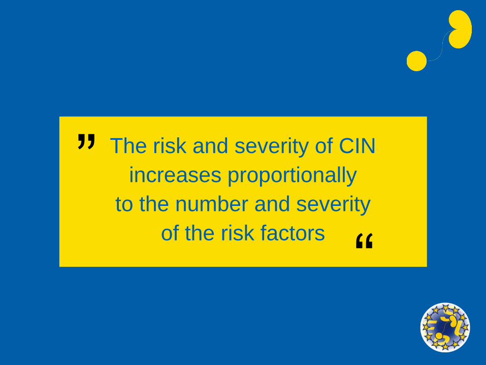
How to reduce the dose of
CM IA administration
• ICM + CO2
• Reducing the iodine concentration
• Focus only on essential diagnostic information

Areas of confusion in prevention of CIN
• Identifying patients at risk
• Hydration regime
• The type of CM (IOCM or LOCM)
• The prophylactic use of acetylcysteine

Identifying patients with renal impairment
Serum Cr measured routinely before contrast injection
or
Selective measuring of Sr Cr using a questionnaire
(history of renal disease, proteinuria, prior kidney surgery, hypertension,
gout or diabetes mellitus)
Morcos SK, Clin Radiol 2004; 59:381-389
Choyke et al., Techniques in Urology 1998; 2:65-69

Serum Creatinine can be used to calculate
eGFR
eGFR < 45 ml/min is a risk factor for CIN
after IV administration
eGFR < 60 ml/min is a risk factor for CIN
after IA administration

Areas of confusion
Hydration
• Type of fluid
• Half-strength saline
• Normal saline
or
• Sodium bicarbonate
• Hydration regime
• Orally
• IV
• Duration

Hydration
Goals
• Diuresis
• Dilute CM in the tubules
• Decrease contact time
• Increase production of prostacycline in renal medulla
• Suppress TGF response
• Volume expansion
• Suppress renine-angiotensine system
• Suppress ADH
• HCO3
• Increase pH of urine and renal medulla
• Suppress production of free radicals

The type of fluid
• A study by Mueller et al. demonstrated that 0.9% saline is superior to 0.45%
preparation in reducing the risk of CIN in patients undergoing coronary
angioplasty
Mueller et al., Arch Intern Med 2002; 162:329-336
• A recent study of 353 patients undergoing coronary angiography
(MEENA trial) showed no benefit of sodium bicarbonate over normal saline
in preventing CIN
Brar et al., JAMA 2008; 300:1038-46

Title

Title
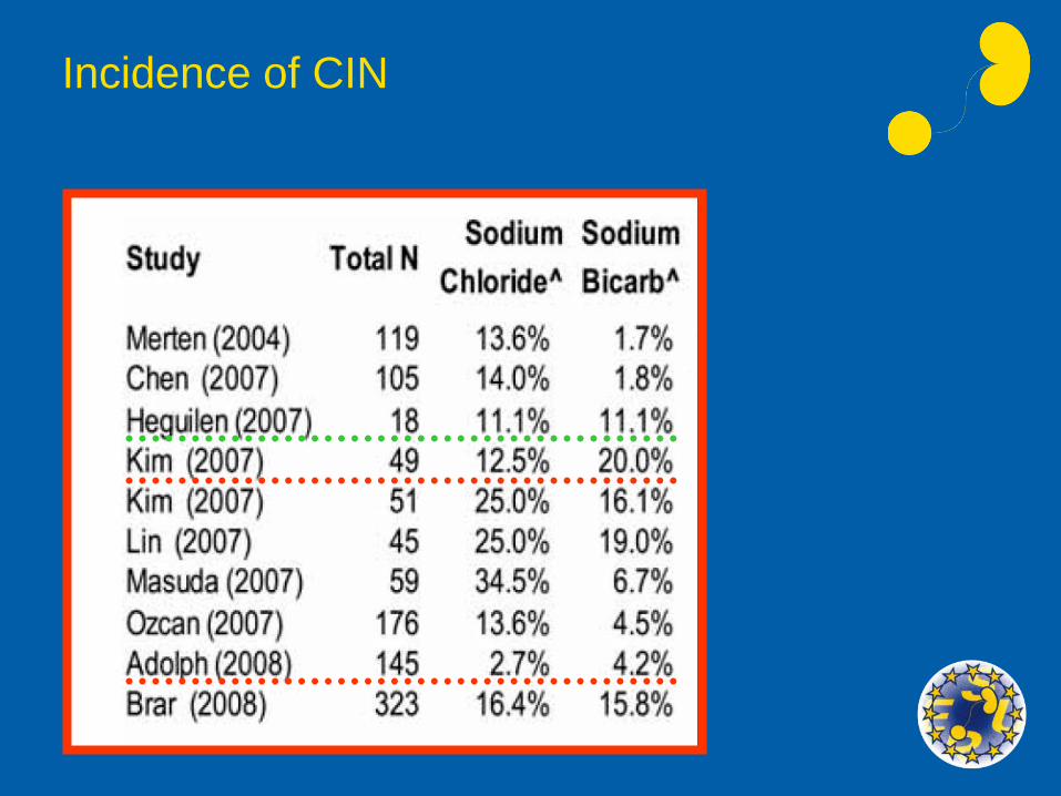
Incidence of CIN

New recommendation from ESUR
• Sodium bicarbonate seems to provide equal or
superior protection against CIN to normal saline.
• Current recommendations
– Dose of N saline: 1.0-1.5 ml/kg/h for at least 6
hours before and after CM administration.
– Dose of sodium bicarbonate: 3ml/kg/h for 1 hour
before contrast medium followed by 1 ml/kg/h for
6 hours after – increase dose until urine
alkanisation is achieved.
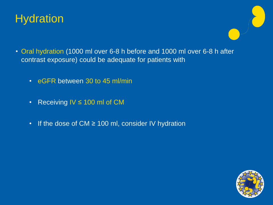
Hydration
• Oral hydration (1000 ml over 6-8 h before and 1000 ml over 6-8 h after
contrast exposure) could be adequate for patients with
• eGFR between 30 to 45 ml/min
• Receiving IV ≤ 100 ml of CM
• If the dose of CM ≥ 100 ml, consider IV hydration

Hydration
100 ml/hr of normal saline IV for 4-6 hours before and after CM injection in
addition to encouraging oral fluid intake
• IA administration of CM
• eGFR < 60 ml/min
• IV injection of CM
• > 100 ml + renal impairment
(eGFR 30 to 45 ml/min)
or
eGFR < 30 ml/min

Type of CM
Are iso-osmolar CM less nephrotoxic in comparison to LOCM after IV administration?

Metaanalysis – 1950-Aug 2007
• Based on 25 trials
• Iodixanol is not associated with a significantly reduced risk of CIN compared
with the LOCM pooled together.
• However, in patients with intraarterial administration and renal insufficiency,
iodixanol is associated with a reduced risk of CIN compared with iohexol,
whereas no significant difference between iodixanol and other LOCM could
be found.
Heinrich et al., Radiology 2009; 250:68-86

IOCM Vv LOCM angiography, diabetic
patients with renal impairment
Comparing Iodixanol Vv Iopamidol in coronary angiography
• Incidence of CIN
• Iopamidol 09.8%
• Iodixanol 11.2%
Laskey et al. (Nephric 2), Am Heart J 2009; 158:822-823
No significant difference in the risk of CIN
between iopamidol and iodixanol

CIN Comparative Studies after IV Injection
High risk patients (20-60 ml/min)
Study LOCM
(monomers)
Iodixanol Criteria
Carraro et al (1998) 0/32 (iopromide) 1/32 50% SCr
Nguyen et al 2008 10/65 (iopromide) 3/61 44 mol/L SCr
Kolehmainen et al
(2003)4/25 (iobiditrol) 4/25 44 mol/L SCr
Barrett et al (2006) 0/77 (iopamidol) 2/76 44 µmol/L SCr
Thomsen et al (2008) 0/76 (iomeron) 5/72 44 µmol/L SCr
Kuhn et al (2008) 7/125 (iopamidol) 6/123 25% SCr
F-R Chuang (2009)(intravenous urography)
1/25 (iohexol) 1/25 25% SCr
TOTAL 22/425
(5.18%)
22/418
(5.26%)
NO
DIFFERENCE
Patients with pre-existing renal
impairment are at risk of
CIN with all classes of CM
Morcos SK, Clin Rad 2009

Acetylcysteine
• Low cost
• Ease of administration
• Limited side-effects

Acetylcysteine
Pharmacology
• Low oral bioavailability (6-10%)
• Other substances used in the oral formulation or administered concomitantly
may combine with the sulfhydryl group, which is the key to its mechanism
of action
Shalansky et al., Heart 2005; 91:997-999

Acetylcysteine
Disadvantages
• Optimal dosage remains uncertain (600 mg b.d or
1200 mg b.d for 48 hr)
• Activity varies between oral products
• Noxious smell and taste of liquid preparations
• Exact mechanism to prevent CIN is unknown
• Induces creatininuria leading to a reduction in serum
creatinine independent of a change in GFR

Acetylcysteine
• Results of clinical studies are remarkably inconsistent
• Many uncertainties remain concerning its effectiveness
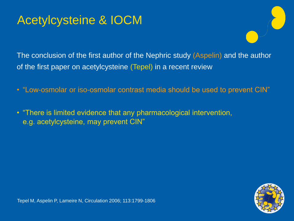
Acetylcysteine & IOCM
The conclusion of the first author of the Nephric study (Aspelin) and the author
of the first paper on acetylcysteine (Tepel) in a recent review
• “Low-osmolar or iso-osmolar contrast media should be used to prevent CIN”
• “There is limited evidence that any pharmacological intervention,
e.g. acetylcysteine, may prevent CIN”
Tepel M, Aspelin P, Lameire N, Circulation 2006; 113:1799-1806

CONTRAST MEDIA NEPHROTOXICITY
ESUR GUIDELINES
Risk factors
Look for:
• High serum creatinine levels, particularly secondary to diabetic nephropathy
• Dehydration
• Congestive heart failure
• Age over 70 years old
• Concurrent administration of nephrotoxic drugs, e.g. non-steroidal
anti-inflammatory drugs

CONTRAST MEDIA NEPHROTOXICITY
ESUR GUIDELINES
In patients with risk factor(s)
Do:
• Make sure that the patient is well hydrated
• Use non-ionic contrast media
• Stop administration of nephrotoxic drugs for at least 24 h
• Consider alternative imaging techniques which do not require the
administration of iodinated contrast media
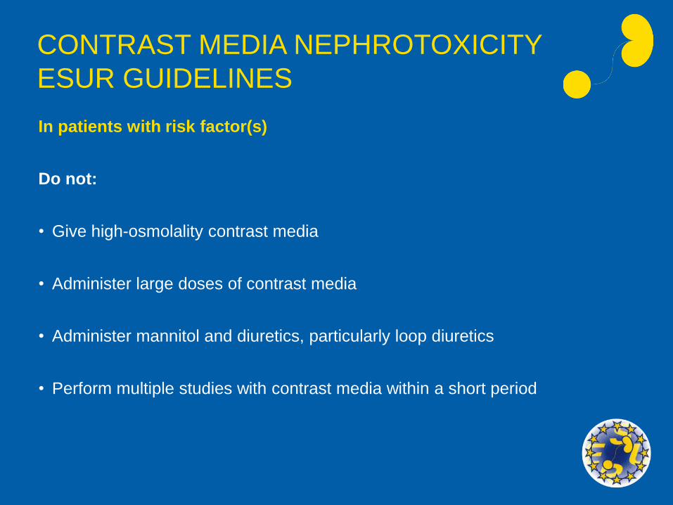
CONTRAST MEDIA NEPHROTOXICITY
ESUR GUIDELINES
In patients with risk factor(s)
Do not:
• Give high-osmolality contrast media
• Administer large doses of contrast media
• Administer mannitol and diuretics, particularly loop diuretics
• Perform multiple studies with contrast media within a short period

2.1 Renal adverse reactions to iodinated
contrast media
Risk factors for contrast medium induced nephropathy
Patient related eGFR less than 60 ml/min/1.73m2
before intraarterial administration eGFR less than 45 ml/min/1.73 m2
before intravenous injection
In particular in combination with: Diabetic nephropathy Dehydration Congestive heart failure Gout Age over 70 Concurrent administration of
nephrotoxic drugs e.g. non-steroid anti-inflammatory drugs.
Procedure-related Intraarterial administration of contrast medium
High osmolality agents Large doses of contrast medium Multiple contrast medium
administrations within a few days
Department of Diagnostic Sciences, CPH University
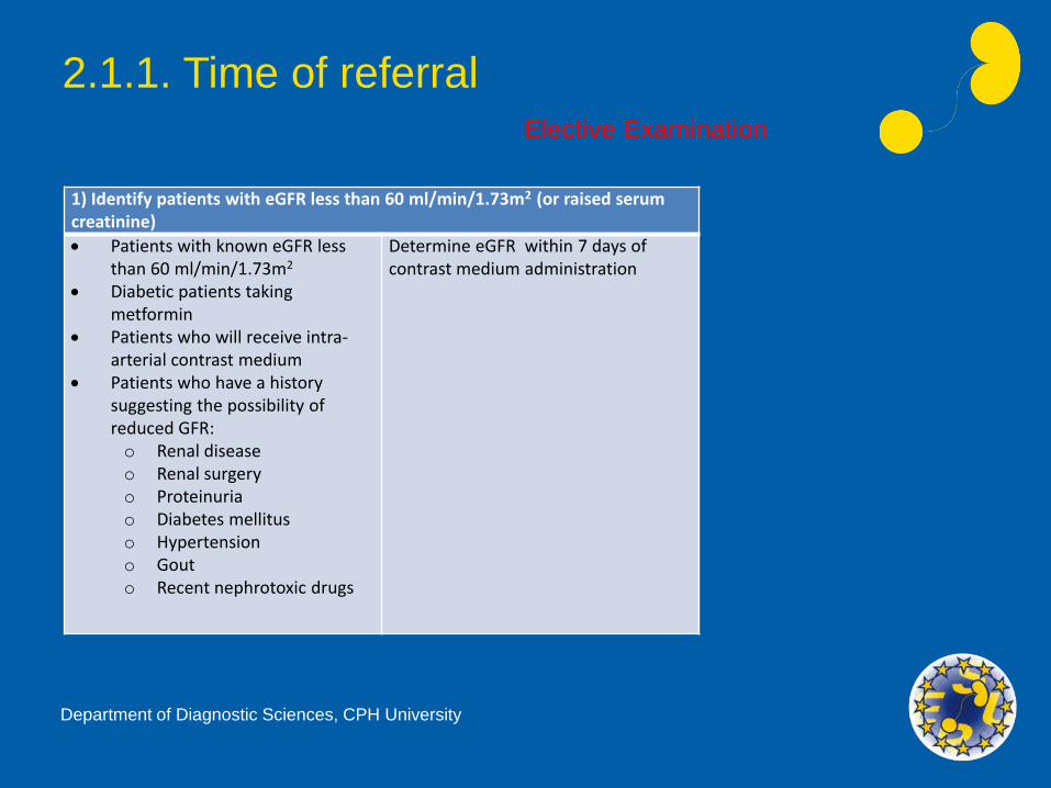
2.1.1. Time of referral
1) Identify patients with eGFR less than 60 ml/min/1.73m2 (or raised serum creatinine)
Patients with known eGFR less than 60 ml/min/1.73m2
Diabetic patients taking metformin
Patients who will receive intra-arterial contrast medium
Patients who have a history suggesting the possibility of reduced GFR:o Renal diseaseo Renal surgeryo Proteinuriao Diabetes mellituso Hypertensiono Gouto Recent nephrotoxic drugs
Determine eGFR within 7 days of contrast medium administration
Elective Examination
Department of Diagnostic Sciences, CPH University
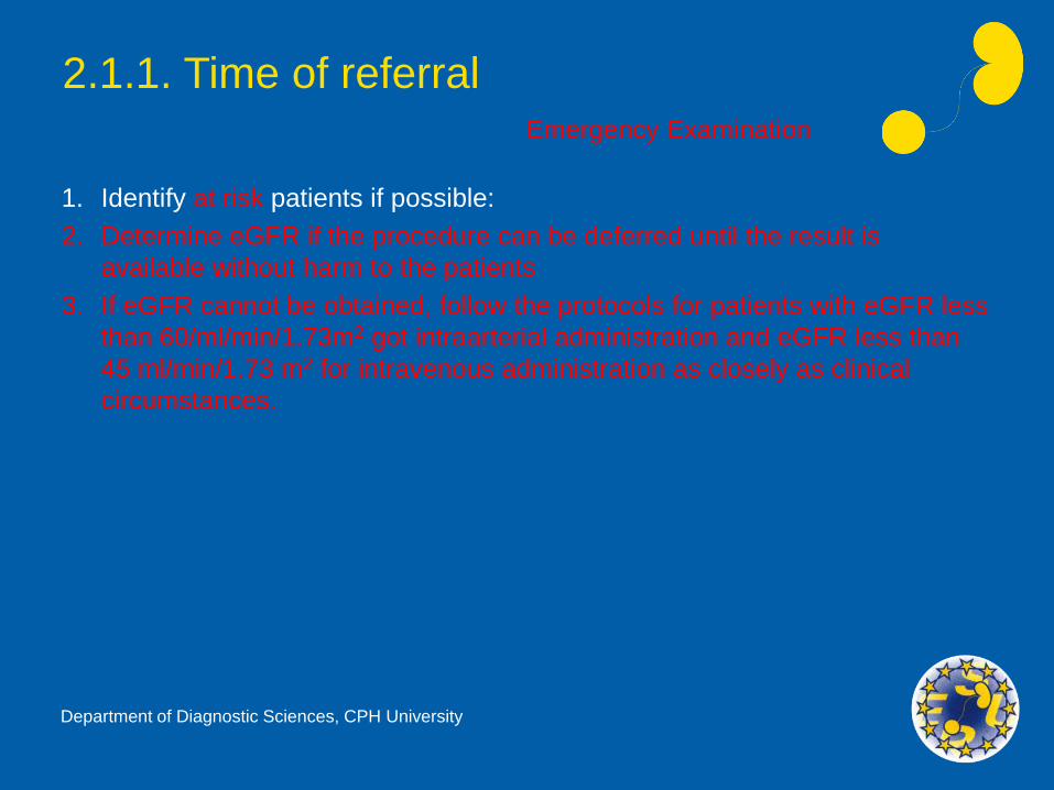
2.1.1. Time of referralEmergency Examination
1. Identify at risk patients if possible:
2. Determine eGFR if the procedure can be deferred until the result is
available without harm to the patients
3. If eGFR cannot be obtained, follow the protocols for patients with eGFR less
than 60/ml/min/1.73m2 got intraarterial administration and eGFR less than
45 ml/min/1.73 m2 for intravenous administration as closely as clinical
circumstances.
Department of Diagnostic Sciences, CPH University

2.1.2. Before the examinationElective Examination
At risk patients Consider an alternative imaging method not using iodinated contrast media.
Discuss the need to stop nephrotoxic drugs with the referring physician.
Start volume expansion. A suitable protocol is intravenous saline, 1.0-1.5 ml/kg/h, for at least 6 h before and after contrast medium. An alternative protocol is intravenous sodium bicarbonate, 3ml/kg/h for 1 h before contrast medium and l ml/kg/h for 6 hafter contrast medium.
Department of Diagnostic Sciences, CPH University

2.1.2. Before the examinationEmergency Examination
At risk patients Consider an alternative imaging method not using iodinated contrast media.
Start volume expansion as early as possible before contrast medium administration.
Department of Diagnostic Sciences, CPH University
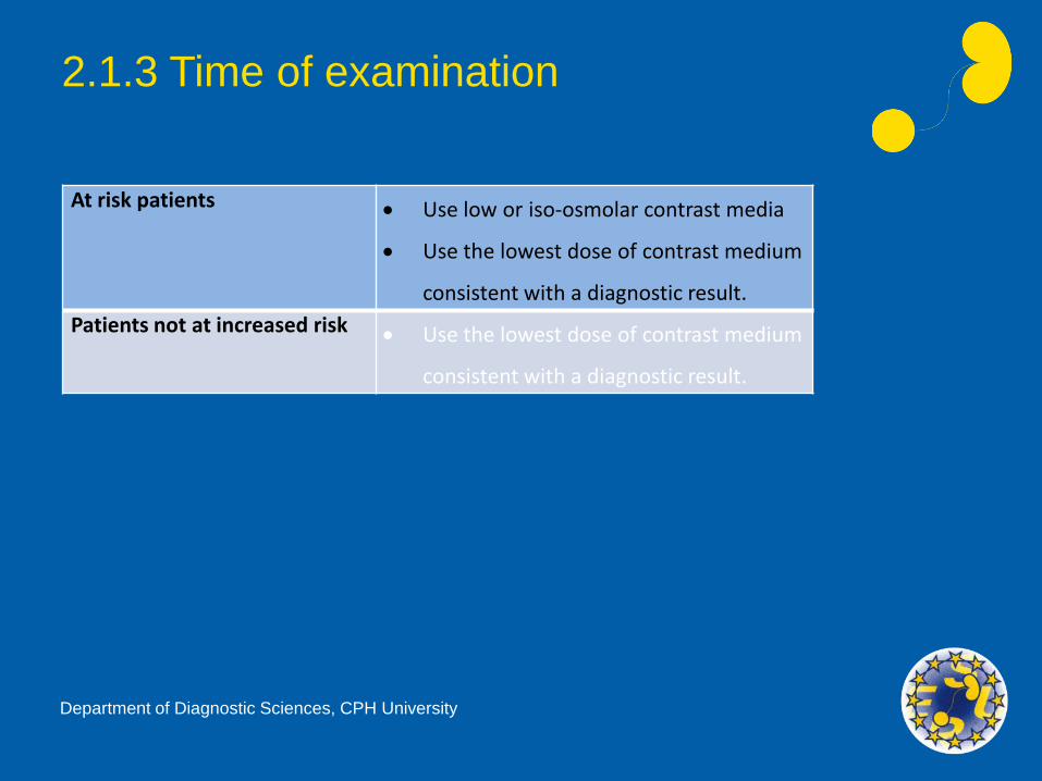
2.1.3 Time of examination
At risk patients Use low or iso-osmolar contrast media
Use the lowest dose of contrast medium
consistent with a diagnostic result.
Patients not at increased risk Use the lowest dose of contrast medium
consistent with a diagnostic result.
Department of Diagnostic Sciences, CPH University

2.1.3 Time of examination
At risk patients Use low or iso-osmolar contrast media
Use the lowest dose of contrast medium
consistent with a diagnostic result.
Patients not at risk Use the lowest dose of contrast medium
consistent with a diagnostic result.
Department of Diagnostic Sciences, CPH University
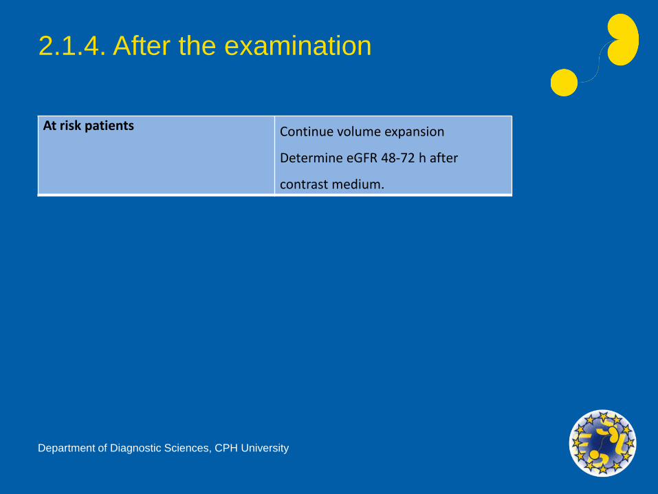
2.1.4. After the examination
At risk patients Continue volume expansion
Determine eGFR 48-72 h after
contrast medium.
Department of Diagnostic Sciences, CPH University

Note
• No pharmacological manipulation (with renal vasodilators,
receptor antagonists of endogenous vasoactive mediators or
cytoprotective drugs) has yet been shown to offer consistent
protection against contrast medium induced nephropathy.
Department of Diagnostic Sciences, CPH University

Thank you
for your attention


![Penanganan Anuria[1]](https://img.pdfslide.net/doc/110x75/55721436497959fc0b94076f/penanganan-anuria1.jpg)