Embed Size (px)
Citation preview

Omurtag et al. International Journal of Emergency Medicine 2012, 5:35http://www.intjem.com/content/5/1/35
ORIGINAL RESEARCH Open Access
Technical and clinical analysis of microEEG: aminiature wireless EEG device designed to recordhigh-quality EEG in the emergency departmentAhmet Omurtag1, Samah G Abdel Baki1, Geetha Chari2, Roger Q Cracco3, Shahriar Zehtabchi4,André A Fenton5 and Arthur C Grant6*
Abstract
Background: We describe and characterize the performance of microEEG compared to that of a commerciallyavailable and widely used clinical EEG machine. microEEG is a portable, battery-operated, wireless EEG device,developed by Bio-Signal Group to overcome the obstacles to routine use of EEG in emergency departments (EDs).
Methods: The microEEG was used to obtain EEGs from healthy volunteers in the EEG laboratory and ED. Thestandard system was used to obtain EEGs from healthy volunteers in the EEG laboratory, and studies recorded frompatients in the ED or ICU were also used for comparison. In one experiment, a signal splitter was used to recordsimultaneous microEEG and standard EEG from the same electrodes.
Results: EEG signal analysis techniques indicated good agreement between microEEG and the standard system in66 EEGs recorded in the EEG laboratory and the ED. In the simultaneous recording the microEEG and standardsystem signals differed only in a smaller amount of 60 Hz noise in the microEEG signal. In a blinded review by aboard-certified clinical neurophysiologist, differences in technical quality or interpretability were insignificantbetween standard recordings in the EEG laboratory and microEEG recordings from standard or electrode capelectrodes in the ED or EEG laboratory. The microEEG data recording characteristics such as analog-to-digitalconversion resolution (16 bits), input impedance (>100MΩ), and common-mode rejection ratio (85 dB) are similar tothose of commercially available systems, although the microEEG is many times smaller (88 g and 9.4 × 4.4 × 3.8 cm).
Conclusions: Our results suggest that the technical qualities of microEEG are non-inferior to a standardcommercially available EEG recording device. EEG in the ED is an unmet medical need due to space and timeconstraints, high levels of ambient electrical noise, and the cost of 24/7 EEG technologist availability. This studysuggests that using microEEG with an electrode cap that can be applied easily and quickly can surmount theseobstacles without compromising technical quality.
Keywords: Electroencephalography (EEG), EEG technology, EEG machine, Signal analysis, Emergency department
BackgroundObtaining rapid EEGs in the ED could improve patientcare by narrowing the differential diagnosis and avoid-ing unnecessary tests, procedures, admissions, andcosts. Approximately two to ten percent of all patientspresenting to US emergency departments (EDs) presentwith altered mental status (AMS), with the most
* Correspondence: [email protected] of Neurology, and Physiology & Pharmacology, StateUniversity of New York, Downstate Medical Center, Brooklyn, USAFull list of author information is available at the end of the article
© 2012 Omurtag et al.; licensee Springer. This iAttribution License (http://creativecommons.orin any medium, provided the original work is p
frequent underlying cause being neurological disease[1]. Studies show that ED patients with AMS whose ini-tial evaluation includes EEG are diagnosed more accur-ately and sooner than those without an EEG [2-9].Despite its utility, routine use of EEG in the ED facesnumerous obstacles. Hospital EEG laboratories arerarely open around the clock [10,11]. An informalInternet-based survey found only 2% of EDs areequipped with EEG machines or have a technologistwho can properly apply EEG electrodes, troubleshootproblems, and record a technically adequate study.
s an Open Access article distributed under the terms of the Creative Commonsg/licenses/by/2.0), which permits unrestricted use, distribution, and reproductionroperly cited.

Omurtag et al. International Journal of Emergency Medicine 2012, 5:35 Page 2 of 10http://www.intjem.com/content/5/1/35
Attaching a full set of EEG electrodes can take up to30 min and even longer with an uncooperative or agi-tated patient. The long wires leading from the electro-des to the traditional EEG machine act as antennas andoften pick up relatively high-voltage ambient electricalnoise because of the large number of noise sources inthe ED environment. The electrode wires may also con-strain movement and limit access of medical personnelto the patient in the typically cramped emergency de-partment setting. Other reasons for the infrequent useof EEG in the ED include lack of space, cost of EEGmachines, and the difficulty of finding skilled EEGinterpreters available 24/7 [12].
Figure 1 The microEEG system. The recorder and transmitter (top) and tcontrol the microEEG, view the signals, adjust the display scale and filters, c
This article describes a new EEG device (“microEEG”)that can potentially overcome these limitations (Figure 1).microEEG is a miniature, portable, battery-powered, andwireless EEG device. Although each of these qualities isnot in itself unique, their combination in a single devicethat can record high-quality EEG signals from high andunbalanced impedances sets microEEG apart from otheravailable wireless EEG machines. microEEG was devel-oped by Bio-Signal Group, optimized for obtaining high-quality EEG recordings in the ED, and has been certifiedto meet electromagnetic compatibility (EMC) and med-ical safety standards. Since the initial review of this art-icle, the microEEG device has received FDA 510(k)
he interface of the software running on a PC that allows the user toheck the battery, and enter annotations (bottom).
Omurtag et al. International Journal of Emergency Medicine 2012, 5:35 Page 3 of 10http://www.intjem.com/content/5/1/35
approval. The aims of this study were to evaluate boththe feasibility of the microEEG for routine use in EDsand the quality of its signals relative to those acquiredby a standard, commercially available EEG machine.
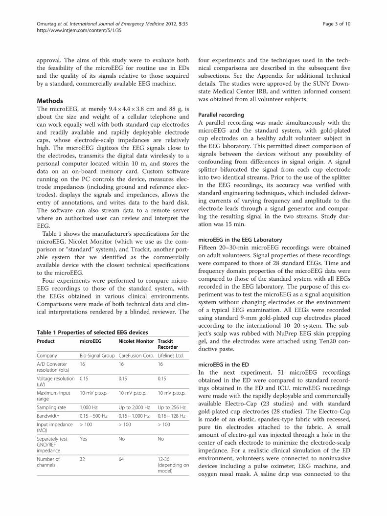
MethodsThe microEEG, at merely 9.4 × 4.4 × 3.8 cm and 88 g, isabout the size and weight of a cellular telephone andcan work equally well with both standard cup electrodesand readily available and rapidly deployable electrodecaps, whose electrode-scalp impedances are relativelyhigh. The microEEG digitizes the EEG signals close tothe electrodes, transmits the digital data wirelessly to apersonal computer located within 10 m, and stores thedata on an on-board memory card. Custom softwarerunning on the PC controls the device, measures elec-trode impedances (including ground and reference elec-trodes), displays the signals and impedances, allows theentry of annotations, and writes data to the hard disk.The software can also stream data to a remote serverwhere an authorized user can review and interpret theEEG.Table 1 shows the manufacturer’s specifications for the
microEEG, Nicolet Monitor (which we use as the com-parison or “standard” system), and Trackit, another port-able system that we identified as the commerciallyavailable device with the closest technical specificationsto the microEEG.Four experiments were performed to compare micro-
EEG recordings to those of the standard system, withthe EEGs obtained in various clinical environments.Comparisons were made of both technical data and clin-ical interpretations rendered by a blinded reviewer. The
Table 1 Properties of selected EEG devices
Product microEEG Nicolet Monitor TrackitRecorder
Company Bio-Signal Group CareFusion Corp. Lifelines Ltd.
A/D Converterresolution (bits)
16 16 16
Voltage resolution(μV)
0.15 0.15 0.15
Maximum inputrange
10 mV p.to.p. 10 mV p.to.p. 10 mV p.to.p.
Sampling rate 1,000 Hz Up to 2,000 Hz Up to 256 Hz
Bandwidth 0.15− 500 Hz 0.16− 1,000 Hz 0.16− 128 Hz
Input impedance(MΩ)
> 100 > 100 > 100
Separately testGND/REFimpedance
Yes No No
Number ofchannels
32 64 12-36(depending onmodel)
four experiments and the techniques used in the tech-nical comparisons are described in the subsequent fivesubsections. See the Appendix for additional technicaldetails. The studies were approved by the SUNY Down-state Medical Center IRB, and written informed consentwas obtained from all volunteer subjects.
Parallel recordingA parallel recording was made simultaneously with themicroEEG and the standard system, with gold-platedcup electrodes on a healthy adult volunteer subject inthe EEG laboratory. This permitted direct comparison ofsignals between the devices without any possibility ofconfounding from differences in signal origin. A signalsplitter bifurcated the signal from each cup electrodeinto two identical streams. Prior to the use of the splitterin the EEG recordings, its accuracy was verified withstandard engineering techniques, which included deliver-ing currents of varying frequency and amplitude to theelectrode leads through a signal generator and compar-ing the resulting signal in the two streams. Study dur-ation was 15 min.
microEEG in the EEG LaboratoryFifteen 20–30-min microEEG recordings were obtainedon adult volunteers. Signal properties of these recordingswere compared to those of 28 standard EEGs. Time andfrequency domain properties of the microEEG data werecompared to those of the standard system with all EEGsrecorded in the EEG laboratory. The purpose of this ex-periment was to test the microEEG as a signal acquisitionsystem without changing electrodes or the environmentof a typical EEG examination. All EEGs were recordedusing standard 9-mm gold-plated cup electrodes placedaccording to the international 10–20 system. The sub-ject's scalp was rubbed with NuPrep EEG skin preppinggel, and the electrodes were attached using Ten20 con-ductive paste.
microEEG in the EDIn the next experiment, 51 microEEG recordingsobtained in the ED were compared to standard record-ings obtained in the ED and ICU. microEEG recordingswere made with the rapidly deployable and commerciallyavailable Electro-Cap (23 studies) and with standardgold-plated cup electrodes (28 studies). The Electro-Capis made of an elastic, spandex-type fabric with recessed,pure tin electrodes attached to the fabric. A smallamount of electro-gel was injected through a hole in thecenter of each electrode to minimize the electrode-scalpimpedance. For a realistic clinical simulation of the EDenvironment, volunteers were connected to noninvasivedevices including a pulse oximeter, EKG machine, andoxygen nasal mask. A saline drip was connected to the

Omurtag et al. International Journal of Emergency Medicine 2012, 5:35 Page 4 of 10http://www.intjem.com/content/5/1/35
subject by taping a blunt needle to his or her arm. Bloodpressure (BP) monitoring was done by an automatedpressure cuff placed around the subject's arm for BP de-tection every 10 min during the EEG recording. Elec-trode impedances were measured at the beginning andend of each recording. Standard recordings consisted of7 ED studies and 11 ICU studies randomly selected fromall of the ED and ICU studies obtained in the prior 1 yearat SUNY Downstate Medical Center.
Assessment by a clinical neurophysiologistA board-certified clinical neurophysiologist reviewed 37de-identified 30-min EEGs obtained with either themicroEEG or the standard system. The data set con-sisted of 14 microEEG studies recorded in the ED withthe Electro-Cap, 13 microEEG studies recorded in theED with cup electrodes, 8 standard EEGs recorded inthe EEG laboratory, and 2 standard EEGs recorded inthe ICU. All recordings were reviewed using Insight II,Persyst Development Corp. (Prescott, AZ). The reviewerdetermined whether each recording was technically ad-equate for clinical interpretation, i.e., was not substan-tially obscured by artifacts that rendered the studyuninterpretable for clinical purposes.
Measures of agreementThe simultaneous parallel recording with the microEEGand the standard system provided a unique opportunityto compare the recorded signals in the time domain. Wecomputed the short time correlations between the twosignals and examined their values throughout therecording on all channels. The correlations were alsoused as a guide to focus visual inspection on specificsegments of the recording. Standard deviations, higherorder statistics, and Hjorth mobility and complexityparameters [13-15] for each system’s signals were alsoexamined.Frequency domain measures were used to compare
microEEG and standard system recordings in all of theexperiments. We began by computing the power spec-tral density (PSD) of each channel over a 500-s interval.Spectral properties were derived from the PSD data.Combinations of such indicators have been used with
varying levels of success to detect both normal and ab-normal EEG findings, as well as artifacts. Examples in-clude detection of rhythmic discharges in newborns [16],multi-morphologic ictal patterns in the human long-term EEG [17], muscle and electrical noise artifacts [18],seizure prediction (reviewed by [19]), early patient-specific seizure detection [20], classification of sleepstages [21,22], and identification of resting state [23] orepileptic [24] brain networks.When sampling rates for the microEEG and standard
system were unequal, the microEEG signal was resampled
to the time points of the standard system using cubicspline interpolation [25]. Whenever needed (as indicatedin the Appendix), the data were bandpass filtered withzero phase shift by a sixth order digital Butterworth filter.This diverse set of measures from both the time and fre-quency domains provided a comprehensive measure oftechnical performance.
ResultsVisual inspection of the parallel recordings with an APbipolar longitudinal montage did not reveal any clinicallysignificant difference between the studies. Figure 2shows a section of the parallel recordings displayed withInsight II (Persyst Corp.). Low-pass and notch filtersare off, and the resolution is set to high. The microEEGand standard EEG appear nearly identical, althoughthere is greater high frequency noise in the standardEEG (Figure 2, bottom panel). Visual inspection of theremaining microEEG studies revealed that none of thesignals contained unexplained artifacts or expected arti-facts (e.g., due to muscle, movement, EKG) at levelsgreater than those found in the standard recordings.
Parallel recordingFigure 3 illustrates the superimposition of 0.7 s of simul-taneous signal recorded from the microEEG and thestandard system at electrodes T6 and O1. These exam-ples were selected to show segments with low (r� 0.5)and high (r� 1) correlation. The T6 segment illustratessuppression of 60-Hz noise by microEEG. Close examin-ation of all 20 channels throughout the recordingsrevealed that imperfect correlation was due primarily torelatively greater 60-Hz noise in the standard system.Table 2 summarizes the statistics of correlation for the
entire recording over all electrodes. As expected, correl-ation was proportional to the amplitude of EEG signalsand inversely proportional to 60-Hz noise. For example,the average correlation for channel O2 during an eyes-closed segment of the EEG (when there is relatively highposterior alpha activity) was 0.960 ± 0.047 (mean ±standard deviation).Table 3 illustrates the sensitivity of time domain para-
meters to high-frequency noise. Again, the data wereobtained from analyzing the signal on all electrodesthroughout the recording. In particular, Hjorth mobilitywas higher in the standard EEG than microEEG andhighly sensitive to high frequency noise. This sensitivityis expected since it is interpretable as the standard devi-ation of the PSD [14]. By contrast, higher order statisticsand Hjorth complexity were unchanged when 60-Hznoise was removed from the signals. The skewness wasnot significantly different from zero and is not shown.Note that we have defined kurtosis as the fourth central

Figure 2 Typical segment from the EEG signals recorded by microEEG (top) in parallel with the standard system (bottom) shown in astandard EEG viewing environment (Insight II from Persyst Development Corp.). Insight's high-pass filter is on, and the resolution is set tohigh. Note that time and amplitude scales as shown in the viewer controls above the EEG traces are, respectively, 5 s and 5 μV.
Omurtag et al. International Journal of Emergency Medicine 2012, 5:35 Page 5 of 10http://www.intjem.com/content/5/1/35

Figure 3 Superimposed segments of microEEG and standardEEGs recorded in parallel from the same electrodes with asignal splitter. Electrode and correlation coefficient between thesignals during each segment is shown above the plots. Thesegment from T6 illustrates significantly lower 60-Hz line noise inthe microEEG compared to the standard EEG. Filters: 0.50-70 Hzbandpass.
Table 3 Time and frequency domain properties
Time domain Spectral
Stdev Kurt Mob(Hz)
Comp MF(Hz)
SEF75(Hz)
SE(μV)
mEEG (Notch off) 67.9 9.1 108.5 2.9 9.3 9.4 11.8
STD (Notch off) 74.7 6.9 149.4 2.3 15.6 29.0 11.4
mEEG (Notch on) 65.6 10.0 69.0 3.5 6.0 7.9 11.9
STD (Notch on) 67.9 8.9 71.8 3.2 6.8 9.4 12.0
microEEG and standard EEG recorded in parallel. Filters 0.5-70-Hz bandpass.Data shown are averages over all channels. mEEG,microEEG; STD, standard;Stdev, standard deviation; Kurt, kurtosis; Mob,mobility; Comp, complexity;MF,mean frequency; SE, spectral entropy. Kurtosis, mobility, complexity, andspectral entropy are dimensionless.
Omurtag et al. International Journal of Emergency Medicine 2012, 5:35 Page 6 of 10http://www.intjem.com/content/5/1/35
moment of the signal divided by the square of the vari-ance so that kurtosis equals 3 for a normal distribution.Figure 4 shows the PSD of the signal recorded by the
microEEG and standard system on two channels, T6 andO2. These channels contained a significant difference in10-Hz activity and were selected to illustrate this differ-ence. The curves are smoothed for better visibility ex-cept in the neighborhood of 60 Hz. Inspection of thePSD for all channels showed that the standard signal’s
Table 2 Averaged short time correlations
Notch filter Overall Eyes open Eyes closed
Off 0.860 ± 0.106 0.849 ± 0.109 0.877 ± 0.099
On 0.911 ± 0.070 0.902 ± 0.076 0.926 ± 0.058
Correlations between themicroEEG and standard EEG computed for 1-swindows and averaged over the recording for all electrodes. Filters: 0.5-70 Hzbandpass.
spectrum agreed well with the microEEG spectrum andwas within the confidence limits in all channels and fre-quencies. Quantities derived from the PSD also had arange of sensitivities to high frequency noise as demon-strated by the results in Table 3. Note that notch filter-ing both signals drastically reduced the difference inSEF75 and resulted in good agreement.
microEEG in the EEG LaboratoryComparison of the band power of the microEEG andstandard system indicated that there was good agree-ment in all frequency ranges except in the lowest range,
Figure 4 Examples of the power spectral density of microEEGand standard EEG signals recorded in parallel. The curves weresmoothed except near 60 Hz. The shaded zone represents the 95%confidence range for the microEEG signal (the 95% confidencerange for the standard EEG was essentially the same and is notshown). Filters: 0.50-70-Hz bandpass.

Omurtag et al. International Journal of Emergency Medicine 2012, 5:35 Page 7 of 10http://www.intjem.com/content/5/1/35
0–4 Hz, and at 60 Hz. The mean band power of eachchannel averaged across recordings was also examinedand found to have a similar agreement between the twodevices. In the ranges alpha–beta4, the two systems’band powers were nearly equal, while in the theta andbeta5 ranges they were within one standard deviation ofeach other. The standard deviations were calculatedfrom the variability across recordings and channels.Lower power in the 0–4 Hz band in the microEEGrecordings was due to the difference in the hardwarehigh-pass filters: the low frequency cutoff in the micro-EEG was set at 1 Hz in these experiments compared to0.16 Hz in the standard device. Lower 60-Hz noise inthe microEEG compared to the standard system likelyresulted from the shorter EEG electrode cable lengths asthe common mode rejection ratio of the two systems isthe same. This experiment demonstrated that microEEGsignals do not contain activity at levels that are unex-pectedly different in any frequency range from thoserecorded by the standard device.
microEEG in the EDFigure 5 shows that there was good agreement betweenthe microEEG and the standard system in all frequencybands except at 60 Hz, where the microEEG power wasless. The agreement at 0–4 Hz is much better than inthe recordings from the EEG laboratory because themicroEEG hardware high-pass cutoff was set to 0.15 Hz.The microEEG recordings with the cup electrodes hadresults (not shown) similar to those with the Electro-Cap with the exception of somewhat higher power near60 Hz. The mean impedance of Electro-Cap electrodeswas 15.9 ± 17.6 kΩ with median 8 kΩ and range 0.9 to80 kΩ. The mean impedance of cup electrodes was5.3 ± 4.2 kΩ with median 4.3 kΩ and range 1.2 to 33.3kΩ. The impedance of each channel is taken as the
Figure 5 Band power for the microEEG and standard system.microEEG with Electro-Cap in the ED (28 recordings), standardsystem in the ICU (11 recordings), and standard system in the ED(7 recordings). Error bars indicate the standard deviation of variabilitydue to differences among channels and recordings. Filters 0.5-70 Hzbandpass.
mean of the impedance at the start and end of eachrecording. The inter-recording variability of impedancewas greater than the intra-recording variability. For eachrecording, the value of the impedance at the end of arecording was strongly correlated to the value at thestart [r= 0.84 (Electro-Cap) and 0.83 (cup electrodes)].We note that good agreement between the microEEG inthe ED and the standard EEG was achieved even thoughthe electrode cap electrode impedances were generallysubstantially greater than the < 5 kΩ impedance IFCNstandard for digital EEG machines [26].
Assessment by a clinical neurophysiologistTwo board-certified clinical neurophysiologists inde-pendently evaluated the technical quality of ten ran-domly selected microEEG recordings done in the EDand described in this article (equally divided betweencup electrodes and Electro-Cap); they found all of themsuitable for making clinically significant interpretations.We then designed the blinded study described in Meth-ods. The blinded reviewer was asked, “Is the recordingclinically acceptable?" The results show that the micro-EEG in the ED performed better than the standard sys-tem in the ICU but not as well as the standard system inthe EEG laboratory (Table 4). The percentage of Yesanswers for the microEEG was significantly lower thanfor the standard EEG from the EEG laboratory (z = 4.6,p < 0.05 test of proportions) but not different from thestandard EEG if the EEG laboratory and ICU recordingswere not distinguished (z = 1.21, p > 0.05 test of propor-tions). Reasons given for clinically unacceptable record-ings included wavering baseline, sharply contouredartifacts especially in T5 and T6, excessive EKG artifacts,60-Hz artifacts, bursts of diffuse artifacts, and muscleartifacts on all leads.
DiscussionOver the past 2 decades, EEG technology hasimproved dramatically. Large analog machines withpaper recordings have been replaced by much smaller,computer-based digital machines, with all the asso-ciated advantages of digital recording and data storage.Preamplifier input impedances have risen without sac-rificing CMRR. Despite these advances, recording a
Table 4 Responses given to the question "Is therecording clinically acceptable?"
Yes (%) Maybe (%) No (%)
microEEG/E-Cap/ED 60 27 13
microEEG/Cup/ED 58 34 8
Standard/EEG lab 88 12 0
Standard/ICU 0 50 50
Blinded evaluation by a neurologist of a set of recordings that contained bothmicroEEG and standard recordings.

Omurtag et al. International Journal of Emergency Medicine 2012, 5:35 Page 8 of 10http://www.intjem.com/content/5/1/35
technically acceptable EEG in electrically hostile envir-onments such as the ED remains a challenge, espe-cially with uncooperative patients in the crampedconfines of a busy ED. The most significant challengesare the following: (1) high line noise (60 Hz) in therecorded signals due to high ambient noise levels, longelectrode wires, and relatively high electrode impedancesand inter-electrode impedance differences; (2) timeneeded to attach a full set of EEG electrodes and achievelow electrode-scalp impedances; (3) around the clockavailability of trained EEG technologists; and (4) limitingphysical access to the patient with the electrode wires andEEG equipment.This study demonstrates that the microEEG can over-
come all of these obstacles. Its miniature size, a built-inrechargeable battery power source, and wireless trans-mission of digitized EEG data eliminate the physical ac-cess problems and space requirements associated withEEG electrode wires, recording equipment, and powercables. The engineering specifications of the microEEG(e.g., A/D converter resolution, sampling rate, input im-pedance, CMRR, number of channels, etc.) are compar-able to currently available commercial systems. However,because the microEEG is wireless and small enough tobe rigidly attached to the patient, for example on thehead using a headband or on an electrode cap, it can beimplemented with very short electrode wires. The shortcables, combined with the on-board DC power source,resulted in microEEG signals having less contaminationwith 60-Hz noise than did those of the standard system.The lower line noise in microEEG signals was apparentin the simultaneous parallel recording from a volunteersubject with standard cup electrodes (Figures 2, 3, 4). Itwas also seen across 28 recordings obtained in the EDwith the microEEG and the Electro-Cap compared to 18recordings made with the standard system and cup elec-trodes in the ED or ICU (Figure 5).These experiments also demonstrated high concord-
ance between the microEEG and the standard system ofspectral properties in the frequency range of physiologicEEG activity (Figures 4 and5 and Table 3, “spectral” col-umns with notch filter on). Not surprisingly, there wasalso high agreement in the time domain between themicroEEG and standard system recordings when the dif-ference in 60-Hz noise was reduced with the notch filter(Table 2 and Table 3, “time-domain” columns, notch fil-ter on). The fact that notch filtering the signals causedthe mean levels of all measures to become nearly equalbetween the two systems (Table 3), combined with thedata shown in Figure 3, demonstrates that the source ofdifferences between the systems was the relatively higherline noise in the standard system. In other words, com-prehensive frequency and time domain analyses of EEGsignals recorded with the microEEG and standard
system did not reveal any device-specific differencesother than the generally higher 60 Hz noise in the stand-ard system. Thus, advantages deriving from the micro-EEG’s ease of use can be obtained without compromiseby substituting the microEEG for a standard EEG ma-chine when≤ 26 recording channels are needed.The absence of systematic differences in signal proper-
ties between the microEEG and standard system isreflected in the blinded assessment of EEGs from bothsystems by an experienced clinical neurophysiologist. Asshown in Table 4, the fraction of microEEG studiesrecorded in the ED with either cup electrodes or theElectro-Cap considered definitely acceptable for clinicalinterpretation was 58 and 60%, respectively, comparedto 88% for EEGs recorded with the standard system andcup electrodes in the EEG laboratory and 0% of standardsystem studies from the ICU. Perhaps equally important,the fraction of clinically acceptable (as well as possiblyacceptable) microEEG studies in the ED did not differsignificantly between those recorded with cup electrodesand the Electro-cap. In a separate prospective study ofED patients presenting with altered mental status at ourinstitution, patients receive both a standard EEG withcup electrodes and a microEEG with the Electro-Cap.Data from the microEEG recordings in the prospectivestudy are not yet available for analysis.These data reveal a significant additional advantage of
the microEEG – its ability to generate high-quality EEGfrom electrodes with high electrode-scalp impedances.Specifically, when used with the Electro-Cap, the micro-EEG performed well with electrode impedances substan-tially higher than the 5 kΩ recommended byprofessional societies, i.e., mean 15.9 ± 17.6 kΩ (range0.9 to 80 kΩ) [26] and with interelectrode impedancedifferences within an EEG much higher than the 2 kΩreported to degrade CMRR, i.e., mean 8.7 ± 11.2 kΩ(range 0 to 60 kΩ) [27]. This result provides a mechan-ism to overcome the two remaining obstacles listedabove to achieving quality EEG recordings in the ED en-vironment. In the prospective study mentioned above,the mean setup time (i.e., time between initial contactwith the patient and beginning the EEG recording) was13 ± 7 min for the first 50 patients, nearly all of whomwere uncooperative and many of whom were agitated.There is also no risk of an agitated patient accidentallydislodging or pulling off electrode wires after the elec-trodes are attached. In a later phase of this study, themicroEEG and Electro-Cap hardware will be used bypersonnel with minimal formal training in EEG elec-trode placement and EEG equipment (e.g., EKG techni-cians) to obtain EEGs on ED patients with AMS.However, the data already obtained with EEG technolo-gists in the ED [28], and with non-EEG technologists ap-plying EEG electrodes in other research studies on

Omurtag et al. International Journal of Emergency Medicine 2012, 5:35 Page 9 of 10http://www.intjem.com/content/5/1/35
healthy subjects [29], strongly suggest that the micro-EEG technology in combination with readily availableand rapidly deployable electrode arrays will permit rapidacquisition of EEGs in the ED by personnel without thetraining and experience of EEG technologists. Since theED is one of the most challenging environments forrecording EEG, we expect that these results will be rele-vant for recording EEG in other challenging environ-ments such as ICUs, as well as in the EEG laboratory.A limitation of this study was that it used recordings
obtained only from healthy adult volunteers and conse-quently involved no abnormal EEG patterns. This fol-lowed from the fact that it was designed as an initialpilot study to demonstrate the feasibility and safety ofthe microEEG as a prerequisite to a separate study of itsdiagnostic accuracy with patients.
ConclusionsmicroEEG is a miniature, wireless, battery-powered EEGdevice with engineering specifications equivalent tothose of much larger commercially available EEGmachines.The analog EEG signals are amplified and digitized
within the device, and then transmitted wirelessly to alaptop computer within 10 meters. Comprehensivetime and frequency domain analyses of microEEGrecordings from normal volunteers in an EEG labora-tory and emergency department revealed neither unex-pected signals nor significant differences from EEGsrecorded with a Nicolet Monitor machine. microEEGis typically placed on or near the patient’s head andcan record high-quality EEG from electrodes with highelectrode-scalp impedances. Its noise immunity isenhanced by the short length of electrode wires fromscalp to device. This feature is particularly useful whenthe device is used with an electrode cap, or in a set-ting such as the emergency department where timeand space constraints may limit a technologist’s abilityto achieve and maintain low electrode impedancesthroughout the recording.
Appendix AThe following 20 channels were used in the parallelrecording: FP1, FP2, F3, F4, C3, C4, P3, P4, O1, O2, F7,F8, T3, T5, T6, FZ, CZ, PZ, LOC, and ROC. Samplingrates for the microEEG and standard system were 200and 500 Hz, respectively. For the recordings with themicroEEG in the EEG laboratory, we used the following16 channels: FP1, FP2, F3, F4, C3, C4, P3, P4, O1, O2,F7, F8, T3, T4, T5, and T6. The sampling rate was250 Hz. Signals from electrodes F7, F8, T5, and T6 wereused in the analyses for the microEEG in the ED. Incomputing the power spectral density (PSD), the multi-taper method [30] with six orthogonal tapers was used,
with the size of the time series defined as the length ofthe FFT (Matlab’s function pmtm). The spectral proper-ties derived from EEG data were as follows: band power:the integral of the PSD over specified frequency ranges.The ranges used were delta (0 ≤ f < 4 Hz), theta(4 ≤ f < 8 Hz), alpha (8 ≤ f < 12 Hz), beta1 (12 ≤ f < 14 Hz),beta2 (14 ≤ f < 16 Hz), beta3 (16 ≤ f < 20 Hz), beta4(20 ≤ f < 24 Hz), beta5 (24 ≤ f < 32 Hz), high frequency(32 ≤ f < 70 Hz), and power line noise (59 ≤ f < 61 Hz).Band power can be considered as a frequency-specificcontribution to the overall variance of the signal. Meanfrequency: the first moment of the normalized PSD.SEF75: The 75% spectral edge frequency, that is, the fre-quency that contains 75% of all the power in thespectrum (starting at f = 0 Hz). Spectral entropy: the en-tropy of the normalized PSD interpreted as a discreteprobability distribution. This ranges between 0 for amaximally concentrated PSD tothe value log2(N), for auniform distribution, where N is the total number ofdiscrete frequencies in the PSD (N was constant for allchannels and for both systems).
Competing interestsSupported by NIH grant 1RC3NS070658 to Bio-Signal Group (BSG), with asubcontract to SUNY Downstate Medical Center. André Fenton is a founderof BSG. Samah Abdel-Baki and Ahmet Omurtag are employees of Bio-SignalGroup and with André Fenton are co-inventors listed on a patent applicationbased on the microEEG (pub. no. WO/2010/129026. Title: EEG Kit). ACGserves on the BSG advisory board. All income derived from this position isdonated directly from BSG to the Downstate College of MedicineFoundation.
Authors’ contributionsDrs. Abdel Baki and Omurtag contributed equally to this paper. All authorsread and approved the final manuscript.
AcknowledgementsThe authors would like to thank Dr. Shweta Malhotra for her invaluableassistance in data collection and Dr. Richard Sinert for generously making EDresources available for this study.
Author details1Bio-Signal Group, 760 Parkside Avenue, Ste 206, Brooklyn, NY 11226, USA.2Departments of Neurology and Pediatrics, State University of New York,Downstate Medical Center, Brooklyn, USA. 3Department of Neurology, StateUniversity of New York, Downstate Medical Center, Brooklyn, USA.4Department of Emergency Medicine, State University of New York,Downstate Medical Center, Brooklyn, USA. 5Center for Neural Science, NewYork University, New York and Department Physiology & Pharmacology, StateUniversity of New York, Downstate Medical Center, Brooklyn, USA.6Departments of Neurology, and Physiology & Pharmacology, StateUniversity of New York, Downstate Medical Center, Brooklyn, USA.
Received: 9 April 2012 Accepted: 31 August 2012Published: 24 September 2012
References1. Kanich W, Brady WJ, Huff JS, Perron AD, Holstege C, Lindbeck G, et al:
Altered mental status: evaluation and etiology in the ED. Am J EmergMed 2002, 20:613–617.
2. Riggio S: Psychiatric manifestations of nonconvulsive status epilepticus.Mt Sinai J Med 2006, 73:960–966.

Omurtag et al. International Journal of Emergency Medicine 2012, 5:35 Page 10 of 10http://www.intjem.com/content/5/1/35
3. Ziai WC, Schlattman D, Llinas R, Venkatesha S, Truesdale M, Schevchenko A,Kaplan PW: Emergent EEG in the emergency department in patients withaltered mental states. ClinNeurophysiol 2012, 123(5):910–917.
4. Saengpattrachai M, Sharma R, Hunjan A, Shroff M, Ochi A, Otsubo H, Cortez MA,Carter Snead O: Nonconvulsive Seizures in the Pediatric Intensive Care Unit.Epilepsia 2006, 47(9):1510–1518.
5. Bautista RE, Godwin S, Caro D: Incorporating abbreviated EEGs in theinitial workup of patients who present to the ER with mental statuschanges of unknown etiology. J ClinNeurophysiol 2007, 24(1):16–21.
6. Moeller JJ, Kurniawan J, Gubitz GJ, Ross JA, Bhan V: Diagnostic accuracy ofneurological problems in the emergency department. Can J NeurolSci2008, 35:335–341.
7. Moulin T, Sablot D, Vidry E, Belahsen F, Berger E, Lemounaud P, Tatu L,Vuillier F, Cochon A, Revenco E, Capellier G, Rumbach L: Impact ofemergency room neurologists on patient management and outcome.EurNeurol 2003, 50:207–214.
8. Roberts K, Costelloe D, Hutchinson M, Tubridy N: What difference does aneurologist make in a general hospital?Ir. J Med Sci 2007, 176(3):211–214.
9. Abdel Baki SG, Omurtag A, Fenton AA, Zehtabchi S: The new wave: time tobring EEG to the emergency department. IntJEmerg Med 2011, 4:36–43.
10. Kaplan PW: Assessing the outcomes in patients with NCSE:nonconvulsive status epilepticus is underdiagnosed, potentiallyovertreated, and confounded by comorbidity. J ClinNeur 1999,16(4):341–342.
11. Drislane FW, Blum AS, Schomer DL: Unsuspected electrographic statusepilepticus in intensive care units. Neurology 1998, 50(suppl 1):A395–A396.
12. Bastani A, Kayyali H, Schmidt R, Qadir R, Manthena P: Wireless brainmonitoring in the emergency department. ConfProc IEEE Eng Med BiolSoc2005, 3:2502–2505.
13. Hjorth B: EEG analysis based on time domain properties.ElectroencephalogrClinNeurophysiol 1970, 29:306–310.
14. Drongelen WV: Signal Processing for Neuroscientists: An Introduction to theAnalysis of Physiological Signals. Burlington MA, USA: Academic Press/Elsevier; 2006:68.
15. Sanei S, Chambers JA: EEG Signal Processing. Wiley, New York, NY, USA:Wiley-Interscience; 2007:51.
16. Faul S, Boylan G, Connolly S, Marnane L, Lightbody G: An evaluation ofautomated neonatal seizure detection methods. ClinNeurophysiol 2005,116:1533–1541.
17. Meier R, Dittrich H, Schulze-Bonhage A, Aertsen A: Detecting epilepticseizures in long-term human EEG: anew approach to automatic onlineand real-time detection and classification of polymorphic seizurepatterns. J ClinNeurophys 2008, 25:119–131.
18. Delorme A, Sejnowski T, Makeig S: Enhanced detection of artifacts in EEGdata using higher order statistics and independent component analysis.Neuroimage 2007, 34:1443–1449.
19. Mormann F, Adrzejak RG, Elger CR, Lehnertz K: Seizure prediction: the longand winding road. Brain 2007, 130:314–333.
20. Minasyan GR, Chatten J, Chatten M, Harner RN: Patient-specific earlyseizure detection from scalp electroencephalogram. J ClinNeurophys 2010,27:163–178.
21. Fell J, Roeschke J, Mann K, Schaeffner C: Discrimination of sleep stages: acomparison between spectral and nonlinear EEG measures.ElectroencephalogrClinNeurophysiol 1996, 98:401–410.
22. Ferri R, Cosentino FI, Elia M, Musumeci SA, Marinig R, Bergonzi P:Relationship between delta, sigma, beta, and gamma EEG bands at REMsleep onset and REM sleep. Clinical Neurophsiol 2001, 112:2046–2052.
23. Mantini D, Perucci MG, Gratta CD, Romani GL, Corbetta GLM:Electrophysiological signatures of resting state networks in the humanbrain. PNAS 2007, 104:13170–13175.
24. Wendling F, Bartolomei F, Senhadji L: Spatial analysis of intracerebralelectroencephalographic signals in the time and frequency domain:identification of epileptogenic networks in partial epilepsy. PhilosTransact A Math PhysEngSci 2009, 367(Wendling F):297–316.
25. Press W, Teukolsky S, Vetterling W, Flannery B: Numerical Recipes. New York,NY, USA: Cambridge University Press; 2007:120–124.
26. Nuwer MR, Comi G, Emerson R, Fuglsang-Frederiksen A, Guerit JM, Hinrichs H,Ikeda A, Luccas FJ, Rappelsburger P: Int Fed ClinNeurophysiol.ElectroencephalogrClinNeurophysiol 1998, 106:259–261.
27. Legatt A: Impairment of common mode rejection by mismatchedelectrode impedances: quantitative analysis. Am J EEG Technol 1995,35:296–302.
28. Zehtabchi S, Grant AC, Abdel Baki SG, Omurtag A, Sinert R, Chari G, et al:Diagnostic accuracy of a novel emergency electroencephalographydevice (microEEG) in identifying non-convulsive seizures and other EEGabnormalities in the emergency department patients with alteredmental status. AcadEmerg Med 2012, 19(S1):s378.
29. Kolls BJ, Olson DM, Gallentine WB, Skeen MB, Skidmore CT, Sinha SR:Electroencephalography leads placed by nontechnologists using atemplate system produce signals equal in quality to technologist-applied, collodion disk leads. J ClinNeurophysiol 2012, 29(1):42–49.
30. Percival DB, Walden A: Spectral Analysis for Physical Applications: Multitaperand Conventional Univariate Techniques. New York, NY, USA: CambridgeUniversity Press; 1998:331–375.
doi:10.1186/1865-1380-5-35Cite this article as: Omurtag et al.: Technical and clinical analysis ofmicroEEG: a miniature wireless EEG device designed to record high-quality EEG in the emergency department. International Journal ofEmergency Medicine 2012 5:35.
Submit your manuscript to a journal and benefi t from:
7 Convenient online submission
7 Rigorous peer review
7 Immediate publication on acceptance
7 Open access: articles freely available online
7 High visibility within the fi eld
7 Retaining the copyright to your article
Submit your next manuscript at 7 springeropen.com