Embed Size (px)
Citation preview
CASM Asia-Pacific
Technical Manual of PIXE target preparation for every type of environmental specimen
Jun Itoh Researcher, Nishina Memorial Cyclotron Center, Japan Radioisotope Association
348 Tomegamori, Takizawa-mura, Iwate-gun, Iwate, Japan 020-0173
Koichiro Sera Director and Professor, Iwate Medical University Cyclotron Center
348 Tomegamori, Takizawa-mura, Iwate-gun, Iwate, Japan 020-0173
Victor B. Maglambayan
Consulting Geologist Philex Mining Corporation
27 Brixton St. Pasig City Philippines 1600
Satoshi Murao
Chairman, CASM Asia-Pacific Assistant Director, Geological Survey of Japan, AIST
1-1-1 Higashi, Tsukuba, Japan 305-8567
First version
CCOP-GSJ/AIST-GAI CASM Asia Workshop on the state-of-the-art of science and technology to protect the environment and people, Bandung.
Introduction This manual has been produced to assist those who are responsible or are willing to work for environmental issues. When faced with environmental degradation, most of the people, except for scientific professionals, often have difficulty to access to reliable chemical analysis. There are lines of reason for the inaccessibility. (1) it is due to the lack of scientific knowledge to select appropriate analytical method; (2) it is due to the lack of experience of sample preparation; and (3) it is because of insufficient budget which people can use. In order to solve these problems, usually suggested are education and training of key persons or fund raising for each environmental incident. Such actions are valuable but are often time-consuming and not proactive. For environmental issues, it is necessary to take proactive actions than reactions. And for such purpose, we should avoid laborious method for chemical analysis of environmental specimens. We must find something which gives us easier, quicker, sensitive and versatile analysis. Also we must establish a scheme in which people can easily cooperate to get analytical result and share idea.
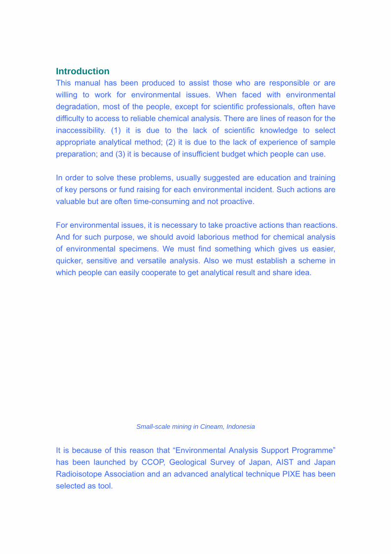
Small-scale mining in Cineam, Indonesia
It is because of this reason that “Environmental Analysis Support Programme” has been launched by CCOP, Geological Survey of Japan, AIST and Japan Radioisotope Association and an advanced analytical technique PIXE has been selected as tool.
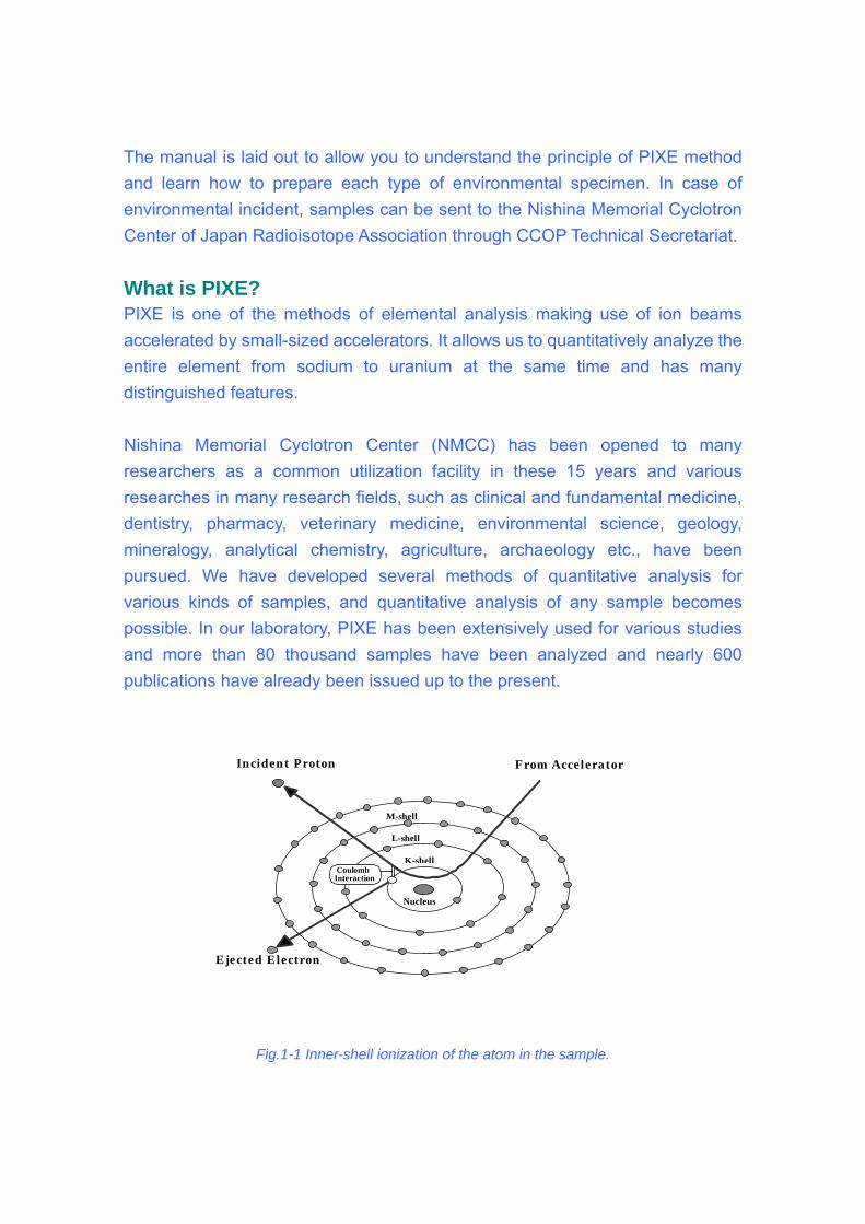
The manual is laid out to allow you to understand the principle of PIXE method and learn how to prepare each type of environmental specimen. In case of environmental incident, samples can be sent to the Nishina Memorial Cyclotron Center of Japan Radioisotope Association through CCOP Technical Secretariat. What is PIXE? PIXE is one of the methods of elemental analysis making use of ion beams accelerated by small-sized accelerators. It allows us to quantitatively analyze the entire element from sodium to uranium at the same time and has many distinguished features. Nishina Memorial Cyclotron Center (NMCC) has been opened to many researchers as a common utilization facility in these 15 years and various researches in many research fields, such as clinical and fundamental medicine, dentistry, pharmacy, veterinary medicine, environmental science, geology, mineralogy, analytical chemistry, agriculture, archaeology etc., have been pursued. We have developed several methods of quantitative analysis for various kinds of samples, and quantitative analysis of any sample becomes possible. In our laboratory, PIXE has been extensively used for various studies and more than 80 thousand samples have been analyzed and nearly 600 publications have already been issued up to the present.
Fig.1-1 Inner-shell ionizatio in the sample.
Incident ProtonN-shel
K-shell
M-shell
L-shell
Nucleus
from accelerator
Electron ejection
CoulombInteraction
From Accelerator Incident Proton
Ejected Electron
n of the atom
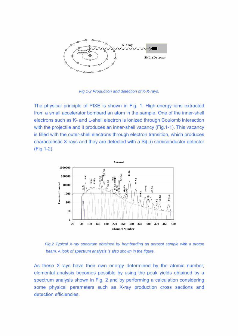
Fig.1-2 Production and detection of K-X-rays.
The physical princip ergy ions extracted
om a small accelerator bombard an atom in the sample. One of the inner-shell
le with a proton
beam. A look of spectrum analysis is also shown in the figure.
As th the atomic number, lemental analysis becomes possible by using the peak yields obtained by a
Eelectron Transition
K-Xray
Si(Li) Detector
le of PIXE is shown in Fig. 1. High-enfrelectrons such as K- and L-shell electron is ionized through Coulomb interaction with the projectile and it produces an inner-shell vacancy (Fig.1-1). This vacancy is filled with the outer-shell electrons through electron transition, which produces characteristic X-rays and they are detected with a Si(Li) semiconductor detector (Fig.1-2).
Aerosol
1
10
100
1000
10000
100000
1000000
20 60 100 140 180 220 260 300 340 380 420 460 500Channel Number
Cou
nts/
Cha
nnel
Fe-
Fig.2 Typical X-ray spectrum obtained by bombarding an aerosol samp
ese X-rays have their own energy determined byespectrum analysis shown in Fig. 2 and by performing a calculation considering some physical parameters such as X-ray production cross sections and detection efficiencies.
Kα
esc
ape
Ti-K
α
Ti- K
βFe
-Kβ
esc
ape
Mn-
Kα
Cr-
Kα
Cr-
Kβ
Fe- K
α
Fe- K
βSi- K
Al-K
S-Kα
Cl- K
α
Ga-
α
Ca-
Kα
K-K
αK
- Kβ
Ca-
Kβ
Zn-
K
Kα
Ni- K
βNi-K
α Cu-
Kα
Cu-
Kβ
Pb-Lα
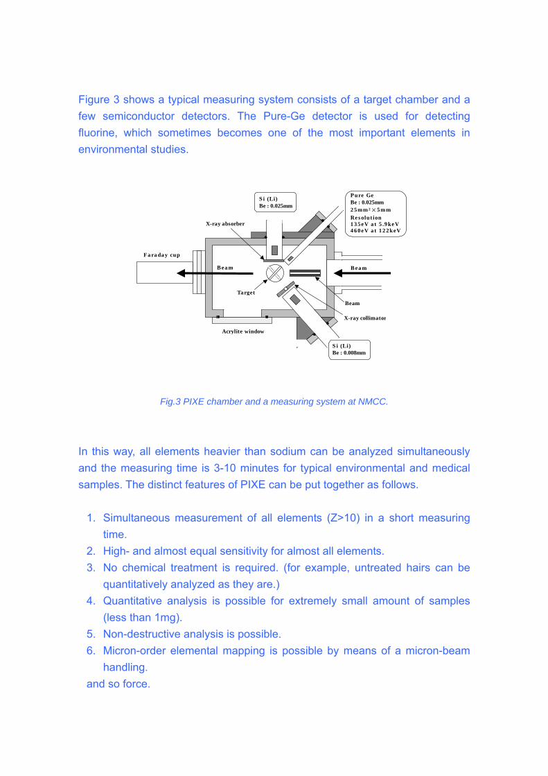
Figure 3 shows a typical measuring system consists of a target chamber and a few semiconductor detectors. The Pure-Ge detector is used for detecting
Fig.3 PIXE chamber and a measuring system at NMCC.
In this way, all e simultaneously and the measuring time is 3-10 minutes for typical environmental and medical samples. The distinct features of PIXE can be put together as follows.
2. High- and almost equal sensitivity for almost all elements.
tatively analyzed as they are.) unt of samples
5.
fluorine, which sometimes becomes one of the most important elements in environmental studies.
Pure GeBe : 0.025mm 2 5 mm 2×5 m m Res olut ion 1 3 5eV a t 5 .9 keV 4 6 0eV a t 1 2 2keV
S i (Li)Be : 0.025mm
X-ray absorber
lements heavier than sodium can be analyzed
1. Simultaneous measurement of all elements (Z>10) in a short measuring
time.
3. No chemical treatment is required. (for example, untreated hairs can be quanti
4. Quantitative analysis is possible for extremely small amo(less than 1mg). Non-destructive analysis is possible.
6. Micron-order elemental mapping is possible by means of a micron-beam handling.
and so force.
S i (Li)Be : 0.008mm
X-ray collimator
Target
Beam
B ea m Bea m
Acrylite window
Fa ra da y cup
2003
2004
1990
1991
1992
1993
1994
1995
1996
1997
1998
1999
2000
2001
2002
Fisc
al y
ear
Life sciences
Environmental
Other fields
Development
0 1000 2000 3000 4000 5000 6000
Number of analyzed samples
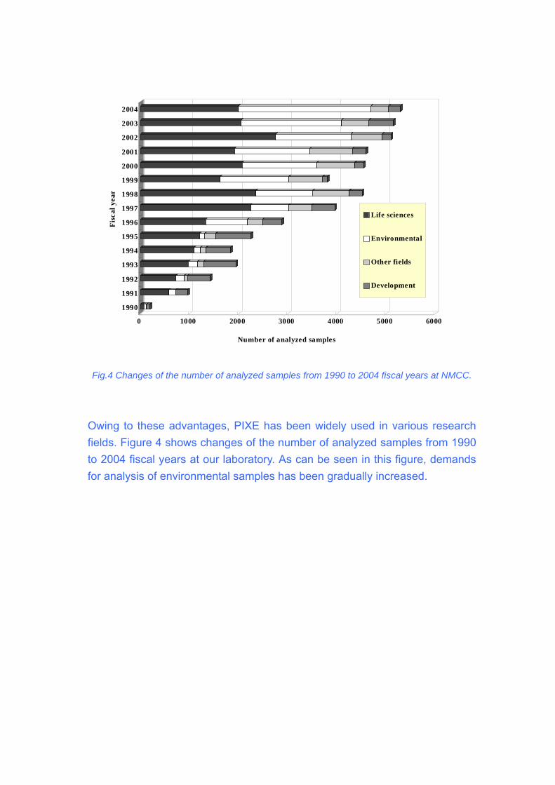
Fig.4 Changes of the number of analyzed samples from 1990 to 2004 fiscal years at NMCC.
Owing to these advantages, PIXE has been widely used in various research f
2004 fiscal years at our laboratory. As can be seen in this figure, demands r analysis of environmental samples has been gradually increased.
ields. Figure 4 shows changes of the number of analyzed samples from 1990tofo
Protocol of sample collection Water Volume should be about 5 cc. This amount should be sealed in micro-tube or
container.
should be preserved in cool condition.
ber and locality shoud be indicated on the surface of the container.
In case of mercury study, you need a careful sampling according to the strict
�・
・ Sample ・ Sample num
・
protocol as shown on the next section.
� A Manual for Sampling Mercury
oth river water and stream sediments will be analyzed for methyl mercury (MeHg) and total
e spectrometer (CV-AFS) method (Bloom,
B
Hg concentration by cold vapor atomic fluorescenc
Crecelius and Fitzgerald, 1988). Additionally, river water will be analyzed for major cations
by inductively coupled plasma mass spectrophotometer (ICP-MS) and for anions by ion
chromatography (IC). The geochemical data generated will enable us to distinguish the
subbasin waters of different chemistries and origin (Fetter, 1994).
Methodology for River Water Sampling
Materials
P) bottles with cap and filled with acid
e, 0.45-µm membrane filter, labels, waterproof pen, Ultrex nitric acid in squeeze
. Select sample site so that you are not standing in water that is being sampled. If you
stand in the creek, syringe water from the upstream section. Avoid sediments from
oves to open the syringe. Fresh hand gloves should be worn in every new
ampling site.
lected in bottles from every site using syringes. Each
Syringe, gloves, high-density polyethylene (HD
preservativ
bottle container
Steps
1
need to
falling into the creek sample site area. Also, do not disturb the sample site prior to
sampling.
2. Use gl
s
3. Four river water samples will be col
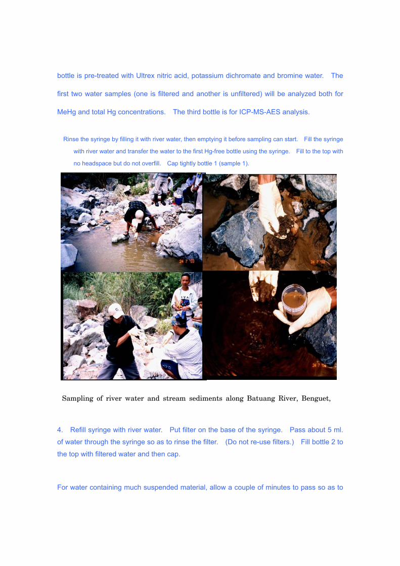
bottle is pre-treated with Ultrex nitric acid, potassium dichromate and bromine water. The
e syringe
with river water and transfer the water to the first Hg-free bottle using the syringe. Fill to the top with
no headspace but do not overfill. Cap tightly bottle 1 (sample 1).
4 l.
of water through the syringe so as to rinse the filter. (Do not re-use filters.) Fill bottle 2 to
aterial, allow a couple of minutes to pass so as to
first two water samples (one is filtered and another is unfiltered) will be analyzed both for
MeHg and total Hg concentrations. The third bottle is for ICP-MS-AES analysis.
Rinse the syringe by filling it with river water, then emptying it before sampling can start. Fill th
Sampling of river water and stream sediments along Batuang River, Benguet,
. Refill syringe with river water. Put filter on the base of the syringe. Pass about 5 m
the top with filtered water and then cap.
For water containing much suspended m
let the suspended material to settle to the bottom of the open end of the syringe. Release
the settled material and water (usually a few ml) into the bottle by putting the filter at the end
of the syringe.
5. Collect the third water sample into a 60 ml clear HDP bottle (third bottle). First, label
to fill your third bottle when the river water is
. Put in 7 drops of Ultrex nitric acid from a squeeze bottle container into your third bottle.
. Use a 60 ml HDP clear bottle to collect a fourth water sample for a single sample site.
ill the fourth bottle with about 5 ml of filtered water that is being sampled and shake the
er to fill a bottle when the water is turbid.
the bottle using waterproof pen and label. Fill your bottle using a syringe in the same
manner as in Step 2. For water with much suspended material, let the suspension settle
for a couple of minutes to the bottom of the syringe. Release this settled material and
water (usually a few ml) and do not collect.
You may need more than one filterNote:
turbid. Once filter gets clogged, remove and save filter after labeling with sample number.
Fill your third bottle with about 5ml of filtered water and shake the bottle with the cap on.6.
Pour out this rinse water. Refill with another 5ml of filtered water that is being samples and
rinse again. Then, fill the bottle with filtered water to about 90% capacity.
7
(Make sure to use gloves when handling nitric acid at all times.)
8
This is going to be for determining the anions in a sample. Label your bottle using
waterproof pen. Fill a new syringe with river water sample. For water containing much
suspended material, let the suspension settle to the bottom of the syringe (open end) for a
couple of minutes. Release this settled material and water (usually a few ml) and do not
collect.
9. F
bottle after putting the cap on. Pour out this rinse water. Refill with another 5 ml of filtered
water that is being sampled and rinse again. Fill the bottle with filtered water to about 90%
capacity. (Do not put acid into this bottle.)
Note: You may need more than one filt
Once filter gets clogged, remove and save filter after labeling. Keep this sample cool.
(This can be stored in ice chest/refrigerator and kept cool until analyzed.)
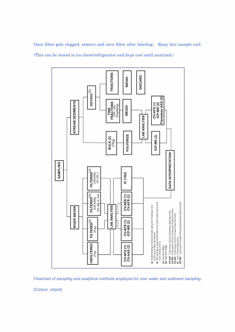
Flowchart of sampling and analytical methods employed for river water and sediment sampling.
(Corpus, unpub)
Methodology for Stream Sediments Sampling
tream sediments can be collected simultaneously with water samples. Each sample is
he stream sediments collected can be split into two samples: bulk (sand size and smaller)
oth bulk sediments and <64 µm fractions can be analyzed for MeHg and total Hg
aterials
h clear bottle with white cap (preferably polypropylene), labels, waterproof pen,
teps
ct area of fine sediment accumulation and try to avoid coarse sediments. Label
2. With gloves on, fill your bottle by scooping sediment directly into it. Decant any water
S
taken from low-velocity river channels along the river or creek bank where fine sediments
accumulate. Composite samples from the surface of the riverbed deposits can be collected
from five randomly selected points within a single sample site.
T
and <64 micron (silt size and smaller) sediment fractions.
B
concentration. The MeHg in sediments is collected by acid bromide/methyl extraction and
analyzed by aqueous phase ethylation, isothermal GC separation and cold vapor atomic
fluorescence (CV-AFS) detection using modified EPA method 1630. Meanwhile, the total
Hg in sediments is prepared by cold aqua regia digestion and analyzed by SnCl2 reduction,
dual amalgamation and CV-AFS detection using modified EPA method 1631.
M
Wide mout
scoop
S
1. Sele
sample bottle with waterproof pen.
after letting sample sit. Fill jar to about 90% capacity. Keep this sample cool if possible.
through a 0.064 mm nylon-mesh sieve.
t 60ºC.
estle the two sample fractions. Place both
properly labeled self-sealing plastic bags.
collected in polyethylene bottles for archive
References
loom, N.S., Crecelius, E. A. and Fitzgerald, W. F., 1988, Determination of volatile mercury
at the picogram level by low temperature gas chromatography with cold
Corpus, T
Fetter,
3. The <64 micron (silt size and smaller) sediment sample can be obtained by wet-sieving
4. Oven-dry the two sample fractions a
5. Grind separately on a porcelain mortar and p
in
6. A separate set of bulk sediments can be
purposes.
B
species
vapor atomic fluorescence detection. Analytica Chimica Acta 208, p. 151-161.
. J. (unpub), Small-scale gold mining in the Ambalanga River Basin, Benguet: An
assessment of anthropogenic mercury contamination in river water and stream
sediments, thesis draft, University of the Philippines Diliman
C.W., 1994, Applied Hydrogeology, 3rd ed., Macmillan College Publishing Company,
Inc., p. 420-423. (Fetter, 1994).
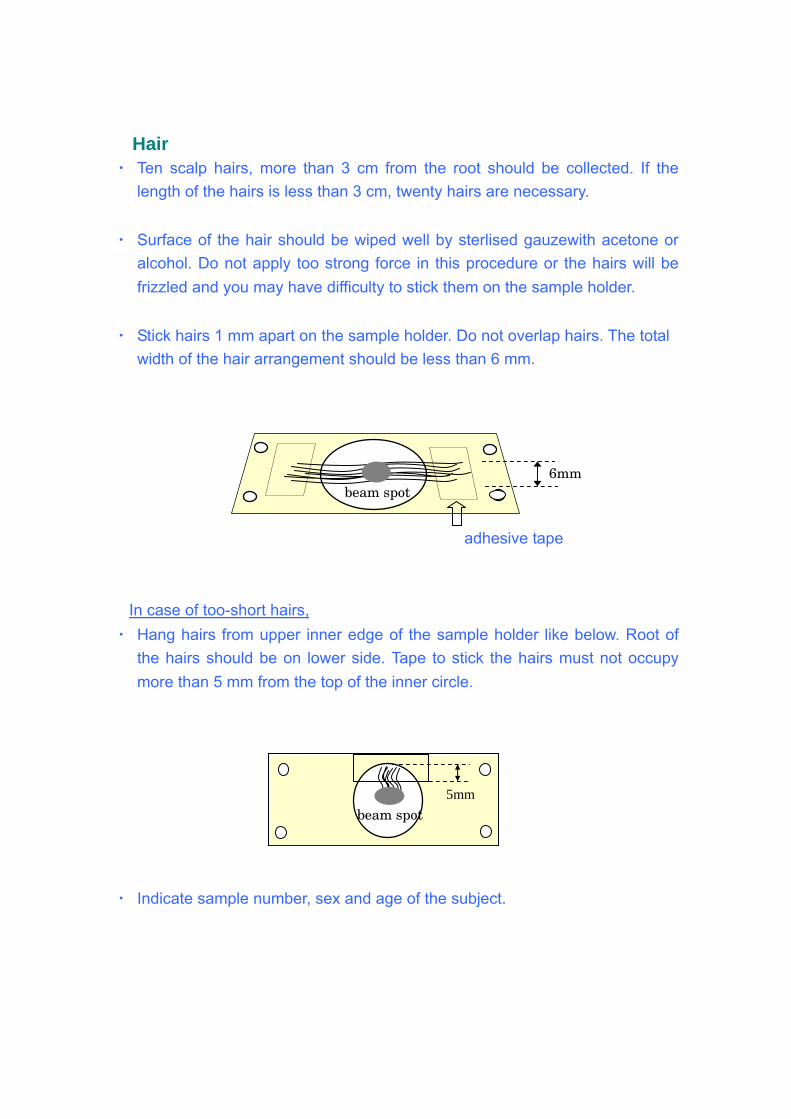
� Hair Ten scalp hairs, more than 3 cm from the root should be collected. If the
of the hairs is less than 3 cm, twenty hairs are necessary.
・ acetone or alcohol. Do not apply too strong force in this procedure or the hairs will be
・ otal width of the hair arrangement should be less than 6 mm.
In case of too-short hairs,
・
length
Surface of the hair should be wiped well by sterlised gauzewith
frizzled and you may have difficulty to stick them on the sample holder.
Stick hairs 1 mm apart on the sample holder. Do not overlap hairs. The t
6mm beam spot
adhesive tape
Hang hairs from upper inner edge of the sample holder like below. Root of wer side. Tape to stick the hairs must not occupy
Indicate sample number, sex and age of the subject.
・
the hairs should be on lomore than 5 mm from the top of the inner circle.
・
5mm beam spot
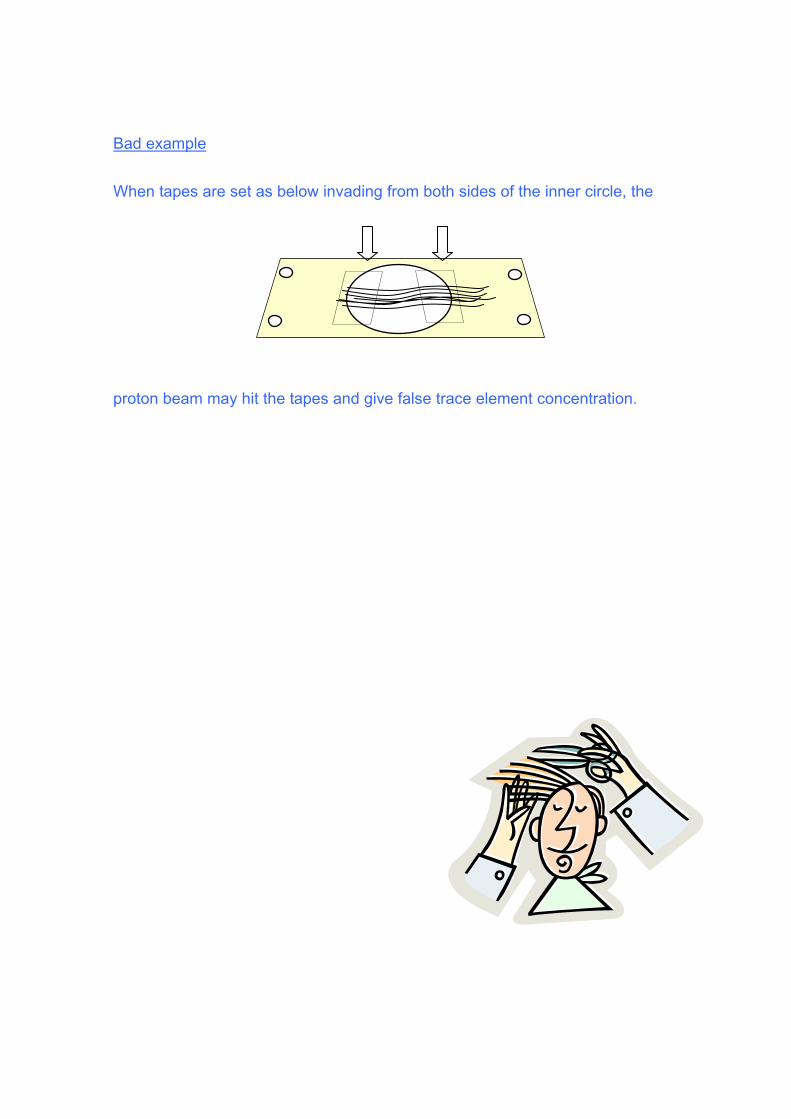
Bad example
from both sides of the inner circle, the
roton beam may hit the tapes and give false trace element concentration.
When tapes are set as below invading
p
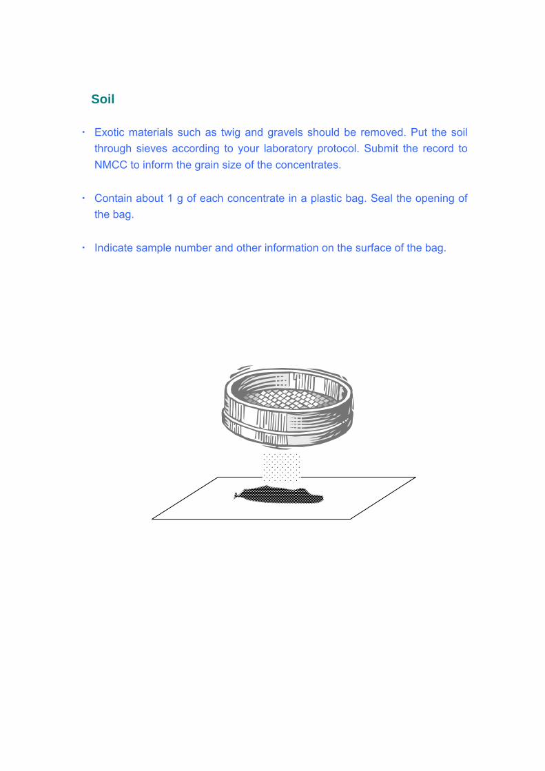
� Soil
materials such as twig and gravels should be removed. Put the soil through sieves according to your laboratory protocol. Submit the record to
・ g. Seal the opening of
the bag.
・ mple number and other information on the surface of the bag.
・ Exotic
NMCC to inform the grain size of the concentrates.
Contain about 1 g of each concentrate in a plastic ba
Indicate sa
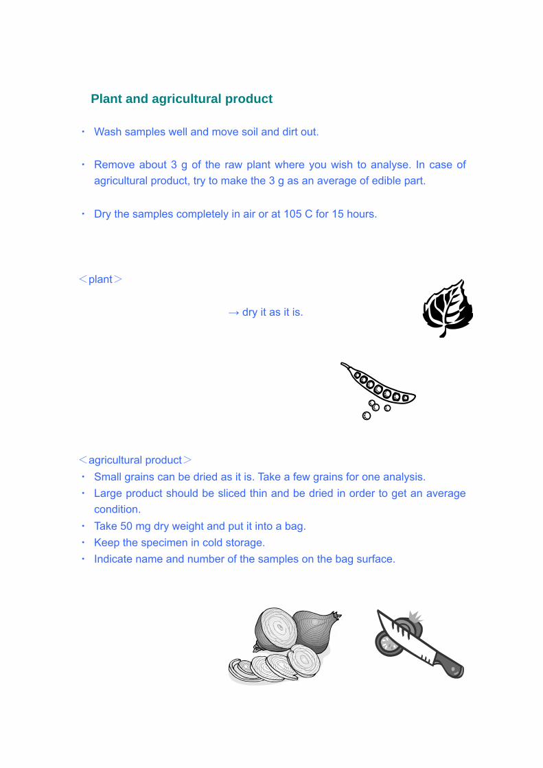
� Plant and agricultural product
dirt out.
wish to analyse. In case of agricultural product, try to make the 3 g as an average of edible part.
・
plant>
→ dry it as it is.
agricultural product> Small grains can be dried as it is. Take a few grains for one analysis.
be sliced thin and be dried in order to get an average
・ pecimen in cold storage. the bag surface.
・ Wash samples well and move soil and ・ Remove about 3 g of the raw plant where you
Dry the samples completely in air or at 105 C for 15 hours.
<
<
・
・ Large product shouldcondition.
・ Take 50 mg dry weight and put it into a bag. Keep the s
・ Indicate name and number of the samples on
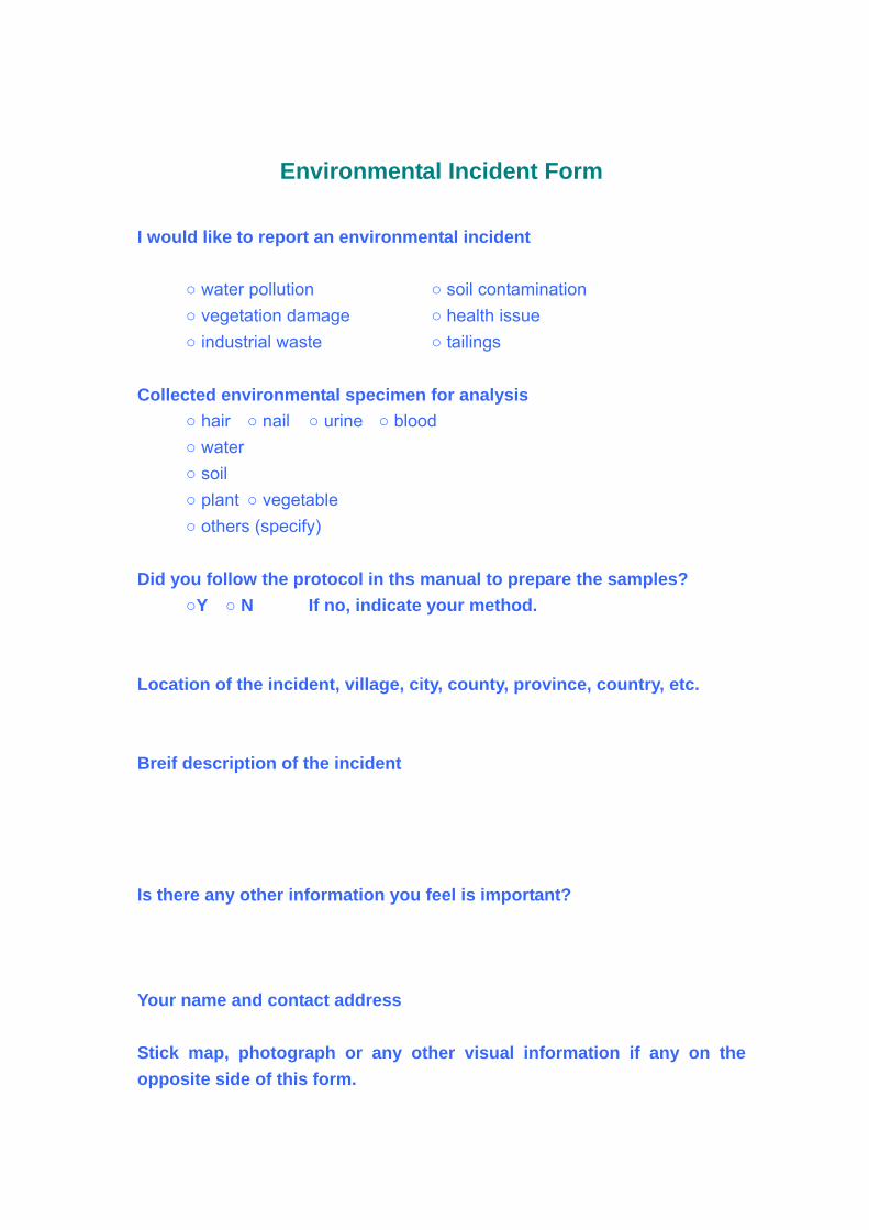
Environmental Incident Form I would like to re
nation ○ vegetation damage ○ health issue
l sp imen is ○ hair ○ nail ○ urine ○ blood
vegetable rs (specify)
id yo col in ths manual to prepare the samples?
our method.
ocation of the incident, village, city, county, province, country, etc.
reif description of the incident
there any other information you feel is important?
our name and contact address
visual information if any on the opposite side of this form.
port an environmental incident ○ water pollution ○ soil contami ○ industrial waste ○ tailings Collected environmenta ec for analys ○ water ○ soil ○ plant ○ ○ othe D u follow the proto ○Y ○ N If no, indicate y L B Is Y
Stick map, photograph or any other
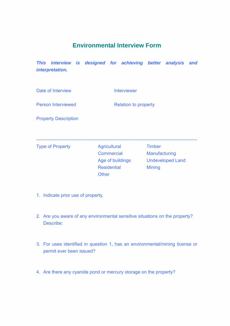
Environmental Interview Form
is designed for achieving better This interview analysis and interpretation.
ate of Interview Interviewer
d roperty
------------------------------------------------------------------------------------------------------ ype of Property Agricultural Timber
ngs oped Land
. Indicate prior use of property,
. Are you aware of any environmental sensitive situations on the property? Describe:
. For uses identified in question 1, has an environmental/mining license or permit ever been issued?
. Are there any cyanide pond or mercury storage on the property?
D Person Interviewe Relation to p Property Description --T Commercial Manufacturing Age of buildi Undevel Residential Mining Other 1 2 3
4
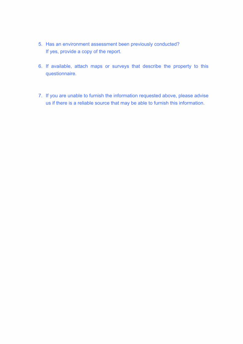
5. Has an environment assessment been previously conducted? If yes, provide a copy of the report.
ys that describe the property to this questionnaire.
. If you are unable to furnish the information requested above, please advise
us if there is a reliable source that may be able to furnish this information.
6. If available, attach maps or surve
7
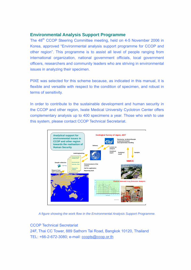
Environmental Analysis Support Programme he 48th CCOP Steering Committee meeting, held on 4-5 November 2006 in
mme for CCOP and
ecause, as indicated in this manual, it is exible and versatile with respect to the condition of specimen, and robust in
to the sustainable development and human security in e CCOP and other region, Iwate Medical University Cyclotron Center offers
TKorea, approved “Environmental analysis support prograother region”. This programme is to assist all level of people ranging from international organization, national government officials, local government officers, researchers and community leaders who are striving in environmental issues in analyzing their specimen. PIXE was selected for this scheme bflterms of sensitivity. In order to contributethcomplementary analysis up to 400 specimens a year. Those who wish to use this system, please contact CCOP Technical Secretariat.
1
10
100
1000
10000
100000
1000000
20 60 100 140 180 22 80 420 460 500
Cha
0 260 300 340 3
nnel Number
Cou
nts/
Cha
nn Fe-Kα
esc
apT
i-Kα
Ti-
K β
Fe-Kβ
esca
p
Mn-
K α
Cr-
K α
Cr-
K β
Fe-
K α
Fe-
K β
Si- K
Al-
K
S-K α
Cl-
K α
Ga-
K α
Ca-
K α
K-Kα
K-Kβ
Ca-
K β
Zn-
K α
Ni-K
β
Ni-K
α
Cu-
K α
Cu-
K β
Pb-
L α
Zn-
K β
Particle Induced X-ray Emission Analysis
NMCC
Environmental incident form
Environmental
Interview form
Request for analysis
CCOP Member Countries
Reviewing incidents/Sample preparation/sample description/data handling
Announcement of the scheme
Call for application
Reporting back
Report to each community
Request from each community
Labeling/packing
Deli
environmental issues in CCOP and other regiontowards the realisation of Human Security
Geological Survey of Japan, AISTAnalytical support for
very
Sample collection
Lecture on analysis
Analytical report Analytical
resultSamples
A figure showing the work flow in the Environmental Analysis Support Programme.
CCO4F, Thai CC Tower, 889 Sathorn Tai Road, Bangkok 10120, Thailand
P Technical Secretariat 2TEL: +66-2-672-3080; e-mail