Embed Size (px)
Citation preview

TECHNICAL REPORT
Risk assessment on Q fever
www.ecdc.europa.eu

ECDC TECHNICAL REPORT
Risk assessment on Q fever

Review group: Frode Forland, Andreas Jansen, Helena de Carvalho Gomes, Hanne Nøkleby, Ana-Belen Escriva, Denis Coulombier, Johan Giesecke
Stockholm, May 2010
ISBN 978-92-9193-210-8
doi:10.2900/28860
© European Centre for Disease Prevention and Control, 2010
Reproduction is authorised, provided the source is acknowledged.


TECHNICAL REPORT Risk assessment on Q fever
iii
Contents Executive summary ..................................................................................................................................................................................... 1 1 Request from the European Commission .................................................................................................................................................... 3 2 Background and methods ......................................................................................................................................................................... 3
Legal authority .............................................................................................................................................................................. 3 Evidence-based public health .......................................................................................................................................................... 3 Evidence-based methodologies ....................................................................................................................................................... 3 Questions from the Commission ...................................................................................................................................................... 4 Search strategies ........................................................................................................................................................................... 4 Assessment of the evidence ............................................................................................................................................................ 5 References: Background and methods ............................................................................................................................................. 5
3 General information on Q fever ................................................................................................................................................................. 6 4 Expert panel ............................................................................................................................................................................................ 7
Participants ................................................................................................................................................................................... 7 Blood ............................................................................................................................................................................................ 7 Pregnant women ........................................................................................................................................................................... 8 Chronic Q fever ............................................................................................................................................................................. 8 Spread and surveillance .................................................................................................................................................................. 8 Judgements, further steps .............................................................................................................................................................. 8
5 Blood ...................................................................................................................................................................................................... 9 Risk for asymptomatic blood donors experiencing bacteraemia ........................................................................................................... 9 Risk from blood donors in other EU countries after travel to the Netherlands ..................................................................................... 11 Risk from collecting blood from cured Q fever cases ........................................................................................................................ 11 Conclusions ................................................................................................................................................................................. 11 References: Blood ........................................................................................................................................................................ 12
6 Chronic Q fever ...................................................................................................................................................................................... 13 Search strategy and selection of studies ......................................................................................................................................... 13 Epidemiology ............................................................................................................................................................................... 13 Clinical features ........................................................................................................................................................................... 13 Conclusions ................................................................................................................................................................................. 14 References: Chronic Q fever ......................................................................................................................................................... 15
7 Vaccination ............................................................................................................................................................................................ 16 Conclusion .................................................................................................................................................................................. 16 References: Vaccination ............................................................................................................................................................... 16
8 Pregnant women .................................................................................................................................................................................... 17 Questions addressed .................................................................................................................................................................... 17 Selection of papers ...................................................................................................................................................................... 17 Evidence summary ....................................................................................................................................................................... 17 Answers to the posed questions .................................................................................................................................................... 19 References: Pregnancy ................................................................................................................................................................. 20
9 Spread and surveillance .......................................................................................................................................................................... 21 Transmission and risk of spread .................................................................................................................................................... 21 Surveillance................................................................................................................................................................................. 21 References: Surveillance ............................................................................................................................................................... 24
Annex 1: Strategies to identify and follow up chronic cases ........................................................................................................................... 26 Strategy 1: Targeted case-finding of persons with known heart valve lesions or vascular grafts ........................................................... 26 Strategy 2: Targeted case-finding for heart valve lesions with echocardiography of all patients with acute Q fever ................................ 26 Cost–benefit of targeted case finding for at-risk patients ................................................................................................................. 27 Strategy 3: Individual follow-up after acute Q fever infection with serology, together with raised awareness among the general population and physicians ............................................................................................................................................................................. 27 Lack of evidence, need for research ............................................................................................................................................... 27 References: Annex 1 .................................................................................................................................................................... 28
Annex 2 - Conflicts of interest .................................................................................................................................................................... 29 Annual Declaration of Interest 2010 ............................................................................................................................................. 29
Annex 3: Search strategies for Q fever ........................................................................................................................................................ 31 Annex 3: Evidence tables ........................................................................................................................................................................... 35
Chronic Q fever ........................................................................................................................................................................... 35 Pregnancy ................................................................................................................................................................................... 38 Surveillance................................................................................................................................................................................. 39

Risk assessment on Q fever TECHNICAL REPORT
iv

TECHNICAL REPORT Risk assessment on Q fever
1
Executive summary A risk assessment was carried out on a request from the European Commission to assess questions on Q fever and its transmission through blood, the health impact of chronic Q fever and the risks for pregnant women. With reference to the ongoing outbreak in the Netherlands, ECDC was also asked to address the question of cross-border spread and the need for better surveillance systems. The risk assessment was performed according to the principles of evidence-based methodologies, by defining search terms for each question, inclusion and exclusion criteria for identified studies and assessing the quality of the evidence. A review of the best available evidence was presented to, and discussed with, an expert panel with representatives from the Netherlands, France, Germany, the UK and the United States. The work has been undertaken simultaneously, and in coordination with, a risk assessment on Q fever from the European Food Safety Authority.
Acute Q fever is typically a mild, self-limiting, flu-like disease, but it sometimes presents with pneumonia, hepatitis and other symptoms. It can usually be successfully treated with a two-week course of doxycycline.
Coxiella burnetii is an obligate intracellular bacterium that can be transmitted through blood and tissues. The risk of such a transmission is low, and there is only one documented case in the literature. During an outbreak, the endemic area should be defined and safety precautions should be considered, such as active surveillance among blood and tissue recipients, screening of donors, and screening of blood and tissue products. For travellers returning from the area within the duration of the incubation period and with asymptomatic bacteraemia (five to seven weeks), deferral from blood donation may be considered until the end of this period. An antibiotic course could be considered for blood recipients at particularly high risk, such as patients with heart valve defects. Donors who have had an acute Q fever infection should be deferred from giving blood for two years following the date of confirmed cure from acute infection. The benefits of implementation of such measures must be carefully considered against the negative impacts they could have on blood supply in the area. A strategy for risk communication should be developed.
Chronic Q fever is a serious complication of an acute Q fever infection that develops in some 2% of acute symptomatic cases, and the fatality rate may vary from 5% to 50%. Chronic Q fever causes endocarditis in risk groups like people with previous heart valve disease, a prosthetic valve or vascular graft. Patients with cancer or those who are immunosuppressed are also at a higher risk. Chronic Q fever must be treated for at least one year, in some cases for the lifetime with more than one antibiotic. Surgical replacement of damaged heart valves might be needed.
Effective detection of, and treatment for, acute Q fever is the best strategy for avoiding chronic cases. Three possible strategies are described: (1) awareness raising among healthcare staff and the public to address the risk groups; (2) active follow-up with serology for known risk groups to detect and treat an acute Q fever infection early; or (3) refer all known acute Q fever patients to echocardiography for active case finding and follow-up.
There is a need to initiate good prospective cohort studies and controlled trials (when ethically feasible) to obtain more robust evidence on how to prevent and inhibit outbreaks of Q fever in the public health field, and on how to diagnose and treat acute and chronic disease at the clinical level.
Evidence on Q fever in pregnancy is very limited and comes mainly from observations and research in domestic and experimental animals, seroprevalence studies, case reports, and one case series including 53 pregnant women over a 15-year period. The risk for pregnant women of severe Q fever outcomes compared with the risk for the general (female) population cannot be quantified based on currently available evidence. Several cases of Coxiella burnetii infection during pregnancy resulting in adverse pregnancy outcomes have been reported. In some of the cases Coxiella burnetii was found in the placenta and in fetal tissue. Coxiella has also been identified in human breast milk but no case of transmission to the breastfed child has been validated.
There is some indication that long-term antibiotic therapy with cotrimoxazole has the potential to prevent severe pregnancy outcomes, but the evidence is based on a case series without randomisation and without controlling for potential biases. As long as no further evidence from high quality treatment studies is available, pregnant women with diagnosed Q fever infection should be treated with antibiotics throughout the remaining pregnancy. However, the scientific basis for this recommendation is weak, and ECDC would strongly recommend that randomised controlled trials are performed to obtain more reliable evidence.
Pregnant women should be advised not to visit farms in affected areas. ECDC does not recommend against breastfeeding except in cases of chronic disease that need long-term treatment of the mother.

Risk assessment on Q fever TECHNICAL REPORT
2
A formaline-inactivated whole-cell Q fever vaccine is produced and licensed in Australia. The vaccine is effective, but pre-vaccination testing is necessary due to high reactogenicity in persons who have earlier been infected with Coxiella burnetii, making the vaccine more suitable for defined risk groups than for general vaccination.
Available evidence suggests an effective range of airborne spread of Coxiella burnetii of less than 5 km. The risk of airborne spread from the Netherlands is therefore limited to neighbouring countries (i.e. Germany, Belgium), and to areas close to outbreak sources. Active surveillance or case finding for acute Q fever in possible risk groups (i.e. pregnant women, patients with heart valve or vascular diseases) on a local level and for a defined period of time is reported feasible and an efficient method for detecting acute infections. In areas adjacent to epidemic settings (≤ 5 km from the source), awareness campaigns among healthcare providers should be initiated. If the area also affects other Member States, the responsible public health authorities need to inform their cross-border counterparts. Sharing of information between public health and veterinary authorities would facilitate an early recognition of an outbreak. Further, the health and veterinary authorities at national and local levels should take the neccessary action to stop an outbreak.

TECHNICAL REPORT Risk assessment on Q fever
3
1 Request from the European Commission With reference to the letter of 3 February 2010 from the European Commission, Directorate-General for Health and Consumers, Luxembourg,
The Commission is concerned about the increase in the number of cases in the Netherlands during the last 2 years. In the Netherlands the human cases appear to have doubled for the year 2009. We are aware of the specific epidemiology of Q fever in the Netherlands, probably related to intensive goat farming in the proximity of densely populated areas, factors that seem to be unique to this country; however a possible spread to other geographical locations (e.g. Belgium and Germany) might be possible. In this perspective we are sure that a package of options will be important in order to facilitate Member States planning measures at national level and to limit the impact of the current outbreak in a coordinated way.
The Commission further asks ECDC to evaluate the risk and safety of blood transfusions, in particular with regard to potential donors who are asymptomatic or still in an incubation phase of the disease. In addition they ask for an assessment of possible elements and procedures to strengthen the surveillance of new cases, particularly in view of the oncoming season of higher incidence, and to provide information about the impact on health of chronic disease and for risk groups like pregnant women.
The Commission suggests a coordinated action and a close cooperation between ECDC and the European Food Safety Authority (EFSA) on this matter.
2 Background and methods Legal authority According to the founding regulation of ECDC, Regulation (EC) No 851/2004i
ECDC shall:
Art 9(2), ‘the Centre may be requested by the Commission, the Member States, third countries and international organisations (in particular the WHO) to provide scientific or technical assistance in any field within its mission. Scientific and technical assistance provided by the Centre shall be based on evidence-based science and technology.’
• search for, collect, collate, evaluate and disseminate scientific data (Art 3(2)(a)); • provide scientific opinions and timely information (Art 3(2)(b),(c)); • exchange information, expertise and best practices (Art 3(2)(e)); and • facilitate the development and implementation of joint actions (Art 3(2)(e)).
Evidence-based public health Evidence-based decision-making in a public health setting is to carefully incorporate the best available scientific evidence from research and other reliable sources with considerations of values, perceived needs and recourses in the given context. Evidence-based medicine is often defined as the integration of expertise, values, and the best available evidence into the decision-making process [1].
A public health decision might be rather complex, and needs to take several determinants of health into account, like genetic factors, lifestyle, physical environment, socio-economic conditions, biological environment and health services at different levels [2]. Most of these factors are relevant to the prevention and control of a Q fever outbreak.
Evidence-based methodologies ECDC has tried to carry out this risk assessment in accordance with the following steps of evidence-based methodologies:
• Formulate questions. • Search for evidence. i Regulation (EC) No 851/2004 of the European Parliament and of the Council of 21 April 2004 establishing a European centre for disease prevention and control. OJ L 142, 30.4.2004, p. 1.

Risk assessment on Q fever TECHNICAL REPORT
4
• Assess the evidence. • Formulate an answer. • Disseminate and implement. • Evaluate.
The Commission required ECDC to work in coordination and close cooperation with EFSA on this topic because it also involves animal health and food safety. This was achieved by sharing documents in progress, by appointing a representative from ECDC to be part of the EFSA expert panel (Howard Needham), and by an EFSA representative participating in ECDC’s expert meeting in Paris (Simon More).
Questions from the Commission These were the rephrased questions posed by the Commission:
• Blood. What is the risk related to safety of blood transfusions, with particular regard to potential donors who are asymptomatic or still in an incubation phase of the disease?
• Chronic. What is the information available on the impact on health of chronic Q fever disease? • Pregnancy. What is the impact on health for risk groups like pregnant women (and other risk groups)? • Surveillance. Is it advisable to strengthen the surveillance of new cases, particularly in view of the
oncoming season of higher incidence? If so, what possible elements and procedures should be recommended (e.g. case definition to implement active surveillance)?
Search strategies To make the questions posed by the Commission searchable in electronic databases, the different questions were split into the following subcategories:
Population: chronic, pregnant and other risk groups including blood recipients.
Intervention: public health measures, prevention and treatment options.
Comparison: between effects of different interventions, risk groups, geographical areas.
Outcome: disease recovery, prevention and control measures.
Some other interesting features were also included in the evidence base, like studies on prevalence, incidence, clinical manifestations, spread, serology, political issues, values, etc.
Reviews and original research articles were retrieved from PubMed and Embase bibliographic databases on 10 March 2010. The search strategies submitted covered different aspects of Q fever: blood, pregnancy, chronic diseases, occupational exposure, transmission and surveillance of the disease.
The concepts used in the search were taken from the controlled vocabulary available in the bibliographic databases (i.e. MeSH and Emtree terms). These were complemented with multiple field search combinations by using natural vocabulary (i.e. keywords). The results were limited to humans and records published from 1970 onwards. The retrieved records were in all languages. A total of 559 abstracts were retrieved and read, and approximately 150 full text articles were selected for inclusion in the evidence base. Finally, some more relevant studies were selected from reading reference lists (see Annex 3 for the full search strategy).
Selections of studies were made according to relevance for the different questions. Selection criteria were decided by a group of reviewers. One reviewer read the articles, but doubts, questions and uncertainties were discussed by a group of reviewers.
Due to time constraints it was not possible to retrieve all possible relevant articles from reference lists, and some relevant articles without abstracts in English as well as reports in the grey literature might also have been missed.
Studies were categorised according to the following study designs: reviews, trials and observational studies. The observational studies were sub-classified into the following categories: cohort studies, case series, case–control studies, case studies, cross-sectional studies, time series, ‘before and after’ studies.
The following sections were included in the evidence table (Annex 4):
Bibliographic citation Type of study Number of patients or size of population Study outcome Strengths of study Limitations of study

TECHNICAL REPORT Risk assessment on Q fever
5
Assessment of the evidence Validity. To assess the validity of a study is to evaluate whether the results of the study are trustworthy. The problems faced in the evaluation of studies on Q fever were connected to lack of control groups, many studies of small numbers (single case descriptions) and possible publication bias (only interesting cases reported).
Generalisability (external validity). To assess external validity or generalisability is to evaluate whether the studies are transferrable to other settings or circumstances. In this assessment the challenges were connected to different strains, different diagnostic methods, different farming methods, different populations and healthcare systems and different testing procedures, making comparisons between different countries and outbreaks difficult.
Grading of evidence according to strength of documentation. Working in an evidence-based way implies trying to draw explicit conclusions, and building on the best available evidence, thus giving more weight to the studies which are of the highest quality and employed the most robust methods. The problems faced in this risk assessment were connected to a lack of trials and systematic reviews. For most questions the reviewers had to start by assessing observational studies, i.e. evidence at the lower level of the evidence hierarchy. Nevertheless, such studies can still be judged according to their quality. A study can be of high quality even if its design indicates that little weight can be given to the evidence.
References: Background and methods 1. Straus SE, et al. Evidence-Based Medicine. How to Practice and Teach EBM. Churchill Livingstone.
2. Gray M. Evidence-based Health Care and Public Health: How to Make Decisions About Health Services and Public Health. 3rd ed. Churchill Livingstone; 2009.

Risk assessment on Q fever TECHNICAL REPORT
6
3 General information on Q fever Q fever is a bacterial zoonosis, caused by the intracellular bacteria Coxiella burnetii. C. burnetii has been identified in a wide range of wild and domestic animals, including arthropods, birds, rodents, cats, and livestock. The most common reservoirs are cattle, sheep and goats. Humans are primarily infected by inhaling aerosols contaminated with C. burnetii.
Q fever has been endemic in large parts of Europe for several decades. Seroprevalence studies from the period 1970–2009 show that 10–30% of rural populations in different parts of Europe have antibodies against C. burnetii. The seroprevalence is higher in farmers working with cattle or sheep, and highest in persons who are in contact with the products of animal births or abortions. Other high-risk groups for infection are veterinarians and personnel in research laboratories working with animals.
Acute Q fever most often presents with unspecific influenza-like symptoms, and the infection may be asymptomatic in about 50% of cases. Headache, rash and arthralgia are common in symptomatic cases. More severe symptoms can be pneumonia, hepatitis and myocarditis. The case fatality rate in acute, symptomatic cases may be as high as 1 or 2%. Starting antibiotic treatment as soon as possible after diagnosis is important to avoid complications.
About 1.5 to 2% of patients develop chronic Q fever, most often seen in persons with underlying disease. Estimates of the case fatality rate for chronic Q fever vary between 5 and 50%. Correct diagnosis and treatment is important. Further information on chronic Q fever can be found in Section 6.

TECHNICAL REPORT Risk assessment on Q fever
7
4 Expert panel A meeting with experts from Europe and the USA was held in Paris on 9 April 2010.
Participants
Surname First name
Institute Country
Asher David United States Food and Drug Administration USA
Bernard Helen Robert Koch Institute Germany
Coutino Roel RIVM (National Institute for Public Health and the Environment)
Netherlands
Daurat Gerald Agence Régionale de Santé France
De Valk Henriette Institut de Veille Sanitaire France
Desenclos Jean-Claude
Institut de Veille Sanitaire France
Holmberg Jerry United States Department of Health and Human Services USA
Kirkbride Hilary Health Protection Agency UK
More Simon University College Dublin Ireland
Scheenberger Peter Jeroen Bosch hospital Netherlands
van der Hoek Wim RIVM (National Institute for Public Health and the Environment)
Netherlands
van der Poel Cees Sanquin blood transfusion organization Netherlands
van Steenbergen
Jim RIVM (National Institute for Public Health and the Environment)
Netherlands
Villanueva Silvia Directorate-General for Health and Consumers, European Commission
Luxembourg
Coulombier Denis ECDC
Forland Frode ECDC
Giesecke Johan ECDC
Jansen Andreas ECDC
Nilsson Monica ECDC
Guichard Catherine Ministry of Health France
Mailles Alexandra Institut de Veille Sanitaire France
Pouchol Elodie French Health Products Agency France
Rousset Elodie French Food Agency France Discussion papers where prepared in advance according to the template described above and the following specific questions for the different topics were addressed.
Blood • Does the scarcity of scientific evidence for Q fever infection after blood transfusion of cell/tissue donation
indicate that the risk is low? • Does a positive polymerase chain reaction (PCR) test on a sample from a blood bag mean that the
contamination is infectious? Which target gene is best for PCR? • Should there be screening of blood donors in areas with high incidence of Q fever and which screening
method would be appropriate?
Risk assessment on Q fever TECHNICAL REPORT
8
• Assuming not all blood donations can be screened, should batches tested negative in screening be prioritised for patients at higher risks of chronic infection, e.g. heart surgery patients?
• Should there be deferral of blood collected from highly endemic areas? • The Blood Directivei
• Should there be deferral of blood donation for visitors to high incidence areas of Q fever?
currently imposes deferral of Q fever cases for two years after ‘cure’. Which criteria could be used to exclude chronic infection for these potential donors?
Pregnant women • Should pregnant women be warned against travelling to (highly) endemic areas, or areas experiencing
acute outbreaks? • Should enhanced surveillance or targeted case-finding among all pregnancies be recommended in the
event of an outbreak? If yes, how often during the pregnancy should tests be performed? Which tests should be used for screening? Do these measures prevent adverse outcomes in pregnant women and adverse pregnancy outcomes?
• Should all pregnant women with serologically proven C. burnetii infection irrespective of symptoms, serological profile, or pregnancy week be treated with long-term antibiotics? Is there enough evidence to perform a risk–benefit assessment?
• Should mothers with serologically proven C. burnetii infection be advised not to breastfeed their children irrespective of symptoms and serological profile?
Chronic Q fever • Who should be included in the risk groups among which targeted case-finding should be undertaken during
an outbreak? • Should all those who have tested positive for Q fever be treated, or should treatment only be given to
symptomatic cases and/or patients belonging to risk groups for chronic disease? • Is it advisable to issue a general warning for people with a heart valve or arterial graft disease against
travel to affected areas during an outbreak of Q fever?
Spread and surveillance • How is an endemic area defined? • Should there be restrictions on animal trade and on products of animal origin (EFSA)?
Judgements, further steps In trying to make a sound judgement, the following factors were taken into consideration when relevant: ethics, appropriateness, economic evaluation, harms and benefits. The reviewers discussed with the panel of experts whether any important studies were missed. The final aim has been to formulate an answer/ guidance/ advice in a language understandable to the recipient. The document was sent for rapid consultation to the participants of the expert meeting.
This risk assessment was requested by the European Commission and it provides an assessment and summary of the best available evidence and suggests possible interventions to prevent and control Q fever. The management of the situation in different countries is a national responsibility. Cross-border interventions are discussed when it comes to surveillance activities. The issue of Q fever has a high public interest, especially in the Netherlands and surrounding countries. A communication strategy should be formulated.
i Commission Directive 2004/33/EC of 22 March 2004 implementing Directive 2002/98/EC of the European Parliament and of the Council as regards certain technical requirements for blood and blood components. OJ L 91, 30.3.2004, p. 25–39.

TECHNICAL REPORT Risk assessment on Q fever
9
5 Blood Risk for asymptomatic blood donors experiencing bacteraemia Q fever can be transmitted through blood and cases have been reported among laboratory personnel and pathologists [1].
The duration of bacteraemia is unknown in the pre-symptomatic phase, for asymptomatic patients and for symptomatic cases after the initial infection. Some authors report having detected C. burnetii DNA up to 12 years after initial infection suggesting persistence of the bacterium for this duration [2]. However, these findings are questioned by other authors concerned by specificity of the target genes used for PCR [3].
Theoretically, a single bacterium, included in one monocyte (in vertebrates, C. burnetii targets monocytes/ macrophages surviving and multiplying intracellularly [4]) among the few hundred remaining in a red cell concentrate can be infective [5]. All blood products, including plasma, can theoretically be contaminated because of the possible breakdown of monocytes and macrophages. The bacteria can remain viable during storage of blood products, even outside the cells. Moreover, the preparation processes of blood-derived products do not eliminate C. burnetii. In particular, leukoreduction does not eliminate all monocytes and macrophages [6, 7]. However, the large proportion of asymptomatic cases, self-limited illness presentations which can be easily misidentified, the unknown duration of the bacteraemia, and the long incubation period [8] make the causal association difficult to establish for this transmission route and to date there is only one documented case of human-to-human transmission via blood transfusion [9]. For confirmed cases, Directive 2004/33/EC [10] establishes temporary deferral of two years following the date of confirmed-cured for donors of allogeneic donation.
Blood donors have been screened for Q fever in many different epidemiological settings in the EU and the rest of the world. In the context of a large epidemic in Germany in 1993, 19 of 171 blood donors (11%) tested positive for C. burnetii-specific IgM antibodies [11].
It is not current practice to screen large groups. However, the Netherlands initiated such testing in a high-incidence area on 15 March 2010 [12]. Although not yet trialed for large groups, PCR targeting the multi-copy htpAB-associated element (also named IS1111) has been demonstrated as an efficient method to detect C. burnetii from blood and other clinical specimens [13]. PCR becomes positive within 10 days of symptoms. One week later immunofluorescence assay (IFA) is positive (mixed IgG/IgM). Then PCR becomes negative when the immune response sets in. An IFA is currently used as the reference method for the serodiagnosis of Q fever, which has the advantage of allowing the screening of a large number of serum samples. For acute Q fever, sensitivity of this test is 58.4 (compared with 100% for chronic infection), whereas specificity is 92.2%. About 90% of patients have detectable antibodies by the third week. Seroconversion is usually detected seven to 15 days after the onset of clinical symptoms [14, 15].
During past outbreaks the risk from blood donation has been assessed and consecutive preventive measures have been taken to minimise this risk when deemed necessary. In France, during an outbreak in 2007, a blood collection had been organised two days before an epidemic was declared in the small town of Florac [5]. A quick risk assessment was conducted which estimated the risk for contamination of blood products using the incidence rate among the donor population (1%), proportion of symptomatic cases (40%), mean duration of bacteraemia (the authors assumed three weeks) and the number of donations (53). All collected blood donations were quarantined, blood collection stopped in the area and samples were screened. Of the 53 donations, three were from persons with acute asymptomatic Q fever (6%). As no available tests were sensitive enough to detect a single bacterium in a donation, it was decided not to release any of the blood products. The outbreak was declared to be over one month after the last case was diagnosed and blood collection was allowed again a month later (i.e. estimated maximum duration of bacteraemia).
Recent studies of patients with chronic Q fever in which PCR was used to detect C. burnetii DNA revealed evidence that the organism persists in human liver, blood monocytes, and most commonly, bone marrow [16]; even in asymptomatic patients. Q fever has been succesfully transmitted via organ donation in animals [17], and one case of transmission from bone marrow transplant in an immunosuppressed patient has also been reported [18]. Donors of organs, cells or tissues are not routinely screened for C. burnetii [19].
The risk of blood-borne transmission of Q fever should be assessed in the epidemic context of the Netherlands, given the fact that at least one case of infection via blood transfusion has been reported in the literature, the low infective dose and the prolonged bacteraemia.

Risk assessment on Q fever TECHNICAL REPORT
10
The calculations for estimating the risk were performed using the method used in France [6], derived from the method developed in the US for West Nile fever infections [20]. The method estimates the risk of collecting blood from an asymptomatic donor experiencing bacteraemia, on the basis of:
• the epidemiological situation: attack rate and duration of the epidemic; and • the characteristics of the disease: estimates of the proportion of asymptomatic cases, of the duration of
bacteraemia for asymptomatic cases, and for symptomatic cases prior to the onset of symptoms.
Two periods of duration of bacteraemia among asymptomatic infected cases were considered, seven days and 21 days, based on the opinion of consulted experts. Epidemiological parameters were derived from the situation in the region of Hart voor Brabant, the Netherlands, between weeks 14 and 31 (119 days). Nine hundred and four cases were notified in the population 20–64 years of age (81.3% of cases). The table below summarises the calculations.
Table 1. Risk for collecting blood from asymptomatic donors experiencing bacteraemia
Observed parameters Value a Duration of the epidemic 119
days
b Number of cases aged 20–64 detected during this period 904 c Total population aged 20–64 in the region 630 000 Estimated parameters d Proportion of asymptomatic cases 0.6 e Bacteraemia among asymptomatic cases 7 days 21 days f Bacteraemia among symptomatic cases prior to onset of
symptoms 7 days
Calculated parameters Formula g Proportion of symptomatic cases 0.4 1 - d h Number of asymptomatic cases 1 356 b x d / g i Number of infected cases 2 260 h + b j Probability that infected donors will give blood during
asymptomatic bacteraemia 5.9% 12.9% ((g x f) + (d x e))
/ a k Attack rate/100 000 359 i x 100 000 / c l Risk of collecting blood from an asymptomatic donor
experiencing bacteraemia /10 000 2.1 4.6 j x k
The result indicates a risk of collecting blood from an asymptomatic donor experiencing bacteraemia of 2.1 for 10 000 donations, when assuming a seven-day bacteraemia among asymptomatic cases, and 4.6 per 10 000 donors when assuming a 21-day bacteraemia period among asymptomatic cases.
The above estimation of the risk presents several limitations due to the lack of available evidence:
• The duration of bacteraemia is unknown in the pre-symptomatic phase and for asymptomatic patients. However, seven days for both parameters seems to be a conservative estimate of this duration.
• The estimated risk of collecting blood from an asymptomatic donor experiencing bacteraemia should not be interpreted as the risk of transfusion-transmitted infection. The contamination of the blood does not imply the infection of the recipient. However, Q fever is known to spread in the body through blood, and therefore it is probable that the introduction of C. burnetii through blood donation would be an effective mode of transmission. The leukodepletion of packed red cells reduces the risk, but does not eliminate it as a few white cells remain that may still carry Coxiella.
• The proportion of asymptomatic cases used in the calculation is 0.6. However, there may be substantial variability in the estimation of this parameter. A proportion of 0.5 (50%) would yield values of risks of 1.7 and 3.4 per 10 000 respectively, rather than the 2.1 and 4.6 observed in the calculations presented in Table 1.
• The model assumes that all symptomatic cases are detected and reported. The sensitivity of surveillance of Q fever in the Netherlands has probably increased in the context of the current (2007–2010) epidemic. As primary infection can be relatively unspecific and under-diagnosed, the real attack rate has probably been higher than the one calculated on the notified cases. For example, considering that only 20% of the real cases are reported through the surveillance system would yield an estimate of the risk of 10.6 and 23.2 per 10 000 rather than the 2.1 and 4.6 observed in the calculations presented in Table 1.
• A recipient of a contaminated blood donation may not develop the disease if he/she is already immune or if he/she was receiving an antibiotic which is effective against C. burnetii at the time of transfusion.
In the present situation in the Netherlands (April 2010), with an estimated eight new cases reported per week in the 20–64 years age group, the risk of collecting an infected blood donation would be between 0.32 and 0.70 per 10 000 donations for seven days and 21 days of bacteraemia among asymptomatic cases, respectively.

TECHNICAL REPORT Risk assessment on Q fever
11
Risk from blood donors in other EU countries after travel to the Netherlands The above calculation represents the risk associated with a 119-day epidemic period. However, the risk that a donor who had been exposed in the affected region for a shorter duration would experience bacteraemia at the time of blood donation, would be lower. For a donor having been exposed for one day in the affected region, it can be approximated using the attack rate by day: 3 per 100 000 per day (k/a in Table 1). Given the duration of the incubation period, considering that bacteraemia starts, on average, 14 days after infection and the duration of bacteraemia for asymptomatic cases, a traveller returning from an affected area should be considered as potentially infected for up to three weeks (assuming seven days of bacteraemia among asymptomatic cases plus 14 days between infection and bacteraemia) or five weeks (assuming 21 days of bacteraemia among asymptomatic cases plus 14 days between infection and bacteraemia). The risk would be proportional to the duration of exposure in the affected region of the Netherlands, for a maximum of 21 days (seven-day bacteraemia for asymptomatic cases) or 35 days (21-day bacteraemia for asymptomatic cases). Therefore, as an example, a traveller who returned two weeks ago after having spent 10 days in an affected area would have a risk of 30/100 000 of experiencing bacteraemia.
In addition, C. burnetii in a large outbreak setting may also pose a risk, and if undetected, complicate bone marrow transplantation [18].
Risk from collecting blood from cured Q fever cases For confirmed cases, Directive 2004/33/EC [10] establishes temporary deferral for two years following the date of confirmed-cured for donors of allogeneic donation. This is based on the fact that the bacteria can remain in the blood for a certain time following initial acute disease. As some of the cases may develop chronic forms of Q fever (see Section 6), it would be useful to consider testing donors who have previously experienced acute confirmed Q fever at the end of the deferral period by a serological test done at the time of the donation. If there are no phase 1 antibodies present after two years, the patient is considered cured. Donors presenting with IgG phase 1 antibodies after two years should be permanently deferred from blood donation, and assessed for potential development of chronic disease.
Conclusions Assuming that a contaminated donation would result in transfusion-transmitted infection, this risk would be lower than the risk of acquiring it through environmental exposures in the regions affected by the epidemic. The risk of transmitting Q fever also exists for donation of cells, tissues and organs involved in the disease, such as bone marrow or sperm [18].
To document the risk of transmission, the following actions could be considered in affected areas:
• Implement active surveillance for recipients of blood transfusion: − look-back and trace-forward investigations to recipients of blood from infected donors; − include transfusion and transplant exposure questions to be added to case report questionnaires; − test blood collected in high-endemic areas in order to document possible contamination.
• Implement screening of donors of cells/tissue/organs and implement active surveillance among recipients. • Increase awareness among physicians of the possibility of transmission through blood transfusion and
cell/tissue/organ donation. • Conduct studies aimed at documenting the duration of bacteraemia. • Study viability and infectivity in blood and blood components using animal models. • Evaluation of efficacy of interventions in reducing infectivity such as leukocyte reduction, pathogen
reduction, and donor deferrals.
Measures aimed at decreasing the risk from blood transfusion and /tissue/cell/organ donation include the following:
• Develop appropriate screening methods for blood products. • Screen donors in endemic areas. • Defer blood donation from high endemic areas. • Define endemic and high endemic areas based on the estimation of risk from an infected blood donation. • Consider a course of antibiotics for blood transfusion recipients at particularly high risk of developing
chronic disease, such as patients presenting with heart valve defects. • Defer donors for two years following the date of confirmed cure, and consider serological testing at the end
of the deferral period using IgG phase I antibodies to rule out sub-clinical chronic infection. • Consider deferring donors for six weeks after returning from a Q fever epidemic area to a low-prevalence
area.

Risk assessment on Q fever TECHNICAL REPORT
12
Any such measures should only be implemented after careful consideration of the estimated risk of transfusion or cell tissue donation-associated infection in relation to the negative impact on blood supply. A strategy for risk communication would need to be developed to anticipate and diffuse any misunderstanding among the public.
References: Blood 1. RIVM. Q-koorts. 2010 23-03-2010 [cited 2010 05/04/2010]; Available from: http://www.rivm.nl/cib/themas/Q-koorts/index.jsp.
2. Marmion BP, Storm PA, Ayres JG, Semendric L, Mathews L, Winslow W, et al. Long-term persistence of Coxiella burnetii after acute primary Q fever. QJM - Monthly Journal of the Association of Physicians. 2005;98(1):7-20.
3. Rolain JM, Raoult D. Molecular detection of Coxiella burnetii in blood and sera during Q fever. QJM. 2005;98(8):615-7; author reply 617-20.
4. Raoult D, Marrie T, Mege J. Natural history and pathophysiology of Q fever. Lancet Infect Dis. 2005;5(4):219-26.
5. Daurat G, Feissel M, Goirand L, Tissot Dupont H, King L, Cicchelero V, et al. Risk of transfusion contamination during an outbreak of Q fever in France in 2007. Blood Transfus 2009; 7 Suppl 1: ABS08.
6. Pillonel J, Brouard C, Laperche S, Barin F, Bernillon P, de Valk H; Groupe de Travail Afssaps, EFS, INTS et InVS. Estimation quantitative du risque de contamination d’un don de sang par des agents infectieux. [Quantitative estimate of the risk of blood donation contamination by infectious agents]. Transfus Clin Biol. 2009 May;16(2):138-45.
7. Q Fever Register developed to address health concern in the meat industry. New South Wales public health bulletin. 2002;13(5):113.
8. Walker DH, Yu X. Rickettsia, orientia, ehrlichia and coxiella: Typhus; spotted fevers; scrub typhus; ehrlichioses; Q fever. In: Greenwood D, Slack R, Peutherer JF, editors. Medical Microbiology: A guide to microbial infections: pathogenesis, immunity, laboratory diagnosis and control.. 16th Ed. New York: Churchill Livingstone ; 2002. p. 369-378.
9. Raoult, D., Canadian Diseases Weekly Report. 1977. Comment on Q fever transmitted by blood transfusion-United States. Can. Dis. Wkly. Rep. 3:210. Clin. Infect. Dis. , 1995. 20: p. 489-496.
10. Commission Directive 2004/33/EC of 22 March 2004 implementing Directive 2002/98/EC of the European Parliament and of the Council as regards certain technical requirements for blood and blood components. OJ L 91, 30.3.2004, p. 25–39.
11. Brandt D, Putzker M, Thode C, Thoele A. Main examination rules for German army blood donors including occurence of epidemic infectious diseases with consequences for the use of the blood products. Clinical Laboratory. 1998;44(12):997-1001.
12. van der Hoek W, Dijkstra F, Schimmer B, Schneeberger PM, Vellema P, Wijkmans C, ter Schegget R, Hackert V, van Duynhoven Y. Q fever in the Netherlands: an update on the epidemiology and control measures. Euro Surveill. 2010;15(12).
13. Fournier PE, Raoult D. Comparison of PCR and serology assays for early diagnosis of acute Q fever. J Clin Microbiol. 2003;41(11):5094-8.
14. Fournier PE, Marrie TJ, Raoult D. Diagnosis of Q fever. J Clin Microbiol. 1998;36(7):1823-34.
15. Vaidya VM, Malik SV, Kaur S, Kumar S, Barbuddhe SB. Comparison of PCR, immunofluorescence assay, and pathogen isolation for diagnosis of q fever in humans with spontaneous abortions. J Clin Microbiol. 2008;46(6):2038-44.
16. Harris RJ, Storm PA, Lloyd A, Arens M, Marmion BP. Long-term persistence of Coxiella burnetii in the host after primary Q fever. Epidemiol Infect. 2000;124(3):543-9.
17. Criley JM, Carty AJ, Besch-Williford CL, Franklin CL.Coxiella burnetii infection in C.B-17 scid-bg mice xenotransplanted with fetal bovine tissue. Comp Med. 2001;51(4):357-60.
18. Kanfer E, Farrag N, Price C, MacDonald D, Coleman J, Barrett AJ. Q fever following bone marrow transplantation. Bone Marrow Transplant. 1988 Mar;3(2):165-6.
19. Commission Directive 2006/17/EC of 8 February 2006 implementing Directive 2004/23/EC of the European Parliament and of the Council as regards certain technical requirements for the donation, procurement and testing of human tissues and cells. OJ L 38, 9.2.2006, p. 40–52.
20. Biggerstaff BJ, Petersen LR. Estimated risk of transmission of the West Nile virus through blood transfusion in the US, 2002. Transfusion. 2003 Aug;43(8):1007-17.

TECHNICAL REPORT Risk assessment on Q fever
13
6 Chronic Q fever Search strategy and selection of studies Some 162 abstracts were retrieved and read and 28 were included in the evidence table (Annex 4).
The studies included refer to a total number of 747 cases of chronic Q fever. Since some studies are from the same authors and refer to the same period of time, some of the cases might have been reported twice.
Epidemiology Prevalence The exact prevalence of chronic Q fever is still uncertain, and there are relatively wide-ranging estimates in the literature. In older studies it has been reported that from a total of 839 confirmed Q fever cases from England and Wales between 1975 and 1981, 11% developed chronic Q fever [1], and 6% in a series of 234 cases from Spain in 1981–1985 [2]. In a study of an outbreak in the French Alps in 2002, it was reported that 5% of patients having had the acute disease became chronic [3]. The American Academy of Pediatrics, in their 2006 Report of the Committee on Infectious Diseases state that approximately 1% of acutely ill patients become chronic [4]. In a study that included 313 confirmed chronic cases between 1985 and 1998, a prevalence of chronic Q fever was estimated at 1.5% [5]. A cumulative point estimate calculated from all the studies included, gives an overall average prevalence for chronic Q fever of 1.86% of acute cases. Chronic Q fever can even develop after an asymptomatic primary infection [6], but there have been few cases reported in the literature so far. Chronic Q fever can also appear as a subclinical infection [7].
Fatality rate Studies report a fatality rate of up to 65% of patients with chronic Q fever [8]. Brouqui P, et al. reports a mortality of 23.5% among patients with endocarditis from France [9]. Depending on the clinical manifestations, treatment options (both medical and surgical) and the long-term follow-up, the fatality rate may vary from 5% to 50% [10]. Early detection and correct treatment for both acute and chronic infections are essential to prevent prolonged morbidity, complications and fatal outcomes.
Clinical features Diagnosis Diagnosis of Q fever is based on isolation of the bacteria in cell culture, its direct detection by PCR or by serology (IFA, ELISA or complement fixation method). Detection of high phase II antibody titres 1–3 weeks after the onset of symptoms and identification of IgM antibodies are indicative of an acute infection. High phase I IgG antibody titres > 1:800 as revealed by immunofluorescence offer evidence of chronic C. burnetii infection [11].
Different diagnostic tests and cut-off values have been used to diagnose acute and chronic Q fever, making it difficult to compare studies of prevalence and incidence across Europe. PCR has the strength of being highly sensitive and is valuable for the purpose of screening blood donors, even if it is probably too sensitive for routine diagnostic use. Different serological tests are widely used and can be calibrated according to set standards. However, new studies from the Netherlands (Wim van der Hoek, et al. [not yet published]) indicate that antibodies vary considerably between individuals in terms of antibody and phase specificity and concentration over time, making it difficult to precisely distinguish between the acute, sub-acute and chronic phases of the disease. Cultivation of bacteria in a laboratory is a final confirmation of the presence of an infection, but is not a very practical procedure due to the necessities of heavy safety precautions in labs (BSL level 3 required).
Clinical manifestations Manifestations of chronic disease are most commonly endocarditis (culture-negative) in patients with underlying heart valve disease, or with prosthetic valves, vascular aneurysms or vascular grafts. Chronic hepatitis is another common feature, as is chronic infection during pregnancy, chronic fatigue syndrome [12] and fever of unknown origin. More rare manifestations are osteomyelitis, pericarditis, meningitis, Guillain–Barre syndrome, osteoarticular infections with tenosynovitis and vertebral infections [13], skin rash and chronic itch [14]. Chronic Q fever, as well as acute Q fever, can have several different clinical manifestations and mimic other diseases, posing a challenge to the clinician and thus delaying diagnosis and treatment.

Risk assessment on Q fever TECHNICAL REPORT
14
There are indications that a non-symptomatic infection can also develop into a chronic stage. Fenollar F, et al. state that no presentation of acute Q fever is more predictive than another of whether chronic disease will develop and how severe it will be [6]. This seems also to be the case for asymptomatic primary infections.
Risk groups Risk factors for developing chronic disease are mainly connected to the host and constitute having a heart valve defect, having heart valve prosthesis or an arterial graft. Disease is more likely to develop in immunocompromised individuals and in patients with cancer or renal failure. Host factors may also play a role in the clinical expression of the acute Q fever infection [15].
Treatment The evidence for the recommended antibiotic treatment for chronic Q fever mostly comes from observational studies. There is a lack of evidence from randomised controlled trials which compare treatment options of drugs, combinations of drugs and duration of treatment of chronic Q fever. The optimal treatment of chronic Q fever is still debated and recommended duration of treatment varies from one year up to a lifetime [16]. Most authors today recommend broad spectrum tetracyclines, preferably doxycycline in combination with hydroxychloroquine for at least 18 months [17]. There might be a need to prolong the treatment to prevent relapses. Treatment is followed up using serology every three months. Treatment should be adapted to the acute or chronic pattern, the presence of a heart valve disease, an aneurysm or a vascular prosthesis, an immunodeficiency and specifically during pregnancy [18] .
Surgical replacement of damaged heart valves or infected arterial grafts or aneurysms might be needed as a lifesaving treatment in serious cases of chronic Q fever.
Due to the seriousness of the disease, doxycycline is also recommended by some authors as treatment for children even after considering the possible risk of dental side effects; others recommend cotrimoxazole. Other treatment options are fluoroquinolones, chloramphenicol, rifampicin and azitromycin. To prevent adverse pregnancy outcomes, women who develop Q fever during pregnancy are recommended to be treated with cotrimoxazole during the whole pregnancy. However, the evidence behind this recommendation is weak (see Section 8).
Prevention and control measures According to Raoult et al., chronic Q fever endocarditis or vascular infection naturally evolves to death if not treated [14]. To avoid long-term morbidity or a potential fatal outcome of the disease, it is important to find the patients at risk to be able to offer curative treatment in time. Three possible strategies for population-based targeted case-finding and individual follow-up to identify patients at risk are described in Annex 1. There is also a rough calculation of the costs associated with these different strategies.
In brief, all three strategies are considered cost efficient, but as discussions in Annex 1 show, it depends on the cumulative incidence of chronic Q fever, the availability of testing facilities and personnel for echocardiography and the definition of an outbreak area.
The three strategies are:
• Serology testing, during an outbreak, of all patients with known heart valve disease or vascular grafts, in order to identify them early and refer them for treatment. The problem with this strategy is that approximately 30% do not know that they have a heart valve disease [19]. Times and intervals for serological testing need to be decided.
• Testing all patients with acute Q fever with echocardiography for heart valve lesions. The drawback with this strategy is that many will not seek medical attention for their acute Q fever illness, since the disease in most cases is mild and self-limiting. Access to echocardiography could also be a limiting factor.
• Individual follow-up after acute Q fever infection with serology, together with raised awareness among the general population and physicians. This strategy is easy to implement and has shown feasible in the Netherlands during the ongoing (2007–2010) outbreak. The problem might be that many patients at potential risk are lost to follow-up.
Conclusions Chronic Q fever is a serious condition which most likely is under-diagnosed and under-reported. Detected cases can be treated effectively.
• Public health interventions such as information campaigns in the affected area and awareness raising among health personnel should be undertaken during an outbreak.

TECHNICAL REPORT Risk assessment on Q fever
15
• Individual follow-up of acute and chronic cases by primary and secondary healthcare services is necessary. • Special attention should be paid to the known risk groups: people with valvulopathies, vascular diseases,
those with cancer or who are immunosuppressed. Targeted case-finding should be considered as an option. • Based on the available evidence and sound judgements from experts, we would advise that people with
known risk factors such as heart valve disease, vascular grafts, cancer or immunosuppression do not visit farms infested with Q fever.
There is an urgent need to initiate good prospective cohort studies and trials with control groups when ethically feasible, to obtain more robust evidence on how to prevent and inhibit outbreaks of Q fever, and on how to diagnose and treat acute and chronic disease at the clinical level.
References: Chronic Q fever 1. Palmer SR, Young SE. Q-fever endocarditis in England and Wales, 1975-81. Lancet. 1982 Dec 25;2(8313):1448-9.
2. Tellez A, Sainz C, Echevarria C, de Carlos S, Fernandez MV, Leon P, et al. Q fever in Spain: acute and chronic cases, 1981-1985. Reviews of infectious diseases. 1988;10(1):198-202.
3. Tissot-Dupont H, Vaillant V, Rey S, Raoult D. Role of sex, age, previous valve lesion, and pregnancy in the clinical expression and outcome of Q fever after a large outbreak. Clinical Infectious Diseases. 2007;44(2):232-7.
4. Pickering LK, Baker CJ, McMillan J, Long S, editors. Red Book: 2006 Report of the Committee on Infectious Diseases. 27th ed. American Academy of Pediatrics; 2006.
5. Raoult D, Tissot-Dupont H, Foucault C, Gouvernet J, Fournier PE, Bernit E, et al. Q fever 1985-1998. Clinical and epidemiologic features of 1,383 infections. Medicine (Baltimore). 2000 Mar;79(2):109-23.
6. Fenollar F, Thuny F, Xeridat B, Lepidi H, Raoult D. Endocarditis after acute Q fever in patients with previously undiagnosed valvulopathies. Clin Infect Dis. 2006 Mar 15;42(6):818-21.
7. Fergusson RJ, Shaw TR, Kitchin AH, Matthews MB, Inglis JM, Peutherer JF. Subclinical chronic Q fever. Q J Med. 1985 Oct;57(222):669-76.
8. Dorko E, Cislakova L, Kizek P. Q fever - Clinical picture. Prakticky Lekar. 2005;85(7):382-4.
9. Brouqui P, Dupont HT, Drancourt M, Berland Y, Etienne J, Leport C, et al. Chronic Q fever. Ninety-two cases from France, including 27 cases without endocarditis. Arch Intern Med. 1993 Mar 8;153(5):642-8.
10. Raoult D, Houpikian P, Tissot Dupont H, Riss JM, Arditi-Djiane J, Brouqui P. Treatment of Q fever endocarditis: comparison of 2 regimens containing doxycycline and ofloxacin or hydrochcloroquine. Arch Intern Med. 1999;159(2):167-173.
11. Kovacova E, Kazar J. Q fever--still a query and underestimated infectious disease. Acta Virol. 2002;46(4):193-210.
12. Wildman MJ, Smith EG, Groves J, Beattie JM, Caul EO, Ayres JG. Chronic fatigue following infection by Coxiella burnetii (Q fever): ten-year follow-up of the 1989 UK outbreak cohort. QJM. 2002 Aug;95(8):527-38.
13. Landais C, Fenollar F, Constantin A, Cazorla C, Guilyardi C, Lepidi H, et al. Q fever osteoarticular infection: four new cases and a review of the literature. Eur J Clin Microbiol Infect Dis. 2007 May;26(5):341-7.
14. Rustscheff S. Q fever as a cause of pure sensory polyneuropathy - The six-year itch: A follow-up of an indigenous Swedish case. Scandinavian Journal of Infectious Diseases. 2005;37(11-12):949-50.
15. Raoult D, Marrie T, Mege J. Natural history and pathophysiology of Q fever. Lancet Infect Dis. 2005 Apr;5(4):219-26.
16. Calza L, Attard L, Manfredi R, Chiodo F. Doxycycline and chloroquine as treatment for chronic Q fever endocarditis. Journal of Infection. 2002;45(2):127-9.
17. Hartzell JD, Wood-Morris RN, Martinez LJ, Trotta RF. Q fever: Epidemiology, diagnosis, and treatment. Mayo Clinic Proceedings. 2008;83(5):574-9.
18. Million M, Lepidi H, Raoult D. [Q fever: current diagnosis and treatment options]. Med Mal Infect. 2009 Feb;39(2):82-94.
19. Nkomo VT, Gardin JM, Skelton TN, Gottdiener JS, Scott CG, Enriquez-Sarano M. Burden of valvular heart diseases: a population-based study. Lancet. 2006 Sep 16;368(9540):1005-11.

Risk assessment on Q fever TECHNICAL REPORT
16
7 Vaccination A safe and effective vaccine could be the best way of reducing the problem of Q fever outbreaks. Three types of Q fever vaccine have been proposed for human use: live attenuated vaccine, sub-unit vaccine and whole-cell vaccine.
A live attenuated vaccine was developed in Russia. However, the vaccine had to be abandoned because of safety concerns. Sub-unit vaccines have been tested pre-clinically (in animals), but so far the immunogenicity of the tested vaccines has been limited. Therefore sub-unit vaccines have not been brought to the stage of clinical trials. The only available vaccine is a whole-cell formalin-inactivated vaccine produced and licensed in Australia.
The whole-cell vaccine is an old vaccine, developed at a time when the routines for clinical trials of vaccines were not what they are today. Its efficacy has been tested in only one blind, randomised controlled study among abattoir workers in Australia, including 200 persons (98 had Q fever vaccine; 102 influenza vaccine) [1]. During the 15 month follow-up there were seven cases in the placebo group and no cases in the vaccine group. Serology testing before the placebo group was offered Q fever vaccine also showed that 24% had seroconverted without symptoms. The efficacy is confirmed by open challenge studies from the USA in the 1950s and 1960s and by retrospective, uncontrolled cohort studies, all showing more than 80% vaccine efficacy (83–100%).
However, the whole-cell vaccine is reactogenic, giving severe local and general reactions in persons who have earlier been infected with C. Burnetii. Negative serology, followed by negative skin testing is necessary before vaccination to avoid severe reactions. In non-immune recipients, however, the reactogenicity is similar to other licensed vaccines [2].
The whole-cell vaccine is used for defined risk groups in Australia but is not licensed or used in any other country. The need for pre-vaccination testing makes the vaccine more suitable for use in defined risk groups than for general vaccination.
There is on-going work to develop new-generation vaccines. However, these efforts will need an identification of key protein antigens and a better understanding of the cytokine responses to the bacterial components. No new-generation vaccines are as yet in clinical trials [3].
Conclusion As Q fever is a serious disease with a possible fatal outcome which affects clearly defined risk groups: people with heart and vascular disease, immunosuppressed patients and patients with cancer, and people being at occupational risk, a vaccination strategy towards risk groups is a feasible option to prevent illness among these groups. An effective vaccine is licensed in Australia. ECDC would recommend that this vaccine is also made available for European countries, while a new-generation vaccine is being developed. The work to develop such a vaccine should be prioritised. The question of whether to vaccinate pregnant women has to be further evaluated.
References: Vaccination 1. Shapiro RA, Siskind V, Schofield FD, Stallman N, Worswick DA, Marmion BP. A randomized, controlled, double-blind, cross-over clinical trial of Q fever vaccine in selected Queensland abattoirs. Epidemiol Infect. 1990;104:267-73.
2. Chiu CK, Durrheim DN. A review of the efficacy of human Q fever vaccine registered in Australia. NSW Public Health Bull. 2007;18:133-6.
3. Zhang G, Samuel JE. Vaccines against Coxiella infection. Expert Rev Vaccines. 2004;3:577-84.

TECHNICAL REPORT Risk assessment on Q fever
17
8 Pregnant women Questions addressed Risk to the pregnant woman Compared with the general (female) population, does pregnancy increase the risk for:
• Coxiella burnetii infection? • severe acute disease? • development of chronic disease?
Risk to the pregnancy Compared with the background rate, or with C. burnetii-negative pregnancies, does C. burnetii infection during pregnancy increase the risk of adverse pregnancy outcomes such as:
• spontaneous abortion; • intrauterine growth retardation (small for gestational age); • oligoamnion; • intrauterine fetal death or stillbirth; • premature delivery (< 37 weeks)?
Risk of vertical and horizontal transmission Is there a risk of:
• vertical transmission to the neonate (during pregnancy, delivery, breastfeeding)? • horizontal transmission to healthcare personnel and other staff during pregnancy or delivery?
Selection of papers In total, the search retrieved 112 publications. Of those, 91 were excluded as irrelevant. Three publications were downloaded as potential background papers with regard to the overall topic (two publications related to Q fever in the Netherlands and one paper summarising Q fever in children).
Among the remaining 18 papers there were:
• four reviews [1,4,7,13]; • five case reports [10,14,16,18,19]; • two case series [9,15]; and • seven cross-sectional studies (seroprevalence studies) [2,3,6,8,11,12,20].
One additional case series was found in the reference lists of the full text articles [5].
None of the retrieved publications directly addressed the questions listed above.
Evidence summary Risk to the pregnant woman The currently available evidence with regard to effects of Q fever infection in pregnant women is limited [1]. Around 50% of C. burnetii infections are asymptomatic. The clinical relevance of asymptomatic infections remains unclear. Acute disease shows non-specific symptoms and under-reporting is therefore probably substantial. There are no indications that these observations differ for pregnant women compared with the general population. Seroprevalence rates in medical literature looking at pregnant women or women after delivery vary between 0.15% (southern France, largest study with 12 716 women tested at the end of the pregnancy) [11], 3.2% (376 pregnant women covered in a one-year enhanced surveillance period following an outbreak in Chamonix valley, France) [6] and 4.6% (London, 269 women after sporadic or recurrent miscarriage and 169 controls after an uneventful pregnancy) [2]. Around 40 case reports of C. burnetii infection have been published so far. In this literature search we did not retrieve all of them, and time limitations did not allow checking in detail all case reports cited in the collected documents. In addition, the inclusion of all single case reports was not perceived as absolutely necessary for the purpose of this review, since additional single case reports would probably not have added any further evidence to to that from the large case series covering 53 cases [5]. As with all topics in medicine, a publication

Risk assessment on Q fever TECHNICAL REPORT
18
bias towards interesting cases with unusual course of infection and severe outcome cannot be ruled out. Several cases of development from acute to chronic infection have been reported in pregnant women, mostly proved by serological findings; less frequently by clinically apparent disease. Very few cases of development of endocarditis during pregnancy have been published. In the so far largest case series, Carcopino et al. describe 28 pregnant women with chronic serological profile, of whom three developed endocarditis [5]. Two of these were diagnosed after delivery and two had a known heart murmur. One of the two women with heart disease died at gestation week 27. In summary, there are indications for severe disease and progress towards chronic infection/disease in pregnant women. To what extent the risk to pregnant women for severe Q fever outcomes differs from the risk of the general (female) population and in comparison with well known risk groups like people with damaged heart valve or heart valve prosthesis cannot be quantified based on the current available evidence.
Risk to the pregnancy Several case reports on adverse pregnancy outcomes associated with maternal Q fever exist [1,10,14,16,17,18,19]. The largest published case series summarising the serological profiles and pregnancy outcomes of 53 women in southern France, found obstetric complications in 70% of all observed pregnancies, and in 81% of the non-treated pregnancies [5]. Currently, the best available evidence with regard to adverse pregnancy outcomes comes from this case series and from several case reports documenting one to two cases. Case reports and case series have methodological limitations since the potential for bias is high and selection and publication of severe outcomes cannot be ruled out. The potential risk of early spontaneous abortion is especially difficult to assess in a reliable way. The background rate is quite high; an estimated 15% of known pregnancies end in spontaneous abortions. In half of the cases chromosomal aberrations and/or embryonic or fetal malformation can be found. For the remaining half, infections are considered responsible for most of the abortions, and a wide range of infectious agents have already been suggested. Although several reports indicate a risk of adverse pregnancy outcomes in women with Q fever, the mechanism leading to adverse outcome remains unclear and the risk cannot be quantified.
Risk of vertical transmission to fetus or neonate There are reports of C. burnetii-positive placentitis and C. burnetii found in cord blood and in fetal tissue [5,8,17,20]. Carcopino et al. report that in all cases of intrauterine fetal death, C. brunetii infection of the placenta could be found. However, this finding did not apply the other way round: placentitis was not always related to an adverse pregnancy outcome or to infection of the fetus or neonate [5]. Placentitis can also be found in cases of normal pregnancy outcome and not all cases of adverse pregnancy outcome present with placentitis. One large Canadian seroprevalence study tested the cord blood of more than 4 000 consecutive deliveries and found 200 positive samples, for which a statistical association with prematurity, current or previous neonatal death, and high parity could be found [8]. The seroprevalance of the mothers was not evaluated. In addition, PCR was performed on placental tissue samples from 98 randomly selected seropositive and 55 seronegative cord blood samples and all PCR results remained negative.
In summary, instances of C. burnetii in fetal tissue after abortion or intrauterine fetal death have been reported, but also in cases of healthy children delivered from infected mothers with placentitis. Transplacental transmission seems to be possible but its association with adverse obstetrical outcomes remains incompletely understood as do the consequences for the child in case of live birth. Coxiella burnetii has been reported in human breast milk from infected mothers but so far no single case of transmission to the breastfed child has been published. Further research is needed.
Risk of horizontal transmission to obstetric healthcare personnel So far, one case of transmission to obstetrical personnel has been reported in the literature [17]. Seven days after an abortion in week 24 in a woman with serologically proven Q fever, the obstetrician presented with pneumonia and C. burnetii antibodies were found shortly afterwards in his serum. This case raised a lot of interest among medical staff in the region. As a consequence, the number of C. burnetii tests of pregnant women increased which could have led to an increased reporting of asymptomatic Q fever in pregnant women in the affected region [15].
Efficacy of long-term antibiotic treatment during pregnancy Again, the largest case series reported so far provides the best available evidence in relation to efficacy of long-term treatment during pregnancy [5]. Some 53 pregnant women were referred for further investigation over a period of 15 years from 1991 to 2005, which could indicate either a very low number of cases or a very high level of under-reporting. Of the referred patients, 21 were asymptomatic, and the referral criteria remain unclear. Sixteen out of the 53 women were diagnosed after delivery; 13 of these after experiencing an adverse pregnancy outcome. A selection bias towards severe acute disease or obstetric complication cannot be ruled out. The efficacy of long-term treatment, defined by the authors of the study as therapy with cotrimoxazole over a period of at least

TECHNICAL REPORT Risk assessment on Q fever
19
five consecutive weeks, was not evaluated in a randomised controlled trial. All pregnant women referred to the centre after 1996 with a serological profile indicative of acute or chronic Q fever infection (19/53) received cotrimoxazole over the remaining duration of the pregnancy. Almost half of these experienced obstetric complications, but no cases of abortion or intrauterine fetal death were observed in the treated group. The publication does not report on the potential harm of long-term cotrimoxazole treatment (e.g. adverse events, development of resistance).
As it is the only publication specifically looking at long-term therapy of Q fever in pregnant women, the results have to be interpreted with caution. Selection bias cannot be ruled out. The statement of the authors that all women were treated when referred to the centre after 1996 could mean that non-treated women with a severe pregnancy outcome like abortion or intrauterine fetal death were referred after experiencing the adverse outcome, which would bias the results and inflate the positive effects of long-term therapy with cotrimoxazole. In summary, there is some indication that long-term antibiotic therapy with cotrimoxazole has the potential to prevent the most severe pregnancy outcomes. However, it does not seem to prevent intrauterine fetal growth retardation or preterm delivery, nor the development of chronic serological profile in pregnant women. There is a clear need for further research to be able to assess and interpret the effects of long-term antibiotic treatment for pregnant women to prevent the adverse outcomes associated with Q fever for both mother and child – preferably using a randomised controlled study design. Any such research should include the potential harm of long-term antibiotic treatment to enable a comprehensive risk–benefit analysis.
Answers to the posed questions • Should pregnant women be warned against travelling to (highly) endemic areas or areas experiencing
acute outbreaks? In the light of high numbers of asymptomatic cases, high levels of under-reporting and high background seropositivity, what are (highly) or endemic areas?
Based on the available evidence and experience the expert panel advised that pregnant women should be recommended not to visit farms in affected areas, but the evidence is not sufficient to warn pregnant women against travelling to affected areas. It was agreed to define epidemic areas as the area covered by a 5 km radius around an affected farm, as long as no further evidence is available that would justify changing this definition.
• Should enhanced surveillance or targeted case-finding among all pregnancies be recommended in the event of an outbreak? If yes, how often during the pregnancy should tests be performed? Which tests should be used for screening? Do these measures prevent adverse outcomes in pregnant women and adverse pregnancy outcomes?
Enhanced surveillance of all pregnancies in an area covered by a 5 km radius around an affected farm was considered useful by the expert panel, with the main objective of increasing knowledge and evidence with regard to Q fever and its effects on pregnancy. Testing twice during pregnancy using currently available tests for diagnosis of Q fever infection (ELISA, IFA) was seen as feasible.
• Should all pregnant women with serologically proven C. burnetii infection irrespective of symptoms, serological profile, or pregnancy week be treated with long-term antibiotics? Is there enough evidence to perform a risk–benefit-assessment?
For the time being, and as long as no further evidence from high quality treatment studies is available, the expert panel agreed that pregnant women with diagnosed Q fever infection should be treated with antibiotics throughout the remaining pregnancy. However, the scientific basis for this recommendation is extremely weak, and ECDC would strongly recommended randomised controlled trials (RCT) to increase the evidence base for a proper risk–benefit analysis of long-term treatment during pregnancy and its potential benefits and harms for the pregnant woman and the unborn child or neonate. The panel strongly supported the plans for an RCT presented by the Netherlands during the meeting.
• Should mothers with serologically proven C. burnetii infection be advised not to breastfeed their children irrespective of symptoms and serological profile?
No case of transmission via breastfeeding has been validated so far. ECDC therefore do not consider it necessary or useful to recommend against breastfeeding except in cases of chronic disease that need long-term treatment of the mother. Like other tetracylines, doxycycline is normally considered contraindicated in children, pregnant women after the second trimester and in breast-feeding mothers.

Risk assessment on Q fever TECHNICAL REPORT
20
References: Pregnancy 1. Carcopino X, Raoult D, Bretelle F, Boubli L, Stein A. Q fever during pregnancy. A cause of poor fetal and maternal outcome. Annals NY Acad Sci. 2009;1166:79-82.
2. Baud D, Peter O, Langel C, Regan L, Greub G. Seroprevalence of Coxiella burnetii and Brucella abortus among pregnant women. Clin Microbiol Infect. 2009;15:499-501.
3. McCaughey C, McKenna J, McKenna C, Coyle PV, O’Neill HJ, Wyatt DE, et al. Human seroprevalence to Coxiella burnetii (Q fever) in Northern Ireland. Zoonoses Public Health. 2008;55:189-194.
4. Delsing CE, Kullberg BJ. Q fever in the Netherlands: a concise overview and implications of the largest ongoing outbreak. Netherl J Med. 2008;66(9):365-367.
5. Carcopino X, Raoult D, Bretelle F, Boubli L, Stein A. Managing Q fever during pregnancy: The benefits of long-term cotrimoxazole therapy. Clin Infect Dis. 2007;45:548-555.
6. Tissot-Dupont H, Vaillant V, Rey S, Raoult D. Role of sex, age, previous valve lesion, and pregnancy in the clinical expression and outcome of Q fever after a large outbreak. Clin Infect Dis. 2007;44:232-237.
7. Parker NR, Barralet JH, Bell AM. Q fever. Lancet. 2006;367:679-688.
8. Langley JM, Marrie TJ, LeBlanc JC, Almudevar A, Resch L, Raoult D. Coxiella burnetii seropositivity in parturient women is associated with adverse pregnancy outcomes. Am J Obstet Gynecol. 2003;189(1):228-232.
9. Raoult D, Fenollar F, Stein A. Q fever during pregnancy. Diagnosis, treatment, and follow-up. Arch Intern Med. 2002;162:701-704.
10. Hellmeyer L, Schmitz-Ziegler G, Slenczka W, Schmidt S. Q-Fieber in der Schwangerschaft: Therapie und Handling des Krankheitsbildes anhand eines Fallberichts – [Q fever in pregnancy: a case report and review of the literature]. Z Geburtshilfe Neonatol. 2002;162(6):701-704.
11. Rey D, Obadia Y, Tissot-Dupont H, Raoult D. Seroprevalence of antibodies to Coxiella burnetii among pregnant women in South Eastern France. Eur J Obstet Gynaecol Reprod Biol. 2000;93:151-156.
12. Numazaki K, Ueno H, Yokoo K, Muramatsu Y, Chiba S, Morita C. Detection of serum antibodies to Bartonella henselae and Coxiella burnetii from Japanese children and women. Microbes Infect. 2000; 2: 1431-1434.
13. Maurin M, Raoult D. Q fever. Clin Microbiol Rev. 1999;12(4):518-553.
14. Tellez A, Sanz Moreno J, Valkova D, Domingo C, Anda P, de Ory F, et al. Q fever in pregnancy: case report after a 2-year follow-up. J Infect. 1998;37(1):79-81.
15. Stein A, Raoult D. Q fever during pregnancy: a public health problem in Southern France. Clin Infect Dis. 1998;27:592-596
16. Bental T, Feigin A, Keysary A, Rzotkiewicz, Oron C, Nachum R, et al. Chronic Q fever of pregnancy presenting as Coxiella burnetii placentitis: successful outcome following therapy with erythromycin and rifampicin. Clin Infect Dis. 1995;21:1318-1321.
17. Raoult D, Stein A. Q fever during pregnancy – A risk for women, fetuses and obstetricians. Letter to the editor. NEJM. 1994;330(371).
18. Dindinaud G, Agius G, Burucoa C, Senet JM, Deshayes M, Magnin G, et al. Fièvre Q et mort foetale in utero. Deux observations. [Q fever and fetal death in utero. Two cases]. J Gynecol Obstet Biol Reprod. 1991;20:969-972.
19. Riechmann N, Raz R, Keysary A, Goldwasser R, Flatau E. Chronic Q fever and severe thrombocytopenia in a pregnant woman. Am J Med. 1988;85(2):253-254.
20. Fiset P, Wisseman CL, El Batawi Y. Immunologic evidence of human fetal infection with Coxiella burnetii. Am J Epidemiol. 1975;101(1):65-69.

TECHNICAL REPORT Risk assessment on Q fever
21
9 Spread and surveillance Transmission and risk of spread Scientific evidence There is good scientific evidence (experimentally, epidemiologically and by use of statistical models) that airborne transmission of C. burnetii is the principal mode of transmission to humans [1,2,3]. Airborne transmission includes long-distance (indirect) transmission of the aerosolised bacteria and direct transmission through inhalation of droplets, aerosols, and dust during contact with infected animals, contaminated animal products (e.g. wool, straw), and contaminated clothing [4,5,6,7]. An association between transmission to humans and environmental factors, i.e. wind speed, dry weather conditions, and vegetation density, has also been established [8,9,10].
The distance infectious particles can spread by air is a point of controversy. Several estimates are provided in the literature from outbreak investigations, starting from approximately 400 m in a German outbreak study, 18 km in an outbreak study from the UK, and up to 40 km in a French observational study [11,12,9]. More sound data was provided form a Dutch GIS study, which demonstrated that the risk of infection is highest within a 5 km radius from the anticipated source [13].
There have only been a few studies that describe food-borne transmission of C. burnetii. These indicated that consumption of contaminated food may lead to seroconversion, but not to clinical disease [14]. Data from experiments in which contaminated milk was fed to healthy volunteers gave no clear evidence about transmission [15,16]. Ticks can carry bacteria and are supposed to play an important role in the transmission between animals, but there is no evidence for transmission to humans by ticks [1]. There is a single report on possible spread by farm transport vehicles [17]. Single case reports indicate a low rate of human-to-human transmission at delivery or through breastfeeding, sexual transmission, transplacental transmission and spread after autopsies [18,19,20,21, 22]. However, the basic reproduction number of Q fever (mean number of secondary cases a typical single infected case will cause in a population with no immunity to the disease in the absence of interventions to control the infection) should be considered to be close to zero. The risk of blood- and tissue-borne infections is addressed in Section 5.
Implications • Available evidence suggests an effective range of airborne spread of C. burnetii of less than 5 km from an
anticipated outbreak source. Based on this, the risk of airborne spread from the Netherlands is limited to neighbouring countries (i.e. Germany, Belgium), and to areas close to outbreak sources.
• Other EU countries are not at risk for indirect (airborne) spread from existing outbreak sources in the Netherlands. Animal trade and selling of manure or other animal products have not been considered in this assessment, and should be addressed by the veterinary health authorities.
• There is no evidence for a considerable spread of Q fever by human-to-human transmission. No conclusions can be drawn from reports describing food-borne transmission of C. burnetii. The risks of transmission through consumption of unpasteurised dairy products should be further addressed by food health authorities.
Surveillance Scientific evidence Available evidence from outbreak reports suggests that active surveillance (i.e. active serological targeted case finding for Q fever independent of clinical symptoms) helped to detect cases of acute Q fever in the general population, in patients with valvular heart diseases or vascular grafts, and in pregnant women [23,24,25,26,11]. In epidemic situations, awareness campaigns addressing both the general public and medical care providers were successfully used to enhance case finding [17,26,27,12]. As incidence of the disease is still low (even in epidemic areas), natural immunity is unlikely to significantly influence the course of the outbreak in the general population.
Coxiella burnetii is a category B bioterrorism agent. Syndromic surveillance systems for Q fever have been developed with regard to potential bioterrorism. The practical application of these systems for detection of non-intentional release of C. burnetii has been suggested, but not been evaluated so far [28,29]. Likewise, surveillance of severe acute respiratory infection was implemented in some countries during the H1N1 pandemic, and it has been suggested that it can be useful for detecting clusters of respiratory illness in various settings [30].

Risk assessment on Q fever TECHNICAL REPORT
22
Several seroprevalence studies for Q fever have been conducted. Generally, high prevalence was found among livestock farmers and veterinarians. Prevalence in the general population showed considerable variance. The results of seroepidemiological studies largely depend on the sampling scheme and diagnostic test (cut-off value) used, and it is difficult to directly compare the values. A summary of the results of seroprevalence studies in the EU and USA is given below.
Table 2. Summary of results from seroprevalence studies conducted in 11 countries (% positive (sample size))
Farmers Veterinarians General population Denmark1 3% (163) 36% (87)
France2,3 37% (168) 25% (12) 7.8% (22 496)
Germany4,5 37% (426) 7.5% (1 036)
Italy6 73.4% (128) 100% (12) 13.6% (280)
Netherlands7,8 68% (94) 84% (221) 2.4% (5 654)
Northern Ireland9 12.8% (2 394)
Poland10 17.8% (90)
Spain11,12 11% (472) 1. 48.6% (595) 2. 23.1% (863)
Sweden13 28% (147) 13%
UK14 27% (385)
United States15,16 22.2% (508) 3.1% (4 437)
Sources: 1. Bosnjak E, Hvass AM, Villumsen S, Nielsen H. Emerging evidence for Q fever in humans in Denmark: role of contact with dairy cattle. Clin Microbiol Infect. 2009 Oct 14. 2. Thibon M, Villiers V, Souque P, Dautry-Varsat A, Duquesnel R, Ojcius DM. High incidence of Coxiella burnetii markers in a rural population in France. Eur J Epidemiol. 1996 Oct;12(5):509-13. 3. Tissot Dupont H, Raoult D, Brouqui P, Janbon F, Peyramond D, Weiller PJ, Chicheportiche C, Nezri M, Poirier R. Epidemiologic features and clinical presentation of acute Q fever in hospitalized patients: 323 French cases. Am J Med. 1992 Oct;93(4):427-34. 4. Brockmann et al. Seroprevalence, risk factors and clinical manifestations of Q fever in Germany. Epidemiology and Infection 2010; accepted. 5. Bernard H, Brockmann S, Kleinkauf N, Klinc C, Wagner-Wiening C, Stark K, Jansen A. 2010, paper in preparation. 6. Monno R, Fumarola L, Trerotoli P, Cavone D, Giannelli G, Rizzo C, Ciceroni L, Musti M. Seroprevalence of Q fever, brucellosis and leptospirosis in farmers and agricultural workers in Bari, Southern Italy. Ann Agric Environ Med. 2009 Dec;16(2):205-9. 7. Richardus JH, Donkers A, Dumas AM, Schaap GJ, Akkermans JP, Huisman J, Valkenburg HA. Q fever in the Netherlands: a sero- epidemiological survey among human population groups from 1968 to 1983. Epidemiol Infect. 1987 Apr;98(2):211-9. 8. National serosurvey Pienter II, 2006/2007; unpublished results. 9. McCaughey C, McKenna J, McKenna C, Coyle PV, O'Neill HJ, Wyatt DE, Smyth B, Murray LJ. Zoonoses Public Health. 2008 May;55(4):189-94. Human seroprevalence to Coxiella burnetii (Q fever) in Northern Ireland. 10. Cisak E, Chmielewska-Badora J, Mackiewicz B, Dutkiewicz J.Cisak et al. Prevalence of antibodies to Coxiella burnetii among farming population in eastern Poland. 2003 Ann Agric Environ Med. 2003;10(2):265-7. 11. Bartolomé J, Riquelme E, Hernández-Pérez N, García-Ruiz S, Luján R, Lorente S, Medrano-Callejas R, Crespo MD. Seroepidemiology of Coxiella burnetii infection among blood donors in Albacete. Enferm Infecc Microbiol Clin. 2007 Jun-Jul;25(6):382-6. 12. Pascual-Velasco F, Montes M, Marimón JM, Cilla G. High seroprevalence of Coxiella burnetii infection in Eastern Cantabria (Spain). Int J Epidemiol. 1998 Feb;27(1):142-5. 13. Macellaro A, Akesson A, Norlander L. A survey of Q-fever in Sweden. Eur J Epidemiol. 1993 Mar;9(2):213-6. 14. Thomas DR, Treweek L, Salmon RL, Kench SM, Coleman TJ, Meadows D, Morgan-Capner P, Caul EO. The risk of acquiring Q fever on farms: a seroepidemiological study. Occup Environ Med. 1995 Oct;52(10):644-7. 15. Whitney EA, Massung RF, Candee AJ, Ailes EC, Myers LM, Patterson NE, Berkelman RL. Seroepidemiologic and occupational risk survey for Coxiella burnetii antibodies among US veterinarians. Clin Infect Dis. 2009 Mar 1;48(5):550-7. 16. Anderson AD, Kruszon-Moran D, Loftis AD, McQuillan G, Nicholson WL, Priestley RA, Candee AJ, Patterson NE, Massung RF. Seroprevalence of Q fever in the United States, 2003-2004. Am J Trop Med Hyg. 2009 Oct;81(4):691-4.
In most outbreak studies, a sensitive case definition was employed, defining probable cases of Q fever as patients with clinical symptoms suggestive for Q fever (i.e. fever, atypical pneumonia, hepatitis) and epidemiologically linked to an anticipated outbreak source [31, 24,11,3]. Within the EU legal framework on communicable disease surveillance and notification, Q fever is one of the 47 communicable diseases for which surveillance is mandatory
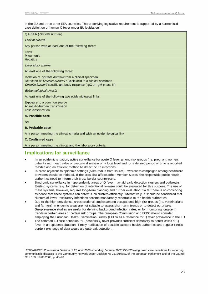
TECHNICAL REPORT Risk assessment on Q fever
23
in the EU and three other EEA countries. This underlying legislative requirement is supported by a harmonised case definition of human Q fever under EU legislationi
Q FEVER (Coxiella burnetii)
:
Clinical criteria
Any person with at least one of the following three:
Fever Pneumonia Hepatitis
Laboratory criteria
At least one of the following three:
Isolation of Coxiella burnetii from a clinical specimen Detection of Coxiella burnetii nucleic acid in a clinical specimen Coxiella burnetii-specific antibody response (IgG or IgM phase II)
Epidemiological criteria
At least one of the following two epidemiological links:
Exposure to a common source Animal-to-human transmission Case classification
A. Possible case
NA
B. Probable case
Any person meeting the clinical criteria and with an epidemiological link
C. Confirmed case
Any person meeting the clinical and the laboratory criteria
Implications for surveillance • In an epidemic situation, active surveillance for acute Q fever among risk groups (i.e. pregnant women,
patients with heart valve or vascular diseases) on a local level and for a defined period of time is reported feasible and an efficient method to detect acute infections.
• In areas adjacent to epidemic settings (5 km radius from source), awareness campaigns among healthcare providers should be initiated. If the area also affects other Member States, the responsible public health authorities need to inform their cross-border counterparts.
• Syndromic surveillance in hyperendemic areas of Q fever may aid early detection clusters and outbreaks. Existing systems (e.g. for detection of intentional release) could be evaluated for this purpose. The use of these systems, however, requires long-term planning and further evaluation. So far there is no convincing evidence that these systems can detect such clusters efficiently. Alternatively, it should be considered that clusters of lower respiratory infections become mandatorily reportable to the health authorities.
• Due to the high prevalence, cross-sectional studies among occupational high-risk groups (i.e. veterinarians and farmers) in endemic areas are not suitable to assess short-term trends or to detect outbreaks. Seroprevalence studies are useful for defining background infection rates, or for monitoring long-term trends in certain areas or certain risk groups. The European Commission and ECDC should consider employing the European Health Examination Survey (EHES) as a reference for Q fever prevalence in the EU.
• The common EU case definition for (possible) Q fever provides sufficient sensitivity to detect cases of Q fever in an epidemic situation. Timely notification of possible cases to health authorities and regular (cross-border) exchange of data would aid outbreak detection.
i 2008/426/EC: Commission Decision of 28 April 2008 amending Decision 2002/253/EC laying down case definitions for reporting communicable diseases to the Community network under Decision No 2119/98/EC of the European Parliament and of the Council. OJ L 159, 18.06.2008, p. 46–90.

Risk assessment on Q fever TECHNICAL REPORT
24
References: Surveillance 1. Maurin M, Raoult D. Q fever. Clin Microbiol Rev. 1999 Oct;12(4):518-53.
2. Parker NR, Barralet JH, Bell AM. Q fever. Lancet. 2006 Feb 25;367(9511):679-88.
3. Wallensten A, Moore P, Webster H, Johnson C, van der Burgt G, Pritchard G, Ellis-Iversen J, et al. Q fever outbreak in Cheltenham, United Kingdom, in 2007 and the use of dispersion modelling to investigate the possibility of airborne spread. Euro Surveill. 2010 Mar 25;15(12).
4. Gonder JC, Kishimoto RA, Kastello MD, Pedersen CE, Larson EW. Cynomolgus monkey model for experimental Q fever infection. J Infect Dis. 1979;139:191–196.
5. Tissot Dupont H, Raoult D, Brouqui P, Janbon F, Peyramond D, Weiller PJ, et al. Epidemiologic features and clinical presentation of acute Q fever in hospitalized patients—323 French cases. Am J Med. 1992;93:427–434.
6. Angelakis E, Raoult D. Q Fever. Vet Microbiol. 2010 Jan 27;140(3-4):297-309. Epub 2009 Aug 8.
7. Varga V. An explosive outbreak of Q-fever in Jedl'ové Kostol'any, Slovakia. Cent Eur J Public Health. 1997 Dec;5(4):180-2.
8. Schulze K, Schwalen A, Klein RM, Thomas L, Leschke M, Strauer BE. A Q-fever pneumonia epidemic in Dusseldorf. Pneumologie. 1996 Jul;50(7):469-73.
9. Tissot-Dupont H, Amadei MA, Nezri M, Raoult D. Wind in November, Q fever in December. Emerg Infect Dis. 2004 Jul;10(7):1264-9.
10. Hunink JE, Veenstra T, van der Hoek W, Droogers P. Q fever transmission to humans and local environmental conditions. RIVM. January 2010. Available from http://www.rivm.nl/en/Images/Q-fever%20transmission_tcm13-67109.pdf
11. Gilsdorf A, Kroh C, Grimm S, Jensen E, Wagner-Wiening C, Alpers K. Large Q fever outbreak due to sheep farming near residential areas, Germany, 2005. Epidemiol Infect. 2008 Aug;136(8):1084-7. Epub 2007 Sep 25.
12. Hawker JI, Ayres JG, Blair I, Evans MR, Smith DL, Smith EG, et al. A large outbreak of Q fever in the West Midlands: windborne spread into a metropolitan area? Commun Dis Public Health. 1998 Sep;1(3):180-7.
13. Schimmer B, Ter Schegget R, Wegdam M, Züchner L, de Bruin A, Schneeberger PM, et al. The use of a geographic information system to identify a dairy goat farm as the most likely source of an urban Q-fever outbreak. BMC Infect Dis. 2010 Mar 16;10:69.
14. Cerf O, Condron R. Coxiella burnetii and milk pasteurization: an early application of the precautionary principle? Epidemiol Infect. 2006 Oct;134(5):946-51.
15. Krumbiegel ER, Wisniewski HJ. Q fever in the Milwaukee area. II. Consumption of infected raw milk by human volunteers. Arch Environ Health. 1970 Jul;21(1):63-5.
16. Benson WW, Brock DW, Mather J. Serologic analysis of a penitentiary group using raw milk from a Q fever infected herd. Public Health Rep. 1963 Aug;78:707-10.
17. Salmon MM, Howells B, Glencross EJ, Evans AD, Palmer SR. Q fever in an urban area. Lancet. 1982 May 1;1(8279):1002-4.
18. Stein A, Raoult D. Q fever during pregnancy: a public health problem in southern France. Clin Infect Dis. 1998 Sep;27(3):592-6.
19. Raoult D, Stein A. Q fever during pregnancy, a risk for women, foetuses and obstetricians. N Engl J Med. 1994;330:371.
20. Gerth HJ, Leidig U, Riemenschneider T. Q-fever epidemic in an institute of human pathology. Dtsch Med Wochenschr. 1982 Sep 17;107(37):1391-5.
21. Milazzo A, Hall R, Storm PA, Harris RJ, Winslow W, Marmion BP. Sexually transmitted Q fever. Clin Infect Dis. 2001 Aug 1;33(3):399-402.
22. Richardus JH, Dumas AM, Huisman J, Schaap GJ. Q fever in infancy: a review of 18 cases. Pediatr Infect Dis. 1985 Jul-Aug;4(4):369-73.
23. Wagner-Wiening C, Brockmann S, Kimmig P. Serological diagnosis and follow-up of asymptomatic and acute Q fever infections. Int J Med Microbiol. 2006 May;296Suppl 40:294-6.
24. Porten K, Rissland J, Tigges A, Broll S, Hopp W, Lunemann M, et al. A super-spreading ewe infects hundreds with Q fever at a farmers' market in Germany. BMC Infect Dis. 2006 Oct 6;6:147.
25. Karagiannis I, Schimmer B, Van Lier A, Timen A, Schneeberger P, Van Rotterdam B, et al. Investigation of a Q fever outbreak in a rural area of The Netherlands. Epidemiol Infect. 2009 Sep;137(9):1283-94.
26. Tissot-Dupont H, Vaillant V, Rey S, Raoult D. Role of sex, age, previous valve lesion, and pregnancy in the clinical expression and outcome of Q fever after a large outbreak. Clin Infect Dis. 2007 Jan 15;44(2):232-7.
27. Taylor R, Hunter I, Tan R. Short report: prevalence of markers of exposure to Q fever in rural central Queensland. Commun Dis Intell. 2001 Nov;25(4):285-7.

TECHNICAL REPORT Risk assessment on Q fever
25
28. Madariaga MG, Rezai K, Trenholme GM, Weinstein RA. Q fever: a biological weapon in your backyard. Lancet Infect Dis. 2003 Nov;3(11):709-21.
29. Buehler JW, Berkelman RL, Hartley DM, Peters CJ. Syndromic surveillance and bioterrorism-related epidemics. Emerg Infect Dis. 2003 Oct;9(10):1197-204.
30. Elliot A. Syndromic surveillance: the next phase of public health monitoring during the H1N1 influenza pandemic? Euro Surveill. 2009 Nov 5;14(44). pii: 19391.
31. Lyytikainen O, Ziese T, Schwartlander B, Matzdorff P, Kuhnhen C, Burger C, et al. Outbreak of Q fever in Lohra-Rollshausen, Germany, spring 1996. Euro Surveill. 1997 Feb;2(2):9-11.

Risk assessment on Q fever TECHNICAL REPORT
26
Annex 1: Strategies to identify and follow up chronic cases Strategy 1: Targeted case-finding of persons with known heart valve lesions or vascular grafts If, during an outbreak there is a population of 100 000 possibly at risk of exposure, and the cumulative incidence of people being infected rises to 10%, (approximately 50% of them being ill, the rest being infected but having an asymptomatic infection), there will be 5 000 people having an acute symptomatic infection. Approximately 2% of the acute cases (100 in this population) will develop into chronic cases with a high fatality rate if not successfully treated. Approximately 80% of chronic cases have a damaged heart valve or a grafted vessel. That means that 80 of these chronic cases would have one of these risk factors.
In a general population, approximately 2.5% have a heart valve disease [4]. Translated to our scenario, this means that 2 500 people are at risk of getting a chronic disease. In other words approximately 31 patients have to be screened to find one case of chronic disease (who would benefit from early detection and treatment).
A serology test costs approximately EUR 30, and has maybe to be taken three times for each risk patient during an outbreak. There will be some additional costs to call patients, for transportation and administrative issues and to follow up drop-outs, amounting to a total of approximately EUR 150 per patient.
This equation can then be adjusted according to the incidence (as shown in Table 3) in a certain region at a certain time during a possible outbreak, to find a potential cut-off point where it could be sensible to undertake targeted case finding according to a cost–utility analysis.
Table 3. Calculation of numbers and costs for patients with known heart valve disease with different cumulative incidence rates
Cumulative incidence 10% 5% 1% Acute symptomatic Q fever 5000 2500 500 2% chronic (80% with HVD) 80 40 8 Chronic/total pop. HVD 80/2500 40/2500 8/2500 Number needed to be tested 31 62 312 Cost to find one case 4650 € 9300 € 46800 €
Strategy 2: Targeted case-finding for heart valve lesions with echocardiography of all patients with acute Q fever This strategy has been proposed by several authors [1,2,3]. Again, consider the same population (100 000) to be exposed to infection during an outbreak, and calculate according to the same figures as above. The cost of performing an echocardiography is estimated to be EUR 100 and additional costs for administrative issues EUR 50, making a total cost of EUR 150 per patient tested. Estimated numbers of patients with a heart valve disease or vascular graft in the population is 2 500. If a heart valve patient gets an acute infection, up to 50% will possibly develop a chronic infection.
Table 4. Calculation of number and costs for echocardiography with different cumulative incidence rates
Cumulative incidence 10% 5% 1% Acute symptomatic Q fever 5000 2500 500
Cost to test all with echocardiography 750 000 € 375 000 € 75 000 €
Patients with HVD possibly affected with acute Q fever 250 125 25
50% of these patients possibly developing chronic Q fever 125 63 13
Cost to find one case 6000 € 6000 € 6000 €
TECHNICAL REPORT Risk assessment on Q fever
27
Cost–benefit of targeted case finding for at-risk patients Considering the serious burden of disease of having endocarditis with possible antibiotic treatments for several years, and a possible surgical heart valve replacement or a substitution of an infected vascular graft, or a possible fatal disease outcome if not treated, both of these strategies seem to be cost-effective. If the incidence is higher, then the first strategy might be the most cost-efficient; if the cumulative incidence is at a level below around 8%, then the second strategy seems to be more cost-efficient.
The total costs of testing all patients with acute Q fever with echocardiography obviously depends on the total number of persons being infected during the outbreak, and the costs to find one case will be the same whatever the incidence, as opposed to the first example. The efficacy of this strategy depends on the people with an acute infection seeking healthcare. Since most acute cases manifest themselves as a mild flu-like illness and it is a self-limiting disease, for which people do not seek medical attention, this may well cause a practical problem. In addition, the availability of personnel and equipment to perform an increased number of echocardiography investigations in an outbreak area might be a limiting factor. An advantage of this strategy is that you will find the cases that have a heart valve disease, who do not know it, but are at risk.
The efficacy of the first strategy, to follow up all patients with known heart valve disease, obviously depends on people knowing that they have such an underlying disease. According to a study from the Mayo clinic, approximately 28% of patients who have a heart valve disease do not know it [4]. A heart valve disease might evolve gradually and at early stages awareness about the diagnosis might not be present, but when it becomes symptomatic, medical attention will be sought. Fenollar F, et al. [1] describe three cases of endocarditis after acute Q fever in patients with previously undiagnosed valvulopathies. Patients with a vascular graft know their diagnosis and could easily be reached.
All other risk groups have a relatively low incidence of chronic Q fever compared with those with a heart or blood vessel disease, and some of these conditions are more frequent among the general population. Targeted case finding seems therefore not to be advisable for these groups, but there should be a raised awareness for individual follow-up among physicians towards all risk groups including pregnant women.
Strategy 3: Individual follow-up after acute Q fever infection with serology, together with raised awareness among the general population and physicians A third alternative is to follow up on an individual patient level with serology testing those who have actively sought medical attention for acute Q fever. This is the proposed strategy from the health authorities in the Netherlands during the ongoing [2010] outbreak and reflects the view of the expert meeting in Paris.
For patients not at risk, they recommend a single follow-up serum sample nine months after seeking medical attention. For patients at risk, the reccomendation is to follow up at three, six and 12 months, with serology combined with PCR. The risk factors being pathologic cardiac valves, aneurysms or vascular surgery, immunosuppression and pregnancy.
In addition to this, the Dutch health authorities have sent a letter to all those who live in an outbreak area, informing them about the outbreak and the risk factors, and to advise people in risk groups to seek medical attention if they get symptoms of an acute Q fever infection.
Treatment of asymptomatic cases has been raised as a potential problem. Currently, there is little evidence to support the theory that asymptomatic cases become chronic, and in any case, as long as these patients do not seek medical attention and do not know about their infection, treatment is not is not a practical option.
Lack of evidence, need for research There does not seem to be any evidence directly supporting any of these three strategies when it comes to outcomes like reduction in the spread of the outbreak, improved treatments, fewer complications and a reduced fatality rate for acute and chronic Q fever. But there is evidence on the feasbility and detection of active case finding, which logically should lead to a better treatment and follow-up of acute and chronic cases [5,6].

Risk assessment on Q fever TECHNICAL REPORT
28
References: Annex 1 1. Fenollar F, Thuny F, Xeridat B, Lepidi H, Raoult D. Endocarditis after acute Q fever in patients with previously undiagnosed valvulopathies. Clin Infect Dis. 2006 Mar 15;42(6):818-21.
2. Raoult D, Marrie T, Mege J. Natural history and pathophysiology of Q fever. Lancet Infect Dis. 2005;Apr;5(4):219-26.
3. Hartzell JD, Wood-Morris RN, Martinez LJ, Trotta RF. Q fever: Epidemiology, diagnosis, and treatment. Mayo Clinic Proceedings. 2008;83(5):574-9.
4. Nkomo VT, Gardin JM, Skelton TN, Gottdiener JS, Scott CG, Enriquez-Sarano M. Burden of valvular heart diseases: a population-based study. Lancet. 2006 Sep 16;368(9540):1005-11.
5. Porten K, Rissland J, Tigges A, Broll S, Hopp W, Lunemann M, van Treeck U, Kimmig P, Brockmann SO, Wagner-Wiening C, Hellenbrand W, Buchholz U. A super-spreading ewe infects hundreds with Q fever at a farmers' market in Germany. BMC Infect Dis. 2006 Oct 6;6:147.
6. Wagner-Wiening C, Brockmann S, Kimmig P. Serological diagnosis and follow-up of asymptomatic and acute Q fever infections. Int J Med Microbiol. 2006 May;296Suppl 40:294-6.

TECHNICAL REPORT Risk assessment on Q fever
29
Annex 2 - Conflicts of interest The expert panel in the Paris meeting was asked to fill in the Annual Declaration of Interest 2010. One member was a speaker in a granted symposium on blood transfusion in 2009. No other relevant conflicts of interest reported.
Annual Declaration of Interest 2010
Information on direct or indirect interests of relevance to the mission of the Centre
Name:
Position:
[ ] Member/Alternate of the Management Board [ ] Member/Alternate of the Advisory Forum [ ] Member of the Scientific Committee [ ] Member of a Panel on:…………………………. [ ] Member of a Working Group [ ] ECDC Director/Senior Management team
The information should cover the 3 years before the date of the current declaration.
(1) Direct interest (financial benefits arising from, for example, employment, contracted work, investments, fees etc.)
(2) Participation into activities supported by grants or contracts concluded in the Framework of the Public Health Programme
(3) Indirect financial interests e.g. grants, sponsorships, or other kind of benefits
(4) Interests deriving from the professional activities of the member or his/her close family Members
(5) Any Membership role or affiliation that you have in organisations/bodies/club with an interest in the work of the Centre

Risk assessment on Q fever TECHNICAL REPORT
30
(6) Other interests or facts that the undersigned considers pertinent
I declare on my word of honour that the information provided above is true and complete.
Done at ________________________ on _________________
Signature: __________________________________________

TECHNICAL REPORT Risk assessment on Q fever
31
Annex 3: Search strategies for Q fever (10 March 2010)
PUBMED:
Search strategies Concept 1: Boolean operator Concept 2: Boolean
operator Concept 3:
OR OR OR
Occupational exposure
"rickettsia burnetii"[Title/Abstract] "rickettsia burnetii infection"[Title/Abstract] "rickettsia diaporic"[Title/Abstract] “rickettsia burnetti"[Title/Abstract] "rickettsiosis infection"[Title/Abstract] "rickettsiosis rickettsia"[Title/Abstract] "australian q fever"[Title/Abstract] "Q Fever"[Mesh] "coxiella burnetii"[mesh]
AND
"Occupational Diseases"[Mesh]
"Environmental Exposure"[Mesh]
Chronic diseases
"rickettsia burnetii"[Title/Abstract] "rickettsia burnetii infection"[Title/Abstract] "rickettsia diaporic"[Title/Abstract] “rickettsia burnetti"[Title/Abstract] "rickettsiosis infection"[Title/Abstract] "rickettsiosis rickettsia"[Title/Abstract] "australian q fever"[Title/Abstract] "Q Fever"[Mesh] "coxiella burnetii"[mesh]
AND "Chronic Disease"[Mesh]
Transmission surveillance and prevention and control
"rickettsia burnetii"[Title/Abstract] "rickettsia burnetii infection"[Title/Abstract] "rickettsia diaporic"[Title/Abstract] “rickettsia burnetti"[Title/Abstract] "rickettsiosis infection"[Title/Abstract] "rickettsiosis rickettsia"[Title/Abstract] "australian q fever"[Title/Abstract] "Q Fever"[Mesh] "coxiella burnetii"[mesh]
AND
"Disease Transmission, Infectious"[Mesh]
"transmission "[Subheading]
AND
"Communicable Diseases, Emerging"[Mesh]
"Communicable Disease Control"[Mesh]
"Disease Notification"[Mesh] "Epidemiology"[Mesh] "Population
Surveillance"[Mesh] "Sentinel Surveillance"[Mesh] "Epidemiologic
Factors"[Mesh] "prevention and
control"[Subheading] "surveillance"[Title/Abstract]
"Q Fever/transmission"[Mesh] ("Coxiella burnetii"[Mesh] AND ("transmission "[Subheading] OR "Disease Transmission, Infectious"[Mesh]))
AND
"Communicable Diseases, Emerging"[Mesh]
"Communicable Disease Control"[Mesh]
Disease Notification"[Mesh] "Epidemiology"[Mesh] "Population
Surveillance"[Mesh] "Sentinel Surveillance"[Mesh] "Epidemiologic Factors"[Mesh] "prevention and
control"[Subheading] "surveillance"[Title/Abstract]

Risk assessment on Q fever TECHNICAL REPORT
32
Search strategies Concept 1: Boolean operator Concept 2: Boolean
operator Concept 3:
"rickettsia burnetii"[Title/Abstract] "rickettsia burnetii infection"[Title/Abstract] "rickettsia diaporic"[Title/Abstract] “rickettsia burnetti"[Title/Abstract] "rickettsiosis infection"[Title/Abstract] "rickettsiosis rickettsia"[Title/Abstract] "australian q fever"[Title/Abstract] "Q Fever"[Mesh] "coxiella burnetii"[mesh]
AND
"Blood Transfusion"[Mesh] "Tissue Donors"[Mesh] "Pregnancy/blood"[Mesh] "Pregnancy
Complications/blood"[Mesh]
“Infection/blood"[Mesh]
Blood transmission
"rickettsia burnetii"[Title/Abstract] "rickettsia burnetii infection"[Title/Abstract] "rickettsia diaporic"[Title/Abstract] “rickettsia burnetti"[Title/Abstract] "rickettsiosis infection"[Title/Abstract] "rickettsiosis rickettsia"[Title/Abstract] "australian q fever"[Title/Abstract] "Q Fever"[Mesh] "coxiella burnetii"[mesh]
AND
"Blood Transfusion"[Mesh] "Tissue Donors"[Mesh] "Pregnancy/blood"[Mesh] "Pregnancy
Complications/blood"[Mesh]
"Infection/blood"[Mesh]
Pregnancy
"rickettsia burnetii"[Title/Abstract] "rickettsia burnetii infection"[Title/Abstract] "rickettsia diaporic"[Title/Abstract] “rickettsia burnetti"[Title/Abstract] "rickettsiosis infection"[Title/Abstract] "rickettsiosis rickettsia"[Title/Abstract] "australian q fever"[Title/Abstract] "Q Fever"[Mesh] "coxiella burnetii"[mesh]
AND
("Pregnancy Complications"[Mesh] OR "Infection"[Mesh] or "Pregnancy"[Mesh]) AND ("Blood"[Mesh] OR "blood "[Subheading])
"rickettsia burnetii"[Title/Abstract] "rickettsia burnetii infection"[Title/Abstract] "rickettsia diaporic"[Title/Abstract] “rickettsia burnetti"[Title/Abstract] "rickettsiosis infection"[Title/Abstract] "rickettsiosis rickettsia"[Title/Abstract] "australian q fever"[Title/Abstract] "Q Fever"[Mesh] "coxiella burnetii"[mesh]
AND
"Chronic Disease"[Mesh] "Carrier State"[Mesh] "Disease Reservoirs"[Mesh] "reservoir host"[Title/Abstract] "reservoir
infection"[Title/Abstract] "reservoir
infections"[Title/Abstract] "Blood Transfusion"[Mesh] "Tissue Donors"[Mesh]
AND
"Pregnancy Complications"[Mesh]
"Pregnancy"[Mesh]
Limit: 1970- and humans

TECHNICAL REPORT Risk assessment on Q fever
33
EMBASE:
Search strategies Concept 1: Boolean operator Concept 2: Boolean
operator Concept 3:
OR OR OR
Occupation exposure
'rickettsia burnetii':ab 'rickettsia burnetii':ti 'rickettsia diaporic':ab 'rickettsia diaporic':ti 'rickettsia burnetti':ab 'rickettsia burnetti':ti 'rickettsiosis infection':ti 'rickettsiosis infection':ab 'rickettsiosis rickettsia':ab 'rickettsiosis rickettsia':ti 'australian q fever':ab 'australian q fever':ti 'coxiella burnetii'/exp 'q fever'/exp
AND
'occupational disease'/exp 'environmental exposure'/exp
Chronic diseases
'rickettsia burnetii':ab 'rickettsia burnetii':ti 'rickettsia diaporic':ab 'rickettsia diaporic':ti 'rickettsia burnetti':ab 'rickettsia burnetti':ti 'rickettsiosis infection':ti 'rickettsiosis infection':ab 'rickettsiosis rickettsia':ab 'rickettsiosis rickettsia':ti 'australian q fever':ab 'australian q fever':ti 'coxiella burnetii'/exp 'q fever'/exp
AND
'chronic disease'/exp
Transmission surveillance and prevention and control
'rickettsia burnetii':ab 'rickettsia burnetii':ti 'rickettsia diaporic':ab 'rickettsia diaporic':ti 'rickettsia burnetti':ab 'rickettsia burnetti':ti 'rickettsiosis infection':ti 'rickettsiosis infection':ab 'rickettsiosis rickettsia':ab 'rickettsiosis rickettsia':ti 'australian q fever':ab 'australian q fever':ti 'coxiella burnetii'/exp 'q fever'/exp
AND
'disease surveillance'/exp 'health survey'/exp 'sentinel surveillance'/exp surveillance:ab surveillance:ti 'prevention and control'/exp 'infection control'/exp
AND
'disease transmission'/exp
Blood transmission
'rickettsia burnetii':ab 'rickettsia burnetii':ti 'rickettsia diaporic':ab 'rickettsia diaporic':ti 'rickettsia burnetti':ab 'rickettsia burnetti':ti 'rickettsiosis infection':ti 'rickettsiosis infection':ab 'rickettsiosis rickettsia':ab 'rickettsiosis rickettsia':ti 'australian q fever':ab 'australian q fever':ti 'coxiella burnetii'/exp 'q fever'/exp
AND
'disease transmission'/exp
AND
'blood'/exp

Risk assessment on Q fever TECHNICAL REPORT
34
Search strategies Concept 1: Boolean operator Concept 2: Boolean
operator Concept 3:
'rickettsia burnetii':ab 'rickettsia burnetii':ti 'rickettsia diaporic':ab 'rickettsia diaporic':ti 'rickettsia burnetti':ab 'rickettsia burnetti':ti 'rickettsiosis infection':ti 'rickettsiosis infection':ab 'rickettsiosis rickettsia':ab 'rickettsiosis rickettsia':ti 'australian q fever':ab 'australian q fever':ti 'coxiella burnetii'/exp 'q fever'/exp
'donor'/exp 'transfusion'/exp (('pregnancy complication'/exp OR 'pregnancy'/exp) AND 'blood'/exp)
Pregnancy
'rickettsia burnetii':ab 'rickettsia burnetii':ti 'rickettsia diaporic':ab 'rickettsia diaporic':ti 'rickettsia burnetti':ab 'rickettsia burnetti':ti 'rickettsiosis infection':ti 'rickettsiosis infection':ab 'rickettsiosis rickettsia':ab 'rickettsiosis rickettsia':ti 'australian q fever':ab 'australian q fever':ti 'coxiella burnetii'/exp 'q fever'/exp :ti OR 'australian q fever':ab OR 'australian q fever':ti OR 'coxiella burnetii'/exp OR 'q fever'/exp
'donor'/exp 'transfusion'/exp 'chronic disease'/exp 'disease carrier'/exp 'heterozygote'/exp 'reservoir host':ab 'reservoir host':ti 'reservoir infection':ab 'reservoir infection':ti 'reservoir infections':ab 'reservoir infections':ti
'pregnant woman'/exp 'pregnancy'/exp 'pregnancy disorder'/exp gravid:ab gravid:ti 'pregnancy':ab 'pregnancy':ti 'pregnant':ab 'pregnant':ti 'pregnancy complications':ab 'pregnancy complications':ti
Limit: 1970- and humans

TECHNICAL REPORT Risk assessment on Q fever
35
Annex 3: Evidence tables Chronic Q fever
Bibliographic citation Type of study
No of patients or population
Study outcome Strengths of study
Limitations of study
Brouqui, P., H. T. Dupont, et al. (1993). "Chronic Q fever. Ninety-two cases from France, including 27 cases without endocarditis." Arch Intern Med
153(5): 642-8.
Case Series 92 patients Demographic, epidemiologic, clinical and lab data
A big study, methods clearly described. Data collected for a long period (9 years)
No control
Chaillon, A., J. L. Bind, et al. (2008). "[Epidemiological aspects of human Q fever in Indre-et-Loire between 2003 and 2005 and comparison with caprine Q fever]." Med Mal Infect
38(4): 215-24.
Retro spective case series
40 total, 6 chronic
Comparing epidemiological findings for human Q fever with data on animal disease and density. This study revealed similar location of human and caprine Q fever. Identifying such geographical correlation may lead to improving prevention and detection.
Data from a 2 year period
Few chronic cases
Raoult, D. (2002). "Q fever: still a mysterious disease." Qjm
95(8): 491-2. Editorial Commenting on two studies of CFS, (not conclusive), and to carefully consider precision of diagnostic methods. (PCR)
A good background article
Cisak, E., J. Chmielewska-Badora, et al. (2003). "Prevalence of antibodies to Coxiella burnetii among farming population in eastern Poland." Ann Agric Environ Med
10(2): 265-7.
Epidemiological study, cross- sectional prevalence study
90 farmers compared to 30 urban blood donors in district of Poland
17,8 % prevalence of Phase II antibodies among farmers, indicative of past infection. No positive tests among urban blood donors. Comparing prevalence with many other countries. Discussing the role of cattle.
Prevalence data from a new area compared to other areas, and urban vs rural settings
Small study
Wildman, M. J., E. G. Smith, et al. (2002). "Chronic fatigue following infection by Coxiella burnetii (Q fever): ten-year follow-up of the 1989 UK outbreak cohort." Qjm
95(8): 527-38.
Retrospective Matched cohort study (Case Control)comparing cases to control
108 cases, 86 controls. Cases followed up at 4 different times. Drop outs counted for
Subjects who were exposed to Coxiella Burnetii in 1989 had more fatigue than did controls, and some fulfilled the criteria for CFS. Whether this is due to ongoing antigen persistence or to the psychological effects of prolonged medical follow-up is uncertain.
10 year since exposure, control group, many cases (108)
High number of fatigue in the general population as well!
Delsing, C. E., C. P. Bleeker-Rovers, et al. (2009). "[Q fever, a potential serious disease]." Ned Tijdschr Geneeskd
153(14): 652-7.
Three cases and review 3
Describing three cases, two with lung infiltrates and one with a mesenterial fat infiltrate
New clinical pictures Few cases
Hartzell, J. D., R. N. Wood-Morris, et al. (2008). "Q fever: Epidemiology, diagnosis, and treatment." Mayo Clinic Proceedings
83(5): 574-579.
Review, background article
No Reporting on Epidemiology, diagnosis, clinical manifestations and treatment
Pedagogical and well written, good background article
No study
Reilly, S., J. L. Northwood, et al. (1990). "Q fever in Plymouth, 1972-88. A review with particular reference to neurological manifestations." Epidemiol Infect
105(2): 391-408.
Case series 61 cases, only 5 chronic
Only 5% of cases had chronic Q fever, but in view of the diverse sequelae observed in this series, we suggest that long-term serological and clinical follow up of all cases of Q fever is fully justified.
Long period of collecting data, 1972 -1988, data well described and good patient follow up
Old data
Sessa, C., L. Vokrri, et al. (2005). "Abdominal aortic aneurysm and Coxiella burnetii infection: report of three cases and review of the literature." J Vasc Surg
42(1): 153-8. Case series
3 cases of Aortic aneurysms
Aortic aneurysm resection is mandatory to cure the chronic infection and must be associated with long-term antibiotic therapy.
Well described Few cases,
Varma, M. P., A. A. Adgey, et al. (1980). "Chronic Q fever endocarditis." Br Heart J
43(6): 695-9 Case series 8
Treatment and prognosis on patients with endocarditis and prosthetic valves. It is suggested that medical treatment is continued until clinically and haematologically there is no evidence of endocarditis and the Q fever phase 1 antibody titre is less than 200.
Good follow up, and good clinical descriptions
Rather old study
Cécile Landais, Florence Fenollar, Franck Thuny, and Didier Raoult; Clinical Infectious Diseases 2007;44:1337–1340 From Acute Q Fever to Endocarditis: Serological Follow Up Strategy
Case series, Retrospective cohort
22 chronic cases with endocarditis
Time to develop chronic infection is measured, mean being 3 months, and follow up on serological testing after acute Q fever is proposed at 3 and 6 months
New data from a reliable source, The French National Ref Centre for Rickettsial Disease

Risk assessment on Q fever TECHNICAL REPORT
36
Bibliographic citation Type of study
No of patients or population
Study outcome Strengths of study
Limitations of study
Fenollar F, Fournier PE, Carrieri MP, Habib G, Messana T, Raoult D. Risks factors and prevention of Q fever endocarditis. Clin Infect Dis 2001; 33:312–6.
Case series 12
To investigate how many develop chronic Q fever after acute Q fever. 0,76 % in this series all with underlying valvulopathy. And how many have a valvular disease? 1,3 % in Minnesota above 35 years. Follow up proposed, all with valvulopathy to be treated 12 months and serological tests for 2 years
Data from French National Centre
Mis calculated figure
Edlinger, E. A. (1987). "Chronic Q fever." Zentralblatt fur Bakteriologie Mikrobiologie und Hygiene - Abt. 1 Orig. A
267(1): 51-56.
Case series, Pasteur institute
36 patients, data collected for 5 years
Clinical description of patients after positive serology. Bordeline between subacute and chronic Q fever is discussed. Older women more resistant than younger women and men. Differences in outcome possible due to strain differences
Data from many centres in France. All patients counted for
Slightly old data
Tellez, A., C. Sainz, et al. (1988). "Q fever in Spain: acute and chronic cases, 1981-1985." Reviews of infectious diseases
10(1): 198-202.
Case series, Spain
15 cases, data from 1981 - 1985
All regions of Spain, most in northern regions and Madrid. (hospitals) Reports 6% chronic cases of the 249 confirmed acute cases. Underdiagnosed disease in Spain
First reports from Spain
Primary data available
Turck, W. P., G. Howitt, et al. (1976). "Chronic Q fever." Q J Med
45(178): 193-217.
Case series 16
Reporting comprehensively on epidemiology, clinical features and and pathology of 16 cases from UK, Discussing treatment and follow up in 1976
Historical interesting article, list of references to all previous reported chronic cases
Old data
Fergusson, R. J., T. R. Shaw, et al. (1985). "Subclinical chronic Q fever." Q J Med
57(222): 669-76. Case series 7 patients
from Scotland
Reporting on subclinical cases where the locus of infection was not found in six cases of seven. Discussing treatment options under such circumstances
Cases well described Few cases
Ellis, M. E., C. C. Smith, et al. (1983). "Chronic or fatal Q-fever infection: a review of 16 patients seen in North-East Scotland (1967-80)." Q J Med
52(205): 54-66. Case series
16 lab confirmed chronic cases
Describes chronic cases associated with extra valvular sites of infection, prematurity, SIDS, emboli, osteomyelitis
Early description of manifestations outside heart and vessels. Comprehensive descriptions of cases
Relevance today?
Raoult, D., P. Brouqui, et al. (1992). "Acute and chronic Q fever in patients with cancer." Clin Infect Dis
14(1): 127-30. Case series
5 cases of patients with Q fever and Cancer
Characteristics of the disease in ca patients. Immunosupression might allow a relapse. Endocarditis is reported in cases without valvulopathies. Testing for Coxiella burnetii in ca pats with fever is recommended
Relevant to our question from the Commission
Few cases
Raoult, D., P. Y. Levy, et al. (1990). "Chronic Q fever: diagnosis and follow-up." Ann N Y Acad Sci
590: 51-60. Case series
40 patients diagnoses betw 1983 and 1988
Epidemiology, clinical features and follow up on 40 cases. Endocaditis, immunosuppressed and bone manifestations
God clinical descriptions
Less relevant for public health
Schimmer B, Morroy G, Dijkstra F, Schneeberger PM, Weers-Pothoff G, Timen A, Wijkmans C, W van der (2008) Large ongoing Q fever outbreak in the south of The Netherlands 2008, Eurosurveillance Vol 13, Issues 7-9.
Outbreak report
Data from 2007 and 2008 in the Netherlands
Updated information, general considerations. Mandatory notifications in ruminants implemented in June 2008. Manure spread banned and visiting to infected farms restricted. Discussions on blood donors and screening of pregnant women
Rapid communication, not specific on chronic disease
Schimmer B, Dijkstra F, Vellema P, Schneeberger PM, Hackert V, Schegget et al: 2009. Sustained intensive transmission of Q fever in the South of the Netherlends. Eurosurveillance Vol 14, Issue 19. 14 May 2009
Outbreak report
Data from the Netherlands 2009
Update on the latest epidemiological data, and an overview over ongoing research
Rapid communication, not specific on chronic disease
Harris, R. J., P. A. Storm, et al. (2000). "Long-term persistence of Coxiella burnetii in the host after primary Q fever." Epidemiol Infect
124(3): 543-9 Lab research
29 patients with chronic sequel to acute Q fever
Reports findings of Coxiela Burnetii DNA in blood (17%), liver biopsies (14%) and bone marrow aspirates (65%) from 0,75 to 5 years after acute infection. Clinical importance unknown
New finding, clinical relevance unknown
Indirect evidence, PCR technique may be too sensitive, false positives possible
Tissot-Dupont, H., V. Vaillant, et al. (2007). "Role of sex, age, previous valve lesion, and pregnancy in the clinical expression and outcome of Q fever after a large outbreak." Clinical Infectious Diseases
44(2): 232-237
Cohort with Control
1064 persons tested during an outbreak. 101 patients had acute Q fever 5 developed a chronic condition
Reporting incidence among risk groups such as immunocompomised patients, pregnant women and patients with valvular disease. The study emphasises the feasibility and importance of active surveillance in postepidemic conditions
New data on active surveillance
Short follow up, only one year

TECHNICAL REPORT Risk assessment on Q fever
37
Bibliographic citation Type of study
No of patients or population
Study outcome Strengths of study
Limitations of study
Maltezou, H. C. and D. Raoult (2002). "Q fever in children." Lancet Infectious Diseases
2(11): 686-691.
Review of all pediatric cases published
Referring 46 cases
Children are less frequently symptomatic than adults, self limited febrile illness or pneumonia being the most common acute clinical feature. Chronic disease manifestations are endocarditits and osteomyelitis. More studies are needed
Summarizing the evidence systematically all studies on primary data included, search strategy included
Raoult D, Tissot-Dupont H, Foucault C, Gouvernet J, Fournier PE, Bernit E, Stein A, Nesri M, Harle JR, Weiller PJ. Q fever 1985–1998: clinical and epidemiologic features of 1,383 infections. Medicine 2000; 79:109–23.
Case series Retrospective analyses
313 chronic cases from hospitals of France during the period 1985 -1998
Reporting clinical and epidemiological features. 1,5 % developing into chronic disease, 81 % having valvular or vascular host characteristics. Other manifestations are chronic hepatitis, osteoaticular infection and peridarditis. Risk of adverse fetal outcome if a woman gets the disease during pregnancy.
The biggest series reported
Fenollar F, Thuny F, Xeridat B, Lepidi H, Raoult D (2006) Endocarditis after acute Q fever in patient with previously undiagnosed valvulopathies. Clinical Infectious Diseases 42: 818-21
Case series 3 patients
Endocarditis after acute Q fever can develop in patients with undiagnosed valvulopathies. 3 weeks of treatment for acute Q fever is not enough to prevent the chronic form. A combination of Doxycycline and Hydroxychloroquine for one year seems to be effective. Active follow up is recommended for patients with minor valvulopathies, Authors suggest all patients with acute Q fever to have an echocardiography.
New information on development of endocarditis after acute Q fever
Few cases, wide ranging and costly proposals. Costs and feasibility not estimated
Palmer SR, Young SEJ, Q-fever endocarditis in England and Wales, 1975 -81.The Lancet, December 25, 1982
Case series 92
Reports of 11% chronic patients with endocarditis in England and Wales. Q fever endocarditis accounts for 3% of all endocarditis cases reported. Most affected young and middle aged men. Only 33% having known underlying heart valve lesion.
A relatively large series. The titers reported
Old lab tests, Complement fixation method.

Risk assessment on Q fever TECHNICAL REPORT
38
Pregnancy
Bibliographic citation [ref no] Type of study No of patients or
population Study outcome Strengths of study Limitations of study
Baud D et al. Clin Microbiol Infect 2009; 15: 499-501 [2]
Cross-sectional - Seroprevalence study
438 sera samples 269 from women with sporadic or recurrent miscarriage, and 169 controls with an uneventful pregnancy, London
Prevalance of C. brunetii antibodies in sera of women with sporadic miscarriage and women with recurrent miscarriage in comparison with women with uneventful pregnancies
Control group
Wide overlapping confidence intervals indicate too low numbers of sera and infections to enable definitive conclusions. Further research needed.
McCaughey C et al. Public Health 2008; 55: 189-194 [3]
Cross-sectional - Seroprevalence study
2394 participants, incl 1209 women
Seroprevalence of C. brunetii antibodies in men compared to women, taking into consideration other demographic factors e.g. occupation
Collection of demographic data. Control for some potential risk factors.
Seroprevalence in pregnant women not the main target of the study. Reporting bias cannot be excluded. No control for other potential risk factors for miscarriage. Age structure of the female population unclear.
Tissot-Dupont H et al. CID 2007; 44: 232-237 [6]
Enhanced serosurveillance in risk groups during 1 year following outbreak
891 samples from 350 pregnant women
Seroprevalence of C. brunetii antibodies in different risk groups
Collection of demographic and anamnestic data.
No follow-up of pregnancies until delivery/ending of pregnancy to assess pregnancy outcome.
Langley JM et al. Am J Obstet Gynecol 2003; 189(1): 228-232 [8]
Cross-sectional - Seroprevalence study
4588 cord blood samples collected after delivery in endemic area in Canada (Nova Scotia) June 1997 to Nov 1998
Seroprevalence study using cord blood, and statistical evaluation of potential associations with different anamnestic parameters
Lab personal blinded. All positives were re-tested a second time. PCR and culture from placenta tissue performed. Collection of pregnancy outcome data.
Maternal seroprevalence unknown. Only cord blood and placenta tested.
Rey D et al. Eur J Obstet Gynaecol Reprod Biol 2000; 93: 151-156 [11]
Cross-sectional - Seroprevalence study
12716 women after ending their pregnancy
Seroprevalence of C. brunetii antibodies
All women of one region ending their pregnancy irrespective of pregnancy outcome included.
Number of infected women too small to allow conclusions on Q fever and risk of abortion, reported preterm births and low-birth-weight could not be linked to the surveyed women, therefore no conclusions with regard to these outcomes possible.
Carcopino X, et al. CID 2007; 45: 548-555 [5]; see also [9], [15] and [17]
Case series 53 pregnant women with Coxiella brunetii infection
Serological profile, progress and pregnancy outcome. Efficacy of long-term cotrimoxazole treatment.
Largest case series so far reported. Exclusion of other infectious agents. Some placental and fetal tissue samples tested. Pregnancy outcome of all cases recorded including partially follow up of cases after delivery.
High potential for selection bias. Efficacy of treatment evaluated in a non-randomised non-controlled way.

TECHNICAL REPORT Risk assessment on Q fever
39
Surveillance Bibliographic citation Type of study
No of patients or population
Study outcome Strengths of study Limitations
Tissot-Dupont et al., 2007
Seroprevalence study, Follow-up (enhanced surveillance for one year)
1064 (incl. 376 pregnant women, 91 patients with valvular heart disease, 19 immunocompromised patients, 578 without risk factors)
101 with acute Q fever (11 pregnant women, 5 patients with VHD, 85 people without risk; 5 with chronic Q fever
Case number, repeated testing, epidemiological data
Bernard et al., 2010 (in preparation)
Seroprevalence study 426 veterinarians from southern Germany
37% seropositive for Q fever Case number, epidemiological data
Sampling bias (voluntary testing of risk group)
Porten et al., 2006
Outbreak investigation, cohort study, case control study. Active Surveillance (for pregnant women, patients with valvular heart disease)
299 11 pregnant women 18 patients with valvular heart disease
4 pregnant women and 2 patients with valvular heart disease show acute Q fever, 1 woman developed chronic Q fever. Proximity to source most important risk factor. Clinical attack rate 20% in adults. Underreporting 50%.
In depth study on epidemiological characteristics of q fever
No follow up of cases.
Lyytikäinen et al., 1999
Outbreak investigation/ Seroprevalence study
120 inhabitants of a 300 inhabitants village
29% IgM-positive (60% clinically), 17% IgG; RF: contact with sheep, walking near sheep farm
Moderate response rate, selection bias, no follow up
Richardus et al., 1986 Seroprevalence study
432 high risk groups incl. farmers, vets, taxidermists, wool spinner) 359 control blood donors
Prevalence in veterinarians 83.7%, taxidermists 70%, wool spinner 58%) Prevalence in controls 24%
Case number Control selection; selection bias
Brockmann et al., 2010 (submitted)
Seroprevalence study/Survey
1036
Seroprevalence 7,8% (0-18) in general population. RF farmer, waste worker, contact to goats. Seropositivity correlating with sheep density
Sample size; population based
Representativeness; selection bias (voluntary testing)
Tissot-Dupont et al., 2004
Environmental study 73 Q fever incidence in correlated with wind speed (Mistral)
Environmental data
No study of other sources; methodology (time series)
Wagner-Wiening et al., 2006
Outbreak investigation/ Serological follow up
263 (incl. 11 pregnant women, 18 patients with valvular heart disease); follow up study in 30 patients
171 tested positive for acute Q fever. Acute Q fever in 4 asymptomatic pregnant women, 2 asymptomatic patients with VHD. Chronic Q fever was diagnosed in 4 patients (incl. pregnant women) during follow up (3 samples).
Prospective study Sampling, Loss of follow up
Karagiannis et al., 2009
Outbreak investigation, case control study
30 cases 443 controls
443 controls:73 with recent infections (25 asymptomatic); RF contact with hay, smoking
Cisak et al.; 2003 Seroprevalence study 90 farmers 30 urban inhabitants (blood donors)
Seropositivity 17,8% farmers, 0% urban inhabitants
? Small sample size, selection bias

Risk assessment on Q fever TECHNICAL REPORT
40
Bibliographic citation Type of study No of patients or population
Study outcome Strengths of study Limitations
Schimmer et al., 2010 GIS, retrospective cohort study
96 cases; 88,000 pop.
Risk of infection significantly related to proximity to goat farm; spread of Coxiella < 5km
Innovative methodology
Sampling bias (underreporting, asymptomatic cases)
Thibon et al.; 1996 Seroprevalence study 208 (168 farmers, 12 vets, 28 lab personnel)
71% in total, farmers>vets>lab personnel
Low numbers in strata, no epidemiological data, only risk groups
Gilsdorf et al., 2008 Outbreak investigation 331 cases 50 asymptomatic people
RF distance of residence to source 10% asymptomatic cases with acute Q fever
Study design with respect to distance of airborne transmission
Low response rate in general population, no follow up of cases
Dupuis et al., 1986 Seroprevalence study 5446 Seropositivity 6,7-31,7%; rural>urban
Sample size No epidemiological data, sampling
Tissot-Dupont et al., 1999
Environmental study 289 (statutorily reported)
Incidence related to wind frequencies which blow from areas with high density of sheep (50 km area)
Sampling (underreporting); low spatial resolution
Hawker et al., 1998 Outbreak investigation, case control study
147 Windborne outbreak (>18km) in an urban area; no other RF
Gonder et al., 1979
Experimental study n.a. Airborne transmission of C. Burnetii to Cynomolgus monkeys
Controlled experimental design
Primate model
Salmon et al., 1994 Outbreak investigation, case-control study
29 Transmission of C. burnetii related to contaminated farm vehicles
No microbiological evidence
Wallensten et al., 2010
dispersion modelling, outbreak investigation
30 Modelling confirms airborne spread of C. burnetii
Innovative methodology
No detailed information on the potential time of release of C. burnetii, release rates, concentrations required for infection
Benson et al., 1963 Experimantal study 120 prisoners from Idaho state prison
Seroconversion, but no clinical disease was observed after ingestion of contaminated milk
Experimental study design
Ethics. Tests used.
Cerf and Condron, 2006
Review on food borne Q fever
n.a. No evidence for transmission of Coxiella through milk leading to clinical disease
n.a. n.a.
Angelakis and Raoult, 2010
Review n.a.
Evidence for transmission through (unpasteurised) milk contradictory. Single case reports on sexual transmission.
n.a. n.a.
Raoult & Stein, 1994 Case report 1 Transmission to an obstetrician during delivery
Single case
Maurin & Raoult, 1999
Review n.a. n.a. n.a.
Parker et al., 2006 Review n.a. n.a. n.a.
Madariaga et al., 2003
Review Q fever and bioterrorism
n.a. n.a. n.a.
Buehler et al., 2003 Review Q fever and bioterrorism
n.a. n.a. n.a.