Embed Size (px)
Citation preview

10/24/2018
1
Ophthalmology Technician conference 2018
UVEITIS
Kelly Mitchell, MD
Uveitis Definitions:
Uvea (Latin = grape)Middle pigmented, vascular layer: iris, ciliary body and choroid (the layer between retina & Sclera)
Itis (Latin = inflammation)
Uveitis can and frequently does involve adjacent structures: Cornea, sclera, vitreous retina, optic nerve

10/24/2018
2
Uveitis Definitions:
Uvea (Latin = grape)Middle pigmented, vascular layer: iris, ciliary body and choroid (the layer between retina & Sclera)
Uveitis Classification:
(Historically: Anatomy, Clinical Course, Etiology, Histology)
SUN Standardization of Uveitis Nomenclature Working Group 2005
EtiologyNoninfectious/ autoimmune
Infectious
Basic 4 anatomical classifications of Uveitis (focus of inflammation)1. Anterior A/C iriits/iridocyclitis/keratouveitis
2. Intermediate Vitreous Pars planitis/posterior cyclitis/hyalitis
3. Posterior Retina and Choroid retinitis, choroiditis, neuroretinitis,
4. Panuveitis A/C//Vitreous//
Retina & Choroid

10/24/2018
3
Uveitis Classification:
SUN Standardization of Uveitis Nomenclature Working Group 2005
Onset: Sudden/Insidious
Duration: Limited less than 3 months
Persistent more than 3 months
Course: Acute sudden onset and limited duration
Recurrent repeated episodes with periods of inactivity greater than 3 months
Chronic Episodes & relapse less than 3 months
Remission: Inactive disease for greater than 3 months after stopping TX
Uveitis Classification:
SUN Standardization of Uveitis Nomenclature Working Group 2005
Anterior Chamber Cells
1X1mm high mag. & high intensity light count the cells
Grade Cells in field
0 <1
0.5+ 1-5
1+ 6-15
2+ 16-25
3+ 26-50
4+ >50
(divide beam ½ if easily over 10 or 20 can help with speed/accuracy)

10/24/2018
4
Uveitis Classification:
SUN Standardization of Uveitis Nomenclature Working Group 2005
Anterior Chamber Flare
Grade
0 none
1+ faint
2+ Moderate (but iris and lens clear)
3+ Marked ( iris and lens hazy)
4+ Intense (fibrin or plasmoid aqueous)

10/24/2018
5
Uveitis Classification:
Vitreous Haze (better indicator of disease activity)NIH/SUN grading Indirect exam with 20D lens
0 = No Flare, 1+ = Clear disc and vessels but hazy NFL2+ = Disc outline clear, but hazy vessels3+ = Only Disc visible with blurred margins and detail, 4+ = NO view of Disc
Vitreous Cell (no current SUN consensus)1X0.5mm high mag. & high intensity light count the cellsCells in vitreous strands are likely inactive, those is clear moving fluid active
0 = 00.5+ 1-51+ = 6-102+ = 11-20 3+ = 21-504+ = >50(divide beam ½ and double if hard to count)

10/24/2018
6
How (and why) Classify inflammation?
1. Granulomatous or NongranulomatousBased on Appearance of precipitates on corneal endothelium
1. Granulomatous KP1. Less common, but more useful
1. Larger, yellowish, glassy or greasy appearance = Mutton or meat Fat keratic precipitates, may have serrated borders, collections of plasma cells, lymphocytes and giant cells
2. Useful because can give diagnostic clue:
Sarcoidosis,, Lens induced uveitis,Vogt-Koyangi-Harada (VKH), Syphilis, TB, Uveitis associated with MS, IOFB, Sympathetic Ophthalmia
2. Nongranulomatous KP1. More common, less useful
1. Fine, white colored collections of plasma cells
2. Can be seen in any form of uveitis

10/24/2018
7
Herpes UveitisAnterior Uveitis is the most common type of uveitis over 80%Herpes simplex or Herpes zoster cause 5-10% of anterior uveitis and is the most common cause of anterior infectious uveitis.Suggestive Clinical Picture:Active Skin lesionsActive keratitis and/or corneal anesthesiaNearly always unilateralSLE:active keratitis/scaring/or normal corneaUsually granulomatousIris atrophy or synchieCan have elevated IOP

10/24/2018
8
Herpes UveitisDiagnosis:Usually Clinical, but can confirm with A/C tap of fluid for PCR or cultureTreatment:1. Topical Steroids and cyclpplegia2. Oral Acyclovir 3. As needed IOP lowering drops
Herpes Related Uveitis

10/24/2018
9
Glaucomatocyclitic crisisPosner SchlossmanClinical Picture/SLE:Adults, unilateralMild, non-granulomatous anterior uveitisPatchy iris atrophy or loss of iris colorPSC cataractMARKEDLY ELEVATED IOPPathology/associations:Elevated levels of protoglandins cause increase in Aqueous productionInflammation causes scaring in TM which decreases aqueous drainageAssociations with Rubella virus//toxocariasis//toxoplasmosisTreatment1. Control IOP (meds or surgery )2. If needed cataract surgery3. Usually steroids not used long-term

10/24/2018
10
Glaucomatocyclitic crisisPosner Schlossman
10/24/2018
11
What is Intermediate Uveitis?
1. IU is defined by inflammation concentrated in the anterior vitreous and the vitreous base overlying the ciliary body and peripheral retina-pars plana and accounts for 15% of all cases of uveitis
1. Anterior vitreous cells are seen1. The cells aggregate = snowballs2. These aggregates coalesce = snowmen3. Inflammatory exudate from the pars plana or snowballs or
snowmen falling onto the pars plana = snowbanking1. Usually associated with more severe disease
2. Retinal phlebitis may occur 3. Anterior cells may occur
1. These are usually spillover from the vitreous
What is Pars Planitis?
Pars Planitis is IU of unknown cause making up 85-90% of the cases of IU
It is the most common type of IU, but is a DX of exclusion
Age range 5-40Bimodal: 5-15 years and 20-40 years
Men = women
Associated with HLADR15Coding for one of the two HLA-DR2 subtypes
MS is associated with HLA-DR2

10/24/2018
12
What are the Clinical Findings of Pars Planitis?
80% bilateral, often asymmetricChildren:
Redness, photophobia, discomfort, a/c reactionTeenagers and adults:
More insidious, usual complaint is floatersSpillover a/c reactionVitreous cells: snowballsInferior peripheral retinal vessel sheathing (phlebitis)
CME (10%) most common cause of ↓ acuityNVE of snowbanking in 5-10%
Can evolve into VH, tractional RD or RRD (10%)PSC cataracts (15%) and ERMs (5-10%)
IU associated with MS
15% of IU eventually develop MSPreceding MS, by 5-10 years
Patients with MSIU or retinal periphlebitis may be seen in 5-20%
Not associated with optic neuritis, systemic exacerbations or disease severity
IU is milder in MS patientsCME less common, treatment less frequently needed
Interferon TX of MS does not appear to effect IU
IU and MS patients share HLA-DR2 haplotype

10/24/2018
13
What in the Differential Diagnosis and work up of Pars Planitis?
DDX:Syphilis, Lyme, Sarcoid, IU associated with MS, and toxocariasis, in older patient remember intraocular lymphoma
W/U:Presence of Clinical Findings
R/O: Sarcoid, Lyme, Syphilis, toxocarasis
Clinical Course:10% short mild course, 30% smoldering up and down course, 60% have mild but chronic course
Pars Planitis

10/24/2018
14
Uveitis associated with Juvenile Rheumatoid Arthritis
Juvenile Rheumatoid Arthritis (JRA)Most common disease associated with iridocylitis in children. JRA has three types20% have Systemic onset (Still Disease).
Age under 5, fever, rash, lymphadenopathy, and hepatosplenomegaly.Joint involvement is minimal or absent initiallyEye disease rare – less than 6%
40% have Polyartiuclar onsetFive or more joints involved within 6 weeksEye disease uncommon – 7-14%
40% have Pauciarticular onsetFour or less joints involved within 6 weeks (some have no joint symptoms)
Type 1: girls under five ANA+ chronic uveitis in 25%Type 2: older boys, 75% are HLA-B27+, usually acute and recurrent rather than chronic

10/24/2018
15
Uveitis associated with Juvenile Rheumatoid Arthritis
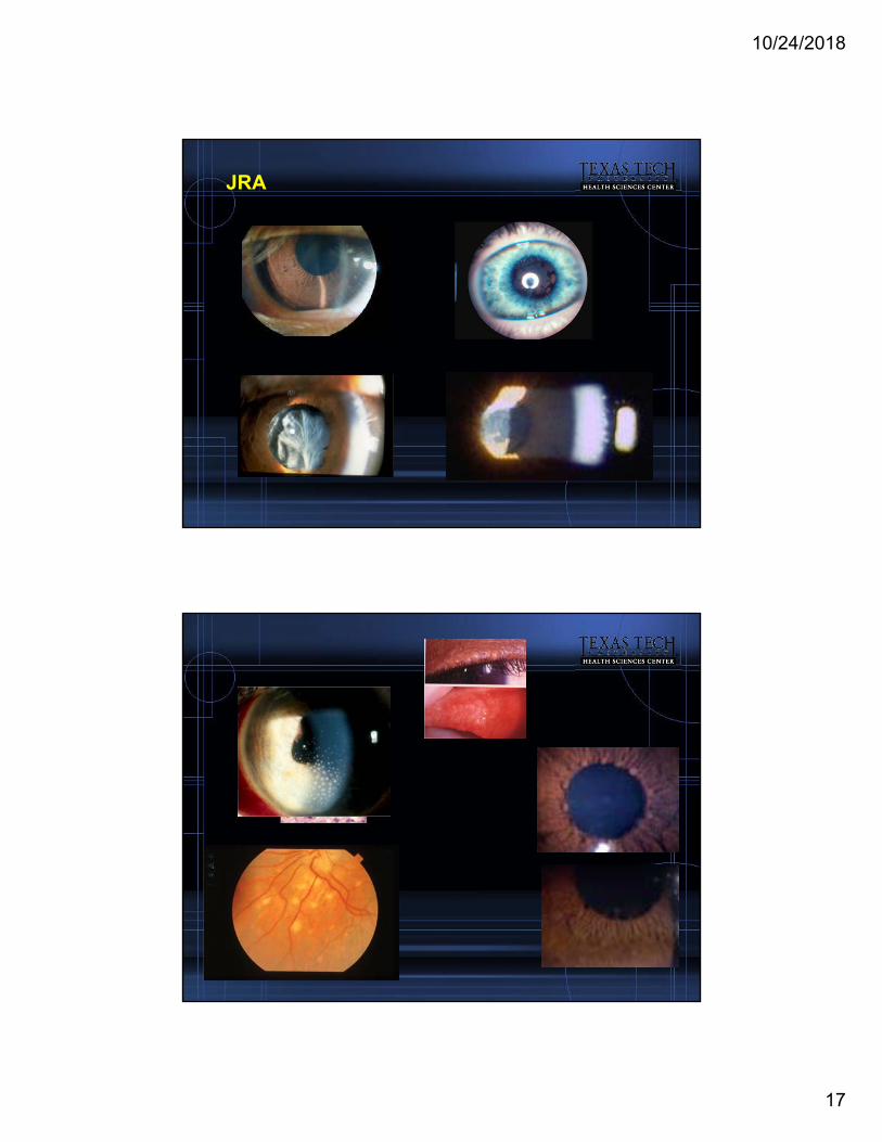
JRA associated UveitisRisk factors for uveitis: female, pauciarticular, ANA+, most are RHF –Eye findings: OFTEN WHITE & QUIET
OFTEN FOUND ON ROUTINE EXAMSFine KP, cell and flare, posterior synechiae cataract, band keratopathy
Symptoms due occur in some: pain, photophobia and ↓ VA
These findings or history in children = workup for JRA, ANA testing and referral to PCP comfortable with this type of evaluation or to Rheumatologist that sees children.
Uveitis associated with Juvenile Rheumatoid Arthritis
Management – Medical Topical Steroids mild cases short course
Treat for cell only, watch for complications Pupil dilation important even with flare, use short acting dilator QHS to prevent synechiae
Periocular steroidsChildren go to OR for this
Systemic steroidsShould be managed by PCP pediatrician
All must be vigilant for systemic and ocular complications
Systemic immunosuppresionManaged by pediatrician or pedi rheum or pedi ocologist or adult rheum
Weekly methotrexate can be PO or IM

10/24/2018
16
Uveitis associated with Juvenile Rheumatoid Arthritis?
Management – Surgical CataractsInflammation controlled for 3 monthsThis will be a challenging surgery
To it do well or refer to someone who canIdeal Referral Surgeon (may not be typical cataract-jock)
Good with parents and kidsComfortable with uveitic cataractsSurgery will likely be done in hospital
Role of IOL??? This is evolvingyoung with active no, older and controlled yesGlaucomaMedical first:
Beta blocker, diamox ok, no alphagan in the very youngSurgery last:
May have to be done with inflammation in order to control IOP Filters with anti-metabolite or valve/tubes
Uveitis associated with Juvenile Rheumatoid Arthritis?
Management – Surveillance
Patients with JRA (girls, ANA +, pauciarticular)
Tables (AAP and AAO)Keys: less than 7, pauciarticular disease, and ANA+
10/24/2018
17
JRA

10/24/2018
18
Uveitis associated with Sarcoidosis
Sarcoidosis accounts for 3-10% of all uveitis15-50% of patients with sarcoidosis have uveitisSarcoidosis is a systemic disease
10x more common is blacksPrimarily thoracic (90%), but can effect brain, bones, joints, liver
Uncommon in young children, but skin/joint >> chest findings
Key pathological lesionnoncaseating epitheloid cell granuloma or tubercle
Epitheloid cell is a polyhedral mononuclear histiocyteTubercle = epitheloid cells + multinucleated giant cells of Langhans type (nuclei at the periphery in an incomplete circle + rim of lymphocyctes
Uveitis associated with Sarcoidosis
Possible genetic link:Increased HLA-DRB1 in biopsy proven cases
Presence of peripheral anergy
Due to depression in delayed-type hypersensitivity
However, at the target organ site (eye)Active T-lymphocyte (CD4+) and MΦ response leading to granuloma
10/24/2018
19
Uveitis associated with Sarcoidosis
Acute Systemic Sarcoidosis and iridocyclitisLöfgren syndrome
Erythema nodosum, febrile arthropathy, bilateral hilar adenopathy, acute iritis
responsive to systemic steroids
Heerfordt Syndrome (uveoparotid fever)Uveitis, parotitis, fever and facial palsy
Chronic Sarcoidosis2 years duration, thoracic findings, chronic uveitis
Thoracic/pulmonary disease major cause of morbidity
Mortality is 5-10% (neurosarcoidosis)
Uveitis associated with Sarcoidosis
Eye FindingsSkin involvement is common
Orbital and eyelid granulomas
Conjunctival nodules
Lacrimal gland infiltration cause of dry eye
UveitisCan involve all structures
May begin as acute nongranulomatousbut 60% become chronic granulomatous

10/24/2018
20
Uveitis associated with Sarcoidosis
Main Uveitic FindingsMutton-fat KPKoeppe and Busacca iris nodulesSnowballs in the inferior anterior vitreous
Other findingsCorneal: nummular infiltrates & endothelial opacification
Angle: posterior and anterior synchiae
Posterior Segment findings – less common than A/CRetinal and choroidal granulomas usually ¼ DD in sizeIrregular nodules along venules = candelwax drippings or taches de bougieRetinal periphelbitis presents as sheathingCME is commonDisc edema, NVD,NVE can occur
Uveitis associated with Sarcoidosis?
DiagnosisBecause of its variety of presentation and frequency, it should be considered in every case of uveitis
If suspicion is strongCXR best screening test (positive in 90%) or CT
Lysozyme, ACE are supportive but not diagnositic
If clinical picture is strong and above are inconclusive considerGallium scan of head and neck especially used with ACE
Steroid treatment make this unreliable
Biopsy is useful especially of conjunctival or lacrimal nodules, which are easier than bronchial sample

10/24/2018
21
Uveitis associated with Sarcoidosis?
1. Treatment1. Steroids: (Topical, Periocular and Systemic)
2. Systemic immunosuppresion1. MTX or azathioprine, cellcept, infliximab
3. Remember it is a systemic dz, so get PCP involved in evaluation and management of systemic effects
Sarcoidosis

10/24/2018
22
Sympathetic Ophthalmia
Sympathetic Ophthalmia (SO)Rare bilateral, nonnecrotizing granulomatous after injury/surgery to one eye (the exciting eye). Followed by a latent period and the development of uveitis in the uninjured eye (the sympathizing eye)
Lower incidence because we are better surgeonsBetter wound closer and techniques (more enucleations vseviscerations)
Cause of SO is not known, but theories include:Hypersensitivity to melanin and melanin-associated proteinAn infectious causal agentSensitivity to retinal S antigen or retinal or uveal proteins
Possible Genetic risk:HLA-DR4, HLA-DRw53, HLA-DQw3In UK and Japan also see HLA-DRB1*04 and HLA-DQB1*04These and VKH are nearly the same

10/24/2018
23
Sympathetic Ophthalmia
Incidence0.2-0.5% in nonsurgical trauma0.01-0.03% in surgical traumaVitrectomy has now emerged as the main surgical risk for the development of SO
1980’s 0.01-0.06%Now may be as high as 0.06-0.12%
Makes senseBetter surgical care of ocular traumaGreater number of PPV casesBoth trauma and non-trauma
Risk factors:Old: males, children, elderlyNewer: no difference in the sexes, lower in kids, still high in the elderly
Increased history of previous eye surgery and trauma
Timing of onsetTraditional: 80% in 3 months, 90% in one yearRecent: 30% in 3 months, 50% in one year
Sympathetic Ophthalmia
Histopathological FeaturesDiffuse granulomatous uveal involvement Absence of reaction at the choriocapillarisPhagocytosis of uveal pigment by epithelioid cellsExtension of the granulomatous process into scleral canals and the optic disc
Clinical Features, asymmetrical bilateral panuveitis worse in exciting eye
Unaffected eyeCan occur within10 days after injury or surgery, slight redness, mild photophobia, mild problems with near visionProgression to panuveitis
Mutton fat KP, iris nodules, PAS, vitritis, choroidiits, ERD, papillitis
Injured or operated eyeEarly Panuveitis,

10/24/2018
24
Sympathetic Ophthalmia
Diagnosis
Bilateral uveitis following trauma or surgery Think SOInflammation may occur between 10 days to 50 years following the triggering event, but usually within the year
ManagementThe course of SO is chronic with frequent exacerbations
Topical, systemic or periocular steroidsCycloplegiaMay need antimetabolite level immunsuppression: cyclosporin
Overall current treatment for SO is effective and results in good vision over many yearsRemoval of blind exciting eye within two weeks of event has been shown to decrease risk of SO or if eye becomes blind after SO develops removal may decrease severity of SO,However, if exciting eye still sees (even LP) most will leave eye because of effective treatment of SOPrognosis is reasonable with 50% of patients having 20/40 in at least one eye
Sympathetic Ophthalmia

10/24/2018
25
Uveitis and Closing Pearls
When seeing a patient with Uveitis:
1. Could it be an infection?
2. Could it be caused from a systemic disease?
3. With Initial Treatment decisions:
1. Deal with inflammation & IOP
4. With chronic treatment decisions1. Deal with Inflammation
2. Deal with the side effects of the medication to TX the inflammation (toxicity and IOP)
3. Deal with the IOP from uveitis or meds to treat uveitis
4. Deal with side effects of uveitis1. Eye: cataracts and glaucoma
2. Systemic: uveitis or TX of uveitis
Questions??

10/24/2018
26
Vogt-Koyangi-Harada (VKH) Disease
1. Rare cause of Panuveitis even among the mostly commonly effected groups
1. Asian & american Indian’s between 30-50 years of age
2. Dark skinned people: hispanics
2. Cause not known1. However, reaction to melanin-associated protein, melanocytes or
RPE
3. Overall the bilateral granulomatous panuveitis resembles SO, however these patients have no history of ocular injury or surgery

10/24/2018
27
VKH – 4 Phases
1. Prodromal1. Flu symptoms
2. CNS signs: nuchal rigidity, seizures, coma, loss of consciousness, paralysis, optic neuropathy, CSF transient pleocytosis
3. Skin signs in 30% of patients1. Alopecia, vitiligo, poliosis
4. Hearing complaints in 30% of patients1. Deafness or tinnitus
2. Uveitic (1-2 days after CNS signs)
1. Symptoms: photophobia, redness, blurry vsion and ocular pain
2. Signs: bilateral a/c and vitreous cell and Exudative RD – these can be shallow and have a cloverleaf pattern around posterior pole, optic nerve head swelling
VKH – 4 Phases
3. Chronic or convalscent1. Resolution of ERD and gradual depigmentation of the choroid
1. Producing the orange-red discoloration = sunset-glow fundus2. Discrete, round depigmented lesion develop in the inferior
peripheral fundus = resolved Dalen Fuchs nodules3. Peripapillary atrophy can be seen4. Skin changes usually occur weeks to months after the beginning
of the uveitic phase, but can occur at the onset1. Perilimbal vitiligo (Sugiura’s sign) in more than 75% of cases2. Vitligo, alopecia, and poliosis in 30%
4. Recurrent phase1. Increased risk with incomplete treatment of uveitic and chronic
phases2. Signs: granulomatous anterior uveitis: KP, iris nodules and
depigmentation and atrophy3. Vision loss due to: Cataracts 50%, glaucoma 33%, CNVM 10%
10/24/2018
28
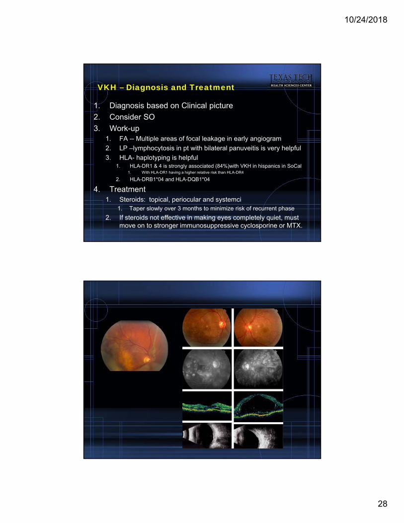
VKH – Diagnosis and Treatment
1. Diagnosis based on Clinical picture
2. Consider SO
3. Work-up1. FA -- Multiple areas of focal leakage in early angiogram
2. LP –lymphocytosis in pt with bilateral panuveitis is very helpful
3. HLA- haplotyping is helpful1. HLA-DR1 & 4 is strongly associated (84%)with VKH in hispanics in SoCal
1. With HLA-DR1 having a higher relative risk than HLA-DR4
2. HLA-DRB1*04 and HLA-DQB1*04
4. Treatment1. Steroids: topical, periocular and systemci
1. Taper slowly over 3 months to minimize risk of recurrent phase
2. If steroids not effective in making eyes completely quiet, must move on to stronger immunosuppressive cyclosporine or MTX.

10/24/2018
29
Behcet Disease (Adamantiades-Behcets)
1. Chronic relapsing occlusive vasculitis1. Eastern Mediterranean and Pacific rim of Asia
2. Old Silk Route established by Marco Polo
3. Turkey: 100-300/100,000, Japan:8-10/100,000 in Japan, 0.4/100,000 in US
4. Four classic lesion:1. Aphthous oral lesions
2. Skin lesions, erythema nodosum
3. Genitial lesions
4. Intraocular inflammation
5. HLA typing1. HLA-B51