Embed Size (px)
Citation preview
4/2/2019
1
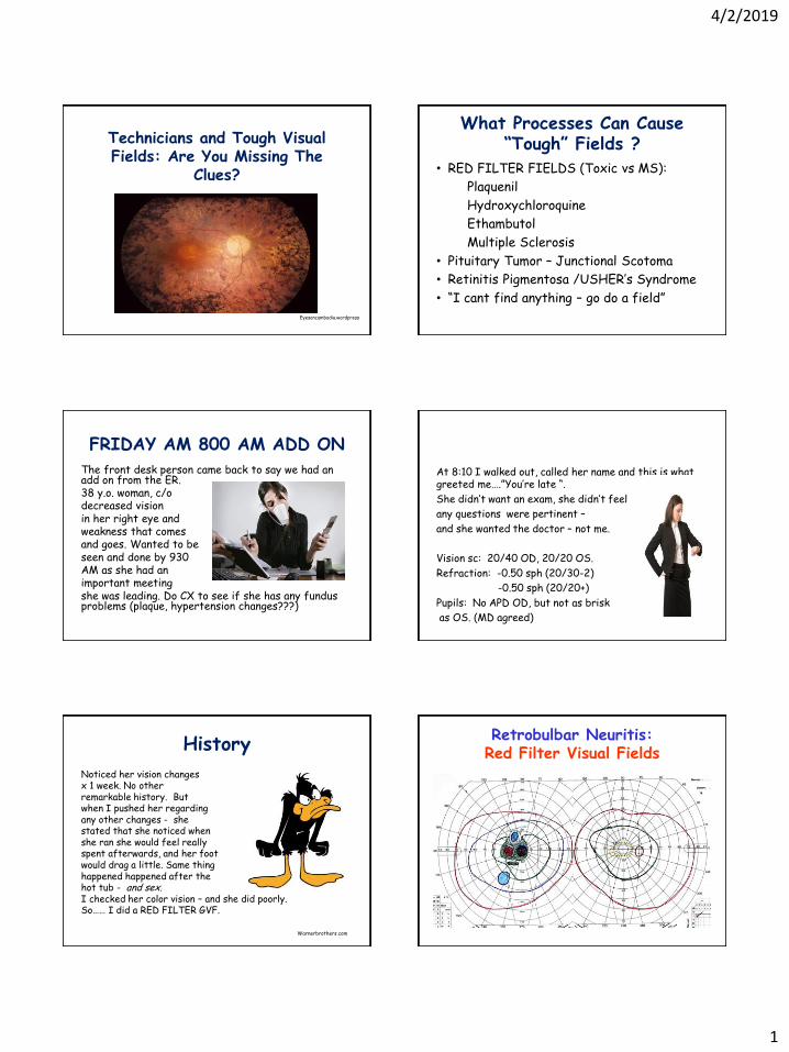
Technicians and Tough Visual Fields: Are You Missing The
Clues?
Eyesoncambodia.wordpress
What Processes Can Cause “Tough” Fields ?
• RED FILTER FIELDS (Toxic vs MS):
Plaquenil
Hydroxychloroquine
Ethambutol
Multiple Sclerosis
• Pituitary Tumor – Junctional Scotoma
• Retinitis Pigmentosa /USHER’s Syndrome
• “I cant find anything – go do a field”
FRIDAY AM 800 AM ADD ON
The front desk person came back to say we had an add on from the ER. 38 y.o. woman, c/o decreased vision in her right eye and weakness that comes and goes. Wanted to be seen and done by 930 AM as she had an important meeting she was leading. Do CX to see if she has any fundus problems (plaque, hypertension changes???)
At 8:10 I walked out, called her name and this is what greeted me….”You’re late “.
She didn’t want an exam, she didn’t feel
any questions were pertinent –
and she wanted the doctor – not me.
Vision sc: 20/40 OD, 20/20 OS.
Refraction: -0.50 sph (20/30-2)
-0.50 sph (20/20+)
Pupils: No APD OD, but not as brisk
as OS. (MD agreed)
History
Noticed her vision changes x 1 week. No other remarkable history. But when I pushed her regarding any other changes - she stated that she noticed when she ran she would feel really spent afterwards, and her foot would drag a little. Same thing happened happened after the hot tub - and sex. I checked her color vision – and she did poorly. So…… I did a RED FILTER GVF.
Warnerbrothers.com
Retrobulbar Neuritis: Red Filter Visual Fields
4/2/2019
2
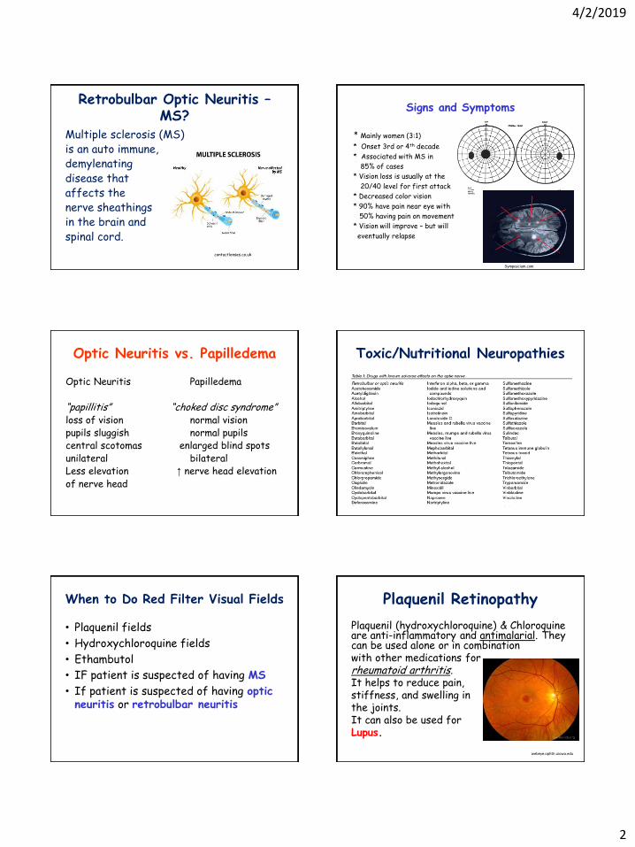
Retrobulbar Optic Neuritis – MS?
Multiple sclerosis (MS) is an auto immune, demylenating disease that affects the nerve sheathings in the brain and spinal cord.
contactlenses.co.uk
Signs and Symptoms
* Mainly women (3:1)
* Onset 3rd or 4th decade
* Associated with MS in
85% of cases
* Vision loss is usually at the
20/40 level for first attack
* Decreased color vision
* 90% have pain near eye with
50% having pain on movement
* Vision will improve – but will
eventually relapse
Symposcium.com
Optic Neuritis vs. Papilledema
Optic Neuritis Papilledema “papillitis” “choked disc syndrome” loss of vision normal vision pupils sluggish normal pupils central scotomas enlarged blind spots unilateral bilateral Less elevation ↑ nerve head elevation of nerve head
Toxic/Nutritional Neuropathies
When to Do Red Filter Visual Fields
• Plaquenil fields
• Hydroxychloroquine fields
• Ethambutol
• IF patient is suspected of having MS
• If patient is suspected of having optic neuritis or retrobulbar neuritis
Plaquenil Retinopathy
Plaquenil (hydroxychloroquine) & Chloroquine are anti-inflammatory and antimalarial. They can be used alone or in combination with other medications for rheumatoid arthritis. It helps to reduce pain, stiffness, and swelling in the joints. It can also be used for Lupus.
webeye.ophth.uiowa.edu

4/2/2019
3
Chloroquine or Hydroxychloroquine Amblyopia
Antimalarial drugs that are also used in the treatment of Lupus and Rheumatoid Arthritis.
Defects are large, central
scotomas. Use Red Filter
Test !
Toxic Macular Disorders
Bull’s Eye Maculopathy with Ethambutol (TB med) or Plaquenil Abnormal ERG with loss of cone function. Your doctor may see a “mottling “ of the fovea area. Important to do COLOR VISION TESTING as well as VF 10-2. IF do a GVF – do a red filter test to hyper check the cones..
retinalphysician.com
Ethambutol Amblyopia
* Ethambutol is used in the
treatment of tuberculosis
• Defects will be central or centrocecal scotomas
of various depths
• Decreased color vision !
• Do RED VISUAL FIELDS
Lead Amblyopia
• Often associated with children of lower socio- economic means living in cities • Also seen with painters and gasoline attendants (leaded gas) • Fumes or paint are absorbed through the skin • Can eventually cause papilledema and optic atrophy due to chronic exposure
Classic Field Defect: Tear Shaped CentrocecalScotoma
Tear shape points at fixation in one eye. IF there is optic nerve damage due to chronic papilledema, the defects will be permanent !
Pituitary Gland (Hypophysis)
Hangs by a “stalk” from under the hypothalamus. Two parts: Anterior pituitary (glandular tissue) and the posterior pituitary (nervous tissue). Called the “master gland” because it controls so many other endocrine glands.
vivo.colostate.edu
4/2/2019
4
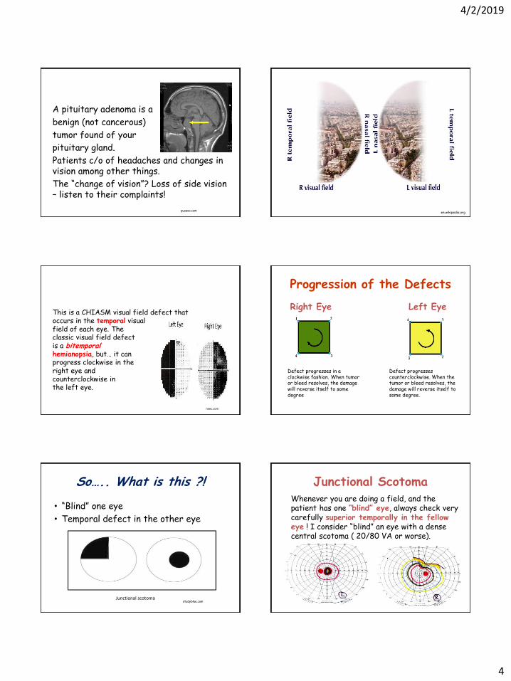
A pituitary adenoma is a
benign (not cancerous)
tumor found of your
pituitary gland.
Patients c/o of headaches and changes in vision among other things.
The “change of vision”? Loss of side vision – listen to their complaints!
quazoo.com en.wikipedia.org
This is a CHIASM visual field defect that occurs in the temporal visual field of each eye. The classic visual field defect is a bitemporal hemianopsia, but… it can progress clockwise in the right eye and counterclockwise in the left eye.
neec.com
Progression of the Defects
Right Eye Left Eye
Defect progresses in a clockwise fashion. When tumor or bleed resolves, the damage will reverse itself to some degree
Defect progresses counterclockwise. When the tumor or bleed resolves, the damage will reverse itself to some degree.
So….. What is this ?!
• “Blind” one eye
• Temporal defect in the other eye
studyblue.com Junctional scotoma
Junctional Scotoma Whenever you are doing a field, and the patient has one “blind” eye, always check very carefully superior temporally in the fellow eye ! I consider “blind” an eye with a dense central scotoma ( 20/80 VA or worse).

4/2/2019
5
Retinitis Pigmentosa
A genetic disorder of the eyes that causes loss of peripheral vision due to ROD dystrophy. Usually occurs in young males. Symptoms include: difficulty with night vision and decreased peripheral vision. Onset is generally gradual. As peripheral vision worsens, patients may experience “tunnel vision".
Complete blindness is uncommon, but central vision can be affected due to cone “drop out” as well .
rpcure.net
Findings in the retina have been characterized as the "ophthalmic triad". This includes : * a “mottled” appearance of the retinal pigment epithelium (RPE) * bony spicule formation • a waxy look to the optic nerve • weakening of blood vessels in the retina
Wikipedia.com
herbal-care-products.com
Normal vs. RP Fundus
dxline.info webeye.ophth.uiowa.edu
Layers of the Retina
101proofsforgod.blogspot.com
A contact lens electrode to the eye is used - a bright light is flashed. Patients with the RP trait would show decreased /delayed electrical response in the
photoreceptors (rods & cones).
If a dim flash ERG is performed on a dark-adapted eye, the response is primarily from the rods. Flash ERGs will reflect the activity of the cones. Bright flashes will show ERGs with an a-wave (initial negative deflection) followed by a b-wave (positive deflection). ( Wikipedia)
Basic ERG in a normal patient
webvision.umh.es

4/2/2019
6
Slideshare.net
When doing a GVF, it is important to NOT start too far “in” or you may miss islands. Very often fields appear “gun barrel” for this very reason –
islands were missed
because the
tech started in
too close.
webeye.ophth.uiowa.edu
“Gun Barrel “ (Tunnel Vision)
healio.com
What The Patient May See
webmd.com
Non Pigmented RP
Atypical cases of
RP are common and
often occur when you
do not see the
usual pigment
changes in
the fundus.
retinagallery.com
Usher’s Syndrome
A rare genetic disorder caused by a mutation in the genes resulting in a combination of hearing loss and visual impairment. It is a leading cause of deaf/blindness and is incurable at this time. The progressive blindness of Usher syndrome results from the retinitis pigmentosa. Testing is the same.
omnieyesurgery.com Webeye.com

4/2/2019
7
Usher’s occurs in roughly 1 deaf person in 23,000 in the United States. People with Usher syndrome represent roughly 1/6th of people with retinitis pigmentosa.
People with Usher are born profoundly deaf and begin to lose their vision in the first 1-2 decades. First visual complaints are often night blindness (nyctalopia) (Wikipedia)
Slideshare.net
Nyctalopia
Slideplayer.com
What Is This ?!
Scotomatous Hemianopsia…….. CHIASM…..temporal! [email protected]