Embed Size (px)
Citation preview
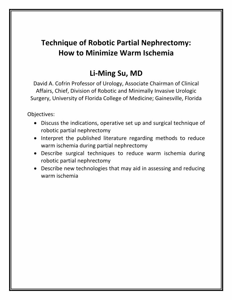
Technique of Robotic Partial Nephrectomy:
How to Minimize Warm Ischemia
Li-Ming Su, MD David A. Cofrin Professor of Urology, Associate Chairman of Clinical Affairs, Chief, Division of Robotic and Minimally Invasive Urologic
Surgery, University of Florida College of Medicine; Gainesville, Florida
Objectives: • Discuss the indications, operative set up and surgical technique of
robotic partial nephrectomy • Interpret the published literature regarding methods to reduce
warm ischemia during partial nephrectomy • Describe surgical techniques to reduce warm ischemia during
robotic partial nephrectomy • Describe new technologies that may aid in assessing and reducing
warm ischemia
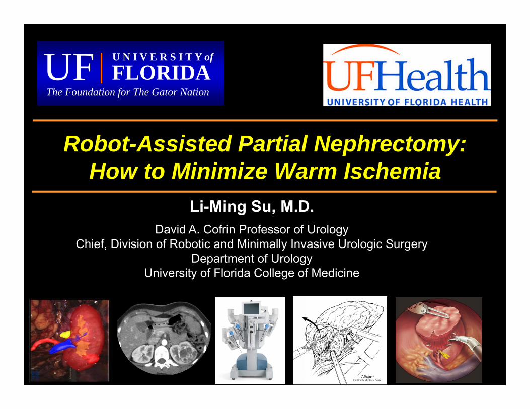
Robot-Assisted Partial Nephrectomy: How to Minimize Warm Ischemia
Li-Ming Su, M.D.David A. Cofrin Professor of Urology
Chief, Division of Robotic and Minimally Invasive Urologic SurgeryDepartment of Urology
University of Florida College of Medicine
U N I V E R S I T Y of
FLORIDA UFThe Foundation for The Gator Nation
Outline
• Indication• Surgical Technique• Techniques to Minimize Warm Ischemia• New Technologies
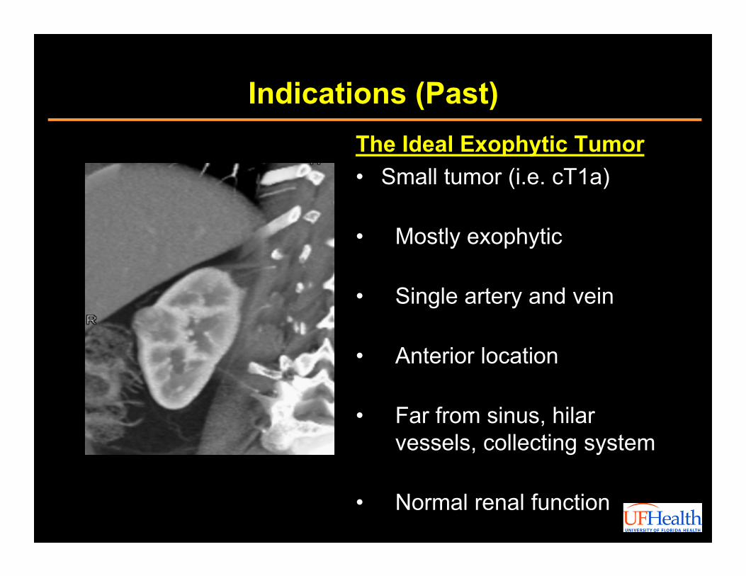
Indications (Past)The Ideal Exophytic Tumor• Small tumor (i.e. cT1a)
• Mostly exophytic
• Single artery and vein
• Anterior location
• Far from sinus, hilarvessels, collecting system
• Normal renal function
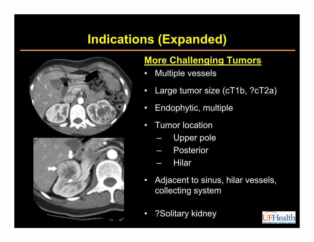
Indications (Expanded)More Challenging Tumors• Multiple vessels
• Large tumor size (cT1b, ?cT2a)
• Endophytic, multiple
• Tumor location– Upper pole– Posterior– Hilar
• Adjacent to sinus, hilar vessels, collecting system
• ?Solitary kidney
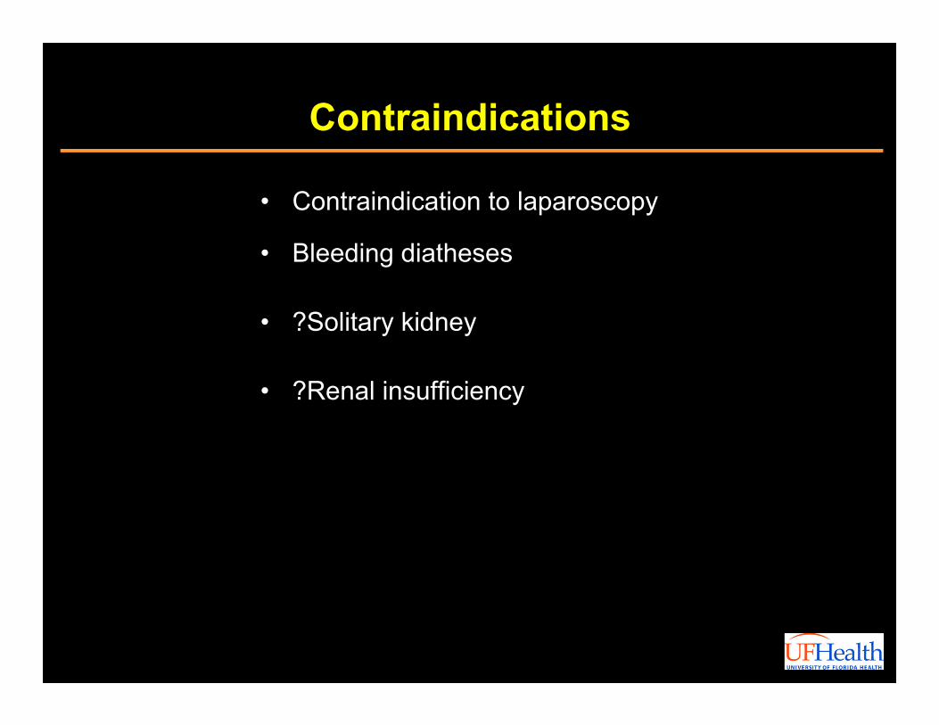
Contraindications
• Contraindication to laparoscopy
• Bleeding diatheses
• ?Solitary kidney
• ?Renal insufficiency
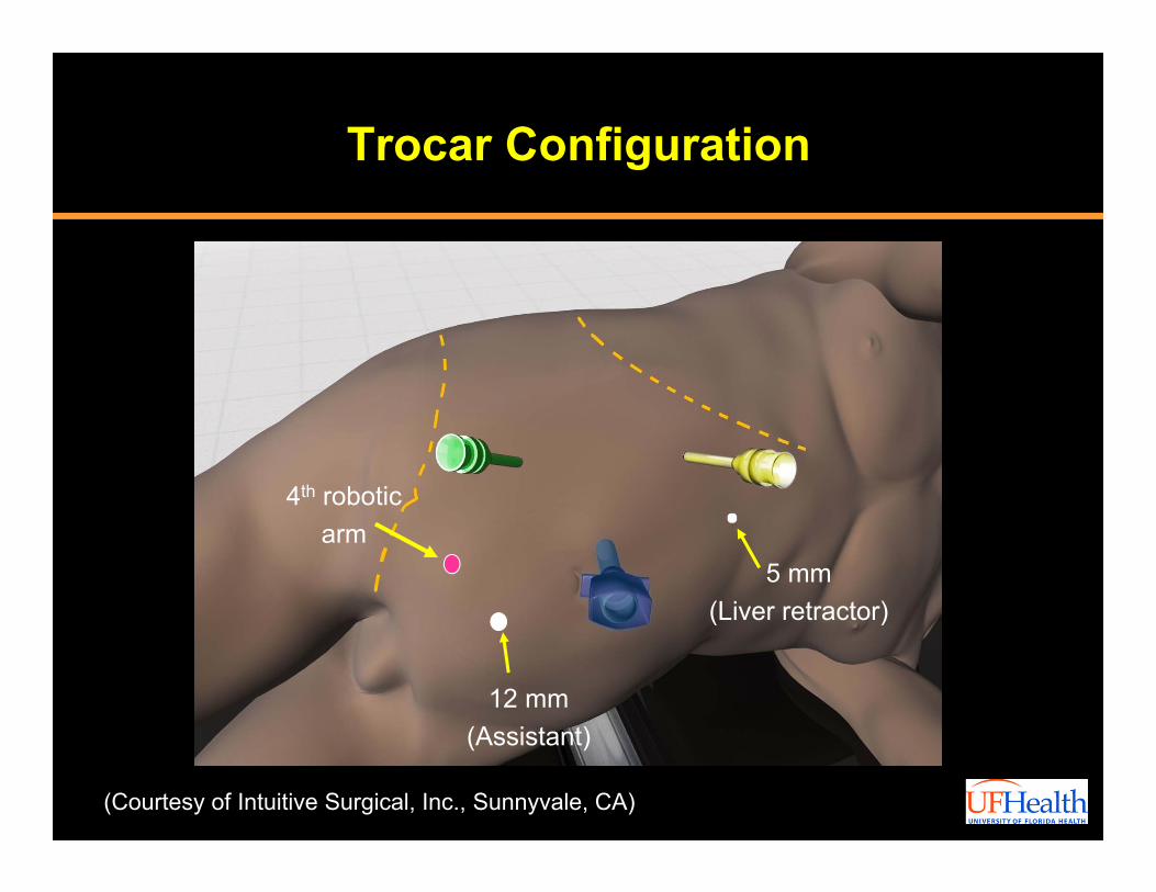
Trocar Configuration
(Courtesy of Intuitive Surgical, Inc., Sunnyvale, CA)
5 mm(Liver retractor)
12 mm(Assistant)
4th roboticarm
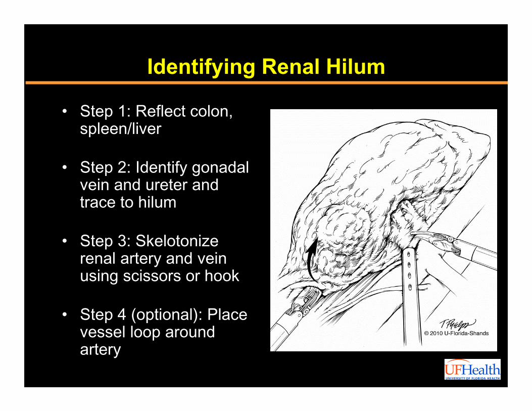
Identifying Renal Hilum
• Step 1: Reflect colon, spleen/liver
• Step 2: Identify gonadal vein and ureter and trace to hilum
• Step 3: Skelotonize renal artery and vein using scissors or hook
• Step 4 (optional): Place vessel loop around artery
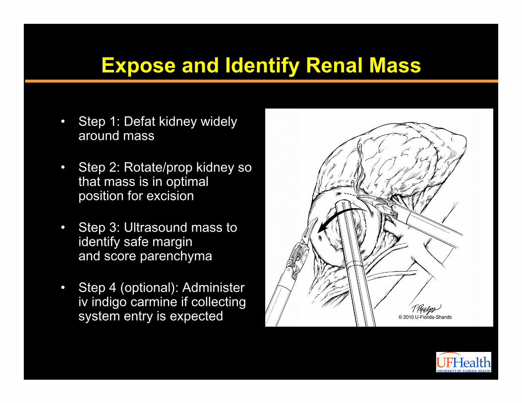
Expose and Identify Renal Mass
• Step 1: Defat kidney widely around mass
• Step 2: Rotate/prop kidney so that mass is in optimal position for excision
• Step 3: Ultrasound mass to identify safe marginand score parenchyma
• Step 4 (optional): Administer iv indigo carmine if collecting system entry is expected
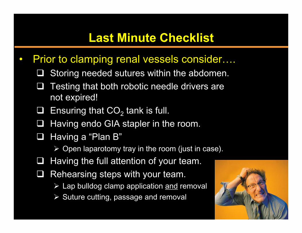
Last Minute Checklist• Prior to clamping renal vessels consider….
Storing needed sutures within the abdomen. Testing that both robotic needle drivers are
not expired! Ensuring that CO2 tank is full. Having endo GIA stapler in the room. Having a “Plan B”
Open laparotomy tray in the room (just in case). Having the full attention of your team. Rehearsing steps with your team.
Lap bulldog clamp application and removal Suture cutting, passage and removal
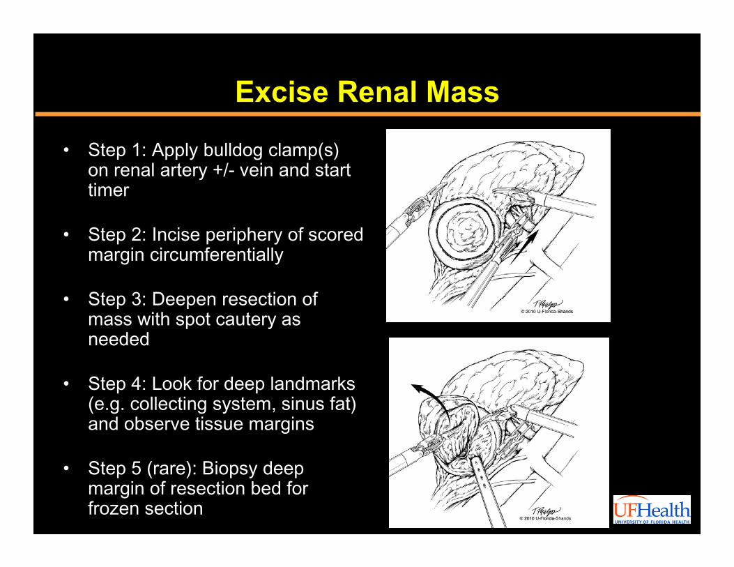
Excise Renal Mass
• Step 1: Apply bulldog clamp(s) on renal artery +/- vein and start timer
• Step 2: Incise periphery of scored margin circumferentially
• Step 3: Deepen resection of mass with spot cautery as needed
• Step 4: Look for deep landmarks (e.g. collecting system, sinus fat) and observe tissue margins
• Step 5 (rare): Biopsy deep margin of resection bed for frozen section
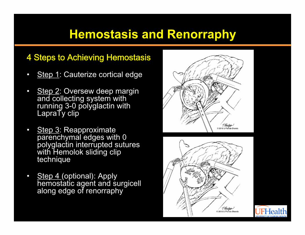
Hemostasis and Renorraphy
4 Steps to Achieving Hemostasis
• Step 1: Cauterize cortical edge
• Step 2: Oversew deep margin and collecting system with running 3-0 polyglactin with LapraTy clip
• Step 3: Reapproximate parenchymal edges with 0 polyglactin interrupted sutures with Hemolok sliding clip technique
• Step 4 (optional): Apply hemostatic agent and surgicell along edge of renorraphy
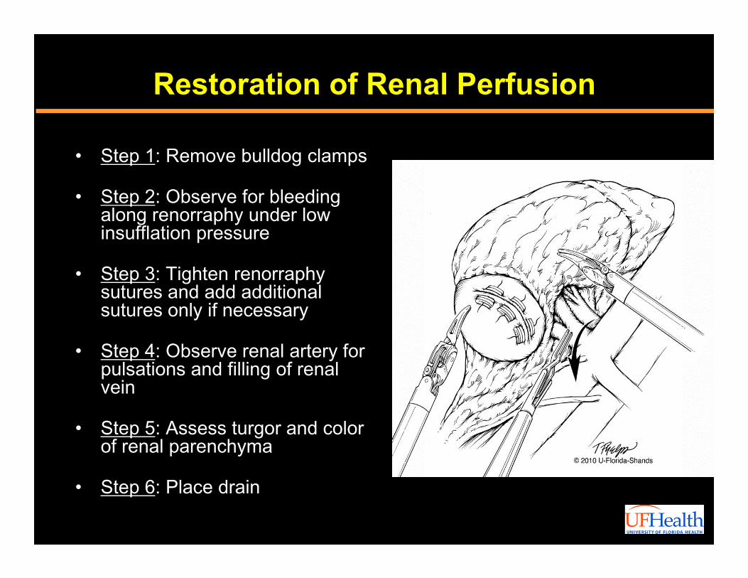
Restoration of Renal Perfusion
• Step 1: Remove bulldog clamps
• Step 2: Observe for bleeding along renorraphy under low insufflation pressure
• Step 3: Tighten renorraphy sutures and add additional sutures only if necessary
• Step 4: Observe renal artery for pulsations and filling of renal vein
• Step 5: Assess turgor and color of renal parenchyma
• Step 6: Place drain
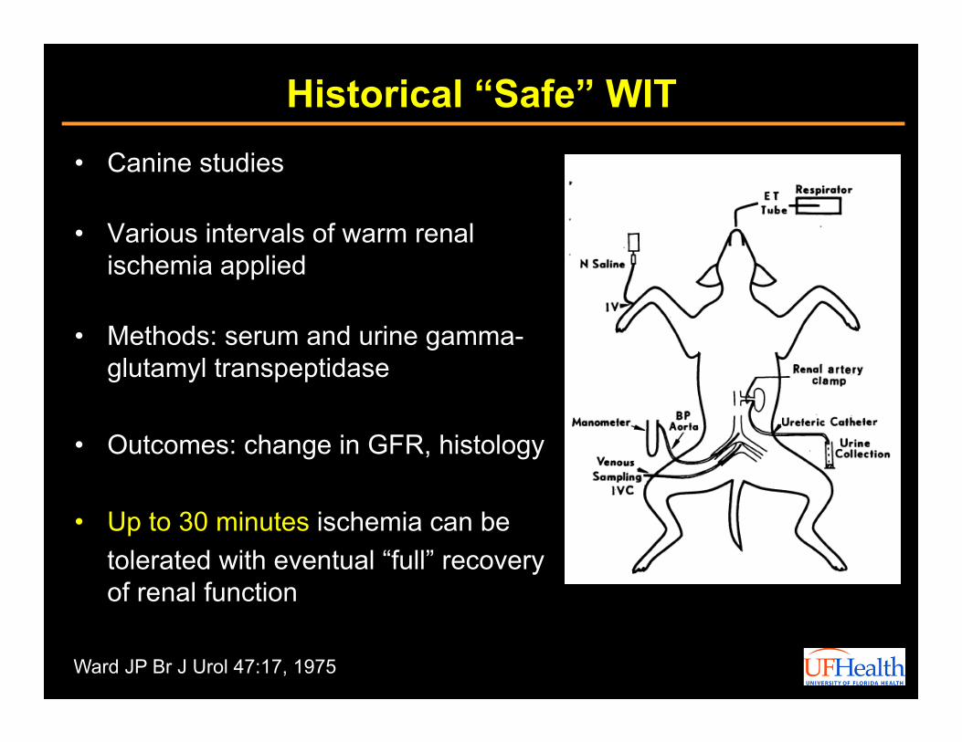
Historical “Safe” WIT• Canine studies
• Various intervals of warm renal ischemia applied
• Methods: serum and urine gamma-glutamyl transpeptidase
• Outcomes: change in GFR, histology
• Up to 30 minutes ischemia can be tolerated with eventual “full” recovery of renal function
Ward JP Br J Urol 47:17, 1975
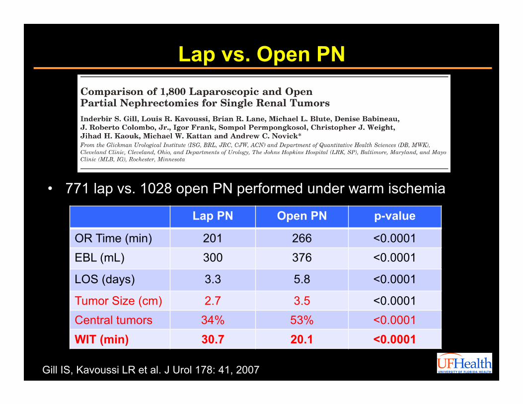
Lap vs. Open PN
• 771 lap vs. 1028 open PN performed under warm ischemia
Gill IS, Kavoussi LR et al. J Urol 178: 41, 2007
Lap PN Open PN p-value
OR Time (min) 201 266 <0.0001EBL (mL) 300 376 <0.0001
LOS (days) 3.3 5.8 <0.0001
Tumor Size (cm) 2.7 3.5 <0.0001Central tumors 34% 53% <0.0001WIT (min) 30.7 20.1 <0.0001
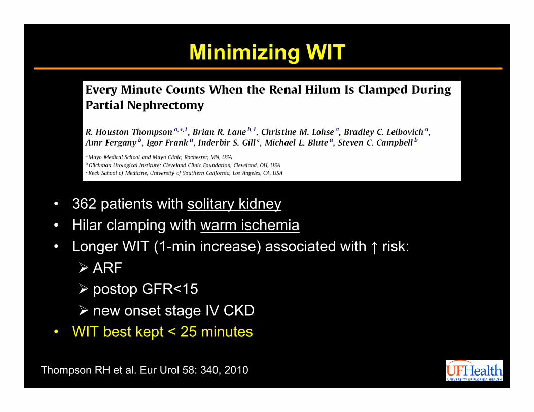
Minimizing WIT
• 362 patients with solitary kidney• Hilar clamping with warm ischemia• Longer WIT (1-min increase) associated with ↑ risk: ARF postop GFR<15 new onset stage IV CKD
• WIT best kept < 25 minutes
Thompson RH et al. Eur Urol 58: 340, 2010
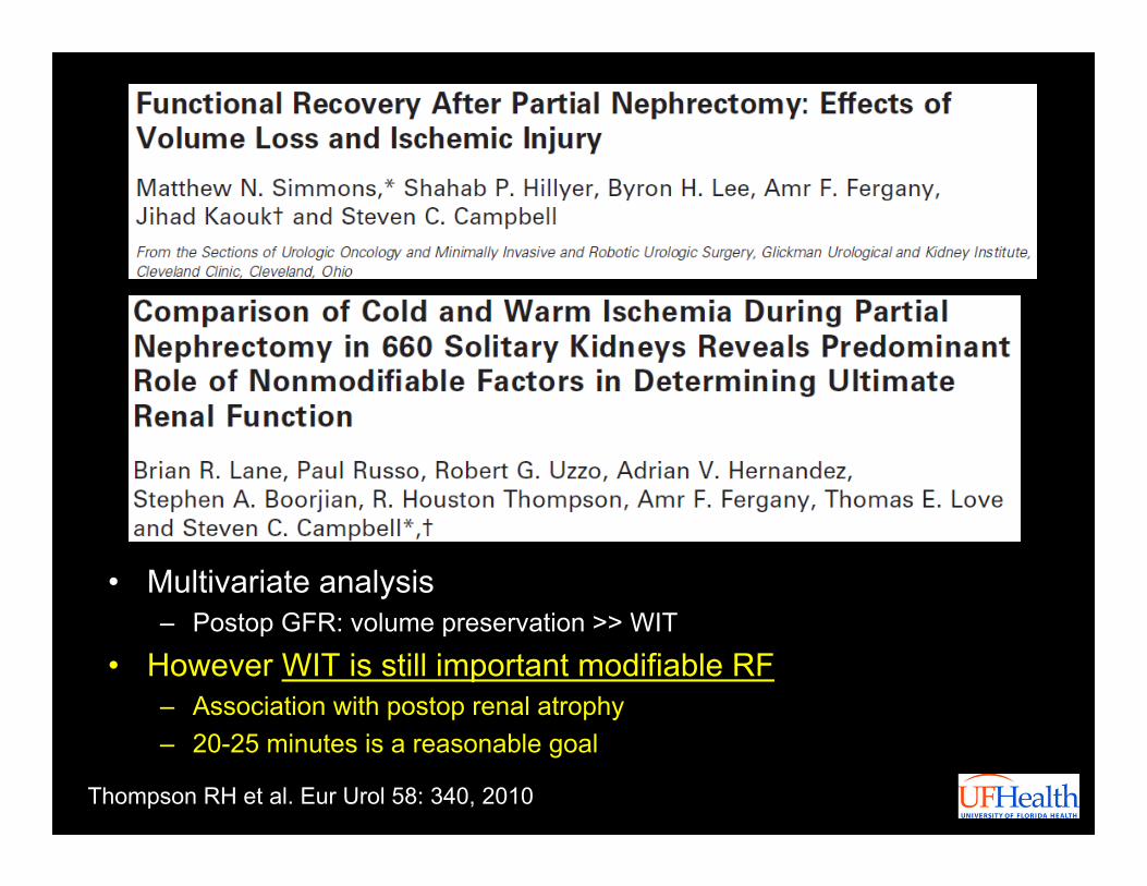
• Multivariate analysis– Postop GFR: volume preservation >> WIT
• However WIT is still important modifiable RF– Association with postop renal atrophy– 20-25 minutes is a reasonable goal
Thompson RH et al. Eur Urol 58: 340, 2010
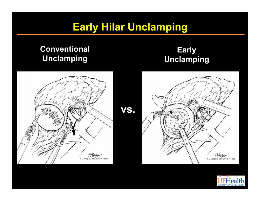
Early Hilar Unclamping
vs.
ConventionalUnclamping
EarlyUnclamping
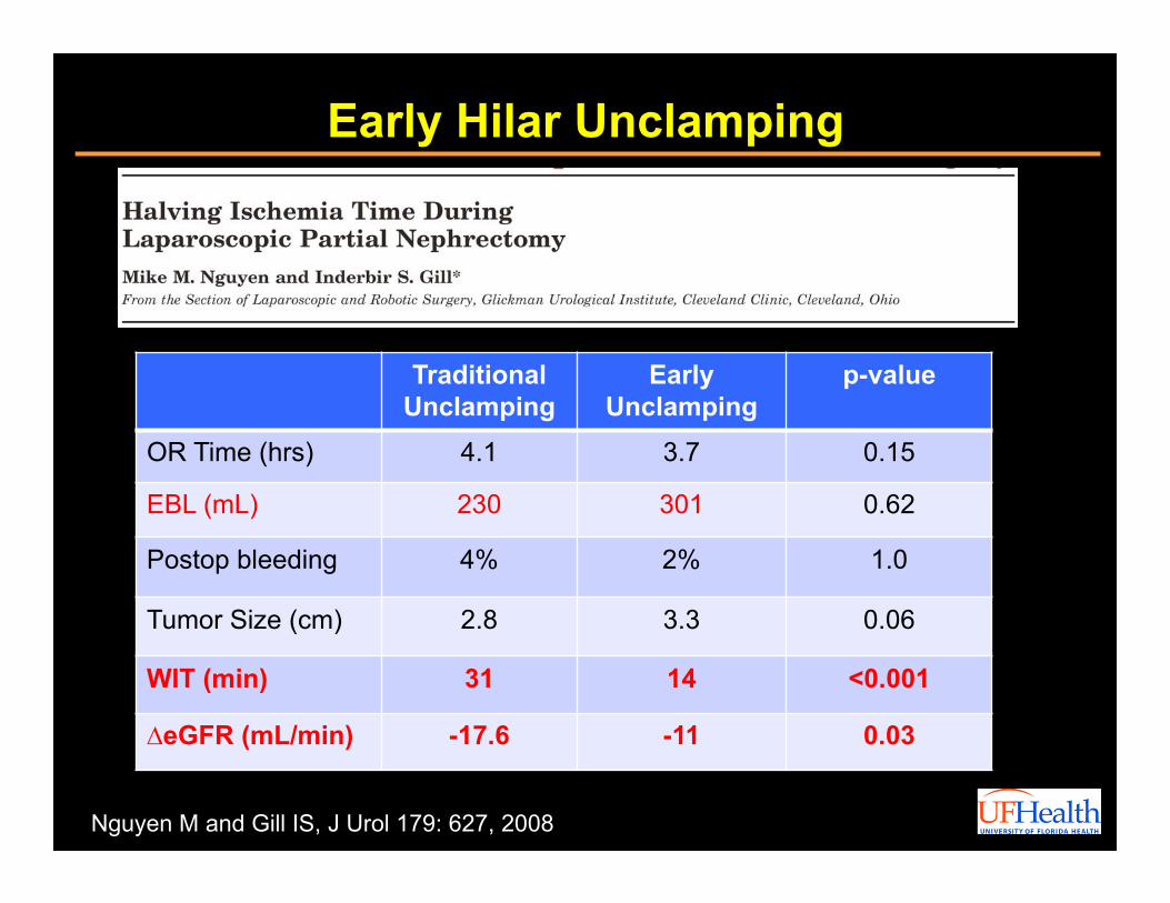
Early Hilar Unclamping
Nguyen M and Gill IS, J Urol 179: 627, 2008
Traditional Unclamping
EarlyUnclamping
p-value
OR Time (hrs) 4.1 3.7 0.15
EBL (mL) 230 301 0.62
Postop bleeding 4% 2% 1.0
Tumor Size (cm) 2.8 3.3 0.06
WIT (min) 31 14 <0.001
∆eGFR (mL/min) -17.6 -11 0.03
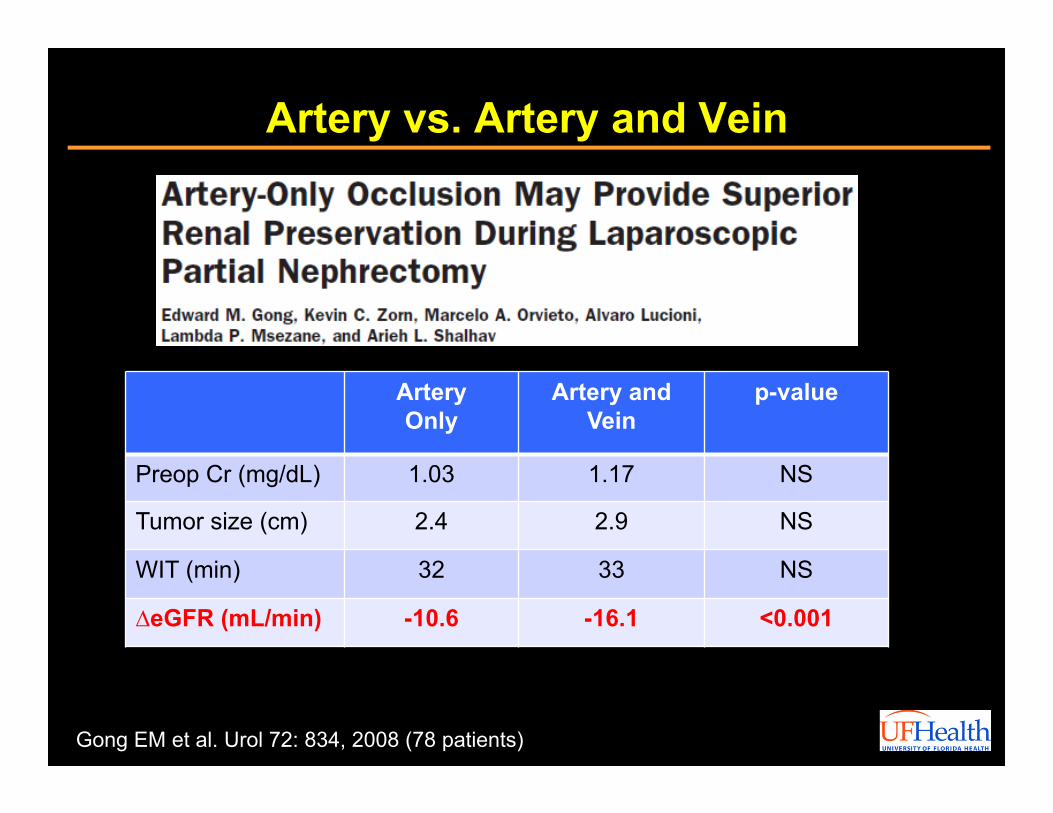
Artery vs. Artery and Vein
Gong EM et al. Urol 72: 834, 2008 (78 patients)
ArteryOnly
Artery andVein
p-value
Preop Cr (mg/dL) 1.03 1.17 NS
Tumor size (cm) 2.4 2.9 NS
WIT (min) 32 33 NS
∆eGFR (mL/min) -10.6 -16.1 <0.001
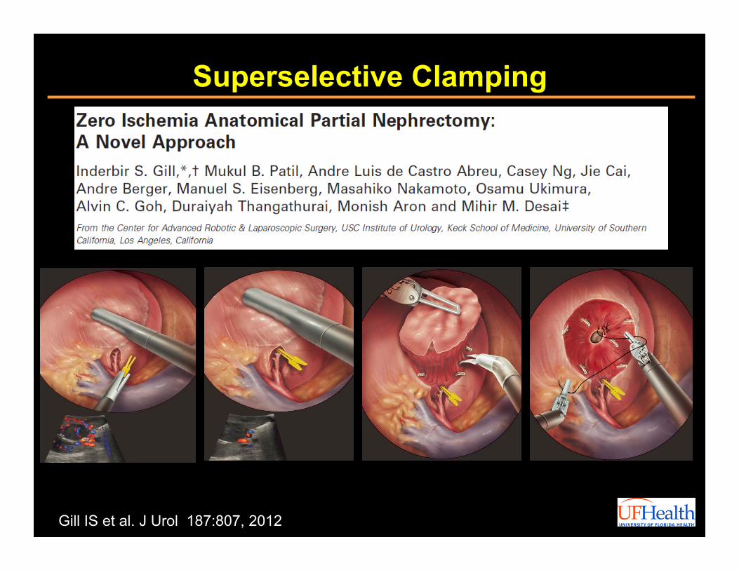
Gill IS et al. J Urol 187:807, 2012
Superselective Clamping
“Zero Ischemia” PN
• 58 patients, WIT 0 min, all negative margins• Tumor size 3.2 cm (0.9-13)• OR time 4.4 hours (1-8)• EBL 206 mL (25-1000)• LOS 3.9 days (2-19)• Complication rate 22.8%:
– Urine leak (3), Renal bleed (0)
• Transfusion rate 21%
Preop D/C 4 mo• SCr (mg/dL) 1.0 1.1 1.3• eGFR 79.6 72.9 61.5
Gill IS et al. J Urol 187:807, 2012
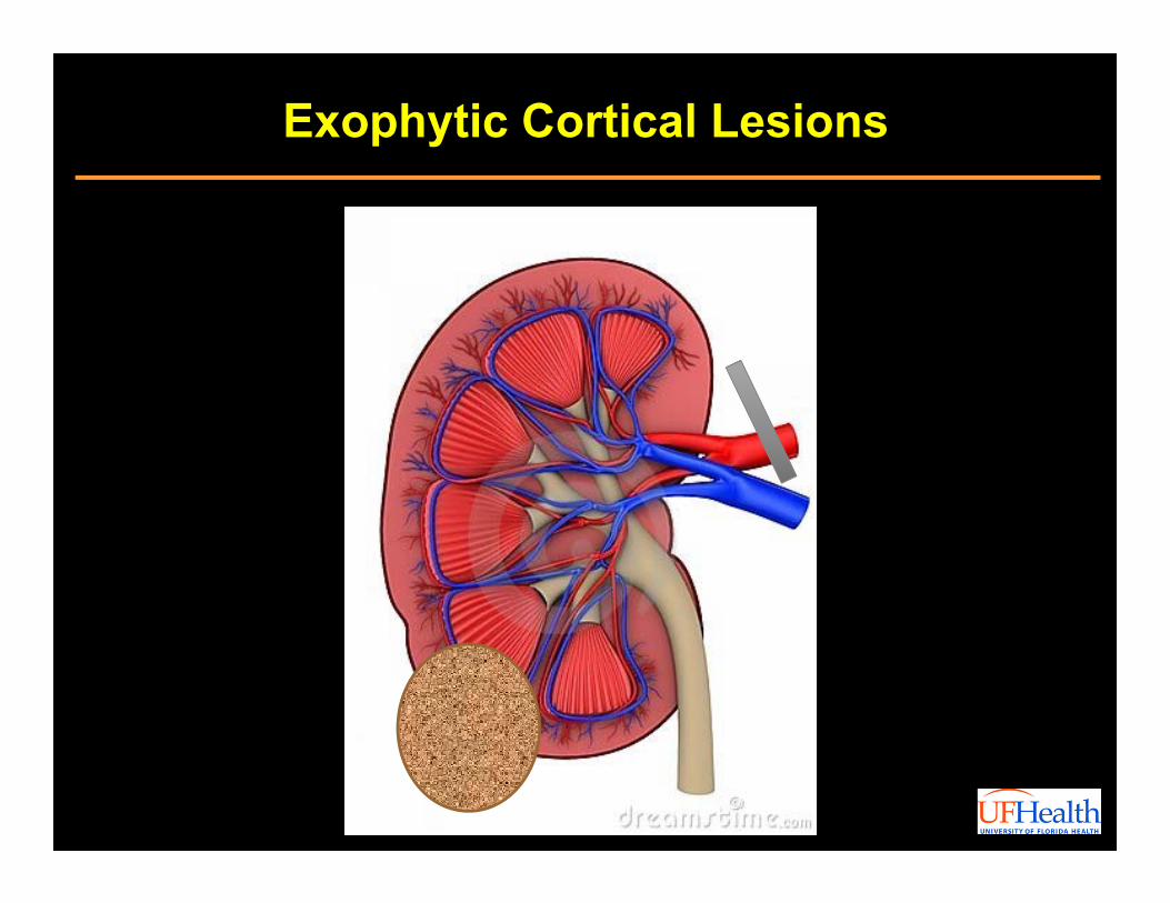
Exophytic Cortical Lesions
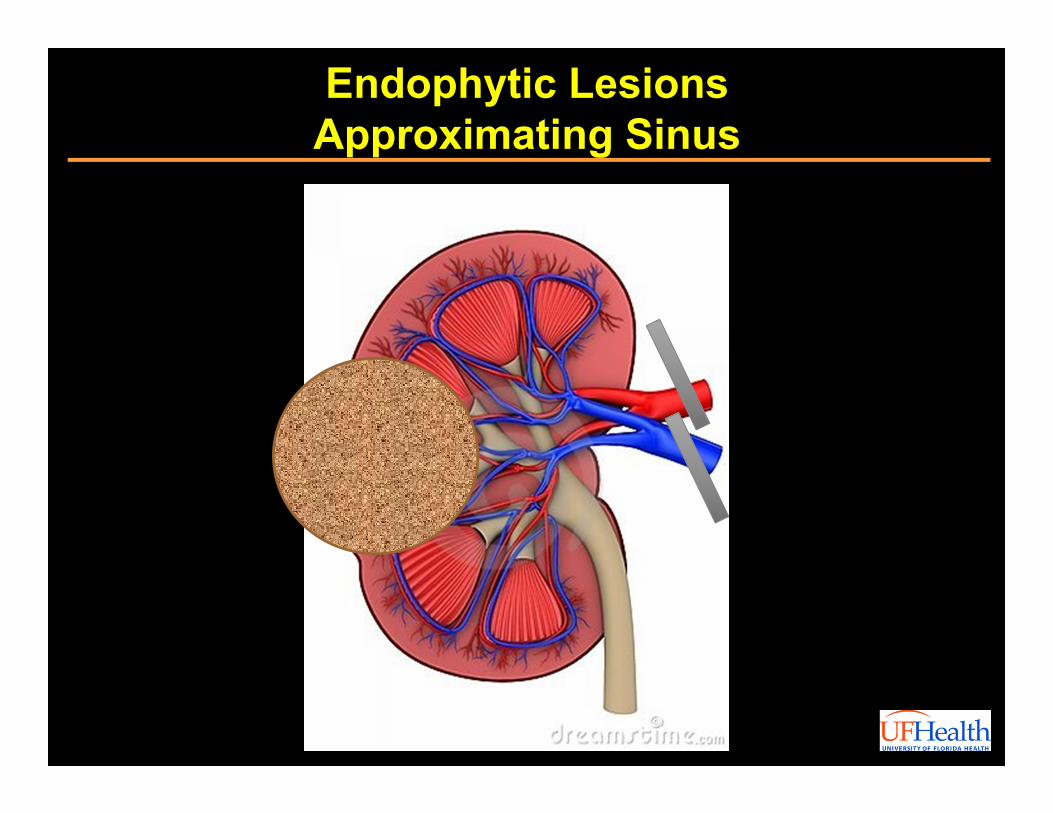
Endophytic Lesions Approximating Sinus
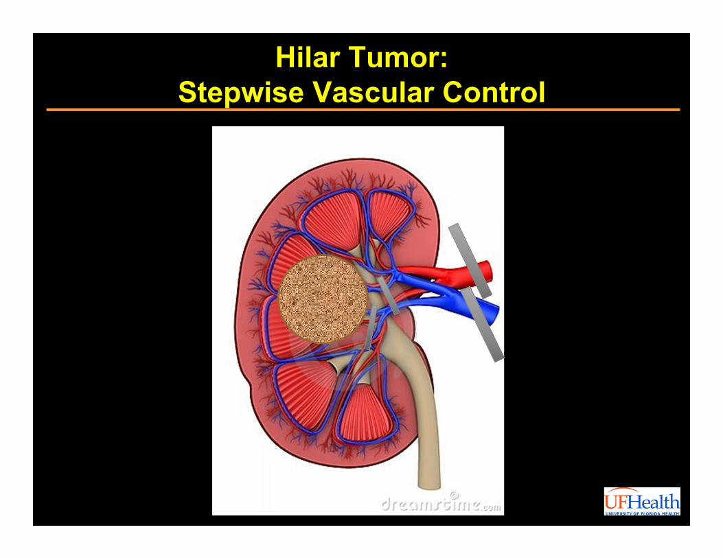
Hilar Tumor:Stepwise Vascular Control
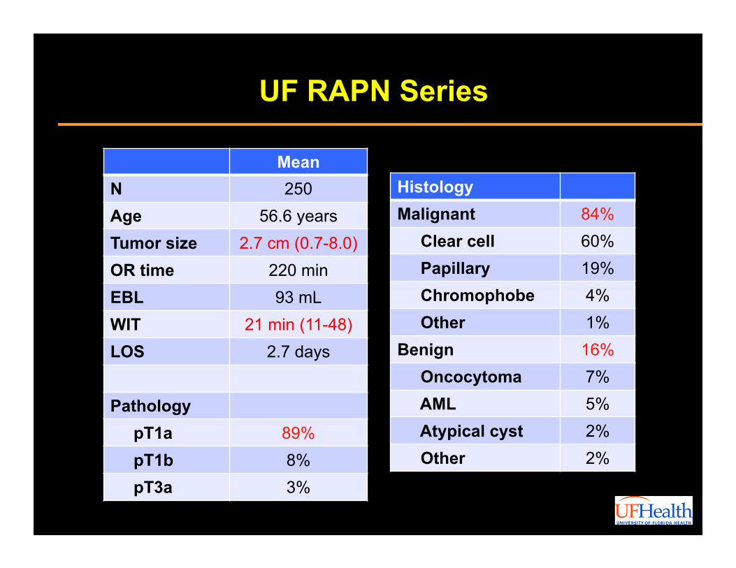
MeanN 250Age 56.6 yearsTumor size 2.7 cm (0.7-8.0)OR time 220 minEBL 93 mLWIT 21 min (11-48)LOS 2.7 days
PathologypT1a 89%pT1b 8%pT3a 3%
UF RAPN Series
HistologyMalignant 84%
Clear cell 60%Papillary 19%Chromophobe 4%Other 1%
Benign 16%Oncocytoma 7%AML 5%Atypical cyst 2%Other 2%
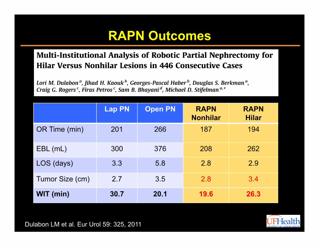
RAPN Outcomes
Dulabon LM et al. Eur Urol 59: 325, 2011
Lap PN Open PN RAPN Nonhilar
RAPNHilar
OR Time (min) 201 266 187 194
EBL (mL) 300 376 208 262
LOS (days) 3.3 5.8 2.8 2.9
Tumor Size (cm) 2.7 3.5 2.8 3.4
WIT (min) 30.7 20.1 19.6 26.3
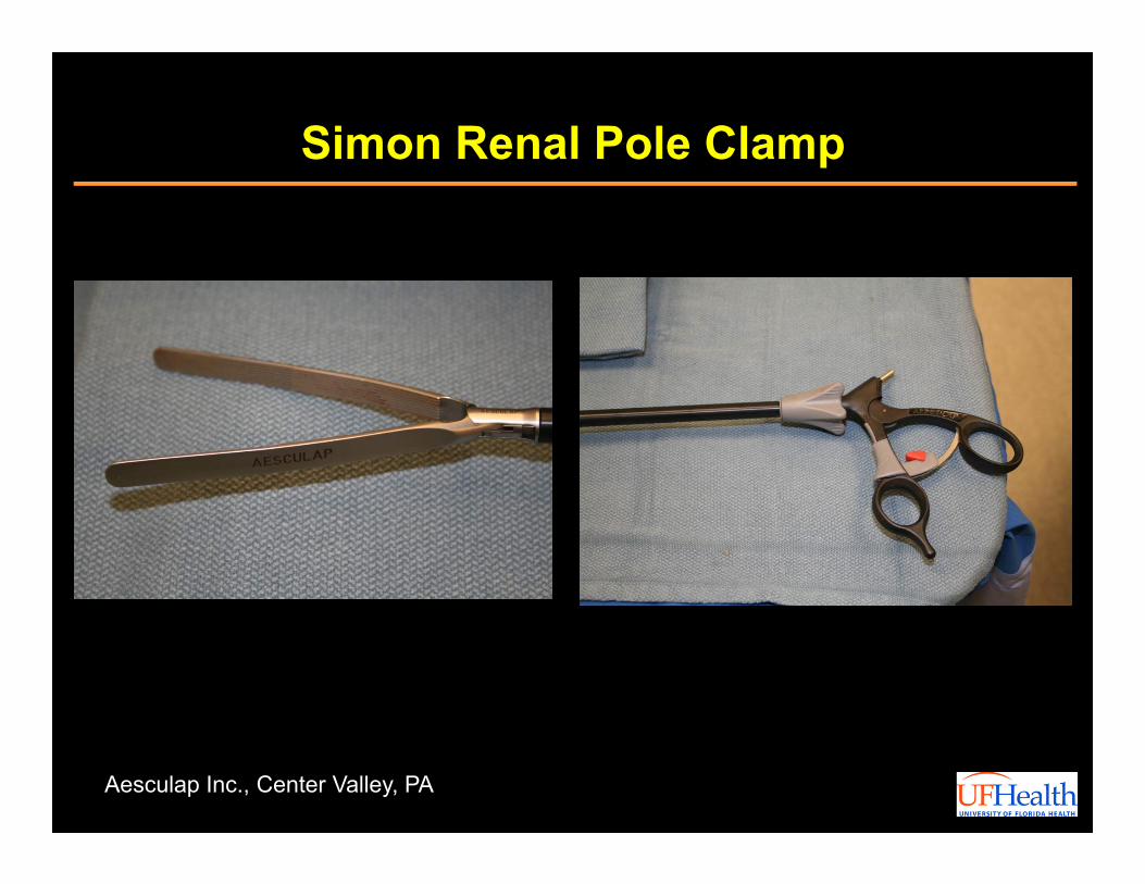
Simon Renal Pole Clamp
Aesculap Inc., Center Valley, PA
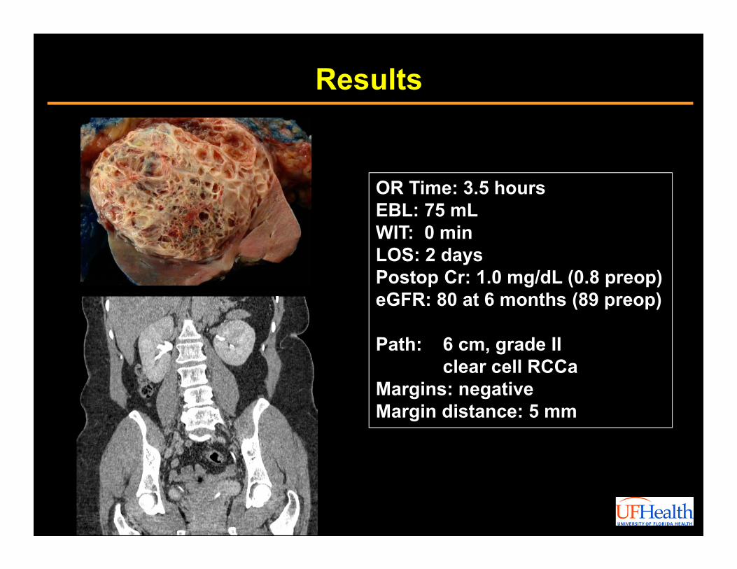
Results
OR Time: 3.5 hoursEBL: 75 mLWIT: 0 minLOS: 2 daysPostop Cr: 1.0 mg/dL (0.8 preop)eGFR: 80 at 6 months (89 preop)
Path: 6 cm, grade IIclear cell RCCa
Margins: negative Margin distance: 5 mm
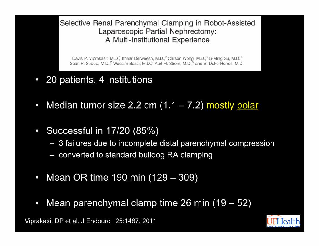
• 20 patients, 4 institutions
• Median tumor size 2.2 cm (1.1 – 7.2) mostly polar
• Successful in 17/20 (85%)– 3 failures due to incomplete distal parenchymal compression– converted to standard bulldog RA clamping
• Mean OR time 190 min (129 – 309)
• Mean parenchymal clamp time 26 min (19 – 52)
Viprakasit DP et al. J Endourol 25:1487, 2011

• Median serum creatinine (mg/dl)Preoperative (range) 0.83 (0.5 – 1.69)Immediate postoperative (range) 0.81 (0.6 – 1.70)At last follow-up (range) 0.81 (0.6 – 1.83)
• Median estimated GFR (ml/min/1.73m2)Preoperative (range) 86 (39 – 118)Immediate postoperative (range) 78 (40 – 124), p = 0.33At last follow-up (range) 78 (36 – 126), p = 0.54
• Mean follow-up (months) 6.1 (1.2 – 11.9)
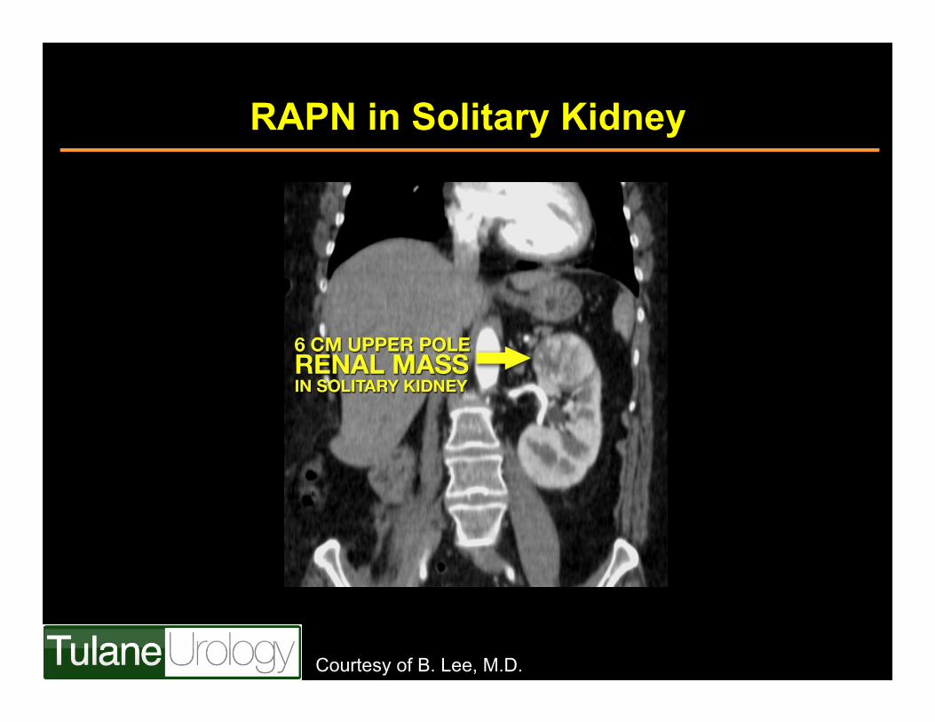
RAPN in Solitary Kidney
Courtesy of B. Lee, M.D.
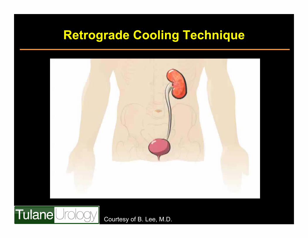
Retrograde Cooling Technique
Courtesy of B. Lee, M.D.
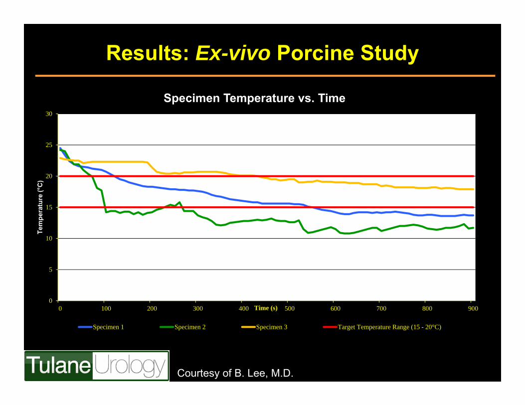
Results: Ex-vivo Porcine Study
0
5
10
15
20
25
30
0 100 200 300 400 500 600 700 800 900
Tem
pera
ture
(°C
)
Time (s)
Specimen Temperature vs. Time
Specimen 1 Specimen 2 Specimen 3 Target Temperature Range (15 - 20°C)
Courtesy of B. Lee, M.D.
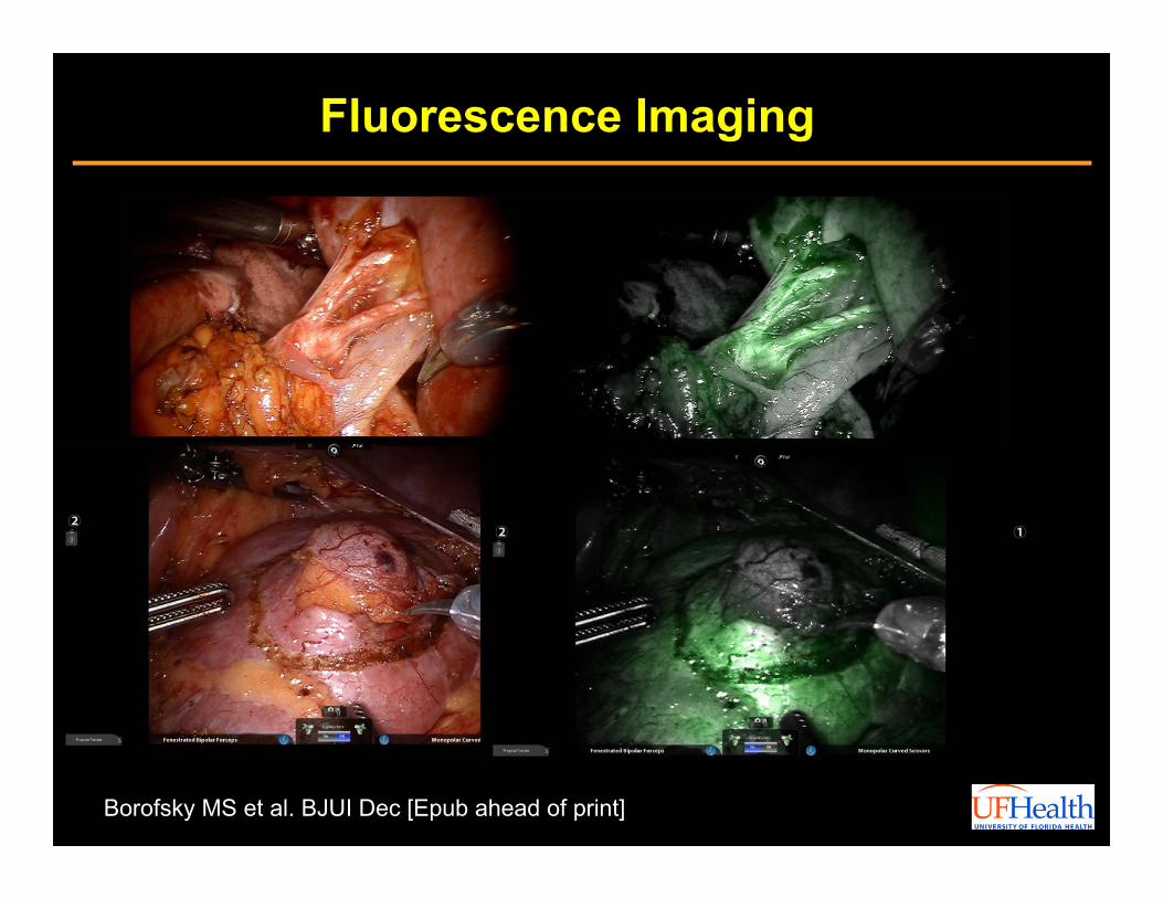
Fluorescence Imaging
Borofsky MS et al. BJUI Dec [Epub ahead of print]
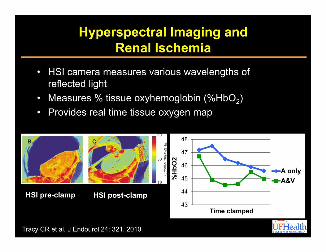
Hyperspectral Imaging and Renal Ischemia
• HSI camera measures various wavelengths of reflected light
• Measures % tissue oxyhemoglobin (%HbO2)• Provides real time tissue oxygen map
Tracy CR et al. J Endourol 24: 321, 2010
HSI pre-clamp HSI post-clamp43
44
45
46
47
48
%H
bO2
Time clamped
A onlyA&V
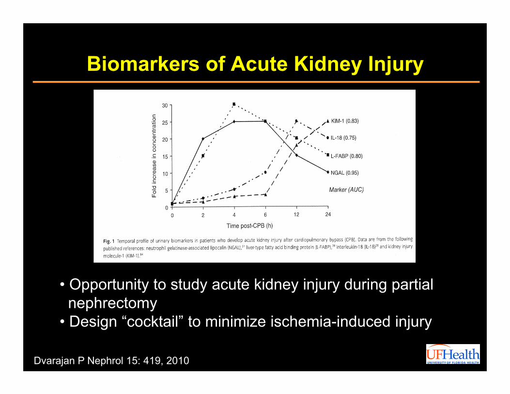
Biomarkers of Acute Kidney Injury
• Opportunity to study acute kidney injury during partial nephrectomy
• Design “cocktail” to minimize ischemia-induced injury
Dvarajan P Nephrol 15: 419, 2010

Conclusions
Robotic Partial Nephrectomy
– Robotics has had an expanding role in the treatment of the small renal mass
– Partial nephrectomies in solitary kidneys under warm ischemia have given us important insights into cutoffs of WIT
– Modifications in surgical technique can help reduce WIT as an important modifiable risk factor of postoperative renal function
– Future technologies may help improve our understanding of risk and prevention of ischemic injury to the kidney

Thank You


![Novel technique: direct access partial nephrectomy ...nuf.nu/NoRenCa/Novel technique direct access... · of its equivalent oncological results compared to radical nephrectomy [3]](https://img.pdfslide.net/doc/110x75/606c80ba9bb7de31a926ad1d/novel-technique-direct-access-partial-nephrectomy-nufnunorencanovel-technique.jpg)