Embed Size (px)
Citation preview
Teledermatology and Common Dermatology
Issues in the Hospitalized Patient
Patricia Meyer, DNP, CRNP, FNP‐BC, AGACNP‐BC, NE‐BC
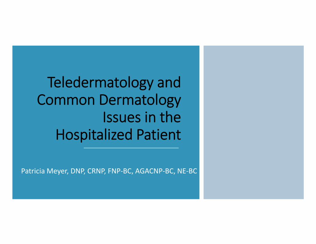
The Rise of Teledermatology
Improve dermatology
access
Shortage of dermatology providers
Ability for dermatologist to see more patients
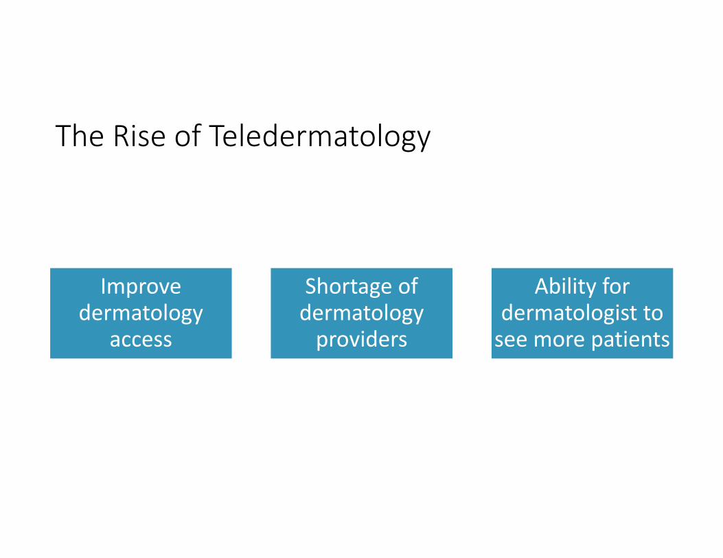
Teledermatology What is Involved
• Obtaining a CC, HPI, ROS, Allergies, Med list , PMH, Social and Family Hx
• A problem‐focused exam• Digital imaging • Uploading of‐ CC, HPI, ROS, Allergies, Med list , PMH, Social and Family Hx, Physical exam, and digital images via secure computer site
• Onsite person to obtain BX if needed
Teledermatology What is Involved
After Info is Uploaded• Dermatologist will form differential diagnosis• Suggest a work up• Formulate and assessment and plan
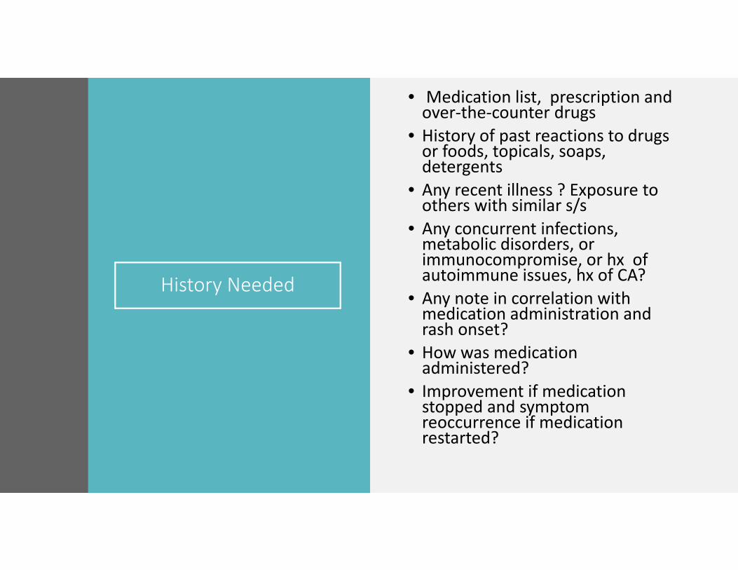
History Needed
• Medication list, prescription and over‐the‐counter drugs
• History of past reactions to drugs or foods, topicals, soaps, detergents
• Any recent illness ? Exposure to others with similar s/s
• Any concurrent infections, metabolic disorders, or immunocompromise, or hx of autoimmune issues, hx of CA?
• Any note in correlation with medication administration and rash onset?
• How was medication administered?
• Improvement if medication stopped and symptom reoccurrence if medication restarted?
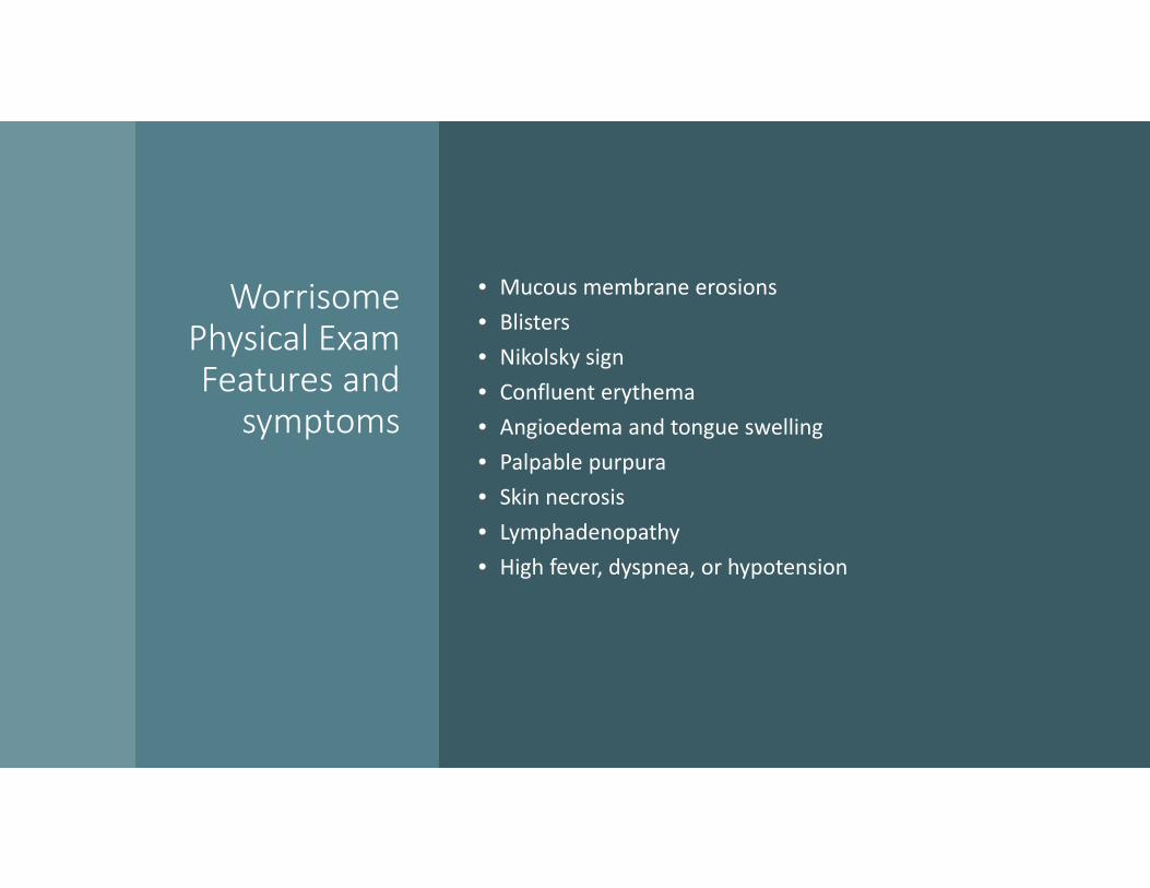
Worrisome Physical Exam Features and
symptoms
• Mucous membrane erosions• Blisters• Nikolsky sign• Confluent erythema• Angioedema and tongue swelling• Palpable purpura• Skin necrosis• Lymphadenopathy• High fever, dyspnea, or hypotension
Case Study One
HJ is a 82 year old male, who was admitted from SNF, due to abd pain. HJ is being treated for diverticulitis with Cipro and Flagyl. Teledermatolgy is consulted due to a “rash.” Per the patient’s RN , “it is unclear if this is a new drug rash”. Upon further review with patient he states that he has had this rash for some time “it is very itchy and often keeps me up at night .” He denies any worsening or improving factors. He denies any family Hx of autoimmune disorders or drug allergies .
Due to patients limited ability to provide in depth Hxof rash onset, the SNF is called , they endorse that the patient has had the rash for some time and was seen by a dermatologist in the out patient setting 1‐2 months ago and was Dx with Dermatitis and has never really got better, rash has been on back, buttocks, groin folds abdomen (esp. around waist) and extremities. The SNF RN denies any new meds , new detergents, or new topicals other than the “creams” the dermatologist provided, she also denies anyone else with a similar rash at the facility
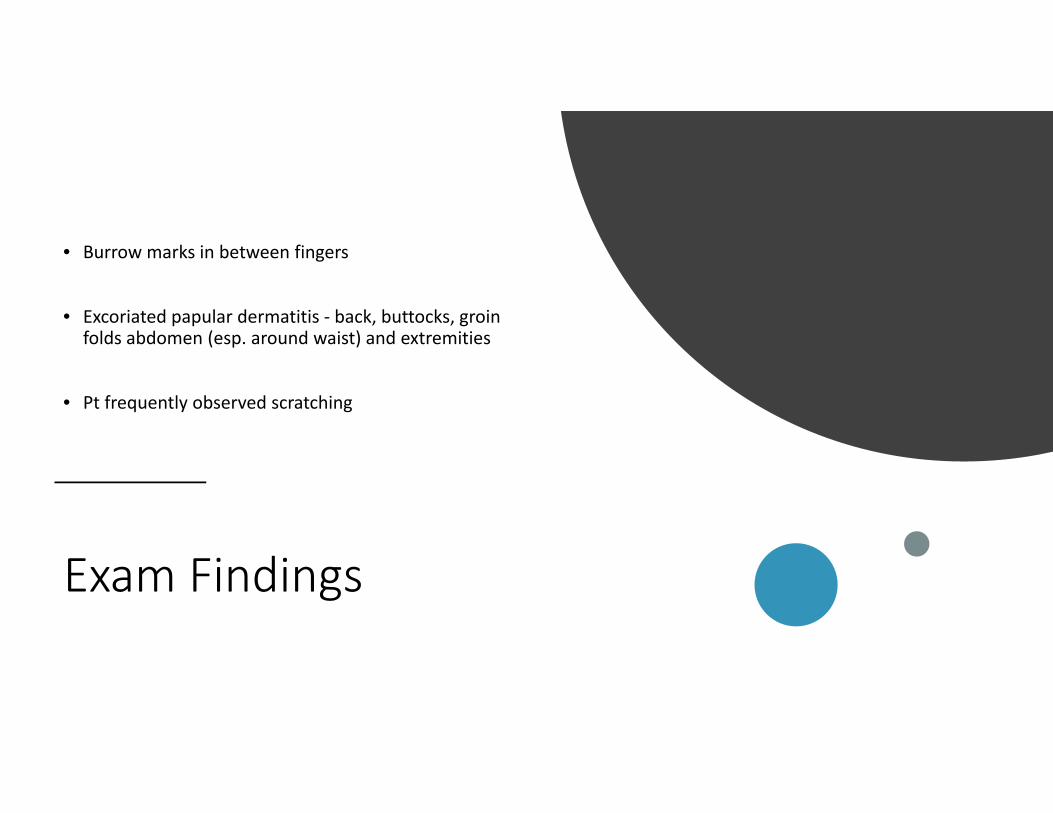
Exam Findings
• Burrow marks in between fingers
• Excoriated papular dermatitis ‐ back, buttocks, groin folds abdomen (esp. around waist) and extremities
• Pt frequently observed scratching
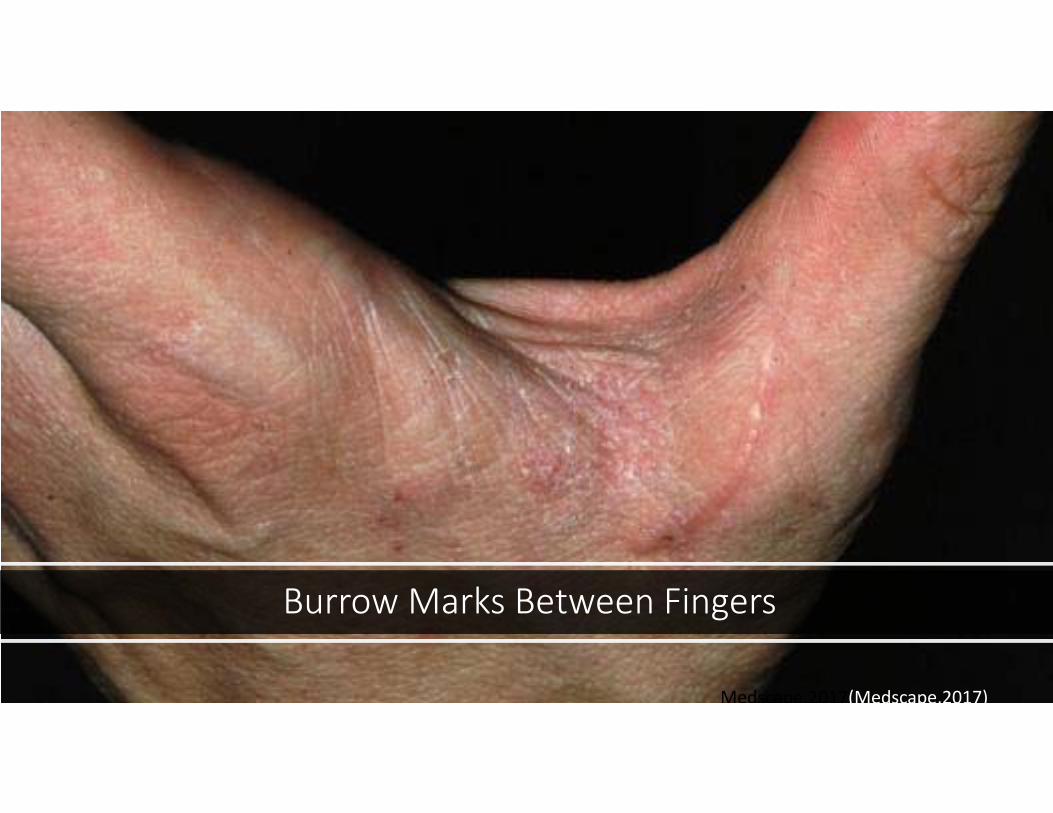
Burrow Marks Between Fingers
Medscape,2017(Medscape,2017)
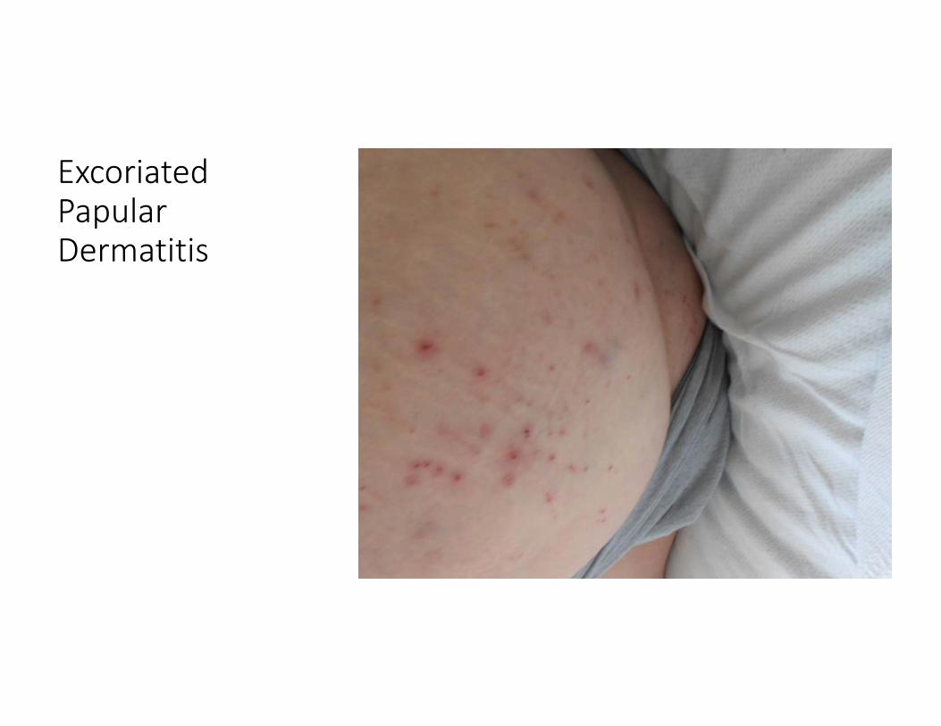
Excoriated Papular Dermatitis
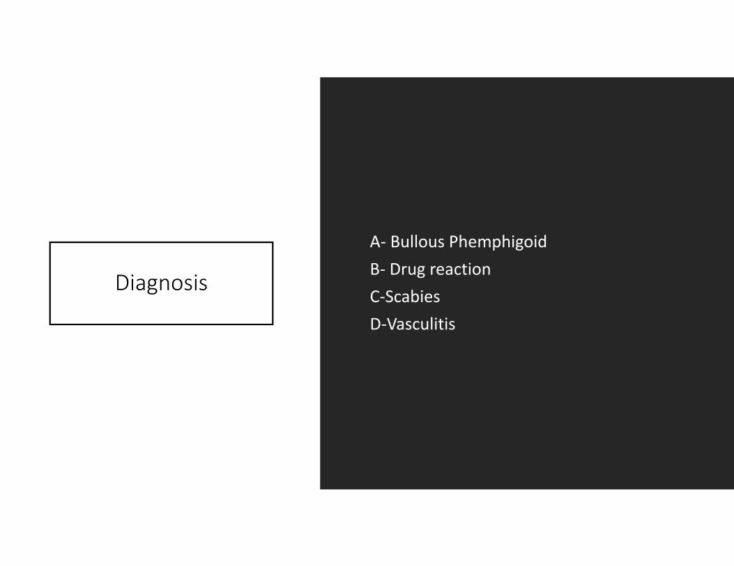
Diagnosis
A‐ Bullous Phemphigoid B‐ Drug reaction C‐ScabiesD‐Vasculitis

Scabies
Why Scabies ?
• It is usually a clinical diagnosis‐ Suspicious history‐ Clinical presentation
• A skin scraping can be obtained to assist with dx , however this is not always accurate

History/ ROS Key Components(CLUES TO ACCURATE DX)
• Intense pruritus• Itching worse at night • Others with close contact have same s/s • SNF Patient
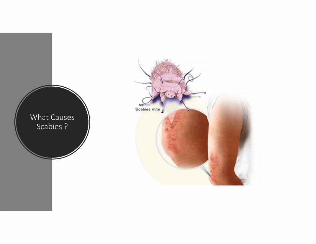
What Causes Scabies ?

Treatment
• Permethrin cream• Ivermectin • Non‐sedating antihistamines
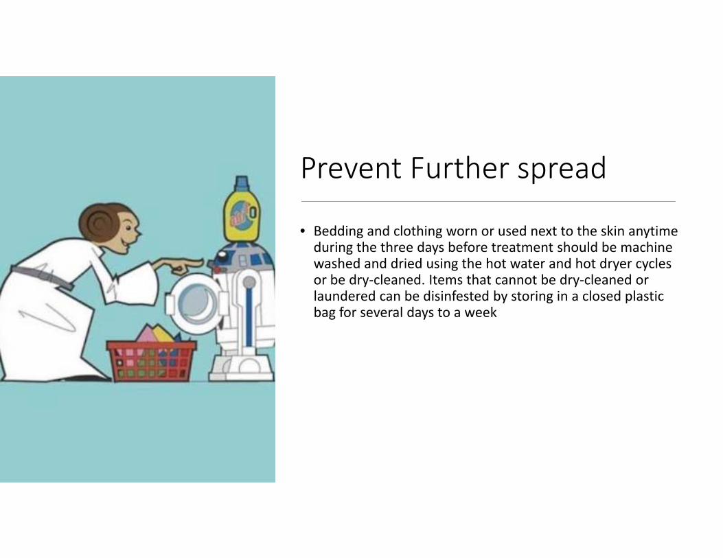
Prevent Further spread
• Bedding and clothing worn or used next to the skin anytime during the three days before treatment should be machine washed and dried using the hot water and hot dryer cycles or be dry‐cleaned. Items that cannot be dry‐cleaned or laundered can be disinfested by storing in a closed plastic bag for several days to a week
Case Study Two
GH is an 81 year old female who is admitted to your facility with a Dx of cellulitis to her lower extremities. Telederm is consulted due to skin with “erythema ulcerations and blisters.” The patient is alert and oriented, she states she has had no medication changes, no new meds, no new detergents , no new topicals. The patient does complain of occasional itching and pain to ulcerative areas. The patient denies a personal history of skin cancer, autoimmune disease or family hx of skin cancer or autoimmune disease. Patient denies being in contact with others with similar symptoms.
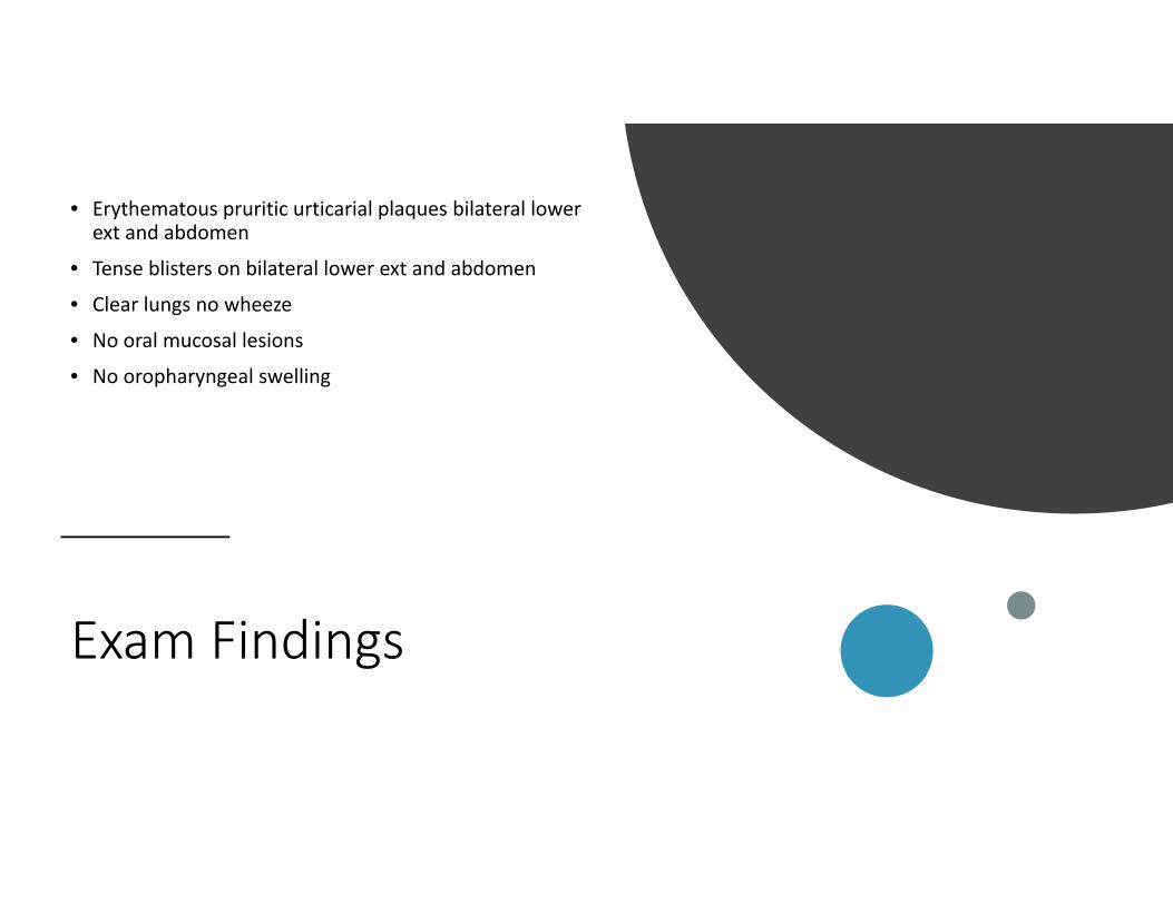
Exam Findings
• Erythematous pruritic urticarial plaques bilateral lower ext and abdomen
• Tense blisters on bilateral lower ext and abdomen
• Clear lungs no wheeze
• No oral mucosal lesions
• No oropharyngeal swelling
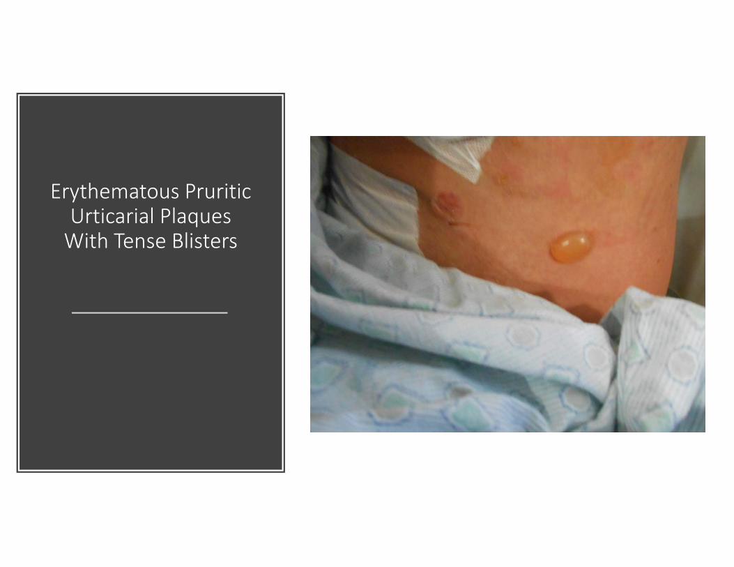
Erythematous Pruritic Urticarial Plaques With Tense Blisters
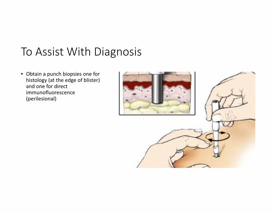
To Assist With Diagnosis
• Obtain a punch biopsies one for histology (at the edge of blister) and one for direct immunofluorescence (perilesional)
Work up
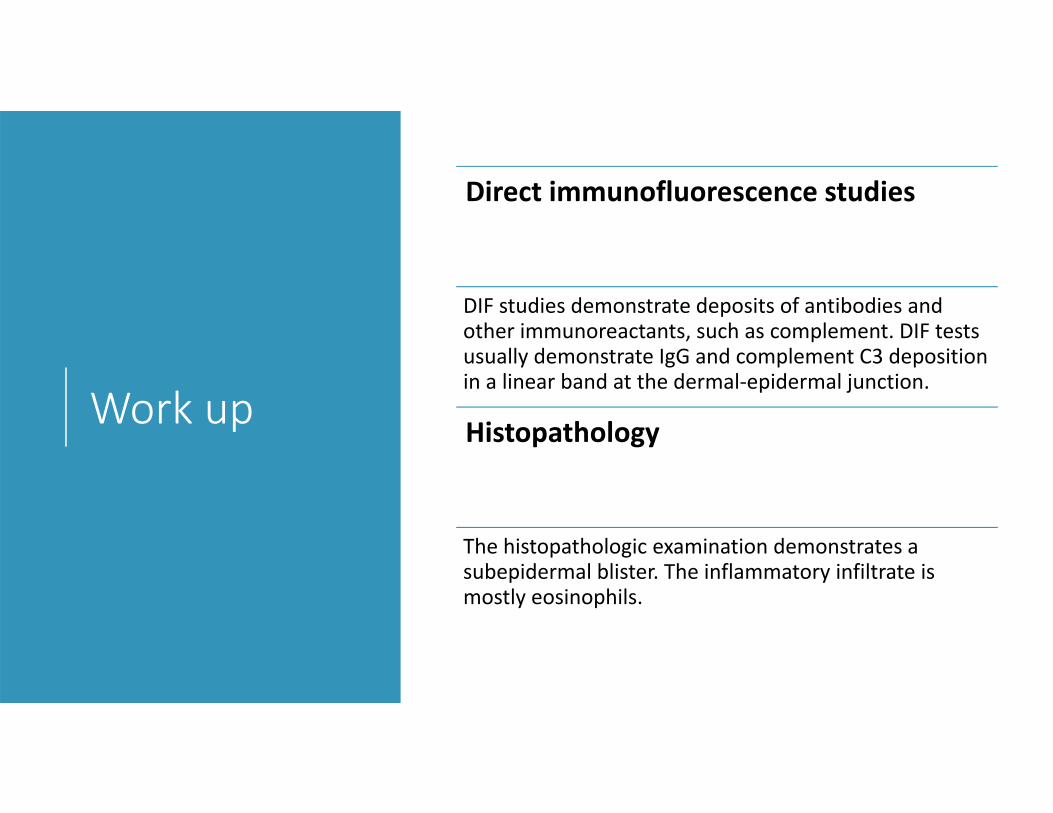
Direct immunofluorescence studies
DIF studies demonstrate deposits of antibodies and other immunoreactants, such as complement. DIF tests usually demonstrate IgG and complement C3 deposition in a linear band at the dermal‐epidermal junction.
Histopathology
The histopathologic examination demonstrates a subepidermal blister. The inflammatory infiltrate is mostly eosinophils.

Diagnosis
A‐ Bullous Pemphigoid B‐ Drug reaction C‐ScabiesD‐Vasculitis
Bullous Pemphigoid
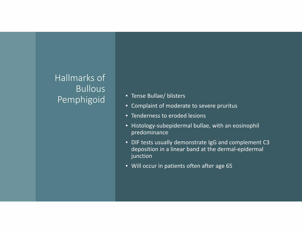
Hallmarks of Bullous
Pemphigoid • Tense Bullae/ blisters
• Complaint of moderate to severe pruritus
• Tenderness to eroded lesions
• Histology‐subepidermal bullae, with an eosinophil predominance
• DIF tests usually demonstrate IgG and complement C3 deposition in a linear band at the dermal‐epidermal junction
• Will occur in patients often after age 65
Cause of Bullous Pemphigoid
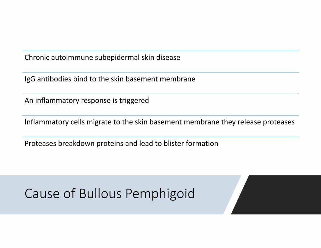
Chronic autoimmune subepidermal skin disease
IgG antibodies bind to the skin basement membrane
An inflammatory response is triggered
Inflammatory cells migrate to the skin basement membrane they release proteases
Proteases breakdown proteins and lead to blister formation

Treatment of Bullous Pemphigoid
• Systemic steroids until clear
• Imuran, Cellcept – To inhibit inflammatory response

Case Study Three
BA is a 80 year old female, who presented to the hospital, with diarrhea and abdominal pain. She has a recent Dx of colitis and had recently been treated with Cipro and Flagyl as an outpatient. She was reordered flagyl for “empiric treatment” due to her s/s and has been on Flagyl x 2 days. Telederm is consulted for a “ rash, to patient’s abdomen and back.” Upon arrival to room the patient is complaining of significant pruritis. Pt reports she first noticed her rash today on her abdomen, back, flank, and chest. Upon exam this patient has a faint, blanchablemaculopapular rash. She can not recall anything that makes the rash worse. She states that Benadryl has been somewhat helpful. She states that, other than Cipro and Flagyl, she has had no new meds, she denies any new topicals or new detergents or soaps . She denies any personal or family Hx of autoimmune diseases or skin cancer. She denies food allergies .
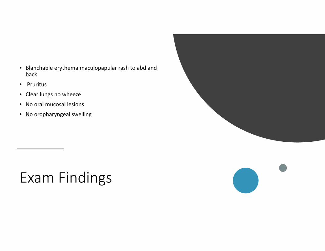
Exam Findings
• Blanchable erythema maculopapular rash to abd and back
• Pruritus
• Clear lungs no wheeze
• No oral mucosal lesions
• No oropharyngeal swelling
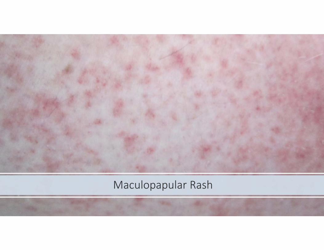
Maculopapular Rash
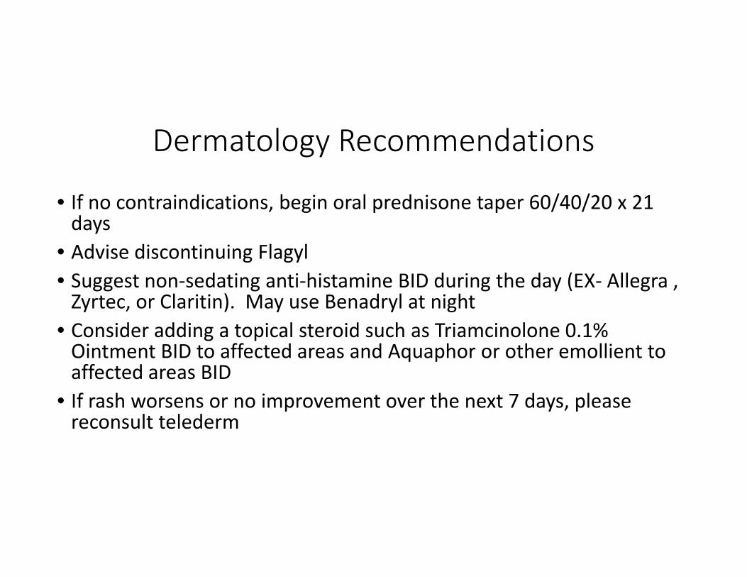
Dermatology Recommendations
• If no contraindications, begin oral prednisone taper 60/40/20 x 21 days
• Advise discontinuing Flagyl• Suggest non‐sedating anti‐histamine BID during the day (EX‐ Allegra , Zyrtec, or Claritin). May use Benadryl at night
• Consider adding a topical steroid such as Triamcinolone 0.1% Ointment BID to affected areas and Aquaphor or other emollient to affected areas BID
• If rash worsens or no improvement over the next 7 days, please reconsult telederm

Called Back to See Patient
Was called back to see patient, due to rash spreading to chest , and increased erythema , with some exfoliation, noted

Exam Findings
• Worsening of a blanchable erythematous papulopustualrash to abd and back, chest and extremities , with some exfoliation
• Pruritus
• Clear lungs no wheeze
• No oral mucosal lesions
• No oropharyngeal swelling
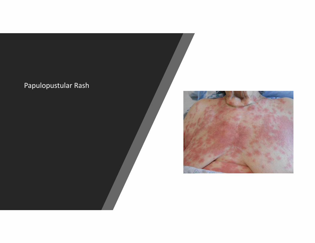
Papulopustular Rash
Work Up
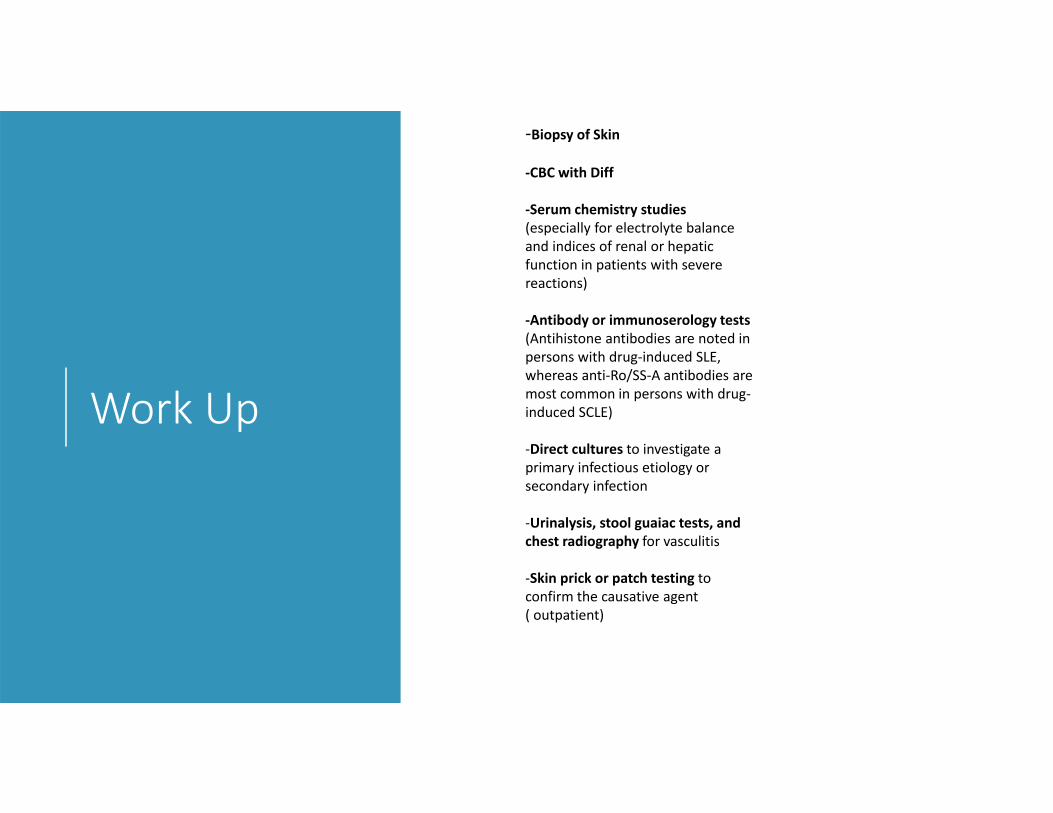
‐Biopsy of Skin
‐CBC with Diff
‐Serum chemistry studies (especially for electrolyte balance and indices of renal or hepatic function in patients with severe reactions)
‐Antibody or immunoserology tests(Antihistone antibodies are noted in persons with drug‐induced SLE, whereas anti‐Ro/SS‐A antibodies are most common in persons with drug‐induced SCLE)
‐Direct cultures to investigate a primary infectious etiology or secondary infection
‐Urinalysis, stool guaiac tests, and chest radiography for vasculitis
‐Skin prick or patch testing to confirm the causative agent ( outpatient)
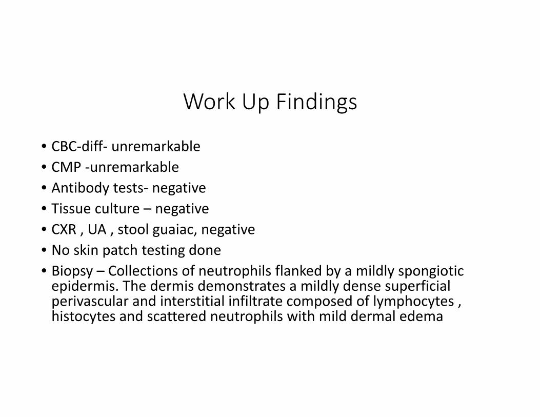
Work Up Findings
• CBC‐diff‐ unremarkable • CMP ‐unremarkable• Antibody tests‐ negative• Tissue culture – negative • CXR , UA , stool guaiac, negative• No skin patch testing done • Biopsy – Collections of neutrophils flanked by a mildly spongiotic epidermis. The dermis demonstrates a mildly dense superficial perivascular and interstitial infiltrate composed of lymphocytes , histocytes and scattered neutrophils with mild dermal edema
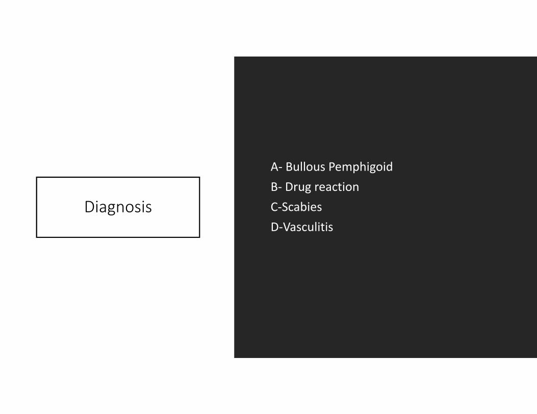
Diagnosis
A‐ Bullous Pemphigoid B‐ Drug reaction C‐ScabiesD‐Vasculitis
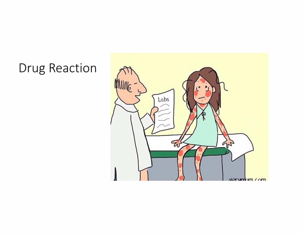
Drug Reaction
Treatment
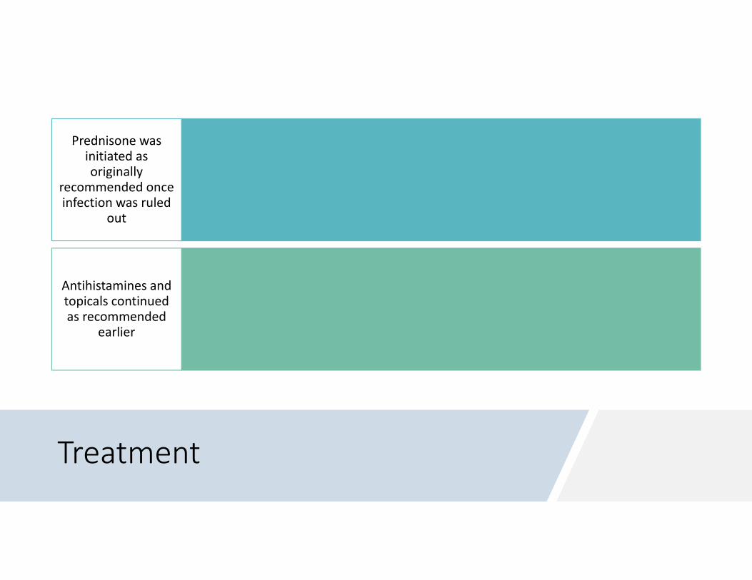
Prednisone was initiated as originally
recommended once infection was ruled
out
Antihistamines and topicals continued as recommended
earlier
Pathophysiology
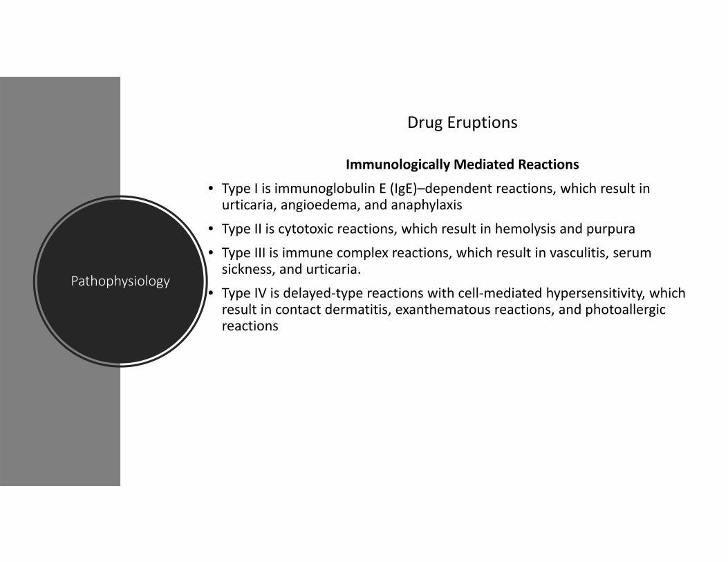
Drug Eruptions
Immunologically Mediated Reactions
• Type I is immunoglobulin E (IgE)–dependent reactions, which result in urticaria, angioedema, and anaphylaxis
• Type II is cytotoxic reactions, which result in hemolysis and purpura
• Type III is immune complex reactions, which result in vasculitis, serum sickness, and urticaria.
• Type IV is delayed‐type reactions with cell‐mediated hypersensitivity, which result in contact dermatitis, exanthematous reactions, and photoallergic reactions
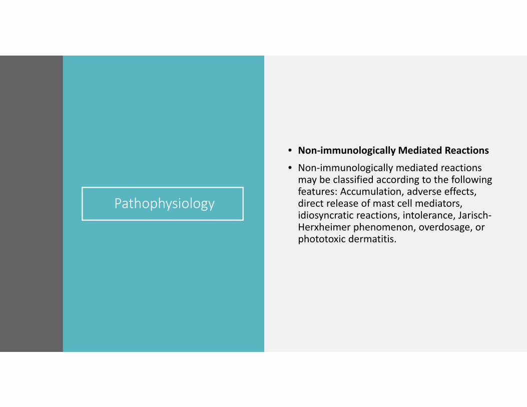
Pathophysiology
• Non‐immunologically Mediated Reactions
• Non‐immunologically mediated reactions may be classified according to the following features: Accumulation, adverse effects, direct release of mast cell mediators, idiosyncratic reactions, intolerance, Jarisch‐Herxheimer phenomenon, overdosage, or phototoxic dermatitis.

Biopsy For Drug Reactions
• Eosinophils in morbilliform eruptions or numerous neutrophils
So I Have Been Told
“ There are no dermatology emergencies”
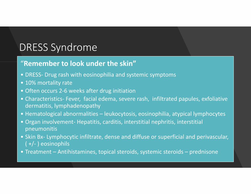
DRESS Syndrome“Remember to look under the skin”• DRESS‐ Drug rash with eosinophilia and systemic symptoms• 10% mortality rate• Often occurs 2‐6 weeks after drug initiation• Characteristics‐ Fever, facial edema, severe rash, infiltrated papules, exfoliative dermatitis, lymphadenopathy
• Hematological abnormalities – leukocytosis, eosinophilia, atypical lymphocytes• Organ involvement‐ Hepatitis, carditis, interstitial nephritis, interstitial pneumonitis
• Skin Bx‐ Lymphocytic infiltrate, dense and diffuse or superficial and perivascular, ( +/‐ ) eosinophils
• Treatment – Antihistamines, topical steroids, systemic steroids – prednisone
DRESS Dx Criteria
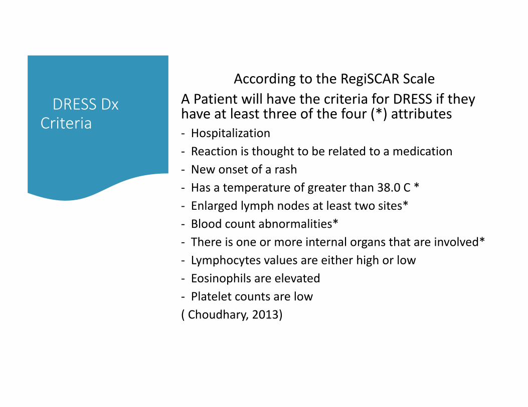
According to the RegiSCAR ScaleA Patient will have the criteria for DRESS if they have at least three of the four (*) attributes‐ Hospitalization‐ Reaction is thought to be related to a medication‐ New onset of a rash‐ Has a temperature of greater than 38.0 C *‐ Enlarged lymph nodes at least two sites*‐ Blood count abnormalities*‐ There is one or more internal organs that are involved*‐ Lymphocytes values are either high or low ‐ Eosinophils are elevated‐ Platelet counts are low ( Choudhary, 2013)
Steven Johnson
Syndrome/ Toxic
Epidermal Necrolysis
(TENS)
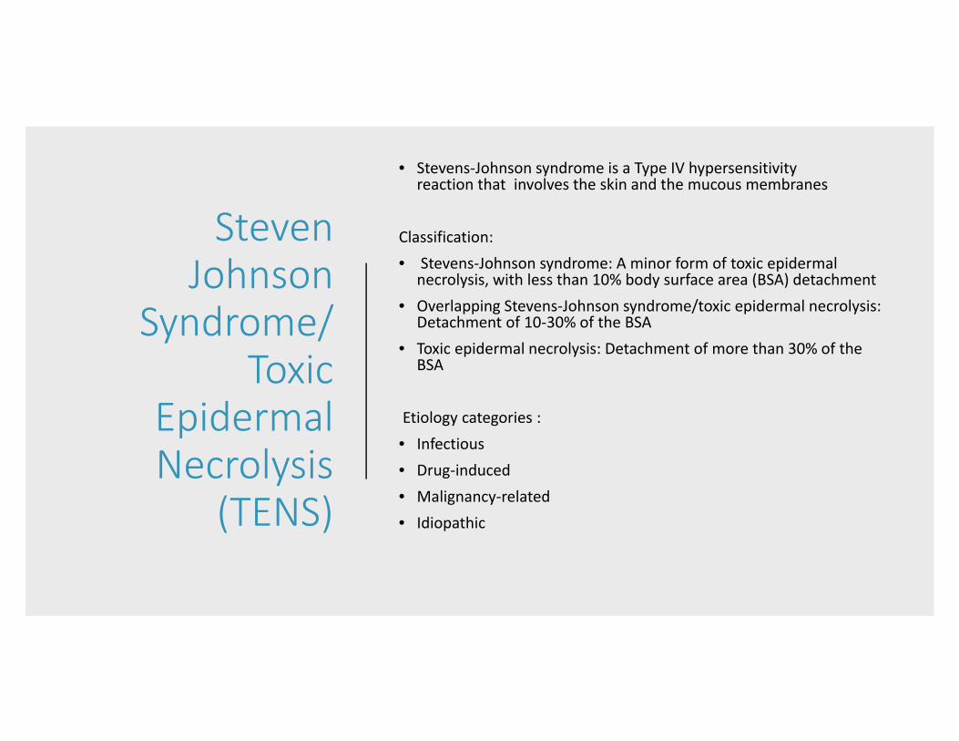
• Stevens‐Johnson syndrome is a Type IV hypersensitivity reaction that involves the skin and the mucous membranes
Classification: • Stevens‐Johnson syndrome: A minor form of toxic epidermal
necrolysis, with less than 10% body surface area (BSA) detachment• Overlapping Stevens‐Johnson syndrome/toxic epidermal necrolysis:
Detachment of 10‐30% of the BSA• Toxic epidermal necrolysis: Detachment of more than 30% of the
BSA
Etiology categories :• Infectious• Drug‐induced• Malignancy‐related• Idiopathic
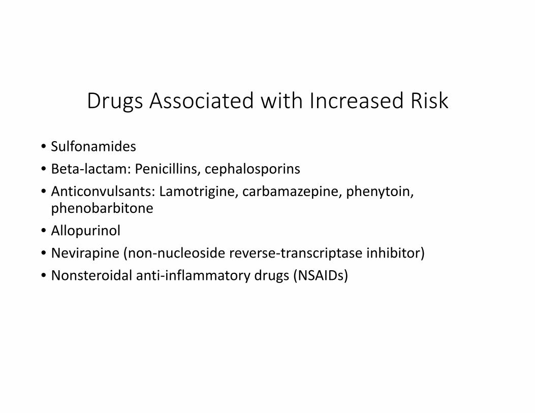
Drugs Associated with Increased Risk
• Sulfonamides• Beta‐lactam: Penicillins, cephalosporins• Anticonvulsants: Lamotrigine, carbamazepine, phenytoin, phenobarbitone
• Allopurinol• Nevirapine (non‐nucleoside reverse‐transcriptase inhibitor)• Nonsteroidal anti‐inflammatory drugs (NSAIDs)
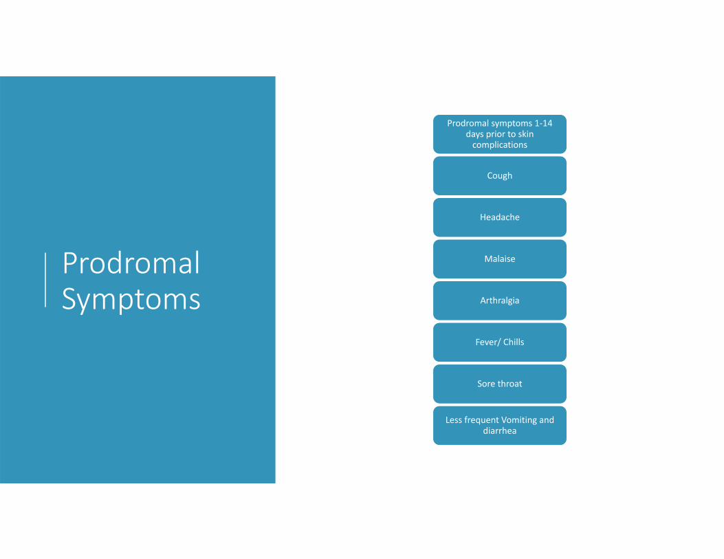
Prodromal Symptoms
Prodromal symptoms 1‐14 days prior to skin complications
Cough
Headache
Malaise
Arthralgia
Fever/ Chills
Sore throat
Less frequent Vomiting and diarrhea
Steven Johnson Syndrome/ Toxic Epidermal Necrolysis (TENS)
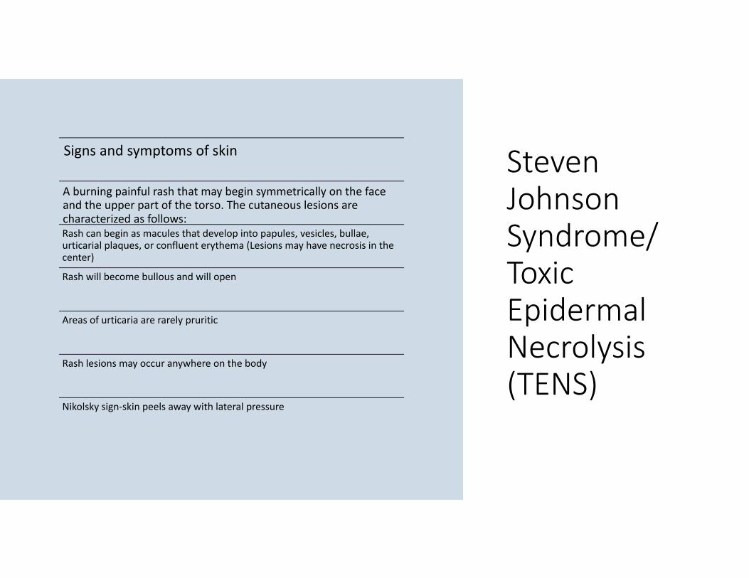
Signs and symptoms of skin
A burning painful rash that may begin symmetrically on the face and the upper part of the torso. The cutaneous lesions are characterized as follows:Rash can begin as macules that develop into papules, vesicles, bullae, urticarial plaques, or confluent erythema (Lesions may have necrosis in the center)
Rash will become bullous and will open
Areas of urticaria are rarely pruritic
Rash lesions may occur anywhere on the body
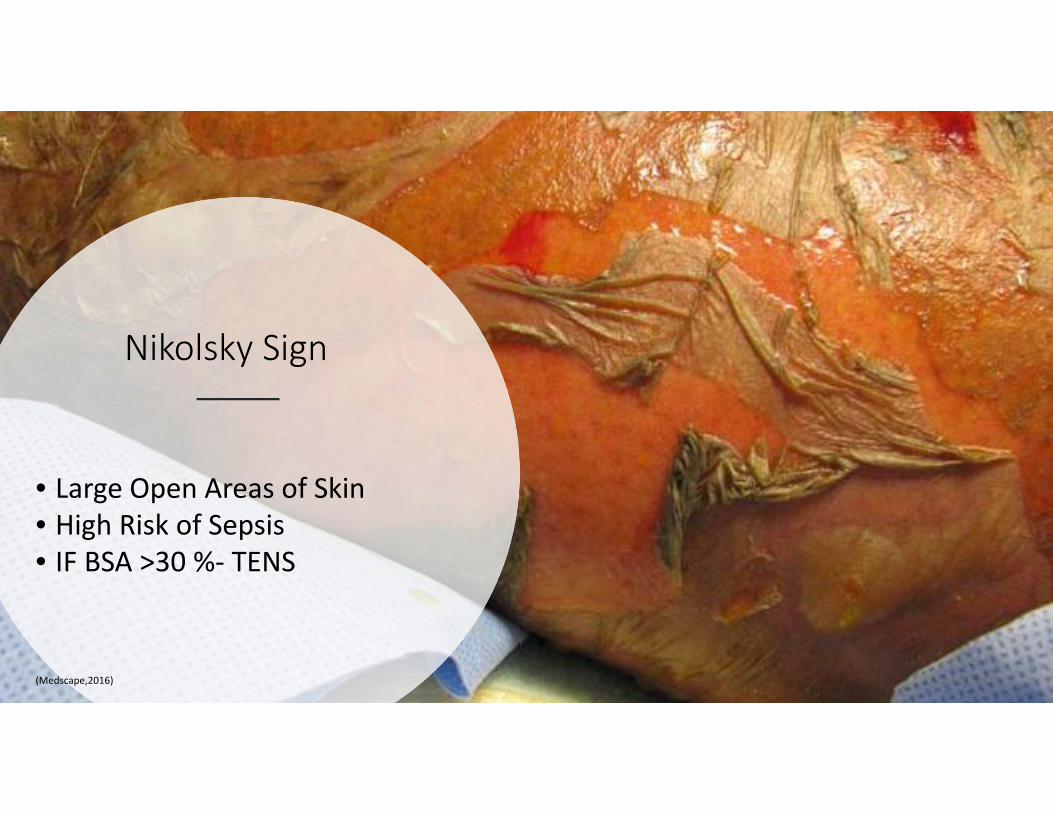
Nikolsky sign‐skin peels away with lateral pressure

Other Signs and Symptoms
• Corneal ulcerations
• Erosive vulvovaginitis or balanitis
• Seizures• Coma• Conjuntival
redness• Entropion• Skin lesions• Nasal lesions• Mouth lesions
• Orthostasis
• Tachycardia
• Hypotension
• Fever
• Altered level of consciousness
• Epistaxis
• Conjunctivitis
Steven Johnson Syndrome/ Toxic Epidermal Necrolysis
(TENS)
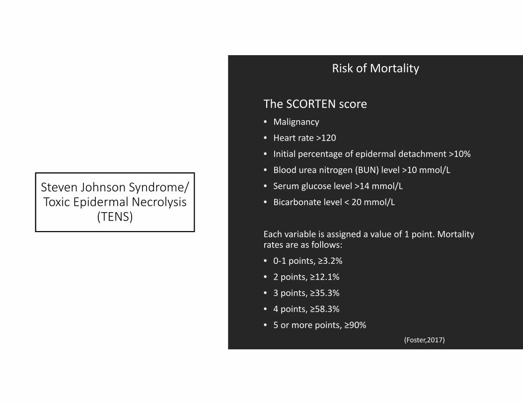
Risk of Mortality
The SCORTEN score • Malignancy
• Heart rate >120
• Initial percentage of epidermal detachment >10%
• Blood urea nitrogen (BUN) level >10 mmol/L
• Serum glucose level >14 mmol/L
• Bicarbonate level < 20 mmol/L
Each variable is assigned a value of 1 point. Mortality rates are as follows:
• 0‐1 points, ≥3.2%
• 2 points, ≥12.1%
• 3 points, ≥35.3%
• 4 points, ≥58.3%
• 5 or more points, ≥90%(Foster,2017)
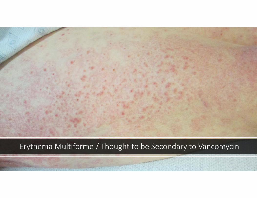
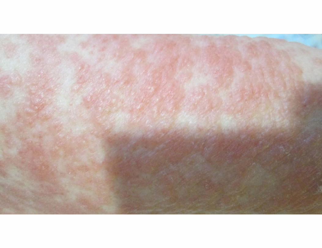
Erythema Multiforme / Thought to be Secondary to Vancomycin
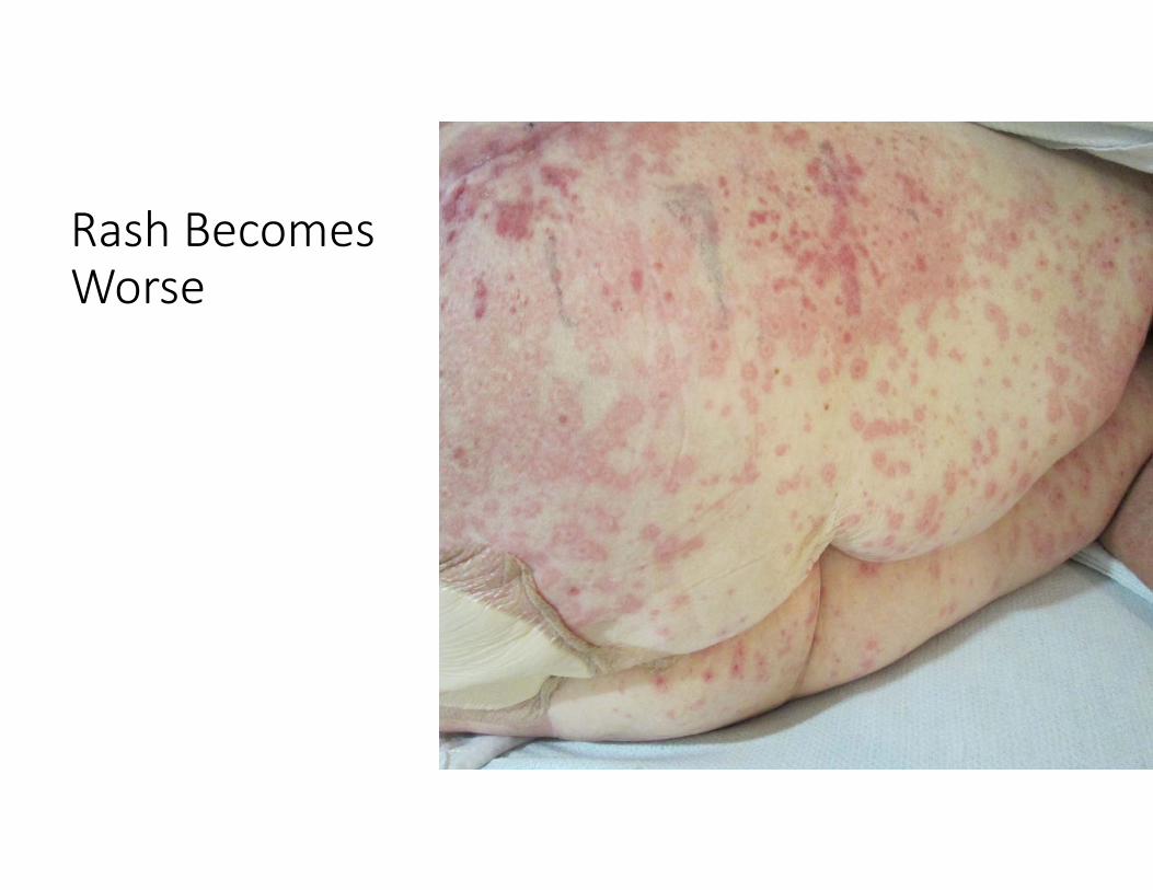
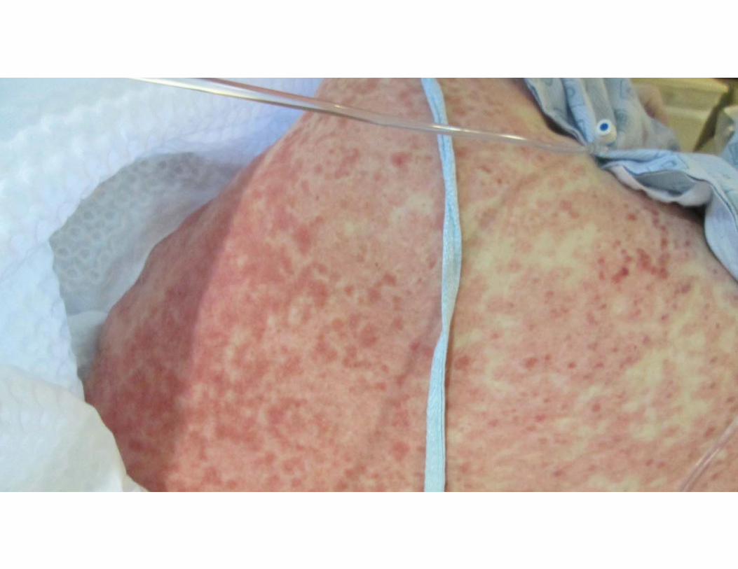
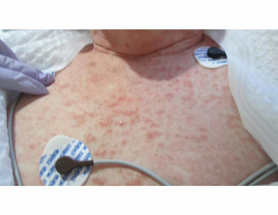
Rash Becomes Worse
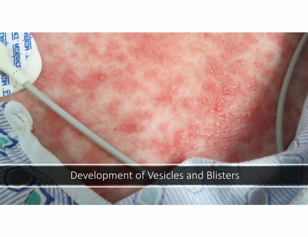
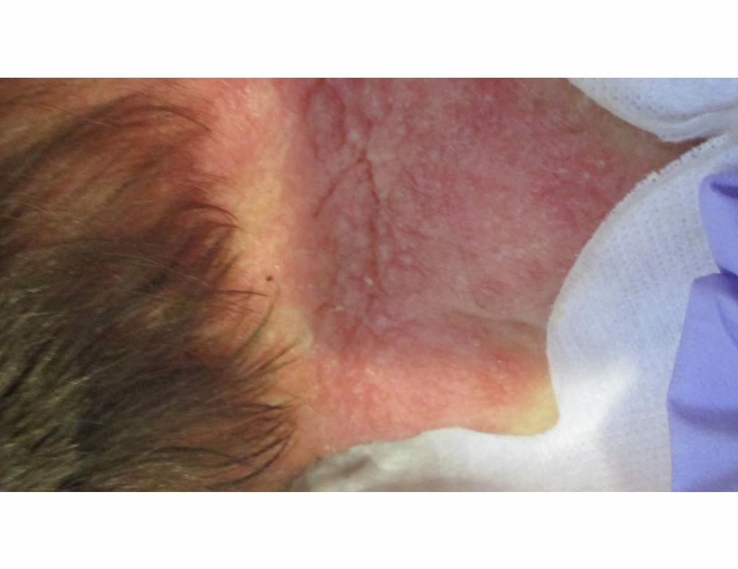
Development of Vesicles and Blisters
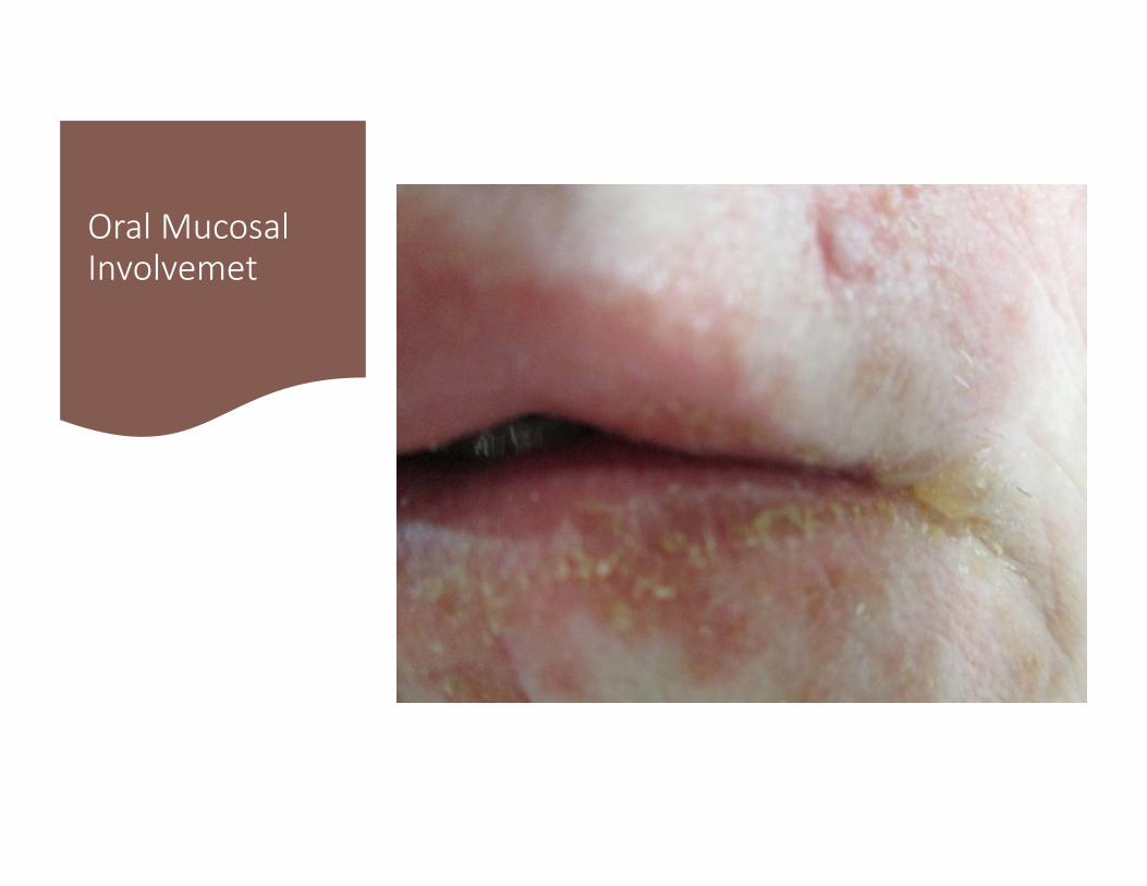
Oral Mucosal Involvemet
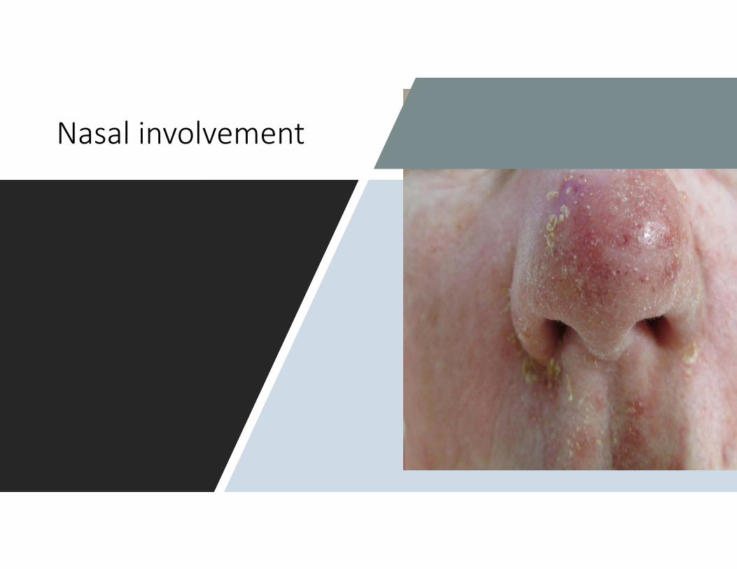
Nasal involvement
• Due to worsening of condition, this PT was sent to burn unit – Skin BX was done at initial hospital was positive for mild SJS
Nikolsky Sign
• Large Open Areas of Skin• High Risk of Sepsis• IF BSA >30 %‐ TENS
(Medscape,2016)

Other Complications
Gastroenterologic ‐Esophageal strictures
Genitourinary ‐ Renal tubular necrosis, renal failure, penile scarring,
vaginal stenosis
Pulmonary ‐Tracheobronchial
shedding with resultant respiratory failure
Cutaneous ‐ Scarring and cosmetic
deformity, recurrences of infection through
slow‐healing ulceration
27‐50% will develop significant visual
problems
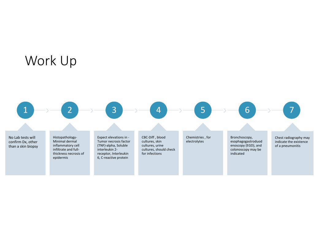
Work Up
1
No Lab tests will confirm Dx, other than a skin biopsy
2
Histopathology‐Minimal dermal inflammatory cell infiltrate and full‐thickness necrosis of epidermis
3
Expect elevations in ‐Tumor necrosis factor (TNF)‐alpha, Soluble interleukin 2‐receptor, Interleukin 6, C‐reactive protein
4
CBC‐Diff , blood cultures, skin cultures, urine cultures, should check for infections
5
Chemistries , for electrolytes
6
Bronchoscopy, esophagogastroduodenoscopy (EGD), and colonoscopy may be indicated
7
Chest radiography may indicate the existence of a pneumonitis
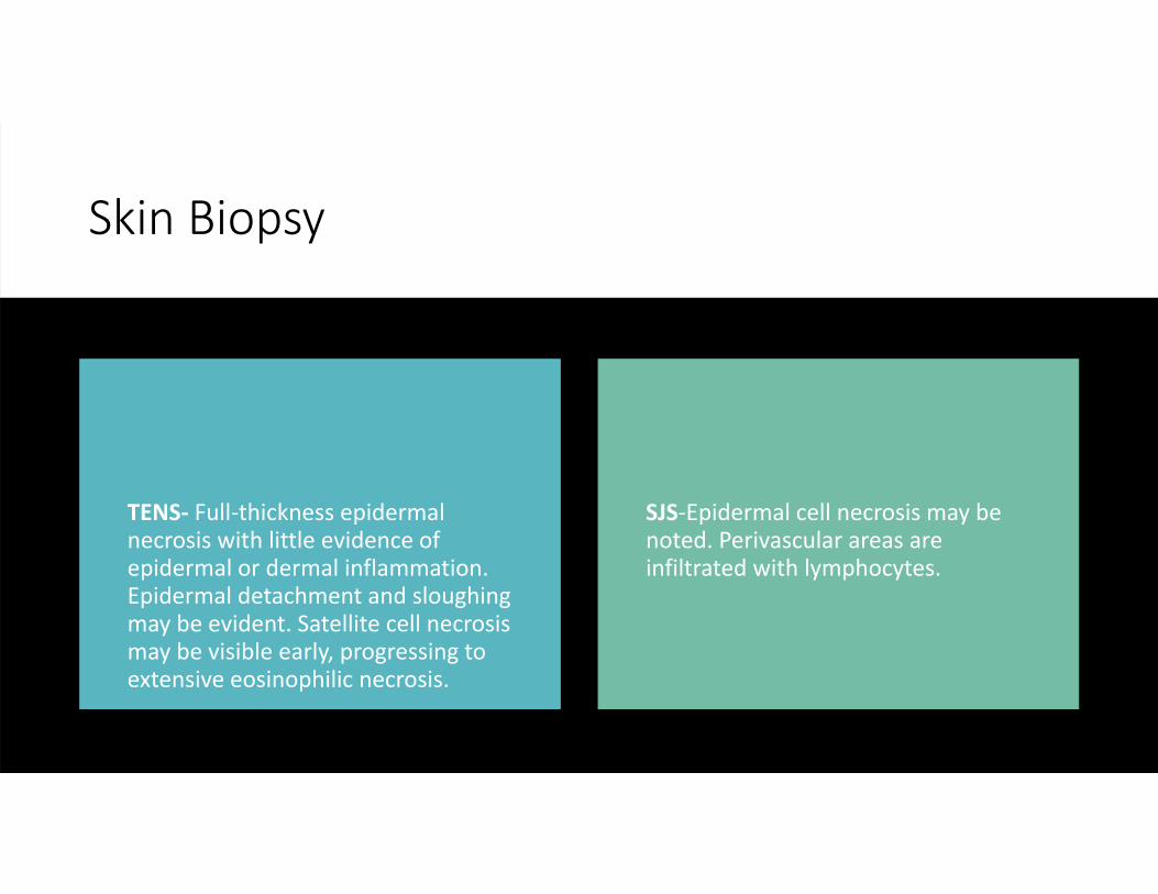
Skin Biopsy
TENS‐ Full‐thickness epidermal necrosis with little evidence of epidermal or dermal inflammation. Epidermal detachment and sloughing may be evident. Satellite cell necrosis may be visible early, progressing to extensive eosinophilic necrosis.
SJS‐Epidermal cell necrosis may be noted. Perivascular areas are infiltrated with lymphocytes.
Treatment
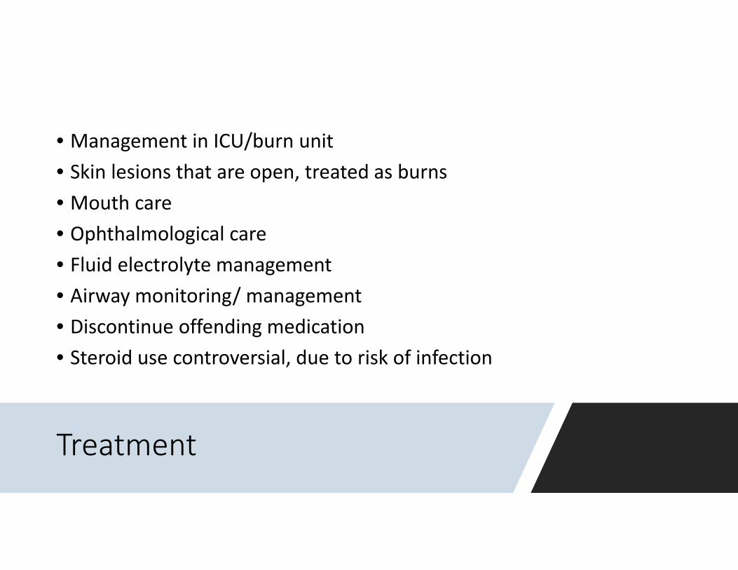
• Management in ICU/burn unit• Skin lesions that are open, treated as burns • Mouth care • Ophthalmological care• Fluid electrolyte management• Airway monitoring/ management• Discontinue offending medication• Steroid use controversial, due to risk of infection

Vasculitis Overview
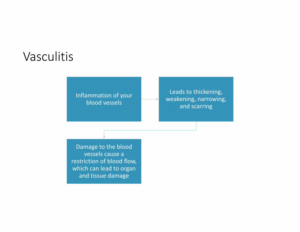
Vasculitis
Inflammation of your blood vessels
Leads to thickening, weakening, narrowing,
and scarring
Damage to the blood vessels cause a
restriction of blood flow, which can lead to organ
and tissue damage
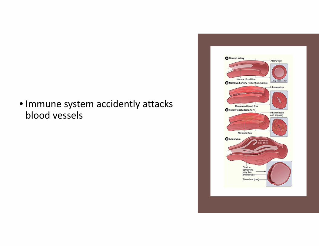
• Immune system accidently attacks blood vessels
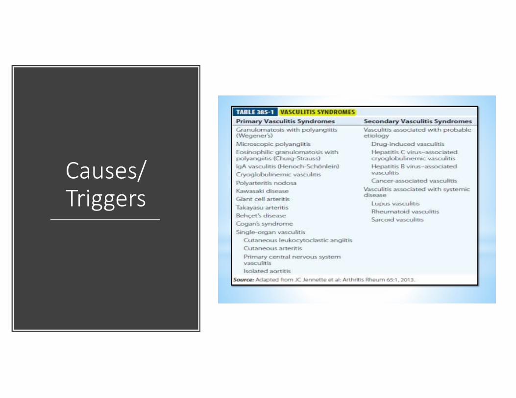
Causes/ Triggers
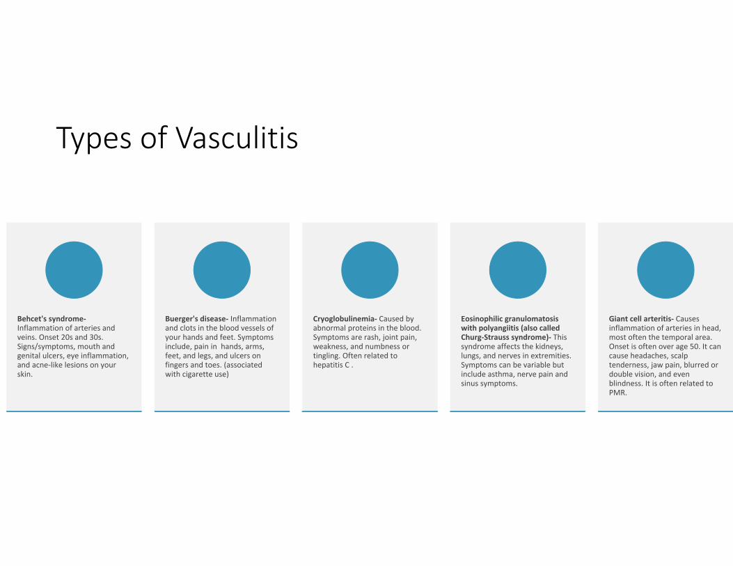
Types of Vasculitis
Behcet's syndrome‐Inflammation of arteries and veins. Onset 20s and 30s. Signs/symptoms, mouth and genital ulcers, eye inflammation, and acne‐like lesions on your skin.
Buerger's disease‐ Inflammation and clots in the blood vessels of your hands and feet. Symptoms include, pain in hands, arms, feet, and legs, and ulcers on fingers and toes. (associated with cigarette use)
Cryoglobulinemia‐ Caused by abnormal proteins in the blood. Symptoms are rash, joint pain, weakness, and numbness or tingling. Often related to hepatitis C .
Eosinophilic granulomatosis with polyangiitis (also called Churg‐Strauss syndrome)‐ Thissyndrome affects the kidneys, lungs, and nerves in extremities. Symptoms can be variable but include asthma, nerve pain and sinus symptoms.
Giant cell arteritis‐ Causes inflammation of arteries in head, most often the temporal area. Onset is often over age 50. It can cause headaches, scalp tenderness, jaw pain, blurred or double vision, and even blindness. It is often related to PMR.
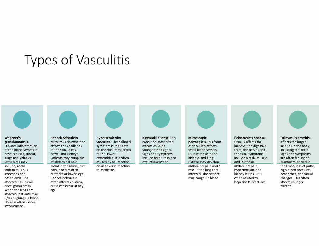
Types of Vasculitis
Wegener's granulomatosis‐Causes inflammation of the blood vessels in nose, sinuses, throat, lungs and kidneys. Symptoms may include, nasal stuffiness, sinus infections and nosebleeds. The affected tissues will have granulomas. When the lungs are affected, patients may C/O coughing up blood. There is often kidney involvement .
Henoch‐Schonleinpurpura‐ This condition affects the capillaries of the skin, joints, bowel and kidneys. Patients may complain of abdominal pain, blood in the urine, joint pain, and a rash to buttocks or lower legs. Henoch‐Schonleinoften affects children, but it can occur at any age.
Hypersensitivity vasculitis‐ The hallmark symptom is red spots on the skin, most often to the lower extremities. It is often caused by an infection or an adverse reaction to medicine.
Kawasaki disease‐This condition most often affects children younger than age 5. Signs and symptoms include fever, rash and eye inflammation.
Microscopic polyangiitis‐This form of vasculitis affects small blood vessels, usually those in the kidneys and lungs. Patient may develop abdominal pain and a rash. If the lungs are affected. The patient, may cough up blood.
Polyarteritis nodosa‐Usually affects the kidneys, the digestive tract, the nerves and the skin. Symptoms include a rash, muscle and joint pain, abdominal pain, hypertension, and kidney issues. It is often related to hepatitis B infections.
Takayasu's arteritis‐Affects the larger arteries in the body, including the aorta. Signs and symptoms are often feeling of numbness or cold in the limbs, loss of pulse, high blood pressure, headaches, and visual changes. This often affects younger women.
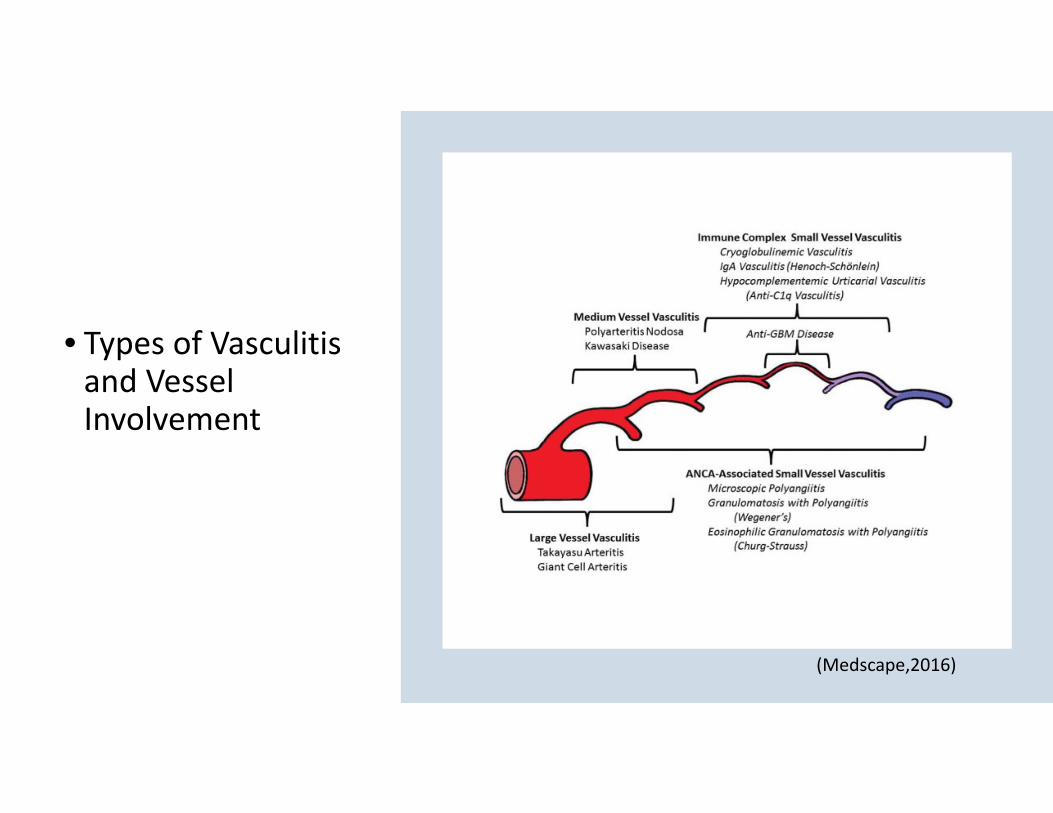
• Types of Vasculitis and Vessel Involvement
(Medscape,2016)
Generalized Symptoms
Fever
Headache
Fatigue
Weight loss
General aches and pains
Night sweats
Rash
Nerve problems, such as numbness or weakness
Loss of a pulse in a limb
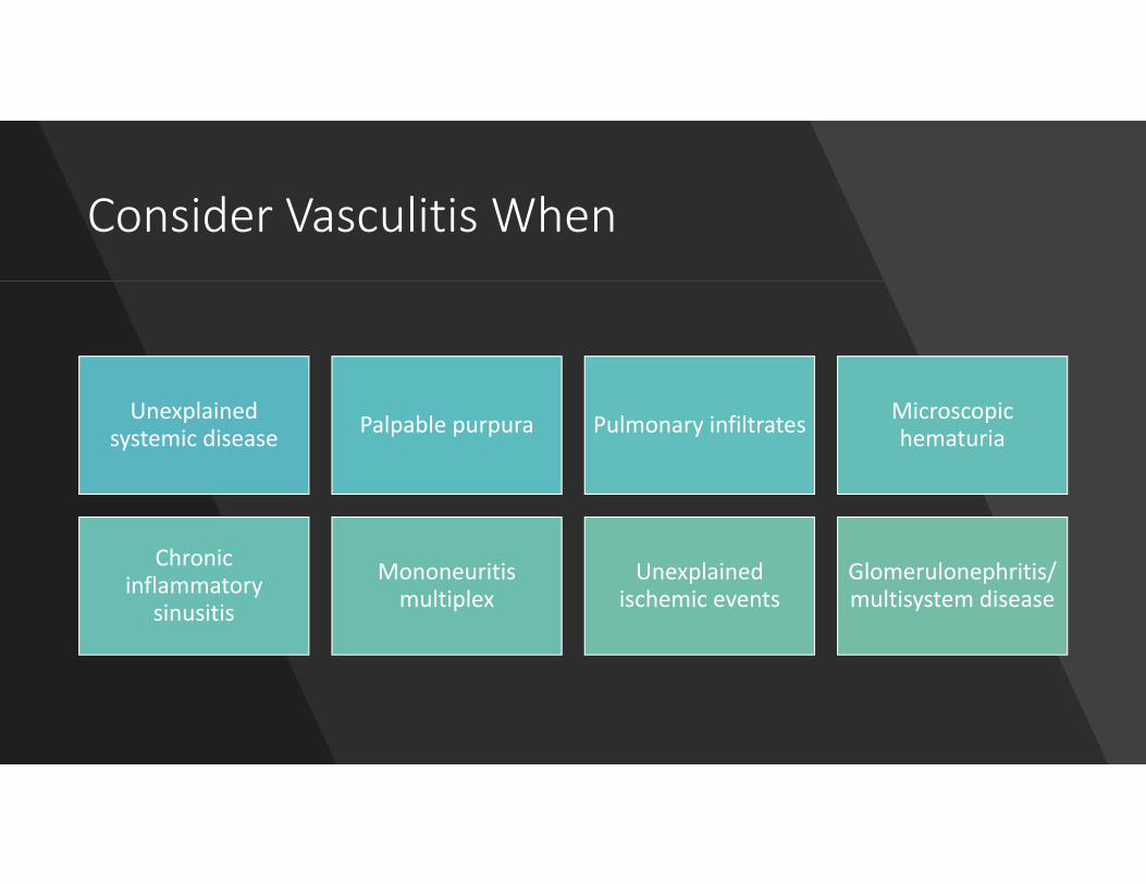
Consider Vasculitis When
Unexplained systemic disease Palpable purpura Pulmonary infiltrates Microscopic
hematuria
Chronic inflammatory
sinusitis
Mononeuritis multiplex
Unexplained ischemic events
Glomerulonephritis/ multisystem disease
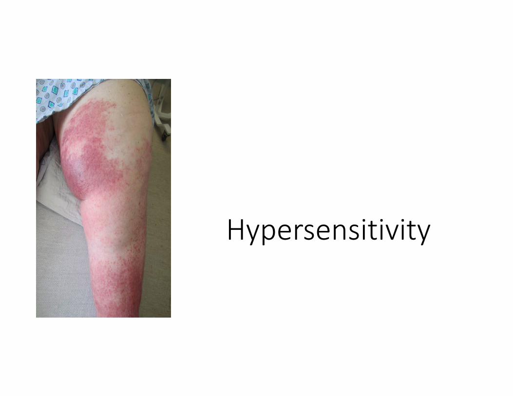
Hypersensitivity
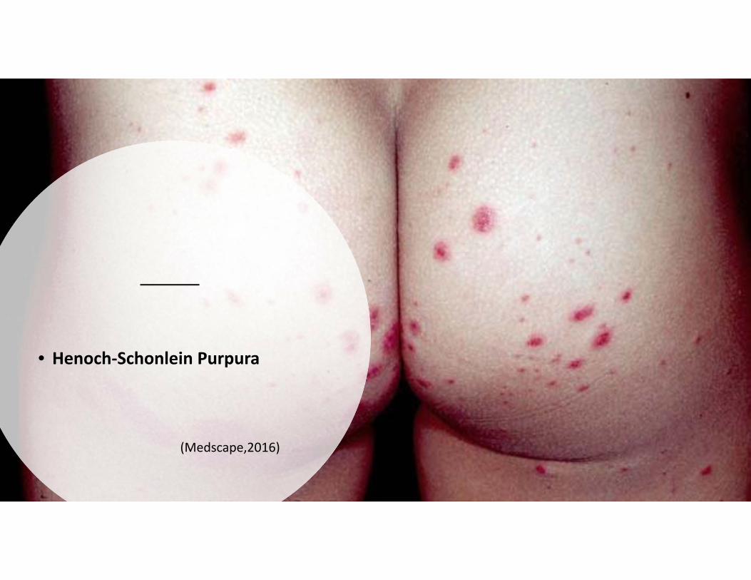
• Henoch‐Schonlein Purpura
(Medscape,2016)
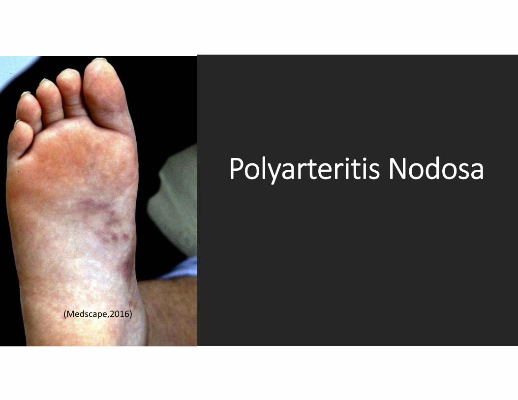
Polyarteritis Nodosa
(Medscape,2016)
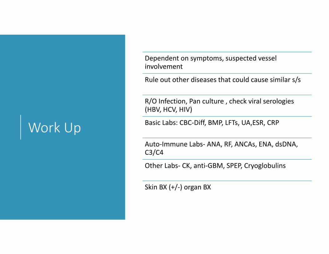
Work Up
Dependent on symptoms, suspected vessel involvement
Rule out other diseases that could cause similar s/s
R/O Infection, Pan culture , check viral serologies(HBV, HCV, HIV)
Basic Labs: CBC‐Diff, BMP, LFTs, UA,ESR, CRP
Auto‐Immune Labs‐ ANA, RF, ANCAs, ENA, dsDNA, C3/C4
Other Labs‐ CK, anti‐GBM, SPEP, Cryoglobulins
Skin BX (+/‐) organ BX
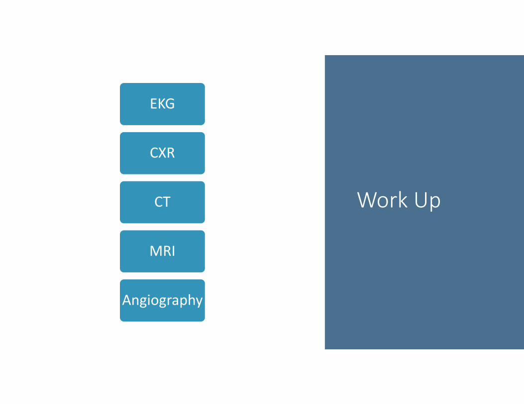
Work Up
EKG
CXR
CT
MRI
Angiography
General Principles of Treatment
• If it is determined that vasculitis is caused by an offending agent such as a medication, it should be discontinued
• If vasculitis is caused by an underlying disease, such as infection, cancer, or connective tissue disease, the underlying disease should be treated
• If vasculitis is secondary to a primary syndrome, treatment should be initiated according to the category of the vasculitis
‐Steroids‐ Immunosuppressive meds
The End

References
• Barry, M. Elston, D., Kauffman,C., Willson,B. (2017, July 20). Scabies.Retrieved from Medscape:http://emedicine.medscape.com/article/1109204‐overview
• Blume, J. Elston, D., Liaqat, A., Ehrich, M., Helm, T., Wilson, B., Rozen, E., Rosh, A. (2017, July 11). Drug Eruptions. Retrieved from Medscape: http://emedicine.medscape.com/article/1197450‐overview
• Chan, L. Stone, O. (2017, July 11). Bullous Pemphigoid. Retrieved from Medscape: http://emedicine.medscape.com/article/1062391‐overview
• Choudary,S., McLeod,M., Romanelli, P. (2013, June 6). Drug Reaction with Eosinophilia and Systemic Symptoms (DRESS).Retrieved from The Journal of Clinical and Aesthetic Dermatology : http//www.ncbi.nlm.nih.gov/pmc/articles/PMC33718748/
• English, J., Huen, A., Patton, T., & Grandinetti, L. (Eds.). (2015). Skin and Systemic Disease a Clinicians Guide (pp. 13‐207). Boca Raton, FL.: CRC Press.
• Foster, S. Ba‐Abbad,R., Letko, E., Parrillo, S. (2016, December 15). Stevens‐Johnson Syndrome. Retrieved from Medscape: http://emedicine.medscape.com/article/1197450‐overview
• Nadia, L. Benselesr, S.(2016, November 15). Vasculitis and Thrombophelebitis.Retrieved from Medscape: http://emedicine.medscape.com/article/1008239‐overview
• NIH. (2014, September 23). NIH. Retrieved from What is Vasculitis?: http://www.nhlib.nih.gov/health/health‐topics/topics/vas
• Smith, C. (2013). Infestation and Bites. In C. H. Soutor, CilincalDermatology (pp. 104‐114). New York, NY.: McGraw Hill
• Soutor, C., & Hordinsky, M. (Eds.). (2013). Clinical Dermatology(pp.22-297). New York: McGraw Hill.
• Thomas, D., & Burkemper, N. (Eds.). (2013). Geriatric Dermatology (pp. 332-541). Philadelphia, PA.: Elsevier.
• William, J., Berger, T., Elson, D., Nehaus, I. (Eds.). (2016). Cutaneous Vascular Disease. In Andrews' Diseases of the Skin Clinical Dermatology (pp. 829‐850). Philadelphia, PA: Elsevier.
• Wolff, K. J. (Eds.). (2009). In Fitzpatricks' Color Atlas & Synopsis of Clinical Dermatology. New York, NY: McGraw Hill.