Embed Size (px)
Citation preview
TELOMERE CORRELATIONS DURING EARLY LIFE IN A LONG-LIVED SEABIRD
A Thesis Submitted to the Graduate Faculty
of the North Dakota State University
of Agriculture and Applied Science
By
Jacob Eli Schmidt
In Partial Fulfillment of the Requirements for the Degree of
MASTER OF SCIENCE
Major Department: Biological Sciences
April 2018
Fargo, North Dakota
North Dakota State University Graduate School
Title
Telomere correlations during early life in a long-lived seabird
By
Jacob Eli Schmidt
The Supervisory Committee certifies that this disquisition complies with North Dakota State
University’s regulations and meets the accepted standards for the degree of
MASTER OF SCIENCE
SUPERVISORY COMMITTEE:
Britt Heidinger, PhD
Chair
Wendy Reed, PhD
Kim Vonnahme, PhD
Approved: 3 April 2018 Wendy Reed, PhD Date Department Chair
iii
ABSTRACT
Telomere dynamics in blood cells have been linked to aging in a variety of organisms.
However, whether blood telomeres correlated with telomeres in other parts of the body is not
well known, especially during early life when telomere loss is expected to be most rapid. We
investigated this question in embryonic and juvenile Franklin’s gulls (Leucophaeus pipixcan).
We measured telomere lengths in blood, heart, liver and skeletal muscle tissues at the end of
embryonic (n = 31) and post-natal development (n=20). In late-stage embryos, blood telomeres
were significantly positively correlated with heart and skeletal muscle, but not liver telomeres.
However, at the end of post-natal development, there were no significant correlations among
blood telomeres and telomeres in any other tissues. In late-stage embryos, heart telomeres were
significantly longer than blood, liver, and skeletal muscle telomeres, but at the end of post-natal
development telomere lengths did not significantly differ among tissues.
iv
ACKNOWLEDGEMENTS
I thank Aubrey Sirman for joint work conducted. I would also like to thank Alexandra
Hett, Grant Edland, Soren Hjort, David Breitbach, and Aaron Kaip for assistance with animal
husbandry, Aurelia Kucera for help with sample collection, Jeff Kittilson for instruction and
support, and the rest of our lab group for support and feedback throughout this research.
I thank Britt Heidinger for her endless patience and encouragement throughout my
graduate school experience. Lastly, I thank my family, friends, military leadership and all others
who encouraged me to complete this thesis.
v
TABLE OF CONTENTS
ABSTRACT ................................................................................................................................... iii
ACKNOWLEDGMENTS ............................................................................................................. iv
LIST OF TABLES ......................................................................................................................... vi
LIST OF FIGURES ...................................................................................................................... vii
1. INTRODUCTION ...................................................................................................................... 1
2. METHODS ................................................................................................................................. 4
3. RESULTS ................................................................................................................................... 8
4. DISCUSSION ........................................................................................................................... 11
REFERENCES ............................................................................................................................. 13

vi
LIST OF TABLES
Table Page
1. Studies examining telomere length correlations between blood and other tissues......................3
2. Relationships between telomeres in blood, heart, liver and skeletal muscle...............................8

vii
LIST OF FIGURES
Figure Page
1. Relationship between blood and heart and blood and skeletal muscle telomeres.......................9
2. Telomere length in embryonic (A) and juvenile (B) Franklin's gulls..........................................9
1
1. INTRODUCTION1
Understanding the mechanisms affecting longevity, such as telomere dynamics, is of
importance to a variety of fields, including evolutionary ecology and biomedicine (Monaghan,
2014). Telomeres are highly conserved, repetitive, non-coding sequences of DNA that together
with other proteins form protective caps at the ends of linear chromosomes (Blackburn, 2005).
Telomeres enhance genome stability by protecting the coding sequences from loss during normal
cell division and by allowing the DNA repair machinery to distinguish double stranded breaks
from chromosomes ends (Vleck et al., 2003). Once telomeres become critically short, cells stop
dividing and can secrete inflammatory compounds (Blackburn, 2005) and both of these
processes are expected to compromise tissue function and contribute to organismal aging (Aubert
and Lansdorp, 2008; Carneiro et al., 2016).
In most vertebrates, telomeres shorten with age (Gomes et al., 2010), but loss is expected
to be greatest during early life when development is most rapid (Frenck et al., 1998; Heidinger et
al., 2012). Early life telomere length has been shown to be positively correlated with lifespan
(Heidinger et al., 2012), and telomere dynamics (both length and loss rate) have been found to be
better predictors of survival and mortality than chronological age in wild populations (Bize et al.,
1 The material in this thesis was co-authored by Jacob Schmidt, Aubrey Sirman, Jeff Kittilson, Wendy Reed, Mark Clark, and Britt Heidinger. Jacob Schmidt had primary responsibility for experimental design, collecting tissue samples, measuring telomeres, conducting statistical analysis and writing and revising this work. Jacob Schmidt and Aubrey Sirman shared animal husbandry duties during this experiment. Jacob Schmidt was the primary developer of the conclusions that are advanced here. Wendy Reed and Mark Clark provided advise on experimental design, expertise working with the study system, and assisted with egg collection. Jeff Kittilson taught Jacob Schmidt how to extract DNA and measure telomeres. Britt Heidinger provided experimental supervision, contributed to revisions and checked the statistics run by Jacob Schmidt.

2
2009). Thus telomeres are expected to be functionally involved in the aging process and/or serve
as biomarkers of an individual’s biological age (Bird et al., 2003; Monaghan, 2010; Carneiro et
al., 2016).
Telomere length is most commonly measured in blood cells because it is a highly mitotic
tissue and it is possible to obtain repeated samples using relatively non-invasive techniques. In
mammals, telomeres are typically measured in leukocytes, whereas in birds and reptiles,
telomeres tend to be measured in erythrocytes as they are nucleated in these organisms.
However, whether blood telomeres are reflective of and correlated with telomeres in other parts
of the body is not well understood.
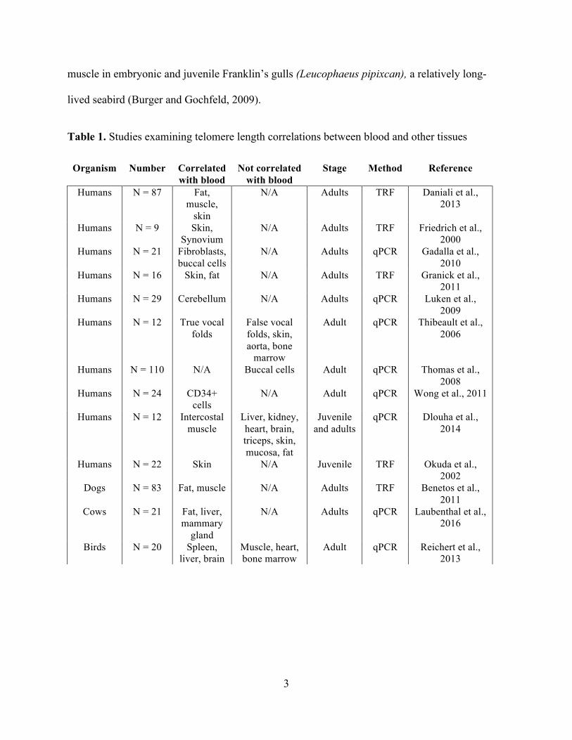
Studies that have addressed this question have typically found positive correlations
between blood and telomere lengths in other somatic tissues (Table 1). However, the results are
not universal and most of these studies have been conducted on adult humans. Only a single
study has addressed this question in a non-mammalian vertebrate and the results were mixed
(Reichert et al., 2013). In zebra finches, small songbirds, blood telomere length was positively
correlated with spleen and brain, but not with bone marrow, heart, or muscle telomere lengths
once a significant outlier was removed (Reichert et al., 2013).
There are reasons to suspect that variation in life-history strategies and developmental
stage could impact telomere correlations among tissues. For example, birds tend to grow faster
and reach adult size earlier than mammals (Ricklefs, 2010; Swanberg et al., 2010), which could
lead to greater variation in proliferation rates and differences in telomere lengths among tissues.
However, to date, no studies have investigated whether blood telomeres are correlated with other
tissues in developing young in vertebrates other than in mammals. Here we investigated whether
blood telomeres were correlated with other somatic tissues including: heart, liver, and skeletal
3
muscle in embryonic and juvenile Franklin’s gulls (Leucophaeus pipixcan), a relatively long-
lived seabird (Burger and Gochfeld, 2009).
Table 1. Studies examining telomere length correlations between blood and other tissues
Organism Number Correlated with blood
Not correlated with blood
Stage Method Reference
Humans N = 87 Fat, muscle,
skin
N/A Adults TRF Daniali et al., 2013
Humans N = 9 Skin, Synovium
N/A Adults TRF Friedrich et al., 2000
Humans N = 21 Fibroblasts, buccal cells
N/A Adults qPCR Gadalla et al., 2010
Humans N = 16 Skin, fat N/A Adults TRF Granick et al., 2011
Humans N = 29 Cerebellum N/A Adults qPCR Luken et al., 2009
Humans N = 12 True vocal folds
False vocal folds, skin, aorta, bone
marrow
Adult qPCR Thibeault et al., 2006
Humans N = 110 N/A Buccal cells Adult qPCR Thomas et al., 2008
Humans N = 24 CD34+ cells
N/A Adult qPCR Wong et al., 2011
Humans N = 12 Intercostal muscle
Liver, kidney, heart, brain, triceps, skin, mucosa, fat
Juvenile and adults
qPCR Dlouha et al., 2014
Humans N = 22 Skin N/A Juvenile TRF Okuda et al., 2002
Dogs N = 83 Fat, muscle N/A Adults TRF Benetos et al., 2011
Cows N = 21 Fat, liver, mammary
gland
N/A Adults qPCR Laubenthal et al., 2016
Birds N = 20 Spleen, liver, brain
Muscle, heart, bone marrow
Adult qPCR Reichert et al., 2013
4
2. METHODS
2.1. Study system and sample collection
Research was conducted on a population of Franklin’s gulls that breed in north-central
North Dakota (Clark and Reed, 2012). Females lay clutches of 1-4 eggs and both males and
females incubate the eggs for 23-26 days and feed the chicks for approximately 35 days prior to
fledging (Burger, 1974). In May and June of 2014 and 2015, we collected first laid eggs (to
control for variation in maternal investment across the clutch) (Engelhardt et al., 2005) and
incubated them in the lab at 65% relative humidity. Eggs collected in May were incubated at a
14:10 light to dark photoperiod, and eggs collected in June were incubated at 18:6, to mimic
naturally occurring photoperiod cues and to be consistent with previous work conducted in this
system (Clark and Reed, 2012).
In 2014, embryos were extracted from eggs near the end of the incubation period (day 18
of incubation) and were promptly weighed and euthanized. In 2015, eggs were allowed to hatch,
and the chicks were subsequently reared in captivity in groups of 3 to 4 birds. Chicks were fed an
ad libitum diet of cat food (Mother and Babycat, Royal Canin USA) from hatching until the end
of post-natal development (approximately 40 days post-hatching), at which point they were
euthanized. To measure telomere lengths in embryos and chicks, we collected a terminal blood
sample and dissected the following tissues: heart, liver and skeletal muscle. Tissue samples were
immediately frozen on dry ice. Blood samples were stored on ice for less than 2 hours and
centrifuged for 10 minutes at 2000 rpm to separate plasma and red blood cells. All samples were
stored at -80oC until being processed for telomere analyses.
5
2.2. Telomere measurement
DNA was extracted using Macherey Nagel Nucleospin Blood and Nucleospin Tissue kits
following the manufacturer’s protocol (Genomic DNA from Blood, Genomic DNA from tissues,
Macherey Nagel). DNA concentration and purity were assessed with a Nanodrop 8000 (Thermo
Scientific) and only samples with 280/260 and 260/230 values above 1.8 were used for telomere
measurement.
Relative telomere length was measured using qPCR (quantitative polymerase chain
reaction) on an Mx3000P (Stratagene) as described in Cawthon (2002) and modified for use in
Franklin’s gulls. The relative telomere length (T/S) of the samples was calculated as the ratio of
the telomere repeat copy number (T) to that of a single copy control gene (S), relative to the
reference sample.
Glyceraldehyde-3-phosphate dehydrogenase (GAPDH) was used as the single copy
control gene. We used the following gull specific GAPDH forward and reverse primers
(Integrated DNA Technologies): 5’-CGGAGCACCGCTTACAATTT-3’ and 5’-
GCATCTCCCCACTTGATGTTG-3’ respectively. Amplified samples were run on a 3% agarose
gel to verify that the amplification was a single product, which yielded a single band at 77 bp as
expected. We used the following telomere primers: TEL 1b: 5’-
CGGTTTGTTTGGGTTTGGGTTTGGGTT-3’ and TEL 2b: 5’-
GGCTTGCCTTACCCTTACCCTTACCCT-3.
The qPCR reactions for GapDH and telomeres were run on separate plates. In both
reactions, the number of PCR cycles (Ct) required for the products to accumulate enough
fluorescent signal to cross a threshold was determined. Individuals with short telomeres took
longer to cross the threshold (i.e., had higher Ct values) than individuals with long telomeres. All
6
reactions used 20 ng of DNA in a final volume of 25 µl containing 12.5 µl of SYBER green
Master Mix, 0.25 µl forward and reverse primer, 6 µl water, and 6 µl of DNA sample. A negative
control of water was run on each plate. All samples were run in triplicate, and average values
were used to determine the T/S ratio according to the following formula: 2∆∆Ct formula, where
∆∆Ct = (CtTelo – Ct
GAPDH) reference − (CtTelo – Ct
GAPDH) (Agilent Technologies, 2012).
In order to assess the efficiencies of each plate, samples were run against a standard curve of 40,
20, 10, 5, and 2.5 ng produced by serially diluting a reference sample. One individual’s tissues
from 2014 were used as the tissue specific reference samples throughout the entirety of the
experiment. Samples were run against tissue specific standard curves. In all cases, plate
efficiencies were in the accepted range (i.e. 100+/-15%) and fell within the bounds of the
standard curve. Average plate efficiencies and standard errors for GAPDH and telomere plates
were 99.02 +/- 3.24, and 90.48 +/- 4.55, respectively. For the 2014 assays, the average intra-plate
variation of the Ct values was 0.12% for the GAPDH assays and 1.13% for the telomere assays,
and the average inter-plate variation for the ΔCt values was 5.20%. For the 2015 assays, the
average intra-plate variation of the Ct values was 0.21% for the GAPDH assays and 1.05% for
the telomere assays, and the average inter-plate variation for the ΔCt values was 6.85%.
2.3. Statistical analysis
To determine whether telomere lengths were correlated among tissues within individuals
we used Pearson’s correlation tests. To examine whether telomere lengths were longer in some
tissues than others in embryonic and juvenile gulls, we used a general linear mixed model (lm).
We included tissue, sex, season, and season*tissue interaction as fixed effects and individual as a
random effect to control for the fact that multiple tissues were collected from the same
individual. All terms were retained in the final model to decrease likelihood of type I errors
7
(Whittingham et. al, 2006). Least squares difference (LSD) tests were used to determine
significant differences in telomere lengths among tissues. We also calculated the repeatability of
telomere lengths across tissues using the formula r = s2A/( s2+ s2
A), as described in Lessells and
Boag (1987). In order to ensure normality, T/S ratios were natural log transformed for all
analyses. All statistical analyses were performed in SPSS (IBM SPSS statistics version 23).
8
3. RESULTS
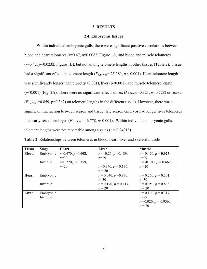
2.4. Embryonic tissues
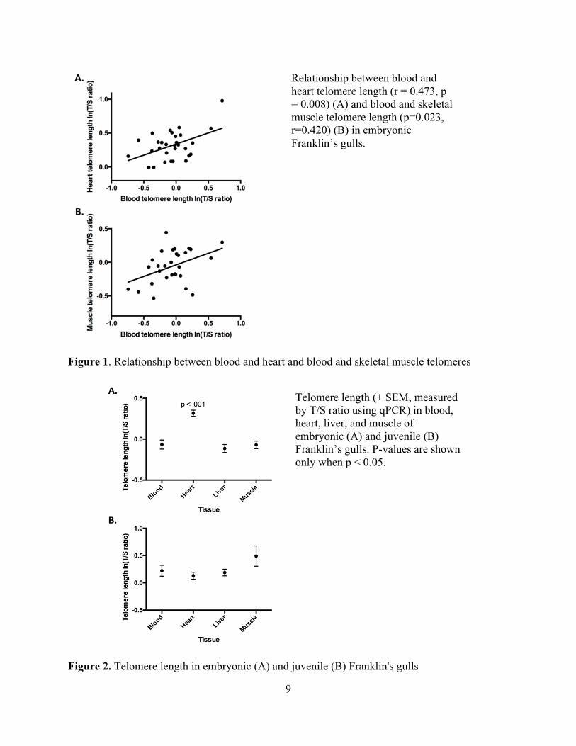
Within individual embryonic gulls, there were significant positive correlations between
blood and heart telomeres (r=0.47, p=0.0083, Figure 1A) and blood and muscle telomeres
(r=0.42, p=0.0232, Figure 1B), but not among telomere lengths in other tissues (Table 2). Tissue
had a significant effect on telomere length (F3,84.661= 25.381, p < 0.001). Heart telomere length
was significantly longer than blood (p<0.001), liver (p<0.001), and muscle telomere length
(p<0.001) (Fig. 2A). There were no significant effects of sex (F1,30.406=0.321, p=0.728) or season
(F1,27.911=0.859, p=0.362) on telomere lengths in the different tissues. However, there was a
significant interaction between season and tissue; late season embryos had longer liver telomeres
than early season embryos (F1, 84.661 = 6.778, p<0.001). Within individual embryonic gulls,
telomere lengths were not repeatable among tissues (r = 0.24918).
Table 2. Relationships between telomeres in blood, heart, liver and skeletal muscle
Tissue Stage Heart Liver Muscle Blood Embryonic
Juvenile
r=0.470, p=0.008, n=30 r=0.220, p=0.359, n=20
r = -0.25, p =0.189, n=29 r =0.340, p = 0.136, n = 20
r = 0.420, p = 0.023, n=29 r = -0.100, p = 0.669, n =20
Heart Embryonic Juvenile
r = 0.040, p =0.830, n=30 r = 0.190, p = 0.417, n = 20
r = 0.260, p = 0.501, n=30 r = 0.050, p = 0.838, n = 20
Liver Embryonic Juvenile
r = 0.190, p = 0.317, n=29 r=-0.020, p = 0.936, n = 20
9
Figure 1. Relationship between blood and heart and blood and skeletal muscle telomeres
Figure 2. Telomere length in embryonic (A) and juvenile (B) Franklin's gulls
Telomere length (± SEM, measured by T/S ratio using qPCR) in blood, heart, liver, and muscle of embryonic (A) and juvenile (B) Franklin’s gulls. P-values are shown only when p < 0.05.
Relationship between blood and heart telomere length (r = 0.473, p = 0.008) (A) and blood and skeletal muscle telomere length (p=0.023, r=0.420) (B) in embryonic Franklin’s gulls.
10
2.5. Post-natal tissues
Within individual gull chicks, there were no significant correlations among telomere
lengths in any tissues (Table 2). In addition, there were no significant effects of tissue (F3,54.00 =
1.911, p = 0.139, Fig 2B), sex (F1,17 = 0.344, p = 0.565), season (F1,17.00 = 0.000, p = 0.994), or
season * tissue interactions (F54.00 = 0.463, p = 0.709) on telomere length. Within individual gull
chicks, telomere lengths were not repeatable among tissues (r = 0.0506).
11
4. DISCUSSION
Blood telomeres are often assumed to be reflective of telomeres in other tissues, but this
has seldom been investigated, particularly in young, developing organisms. Here we report in
Franklin’s gulls, that telomere lengths in blood and other tissues (heart and skeletal muscle, but
not liver) were significantly correlated in late term embryos, but not at the end of post-natal
growth. Thus in contrast to some prior studies, our results suggest that blood telomeres may not
be universally predictive of telomere dynamics in the rest of the body at all stages of
development. In addition, we found that heart telomeres were significantly longer than other
tissues in embryos, but not in chicks.
Telomeres might be expected to be highly correlated in developing organisms because
some evidence suggests that telomerase, an enzyme that adds DNA to the ends of telomeres and
recombination mechanisms act to reset telomere length at fertilization (Eisenhauer, 1997;
Schaetzlein et al., 2004; Vizlin-hodzic et al., 2009). Once the initial telomere length is set,
several factors could contribute to tissue specific differences in telomere shortening rates and
affect telomere correlations among tissues. For example, telomerase activity is expected to be
down-regulated in most somatic tissues after embryonic development (Blackburn, 2005) and this
is known to vary among species (Haussmann et al., 2004). For example, in some long-lived
seabirds, such as Leach’s storm petrels (Oceanodroma leucorhoa) and common terns (Sterna
hirundo), telomerase activity remains elevated in bone marrow and intestine into adulthood
(Haussmann et al., 2007).
Variation in the timing or rate of development could also affect telomere length
correlations among tissues. For example, in adult humans, differences in replication rates during
development contribute to variation in telomere lengths among tissues (Daniali et al., 2013).
12
Variation in cell replication rates may weaken correlations between highly mitotic tissues and
those that are post-mitotic or experience slower proliferation rates. The lack of correlation we
observed in juvenile tissues may be partially due to increasing differences in cell replication rates
across tissues. The idea that less replicative tissues retain longer telomere lengths is consistent
with our finding that heart telomeres were longer than other tissues in embryonic gulls.
Another factor that could affect telomere length correlations among tissues is exposure to
oxidative stress. Oxidative stress, the damage caused to macromolecules by free radicals has
been shown to accelerate telomere loss (von Zglinicki, 2002) and these effects can vary among
tissues (Sahm and Gümüslü, 2007). If either exposure to or the ability to repair oxidative damage
varies among tissues this could affect telomere shortening rates and correlations among tissues.
Thus, organisms may incur variable costs of stress exposure among tissues and as a consequence
these tissues may age at different rates, which may not always be captured by blood telomeres.
For example, Jennings reported that rat pups that were subjected to experimental nutrient
deprivation had shorter kidney telomeres, but telomeres of other tissues (brain and liver) were
unaffected (Jennings et al., 1999).
Blood telomeres have been shown to be predictive of longevity in humans, other
mammals, and in birds (Cawthon, 2003; Fairlie et al., 2015; Bize et al., 2009; Heidinger et al.,
2012) and are increasingly used as a marker of biological aging. Our results in Franklin’s gulls
suggest that telomere length in blood is not always reflective of telomere lengths in other tissues
at all stages of development and highlight the need for additional studies examining the causes
and functional consequences of these tissues specific differences in telomere length.
13
REFERENCES
Agilent Technologies, 2012. Introduction to Quantitative PCR.
Aubert, G., Lansdorp, P.M., 2008. Telomeres and Aging. Physiol Rev 557–579. doi:10.1152/physrev.00026.2007.
Benetos, A., Kimura, M., Labat, C., Buchoff, G.M., Huber, S., Labat, L., Lu, X.B., Aviv, A., 2011. A model of canine leukocyte telomere dynamics. Aging Cell. 10, 991-995. doi: 10.111/j.1474-9726.2011.00744.x.
Bird, J., Ostler, E.L., Faragher, R.G. a, 2003. Can we say that senescent cells cause ageing? Exp. Gerontol. 38, 1319–1326. doi:10.1016/j.exger.2003.09.011
Bize, P., Metcalfe, N.B., Nasir, L., Monaghan, P., Building, G.K., Glasgow, G., 2009. Telomere dynamics rather than age predict life expectancy in the wild. Proc. R. Soc. B Biol. Sci. 1679–1683. doi:10.1098/rspb.2008.1817
Blackburn, E.H., 2005. Telomeres and telomerase: their mechanisms of action and the effects of altering their functions. FEBS Lett. 579, 859–62. doi:10.1016/j.febslet.2004.11.036
Burger, J., 1974. Breeding Adaptations of Franklin’s Gull (Larus Pipixcan) to a Marsh Habitat. Anim. Behav. 521–567.
Burger, J., Gochfeld, M., 2009. Franklin's Gull (Leucophaeus pipixcan), The Birds of North America Online (A. Poole, Ed.). Ithaca: Cornell Lab of Ornithology; Retrieved from the Birds of North America Online: http://bna.birds.cornell.edu/bna/species/116. doi:10.2173/bna.116
Carneiro, M.C., Henriques, C.M., Nabais, J., Ferreira, T., Carvalho, T., Ferreira, M.G., 2016. Short telomeres in key tissues initate local and systemic aging in zebrafish. PLOS Genetics. 12 (1). doi:10.1371/journal.pgen.1005798.
Cawthon, R.M., 2002. Telomere measurment by quantitative PCR. Nucleic Acids Res. 129, 550-557.
Cawthon, R.M., Smith, K.R., O'Brien, E., Sivatchenko, A., Kerber, R.A., 2003. Association between telomere length in blood and mortality in people aged 60 years or older. Lancet 361:393-395.
Clark, M.E., Reed, W.L., 2012. Seasonal interactions between photoperiod and maternal effects determine offspring phenotype in Franklin ’ s gull 948–958. doi:10.1111/j.1365-2435.2012.02010.x
14
Daniali, L., Benetos, A., Susser, E., Kark, J.D., Labat, C., Kimura, M., Desai, K., Granick, M., Aviv, A., 2013. Telomeres shorten at equivalent rates in somatic tissues of adults. Nature Communications. 4. doi:10.1038/ncomms2602.
Dlouha, D., Maluskova, J., Lesna, I.K., Lanska, V., Hubacek, J.A., 2014. Comparison of the Relative Telomere Length Measured in Leukocytes and Eleven Different Human Tissues. Physiol. Res. 63, S343–S350.
Engelhardt, N. Von, Carere, C., Eising, C., Groothuis, T.G.G., Mu, W., 2005. Maternal hormones as a tool to adjust offspring phenotype in avian species. Neurosci. Biobehav. Rev. 29, 329–352. doi:10.1016/j.neubiorev.2004.12.002
Eisenhauer, K.M., Gerstein, R.M., Chiu, C.P., Conti, M., Hsueh, A.J.W., 1997. Telomerase activity in female and male rat germ cells undergoing meiosis and in early embryos. Biol. Repro. 56, 1120-1125.
Fairlie, J., Holland, R., Piklington, J.G., Pemberton, J.M., Harrington, L., Nussey, D.H., 2015. Lifelong leukocyte telomere dynamics and survival in a free-living mammal. Aging Cell. 1-9. doi: 10.1111/acel.12417.
Friedrick, U., Ernst-Ulrich, G., Schwab, M., Fritz, P., Klaus-Peter, T., Klotz, U., 2000. Telomere length in different tissues of elderly patients. Mech. Ageing Dev. 119, 89–99.
Frenck, R.W.J., Blackburn, E.H., Shannon, K.M., 1998. The rate of telomere sequence loss in human leukocytes varies with age. Proc. Natl. Acad. Sci. U. S. A. 95, 5607–5610.
Gadalla, S.M., Cawthon, R., Giri, N., Alter, B.P., Savage, S.A., 2010. Telomere length in blood, buccal cells, and fibrobasts from patients with inherited bone marrow failure syndromes. Aging US. 2, 867-874.
Gomes, N.M. V, Shay, J.W., Wright, W.E., 2010. Telomere biology in Metazoa. FEBS Lett. 584, 3741–51. doi:10.1016/j.febslet.2010.07.031
Granick, M., Kimura, M., Kim, S., Daniali, L., Cao, X., Herbig, U., Aviv, A., 2011. Telomere dynamics in keloids. Open Access J. of Plastic Surgery. 11, 127-136.
Haussmann, M.F., Winkler, D.W., Huntington, C.E., Nisbet, I.C.T., Vleck, C.M., 2004. Telomerase Expression is Differentially Regulated in Birds of Differing Life Span. Ann. N. Y. Acad. Sci. 186–190. doi:10.1196/annals.1297.029
Haussmann, M.F., Winkler, D.W., Huntington, C.E., Nisbet, I.C.T., Vleck, C.T., 2007. Telomerase activity is maintained throughout the lifespan of long-lived birds. Exp. Gerontol. 42, 610-618. doi:10.1016/j.exger.2007.03.004.
15
Heidinger, B.J., Blount, J.D., Boner, W., Griffiths, K., Metcalfe, N.B., Monaghan, P., 2012. Telomere length in early life predicts lifespan. Proc. Natl. Acad. Sci. U. S. A. 109, 1743–8. doi:10.1073/pnas.1113306109
Jennings, B.J., Ozanne, S.E., Dorling, M.W., Hales, C.N., 1999. Early growth determines longevity in male rats and may be related to telomere shortening in the kidney. FEBS. Letts. 448, 4-8. doi:10.1016/S0014-5793(99)00336-1.
Laubenthal, L., Hoelker, M., Frahm, J., Dänicke, S., Gerlach, K., Südekum, K., Sauerwein, H., 2016. Short communication : Telomere lengths in different tissues of dairy cows during early and late lactation. J. Dairy Sci. 1–5. doi:10.3168/jds.2015-10095
Lessells, C.M., Boag, P.T., 1987. Unrepeatable Repeatabilities: A Common Mistake. Auk 104, 116–121.
Luken, J.N., Van Deerlin, V., Clark, C.M., Xie, S.X., Johnson, F.B., 2009. Comparisons of telomere lengths in peripheral blood and cerebellum in Alzheimer's disease. Alzheimers Dement. 5, 463-469.
Monaghan, P., 2014. Organismal stress, telomeres and life histories. J. Exp. Biol. 217, 57–66. doi:10.1242/jeb.090043
Monaghan, P., 2010. Telomeres and life histories: the long and the short of it. Ann. N. Y. Acad. Sci. 1206, 130–142. doi:10.1111/j.1749-6632.2010.05705.x
Okuda, K., Bardeguez, A., Gardner, J.P., Rodriguez, P., Ganesh, V., Kimura, M., Skurnick, J., Awad, G., Aviv, A., 2002. Telomere length in the newborn. Ped. Res. 52, 377-381.
Reichert, S., Criscuolo, F., Verinaud, E., Zahn, S., Massemin, S., 2013. Telomere length correlations among somatic tissues in adult zebra finches. PLoS One 8, 1–7. doi:10.1371/journal.pone.0081496
Ricklefs, R., 2010. Life-history connections to rates of aging in terrestrial vertebrates. Proc. Natl. Acad. Sci. U. S. A. 107, 10314–319.
Sahm, E., Gümüslü, S., 2007. Stress-dependent induction of protein oxidation, lipid peroxidation and anti-oxidants in peripheral tissues of rats: comparison of three stress models (immobilization, cold, and immobilization-cold). Pharmacol. Exp. 34, 425–431. doi:10.1111/j.1440-1681.2007.04584.x
Schaetzlein, S., Lucas-Hahn, A., Lemme, E., Kues, W.A., Dorsch, M., Manns, M.P., Niemann, H., Rudolph, K.L., 2004. Telomere length is reset during early mammalian embryogenesis. Proc. Natl. Acad. Sci. U. S. A. 101, 8034–8038. doi:10.1073/pnas.0402400101.
16
Swanberg, S.E., O’Hare, T.H., Robb, E. a., Robinson, C.M., Chang, H., Delany, M.E., 2010. Telomere biology of the chicken: A model for aging research. Exp. Gerontol. 45, 647–654. doi:10.1016/j.exger.2010.04.002
Thibeault, S.L., Glade, R.S., Li, W., Thibeault, S.L., Al, E.T., 2006. Comparison of Telomere Length of Vocal Folds With Different Tissues : A Physiological Measurement of Vocal Senescence. J. Voice 20, 165–170. doi:10.1016/j.jvoice.2005.04.006
Thomas, P., O’ Callaghan, N.J., Fenech, M., 2008. Telomere length in white blood cells, buccal cells and brain tissue and its variation with ageing and Alzheimer’s disease. Mech. Ageing Dev. 129, 183–190. doi:10.1016/j.mad.2007.12.004
Vizlin-hodzic, D., Ryme, J., Simonsson, S., Simonsson, T., 2009. Developmental studies of Xenopus shelterin complexes : the message to reset telomere length is already present in the egg. FASEB 23, 2587–2594. doi:10.1096/fj.09-129619
Vleck, C.M., Haussmann, M.F., Vleck, D., 2003. The natural history of telomeres: tools for aging animals and exploring the aging process. Exp. Gerontol. 38, 791–795. doi:10.1016/S0531-5565(03)00110-4
Whittingham, M.J., Stephens, P.A., Bradbury, R.B., Freckleton, R.P., 2006. Why do we still use stepwise modelling in ecology and behaviour? Anim. Behav. 75, 1182-1189.
von Zglinicki, T., 2002. Oxidative stress shortens telomeres. Trends Biochem Sci 27, 339–344.
Wong, L.S.M., Huzen, J., de Boer, R.A., van Gilst, W.H., van Veldhuisen, D.J., van der Harst, P., 2011. Telomere length of circulating leukocyte subpopulations and buccal cells in patients with ischemic heart failure of their offspring. PLOS one. 6. doi: 10.137/journal.pone.0023118.


![Intrarenal arteriosclerosis and telomere attrition ...€¦ · Telomere length is a well-established marker of biological age [4]. Although telomere length is partly heritable, there](https://img.pdfslide.net/doc/110x75/5f2629fb310cc83259516f06/intrarenal-arteriosclerosis-and-telomere-attrition-telomere-length-is-a-well-established.jpg)


![Determination of Telomere Length by the Quantitative ... · Telomere intensity assessed by FISH using a PNA probe is known to correlate with telomere length [20]. Therefore, PNA probes](https://img.pdfslide.net/doc/110x75/5f2629add358ac5cd71a88d8/determination-of-telomere-length-by-the-quantitative-telomere-intensity-assessed.jpg)