Embed Size (px)
Citation preview
Multifunctional poly(methacrylate) polyplex libraries: A platform for gene delivery
inspired by nature
Marco Emanuele Favretto, Andreas Krieg, Stephanie Schubert, Ulrich S. Schubert, Roland
Brock*
M.E. Favretto1,2, A. Krieg2,3,4, S. Schubert4,5, U.S. Schubert2,3,4, R.Brock1,2
1 Department of Biochemistry, Radboud University Medical Centre, Radboud Institute for
Molecular Life Sciences, Nijmegen, The Netherlands
2 Dutch Polymer Institute (DPI), Eindhoven, The Netherlands
3 Laboratory of Organic and Macromolecular Chemistry (IOMC), Friedrich Schiller University
Jena, Jena, Germany
4 Jena Center for Soft Matter, Friedrich Schiller University Jena, Jena, Germany
5 Institute of Pharmacy, Pharmaceutical Technology, Friedrich Schiller University Jena, Jena,
Germany
Corresponding Author: Roland Brock,Radboud University Medical Centre, Radboud Institute for
Molecular Life Sciences, 6525 GA Nijmegen, The Netherlands. Phone: +31243666213; Email:
Keywords: Gene delivery, poly(methacrylate)s, screening strategies, blood compatibility,
oligonucleotides, polyplex
1
ABSTRACT
Polymer-based gene delivery systems have enormous potential in biomedicine, but their
efficiency is often limited by poor biocompatibility. Poly(methacrylate)s (PMAs) are an
interesting class of polymers which allow to explore structure-activity relationships of polymer
functionalities for polyplex formation in oligonucleotide delivery. Here, we synthesized and
tested a library of PMA polymers, containing functional groups contributing to the different steps
of gene delivery, from oligonucleotide complexation to cellular internalization and endosomal
escape. By variation of the molar ratios of the individual building blocks, the physicochemical
properties of the polymers and polyplexes were fine-tuned to reduce toxicity as well as to
increase activity of the polyplexes. To further enhance transfection efficiency, a cell-penetrating
peptide (CPP)-like functionality was introduced on the polymeric backbone. With the ability to
synthesize large libraries of polymers in parallel we also developed a workflow for a mid-to-high
throughput screening, focusing first on safety parameters that are accessible by high-throughput
approaches such as blood compatibility and toxicity towards host cells and only at a later stage
on more laborious tests for the ability to deliver oligonucleotides. To arrive at a better
understanding of the molecular basis of activity, furthermore, the effect of the presence of
heparan sulfates on the surface of host cells was assessed and the mechanism of cell entry and
intracellular trafficking investigated for those polymers that showed a suitable pharmacological
profile. Following endocytic uptake, rapid endosomal release occurred. Interestingly, the
presence of heparan sulfates on the cell surface had a negative impact on the activity of those
polyplexes that were sensitive to decomplexation by heparin in solution. In summary, the
screening approach identified two polymers, which form polyplexes with high stability and
transfection capacity exceeding the one of poly(ethylene imine) also in the presence of serum.
2
INTRODUCTION
Polycationic polymers, either from natural or from synthetic origin, have been widely
investigated as vectors for nucleic acid therapeutics, like DNA, siRNA or antisense
oligonucleotides [1]. Prominent examples are the poly(ethylene imines) (PEI) [2] and
polyamidoamides (PAA) [3] that have shown effectivity in vitro and in vivo. Complexation into
so-called polyplexes occurs through electrostatic interactions with the negatively charged nucleic
acids. In these complexes, oligonucleotides are protected from degradation. Furthermore, an
excess of positive charge on the surface of the polyplexes is supposed to facilitate cellular uptake
[4, 5].
To guarantee an efficient delivery, polyplexes have to meet several requirements: Outside the
cells effective condensation of oligonucleotides into monodisperse nanoparticles needs to take
place. For in vivo applications, these polyplexes need to be stable in serum and free of hemolytic
activity. In contact with cells, efficient induction of uptake has to occur.
According to the present model, cellular uptake involves the interaction of the positively charged
polyplexes with the negatively charged glycocalyx. Along with charge, particle size is an
important determinant for endocytic uptake. Ideally, polyplexes should have a size of 200 to 600
nm in order to show effective delivery [6-8]. Engagement of the glycocalyx constituent heparan
sulfate (HS) has also directly been associated with the induction of endocytic cellular uptake [9,
10]. However, given the capacity of negatively charged oligosaccharides to disassemble
polyplexes [11], HS may also sequester the polyplexes on the cell membrane and displace the
DNA from the complexes thereby compromising the uptake of oligonucleotides.
Following endocytosis, endosomal escape is required to guarantee an appropriate delivery of the
cargo into the cytoplasm and nucleus. Endosomal escape can occur through two main
mechanisms. For membrane viruses and liposomes, release occurs by membrane fusion [12, 13].
For cationic polymers, the “proton sponge effect” has been discussed as the main mode-of-action
[14]. Briefly, protonable groups of tertiary amines with a mildly acidic pKa value such as
imizadoles sequester protons, which counteract the endosomal acidification leading to further
import of protons and chloride counterions that will cause an osmotic swelling and the
3
subsequent rupture of the endo(lyso)somes [4]. Also decomposition of polyplexes leads to an
increase in osmolarity [15]. Finally, in the cytoplasm release of the oligonucleotides has to occur.
To further improve internalization efficiency the functionalization of polyplexes with cell-
penetrating peptides (CPPs) has been explored [16]. These peptides have been shown to facilitate
cellular uptake of membrane impermeable macromolecules. Several classes of CPPs can be
distinguished [17]. The well-studied CPP Tat and nonaarginine belong to the class of arginine-
rich CPP. For induction of cellular import arginine is a privileged structure as it can effectively
interact with glycosaminoglycans on the cell surface and form bidentate hydrogen bonds with
negatively charged molecules to partition into lipid bilayers [18].
To improve the transfection efficiency of polyplexes, significant efforts were invested into the
synthesis and screening of polymers that possess a better pharmacological profile. However, only
a limited set of functionalities was tested so far. In contrast, for polypeptides, the role of a variety
of amino acid side chains with respect to oligonucleotide complexation, membrane binding and
intracellular trafficking has been explored. For example, complexation of negatively charged
nucleic acids can be achieved by incorporation of lysines [19], while histidines can induce
endosomal release [20]. Tryptophans can be introduced to promote interaction with the plasma
membrane [21]. The solubility can further be modulated by tuning the ratio between hydrophilic
and hydrophobic residues.
Recently, poly(methacrylate)s (PMAs) have been emerging as an alternative vector class in gene
delivery, either alone [22, 23] or as co-polymers with PEI [24, 25]. Their applications range
from brain-targeted gene delivery [26] to intradermal administration [27]. Their accessible
chemistry allows the synthesis of a variety of monomers which mimic the physicochemistry and
functionality of amino acid side chains. Utilizing controlled radical polymerization methods
(atom transfer radical polymerization (ATRP), reversible addition-fragmentation chain-transfer
(RAFT) polymerization [28]), polymers with tunable solubility, complexation capacity and
transfection efficiency can be formed. Frequently, complexation with nucleic acids is granted by
the presence of dimethylaminoethyl methacrylate (DMAEMA) or diethylaminoethyl
methacrylate (DEAEMA) [22], which also act as a proton sponge and facilitate cytoplasmic
delivery; in addition, endosomal escape is often achieved by the hydrophobic butyl methacrylate
(BMA) [22, 29], which is known to destabilize cellular membranes. In order to prolong the
4
circulation time and to reduce cell toxicity, PEG monomers (PEG-methylether methacrylate,
ethylene glycol dimethacrylate and PEG-DMAEMA) are inserted into the polymer backbone and
act as a protective shell [26, 30].
Thus, given the well-established polymerization chemistry and the capacity to vary the side
chain, PMAs represent a highly interesting class of polymers to explore the structure space for
oligonucleotide delivery.
The aim of this study was two-fold. First, we aimed at identifying the structure-activity
relationship between the polymer structure and the physicochemical and biological properties for
PMA copolymers with functionalities intended to cover major structural characteristics relevant
to oligonucleotide complexation, cellular uptake and endosomal release. Second, we aimed to do
so by establishing a screening strategy that would allow for a rapid and reliable selection of the
most promising candidates for pre-clinical trials. Therefore, toxicity tests that can be performed
in a high-throughput approach and that conventionally are only conducted at a later stage in
testing for those polymers that show transfection activity were introduced early in the test
protocol. Following an assessment of the capacity to form polyplexes with oligonucleotides, the
resulting polyplexes were then tested for their stability in the presence of serum protein and
polyanions, their hemolytic activity and their acute toxicity towards host cells. Finally, the
activity in oligonucleotide delivery was determined. For this purpose we made use of a 2’-O-
methylated (2-OMe)-oligonucleotide that induces splice correction of an aberrant primary
luciferase gene transcript [31]. Moreover, the effect of the presence of HS on host cells was
investigated. Using time-lapse confocal microscopy, we furthermore demonstrate that the most
active polymer induces a wave of massive endosomal uptake followed by a dispersion of
endosomal structures and endosomal release.
MATERIALS AND METHODS
Materials
N,N’-(Dimethylamino)ethyl methacrylate (DMAEMA), oligo(ethyleneglycole) methacrylate
(OEGMA), 2-cyano-2-propyl benzodithioate (CPDB), and 2,2′-azobis(2-isobutyronitrile)
5
(AIBN) were purchased from Sigma-Aldrich. The monomers were passed over a column of
inhibitor remover (Sigma-Aldrich) preliminary to the reaction. AIBN was recrystallized from
methanol whereas CPDB was used without further purification. 2-(N-Imidazol)ethyl
methacrylate (ImEtMA) and but-3-ene-1-yl methacrylate (BEMA) were synthesized by
conversion of the corresponding alcohol with methacrylic acid chloride in
chloroform/triethylamine at 25 °C. An acetylated cysteinyl tetraarginyl amide CPP (Ac-CRRRR-
NH2) was purchased from EMC microcollections, Tuebingen, Germany.
Resazurin, heparin from porcine intestinal mucosa (average MW 5kDa) and 25,000 g/mol
branched PEI were obtained from Sigma-Aldrich, Zwijndrecht, NL. Peri-phosphorothioate Cy5-
labelled 2’-OMe-ON-705 was purchased from Biolegio, Nijmegen, NL. A luciferase assay
system was purchased from Promega, Leiden, NL. Rhodamine-labelled dextran (neutral, 10000
Da) was obtained from Invitrogen, Bleiswijk, NL. Heparinase III was purchased from Ibex,
Montreal, Canada. cDNA encoding for a Rab5-GFP protein was kindly donated by Dr. Sandra de
Keijzer, Dept. of Tumour Immunology, Radboud University Medical Centre, Nijmegen.
Methods
Polymer synthesis
The polymerization conditions are described for the typical example IM-1: In a microwave vial,
505 µL (471 mg, 3 x 10⁻³ mol) DMAEMA, 429 µL (463 mg, 9.75 x 10⁻⁴ mol) OEGMA, and
108 mg (6 x 10⁻⁴ mol) ImEtMA were mixed with 4.4 mg (2 x 10⁻⁵ mol) CPDB and 0.8 mg (5 x
10-6 mol) AIBN. Subsequently, 1.258 mL ethanol were added, and the vial was capped. The
mixture was flushed with argon for 30 minutes to remove the oxygen and placed in an oilbath at
60 °C for 12 hours. After the reaction, the polymer was precipitated into hexane and dried under
vacuum. The polymers were analyzed by size exclusion chromatography (SEC) as well as 1H
NMR spectroscopy. The polymerization of the other polymers was performed in a similar
manner using the corresponding amounts of each monomer (Table S1).
Polymer series IM+V-1-3 was functionalized with a cell-penetrating peptide via UV induced
thiol-ene coupling reaction using 2,2-dimethoxy-2-phenylacetophenone (DMAP) as
photoinitiator. The representative procedure is presented for polymer IM+CPP-1:
6
The polymer IM+V-1 (50 mg, 2 x 10⁻⁶ mol) and Ac-CRRRR-NH2 (3 mg, 4 x 10⁻⁶ mol) were
dissolved in 3 mL ethanol. 2 mg DMAP (8 x 10⁻⁶ mol) were added, and the whole mixture was
flushed with argon for 30 minutes. The reaction solution was irradiated with UV light for
24 hours. Afterwards, the solvent was evaporated and the residues taken up with chloroform,
intensively washed with water to remove unbound Ac-CRRRR-NH2, and dried under vacuum.
Polymer characterization
1H NMR spectra were recorded in CDCl3 or DMSO-d6 on a Bruker AC 300 MHz using the
residual solvent resonance as an internal standard. Size exclusion chromatography (SEC) was
performed on a Shimadzu system equipped with an SCL-10A system controller, an LC-10AD
pump, an RID-10A refractive index detector and both, a PSS Gram30 and a PSS Gram1000
column in series, whereby chloroform or N,N-dimethylacetamide (DMAc) with 5 mmol lithium
chloride (LiCl) were used as an eluent at 1 mL/min flow rate and a column oven temperature of
60 °C (for DMAc). The system was calibrated with polystyrene (370 g/mol – 67,500 g/mol) and
poly(methyl methacrylate) (2000 g/mol – 88,000 g/mol) standards, respectively.
Formation of polyplexes and their characterization
Polyplexes of polymers and oligonucleotides were formed by mixing polymers and Cy5-labelled
2’-OMe-ON-705 in water, in a N/P ratio 3:1, to a final polymer concentration of 200 g/mL.
ON-705 is a splice correcting oligonucleotide, directed to the -globin intron 2, with the
following sequence: 5'-CCUCUUACCUCAGUUACA-3'. Upon mixing and vigorous vortexing
for 30 seconds, polyplexes were left for annealing for 30 minutes at room temperature. After
annealing, the size of ON/polymer complexes in water or upon 1:5 v/v dilution in RPMI 1640
culture medium supplemented with 10% FCS, was measured at 25 °C by dynamic light
scattering (DLS) at a backscatter angle of 173°, using a Malvern Nanosizer ZS (Malvern, UK).
The zeta-potential of the complexes in water was measured by Laser Doppler Anemometry,
using the Malvern Nanosizer ZS.
Polyanion decomplexation assay
7
To probe for polyanion-induced decomplexation, polyplexes were incubated for 30 minutes with
heparin at heparin/ON molar ratios of 0:1, 0.1:1, 0.5:1 and 1:1. Immediately after incubation,
samples were measured by DLS, using a backscatter angle of 173° at 25 °C
Cell culture conditions
HeLa pLuc 705 cells stably transfected with a reporter construct for determination of delivery of
a splice-correcting oligonucleotide [31] were grown as monolayers in RPMI 1640 medium,
supplemented with 10% fetal calf serum (FCS) and 200 mM L-glutamine, at 37 °C, in a
humidified atmosphere containing 5% CO2. Cells were passaged every 2 days or at 80 to 90%
confluency.
Determination of hemolytic activity of polyplexes
Hemolytic activity was determined with red blood cells (RBCs) from RBC units of blood group
0, Rhesus-positive donors that had been collected and processed according to standard Dutch
blood bank protocols, including leukoreduction and storage in saline-adenine-glucose-mannitol.
RBCs were washed and resuspended in complete Ringer solution (125 mM NaCl, 5 mM KCl, 1
mM MgSO4, 32 mM HEPES, 5 mM glucose and 1 mM CaCl2). All experiments with RBCs were
performed at room temperature. In an Eppendorf tube, 3 x 10⁷ RBCs were suspended into 300
µL of Ringer buffer, and polyplexes were added to the cell suspension at the indicated
concentrations. After 2 hours of incubation, RBCs were centrifuged (3000 rpm for 3 minutes)
and the supernatant (containing the free hemoglobin) was collected. Non-treated cells were used
as negative control, while burst cells induced by incubation with water were used as a positive
control. 60 µL of the supernatant were transferred into a 96-well plate and diluted to 300 µL with
water. Absorbance for each sample was measured at 405 nm.
The degree of hemolysis was calculated as follows:
% H emolysis=|sample|−|negative|control
|positive|control ×100
8
Removal of HS from the cell surface
Removal of HS chains from the cell surface was accomplished by a 1-hour incubation at 37°C
with RPMI 1640 culture medium supplemented with 1% FCS and 3 mIU/mL of heparinase III.
Afterwards, cells were washed in order to remove cleaved HS chains in the medium.
Acute cellular toxicity induced by polyplexes
The impact of polyplexes on cell viability, either on untreated cells or after removal of heparan
sulfates (HS), was assessed using the resazurin assay. Briefly, 8 x 10⁴ cells/well were seeded in a
96 well plate. After 24 hours, HS chains were removed, if required, as described above and
polyplexes were added at a concentration of 100 g/mL for an incubation of 2 hours. Cells were
washed, and resazurin (100 g/mL in RPMI 1640 culture medium supplemented with 10% FCS)
was added. Non-treated cells were used as a negative control, culture medium was used as a
blank. After 4 hours, readings of fluorescence intensity (Ex: 540/25 nm, Em: 620/40 nm) were
taken on a BioTek Synergy 2 plate reader. Cell viability was calculated as follows:
% cell viability= Fluorescence Sample−Fluorescence BlankFluorescence Control−Fluorescence Blank
×100
Transfection efficiency/splice correction assay
Polyplexes in a range of concentrations between 10 and 100 g/mL were incubated in a 96-well
plate for 2 hours at 37 °C, with HeLa pLuc 705 cells (8 x 10⁴ cells/well) in RPMI 1640 culture
medium supplemented with 10% FCS. After incubation, polyplex-containing medium was
replaced with fresh medium and cells were incubated for a further 24 hours. Then, the growth
medium was removed and cells were rinsed twice with HBS. 20 L of lysis reagent were added
into each well, and the total protein concentration in each sample was determined by Bradford
assay [32]. Upon normalization to total protein content, lysates were mixed with 100 L of
luciferase assay reagent. Bioluminescence was measured after 3 minutes using a BioTek Synergy
2 plate reader.
9
Intracellular distribution of polyplexes
HeLa pLuc 705 cells were transfected with cDNA encoding for a Rab5-GFP fusion protein as a
marker for early endosomes using Lipofectamine 2000. 24 hours after transfection, cells were
incubated with polyplexes at a concentration of 100 g/mL for 1 hour in RPMI 1640 culture
medium supplemented with 10% FCS.
Colocalization with early endosomes and heparan sulfates
Upon incubation with polyplexes, heparan sulfate chains were stained by immunocytochemistry,
using an anti HS4C3V primary antibody [33] and an anti-vsv-Zenon IgG1-AlexaFluor 546
secondary antibody. Cells were imaged using a TCS SP5 confocal microscope (Leica
Microsystems, Mannheim, Germany), equipped with an HCX PL APO 63x NA 1.2 water
immersion lens. Excitation/detection wavelengths of 488 nm/500-520 nm, 561 nm/570-600 nm
and 633 nm/650-700 nm were used for Rab5-GFP, HS immunofluorescence and Cy5-ON,
respectively. Images were captured sequentially (1: Rab5-GFP and Cy5-ON; 2: HS) to reduce
crosstalk between the fluorescent probes. Images were processed using the FIJI image processing
package (Fiji.sc, version win32-20110307). Colocalization was quantitated using the
Colocalization Threshold plugin in FIJI, using a total of 30 cells per condition from 3
independent experiments (10 cells per experiment).
Colocalization with early endosomes and dextran
Cells were incubated for 1 hour with polyplexes (100 g/mL) and Rhodamine-B labelled dextran
10 kDa (1 mg/mL). Dextran was used as a marker for macropinocytosis. Cells were imaged right
after incubation and after allowing 1, 2 and 3 hours recovery following removal of polyplexes,
using the excitation and detection settings indicated in the previous paragraph.
Statistical analysis
All the data reported in this article are the result of at least three independent experiments.
Statistical significance was calculated by one-way ANOVA or t-test, as appropriate. The
confidence interval was set at 95%. Data were analyzed using The R Project for Statistical
Computing (http://www.r-project.org, version 2.14).
10
RESULTS
Polymer synthesis and characterization
To explore the structure space of polymers for oligonucleotide delivery we chose
poly(methacrylates) as the basic polymer structure. The structural variety of methacrylate
monomers enables the inclusion of all functionalities deemed necessary to provide a powerful
platform for gene delivery. The copolymer series were synthesized by reversible addition–
fragmentation chain transfer polymerizations (RAFT) using CPDB as chain transfer agent and
AIBN as initiator (Scheme 1). RAFT is a controlled radical polymerization technique working
under mild conditions and tolerating a variety of monomer functionalities, thus, enabling the
synthesis of well-defined copolymers with specific monomer composition [28]. The polymer
design was inspired by the knowledge of the physicochemical and biological requirements: At
least one cationic species is required for a successful formation of the polyplexes with anionic
oligonucleotides and the generation of a positive charge surplus for interaction with cell
membranes. In the present study, two amine-species were used for this purpose, namely N,N’-
dimethyl-aminoethyl methacrylate (DMAEMA) and 2-(N-imidazol)ethyl methacrylate
(ImEtMA). The imidazole functionality was also included to provide a significantly different
pKa value and, thus, to promote endosomal escape in analogy to oligohistidines and histidine-
modified polymers [20]. Furthermore, to enhance the overall solubility and to reduce the
membrane-disturbing potential, hydrophilic species like hydroxyethyl methacrylate (HEMA) and
oligoethyleneglycol methacrylate (OEGMA), were incorporated into the polymer. Finally, to
enable the coupling of a tetraarginine (R4) to serve as a model CPP for promoting cellular uptake,
a but-3-ene-1-yl methacrylate (BEMA) monomer was included. This building block allows
selective and efficient coupling of thiol-containing systems to the polymer backbone via a thiol-
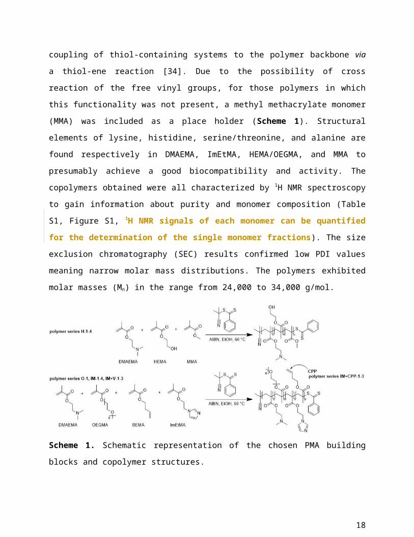
ene reaction [34]. Due to the possibility of cross reaction of the free vinyl groups, for those
polymers in which this functionality was not present, a methyl methacrylate monomer (MMA)
was included as a place holder (Scheme 1). Structural elements of lysine, histidine,
serine/threonine, and alanine are found respectively in DMAEMA, ImEtMA, HEMA/OEGMA,
and MMA to presumably achieve a good biocompatibility and activity. The copolymers obtained
were all characterized by 1H NMR spectroscopy to gain information about purity and monomer
11
composition (Table S1, Figure S1, 1H NMR signals of each monomer can be quantified for
the determination of the single monomer fractions). The size exclusion chromatography
(SEC) results confirmed low PDI values meaning narrow molar mass distributions. The
polymers exhibited molar masses (Mn) in the range from 24,000 to 34,000 g/mol.
Scheme 1. Schematic representation of the chosen PMA building blocks and copolymer
structures.
The functionalization of the vinyl-bearing polymers with the CPP was performed in a click-like
reaction using DMAP as UV initiator. Two equivalents of the tetra-arginine peptide with a
cysteine moiety (with respect to vinyl functionalities) were added to one polymer molecule of
the IM+V-series. The mild conditions allowed a chemoselective conjugation of the CPP to the
polymer backbone. Unfortunately, the low conversion rates could not be quantified via NMR
spectroscopy or other analytical tools since the bulk polymer characteristics are predominant.
Consequently, five series of polymers, which differed in their hydrophilicity and in their degrees
of complexity, were generated: PEI-like polymers (O-1 and H), polymers carrying imidazole
functionalities to promote endosomal escape (IM), polymers including both imidazoles and vinyl
groups to allow the conjugation with a CPP-like molecule (IM+V) as well as polymers carrying a
tetra-arginine peptide (IM+CPP).
A mid-to-high throughput screening of the biological properties of polyplexes
12
The polymethacrylate copolymer synthesis strategy enables the generation of tens to
hundreds of different polymers from lab-scale to industrial products such as EUDRAGIT
[35, 36]. Therefore, not the synthesis but rather the identification of those polymers with the
most desirable physicochemical and biological characteristics constitutes a bottleneck. As a
consequence, we aimed at creating a streamlined workflow, in which assays that can be
conducted in high throughput and with little effort are conducted first, while those that are more
cost and labour intensive are conducted later. Also, those polymers that would fail later in pre-
clinical development due to toxicity should be eliminated early in the selection procedure.
The absence of hemolytic activity has a high predictive power for pre-clinical safety, yet can be
conducted in high-throughput. Nevertheless, in current practice, such a test is only performed for
gene delivery systems that have shown activity in vitro. In line with our synthesis and selection
strategy, testing for hemolytic activity was introduced early in the workflow (Scheme 2). With
each step, the complexity of the polymer collection is reduced so that finally more detailed
mechanistic studies are restricted to those polymers with a beneficial toxicity profile.
Scheme 2: Workflow for the identification of biologically active polymers from large polymer
collections. Assays with predictive power for pre-clinical safety are conducted first, followed by
more labour and cost-intensive tests.
13
Characterization of polyplexes (number of polymers tested: 15)
First, we assessed the capacity of the polymers to form polyplexes, using an N/P ratio of 3:1,
which describes the ratio of moles of the protonable amine groups of cationic polymers to the
phosphates/phosphorothioates in the oligonucleotide. This N/P ratio is commonly used for
PEI-based polyplexes. At this specific N/P, polyplexes of PEI and ON, as well as DNA, show
the ideal complexation, accompanied by relatively small fraction of free PEI, which can
cause toxicity to cells. In our calculation, we took into account only the amine groups of the
DMAEMA monomers and did not include the imidazole-carrying monomer. Polyplexes derived
from polymers of our library and PEI as a reference were characterized in terms of size
distribution by DLS. Their size and stability were assessed in MilliQ water and in culture
medium, supplemented with 10% FCS (Table S2). The lack of activity in the presence of fetal
calf serum is the major shortcoming of most current transfection agents. However, for potential
in vivo use, resistance of polyplexes towards FCS is an absolute requirement. We evaluated the
DLS data with respect to the maintenance of polyplexes as well as with respect to the absence of
aggregate formation.
The formation of polyplexes was observed for all the polymers, except for O-1 and H-3. The size
of ON/polymer complexes in water ranged from 130 nm to 400 nm, with PDI values from 0.118
to 0.256. The low PDI values indicate that the polyplexes formed a relatively monodisperse
population (Table S2). The introduction of R4 as a CPP functionality resulted in a significant
increase in size. Polyplexes derived from the IM+CPP series were larger by a factor of 1.5 to 1.7
than the ones derived from the same polymer backbone lacking the CPP functionality (IM+V),
with the exception of IM+CPP-3/IM+V-3.
Addition of serum had a detrimental effect on most of the polyplexes (Table S2). Polyplexes
from the IM+V series (imidazole-carrying polymers with free vinyl groups) were within a size
range of 350 to 390 nm, which is acceptable for intravenous administration and is expected to be
taken up by cells via endocytosis. Once again, the introduction of the CPP functionality led to an
14
increase in size by 100 to 200 nm with the exception of the IM+CPP-3 polyplexes. However, the
presence of R4 did not affect the stability of polyplexes.
With respect to the structure-activity relationship, there was a decrease in diameter with
increasing MMA content (for the H series) and an increase with increasing imidazole fraction
(for both, the IM and IM+V series). The introduction of the vinyl functionality contributed to a
decrease in particle size and prevented aggregation in serum. This effect may be either due to
hydrophobic interactions occurring between the free vinyl groups the side chains, that
compact the structure and shield the positively charged groups in the polyplexes from
serum protein or from cross linking. Due to the rapid decomplexation of the polyplexes in
the presence of heparin (see below) we favor the hydrophobic interactions as the possible
explanation.
Since one of the aims of this study was to screen polymers which can form polyplexes with
suitable characteristic for in vivo delivery, only polyplexes that showed stability in serum and
had a size that fell in the optimal range of 200 to 600 nm [6-8] were tested further.
Hemolytic activity [number of polymers tested: 7]
Hemolytic activity was assessed for serum-stable polyplexes (H-1, IM+V series, IM+CPP series
and PEI) in a range of concentrations between 10 g/mL and 250 g/mL. As a threshold,
hemolytic activity should not exceed 10% for a formulation to be considered safe [37].
All polyplexes induced hemolysis on RBCs in a dose dependent manner up to 100 µg/mL, while
the increase in hemolytic activity was less evident between 100 g/mL and 250 g/mL. For most
of the polyplexes, the safety threshold of 10% was reached at a concentration of 100 g/mL.
There was no clear-cut correlation between molecular structure and hemolytic activity. However,
the introduction of the tetraarginine led to some variations in the behavior of the polyplexes.
While the degree of hemolysis induced by the IM+V-2 and the IM+CPP-2 polyplexes was quite
comparable, IM+CPP-1 polyplexes showed significantly less hemolysis than IM+V-1
polyplexes, and IM+CPP-3 polyplexes induced a higher degree of hemolysis than polyplexes
derived from the parent IM+V-3 polymer in particular at high concentrations. This observation
seems counterintuitive, as it is known that introduction of protonable (and protonated) groups is
often correlated with a higher degree of hemolysis [37]. Nevertheless, it has to be highlighted
15
that all the polyplexes derived from the PMA polymers showed an improved safety over PEI
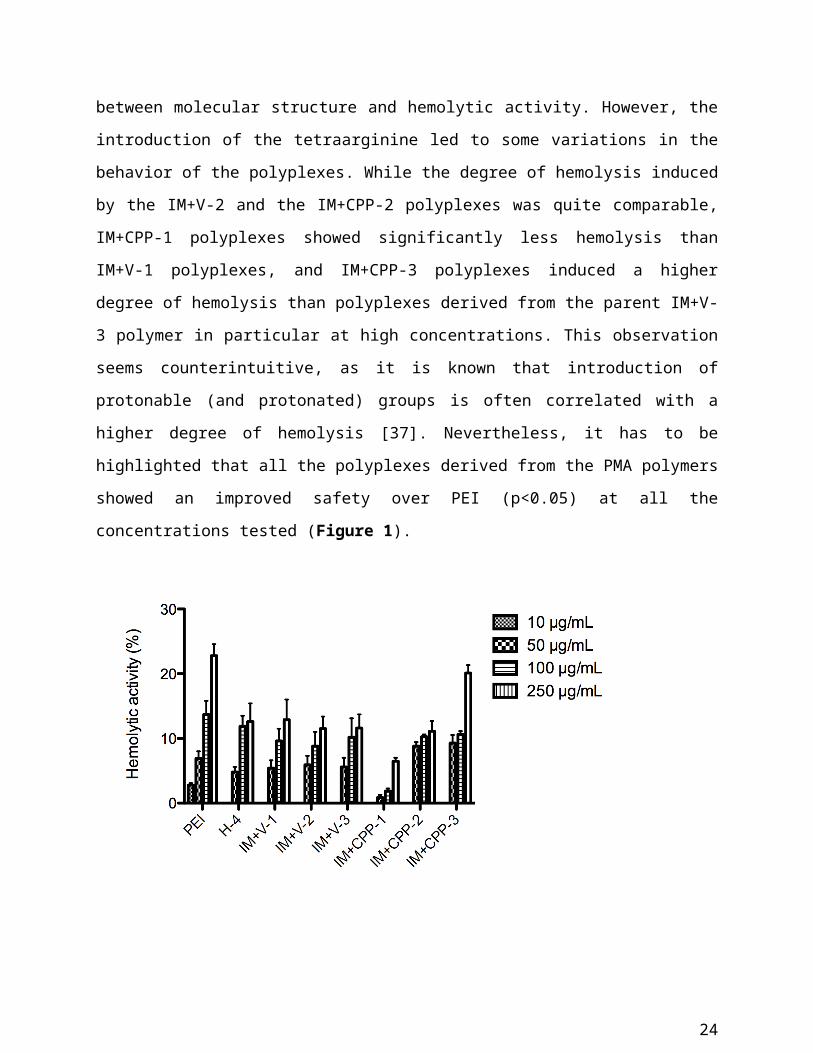
(p<0.05) at all the concentrations tested (Figure 1).
Figure 1: Hemolytic activity induced by polyplexes at 10 g/mL, 50 g/mL, 100 g/mL and
250 µg/mL. Results are expressed as mean values ± SEM (n=3).
Acute toxicity towards HeLa cells and dependence on HS (number of polymers tested: 7)
Next, we tested for acute toxicity on HeLa cells. At this time, we also explored the effect of
proteoglycans on the cell surface by removing HS chains.
In the presence of HS on the plasma membrane, PEI-like polyplexes (H-4) and CPP-carrying
polyplexes with a high fraction of imidazole monomers (IM+CPP-2 and IM+CPP-3) were
extremely toxic, as they reduced the mitochondrial activity by 55 to 75% after 2 hours of
incubation at 100 g/mL. On the other hand, polyplexes derived from the IM+V series and the
IM+CPP-1 polymer had only a moderate, sometimes negligible, effect on cell viability (Figure
2A). In general, all the PMA polyplexes were less toxic than the ones derived from PEI (p<0.01).
Moreover, the presence of the OEGMA building block, which is missing in H-4, seemed to
shield the complexes and protect the cells, as proposed by Mathew and coworkers [30].
16
Upon enzymatic removal of HS, we had expected an increase in cell toxicity, as the presence of
HS could shield the cells from a potentially detrimental effect of the polyplexes. However, this
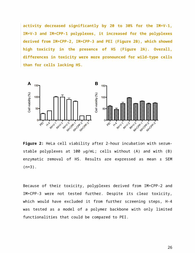
was not always the case. While upon removal of HS mitochondrial activity decreased
significantly by 20 to 30% for the IM+V-1, IM+V-3 and IM+CPP-1 polyplexes, it increased
for the polyplexes derived from IM+CPP-2, IM+CPP-3 and PEI (Figure 2B), which showed
high toxicity in the presence of HS (Figure 2A). Overall, differences in toxicity were more
pronounced for wild-type cells than for cells lacking HS.
Figure 2: HeLa cell viability after 2-hour incubation with serum-stable polyplexes at 100
g/mL; cells without (A) and with (B) enzymatic removal of HS. Results are expressed as mean
± SEM (n=3).
Because of their toxicity, polyplexes derived from IM+CPP-2 and IM+CPP-3 were not tested
further. Despite its clear toxicity, which would have excluded it from further screening steps, H-
4 was tested as a model of a polymer backbone with only limited functionalities that could be
compared to PEI.
Transfection efficiency and dependence on HS (number of polymers tested: 5)
The remaining set of polyplexes (H-4, IM+V series and IM+CPP-1) were tested for their ability
to deliver an antisense oligonucleotide to HeLa pLuc 705 cells, which express an aberrant
luciferase transcript. Upon successful delivery of the ON, a fully functional luciferase transcript
17
is restored through the process of splice correction, in which the oligonucleotide redirects the
activity of the splicing machinery. The efficiency of the correction can be tested by measuring
the bioluminescence emitted by cell lysates after addition of the luciferase substrate. The
transfection efficiency was compared to the one of PEI. In these analyses we again also included
cells, on which heparan sulfates had been enzymatically removed to assess a role of these
glycosaminoglycans in the delivery of the polyplexes. To provide a solid comparison between all
the polyplexes and correct for potential differences in toxicity, all the samples were normalized
to the total protein content (10 g/mL).
At concentrations lower than 100 g/mL, polyplexes showed negligible transfection efficiency,
regardless of the presence of HS on cell surface (Figure S2).
At a concentration of 100 g/mL, the activity of the polyplexes on HS-containing cells was
comparable to the one of PEI (p<0.05). In particular, polyplexes derived from H-4 and IM+CPP-
1 showed the highest transfection efficiency. This is remarkable, as the structure of the two
polymers is rather different; while H-4 contains only one cationic group (DMAEMA) and two
modulators of solubility (HEMA and MMA), IM+CPP-1 contains all the functionalities that
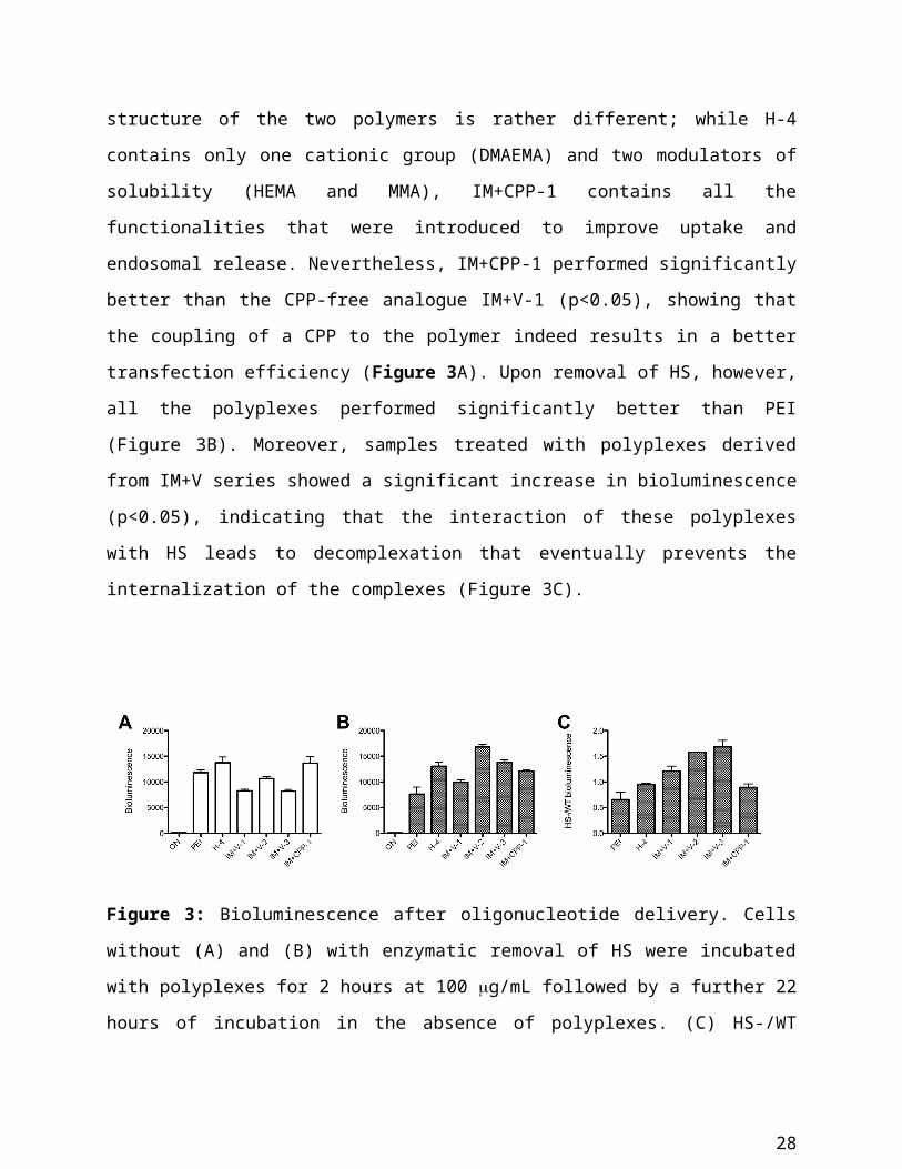
were introduced to improve uptake and endosomal release. Nevertheless, IM+CPP-1 performed
significantly better than the CPP-free analogue IM+V-1 (p<0.05), showing that the coupling of a
CPP to the polymer indeed results in a better transfection efficiency (Figure 3A). Upon removal
of HS, however, all the polyplexes performed significantly better than PEI (Figure 3B).
Moreover, samples treated with polyplexes derived from IM+V series showed a significant
increase in bioluminescence (p<0.05), indicating that the interaction of these polyplexes with HS
leads to decomplexation that eventually prevents the internalization of the complexes (Figure
3C).
18
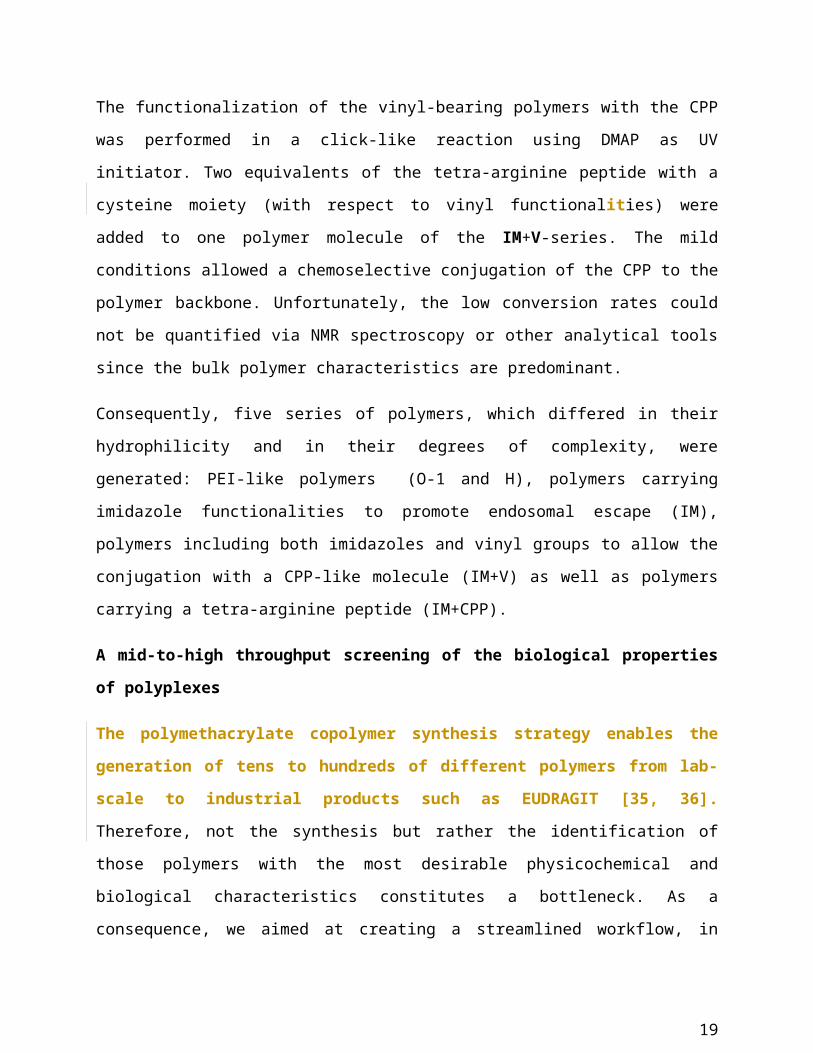
Figure 3: Bioluminescence after oligonucleotide delivery. Cells without (A) and (B) with
enzymatic removal of HS were incubated with polyplexes for 2 hours at 100 g/mL followed by
a further 22 hours of incubation in the absence of polyplexes. (C) HS-/WT bioluminescence
ratio. Samples were normalized to the total protein content (10 g/mL). Results are expressed as
mean ± SEM (n=4).
In order to address this hypothesis, polyplexes were tested for their stability in the presence of
heparin, a polyanion, structurally similar to heparan sulfate on the cell surface, that can compete
with the ON for the formation of complexes with the positively charged polymers. Polyplexes
were incubated for 30 minutes at heparin/polymer molar ratios of 0:1, 0.1:1, 0.5:1 and 1:1.
Immediately after incubation, size distributions were measured by DLS.
Polyplexes derived from H-4 and IM+CPP-1 formed stable complexes with the oligonucleotide
even in the presence of heparin (0.5:1), while IM+V-2 and IM+V-3 polyplexes underwent
decomplexation at a lower concentration of heparin (0.1:1). Polyplexes derived from IM+V-1
were stable at low concentration of heparin (0.1:1) but showed decomplexation at higher
concentrations (0.5:1) (Figure S3).
These results correlate with the observations in the transfection assays. Indeed, delivery
efficiency of H-4, IM+V-1 and IM+CPP-1 polyplexes did not change after removal of HS from
the cells, while IM+V-2 and IM+V-3 polyplexes performed significantly better (up to 1.8-fold)
in the absence of HS. Transfection efficiency for IM+V-1 polyplexes was affected less by HS
removal than the one of the more heparin-sensitive polyplexes.
19
While for the PEI polyplexes the impact of HS removal has been related to a reduced uptake, our
results identify HS-induced decomplexation as a critical factor, by which the glycocalyx may
have a negative impact on the biological activity of polyplexes.
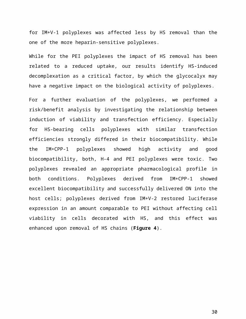
For a further evaluation of the polyplexes, we performed a risk/benefit analysis by investigating
the relationship between induction of viability and transfection efficiency. Especially for HS-
bearing cells polyplexes with similar transfection efficiencies strongly differed in their
biocompatibility. While the IM+CPP-1 polyplexes showed high activity and good
biocompatibility, both, H-4 and PEI polyplexes were toxic. Two polyplexes revealed an
appropriate pharmacological profile in both conditions. Polyplexes derived from IM+CPP-1
showed excellent biocompatibility and successfully delivered ON into the host cells; polyplexes
derived from IM+V-2 restored luciferase expression in an amount comparable to PEI without
affecting cell viability in cells decorated with HS, and this effect was enhanced upon removal of
HS chains (Figure 4).
Figure 4: Transfection efficiency vs. cell viability of polyplexes in (A) the presence or (B)
absence of HS chains. Mean values of three independent experiments.
Intracellular distribution of polyplexes
Having seen clear differences in the response of the polyplexes to HS removal, next we
addressed whether these characteristics translated into different degrees of colocalization of
20
polyplexes and HS during internalization. For this purpose, the colocalization of polyplexes
derived from H-4, the IM+V series and IM+CPP-1 with HS and endosomes was assessed by
confocal microscopy of HeLa cells, transiently transfected with a GFP-tagged Rab5 as a marker
for early endosomes. Heparan sulfate chains were stained by immunocytochemistry on living
cells. Cells were imaged by multichannel confocal microscopy.
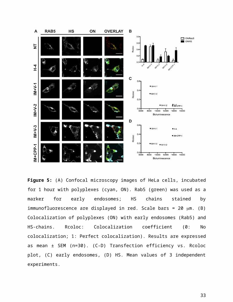
In control untreated cells, endosomes appeared as bright spots in the cytoplasm. Treatment of
cells with polyplexes led to a diffuse cytoplasmic distribution of fluorescence also of Rab5-GFP
(Figure 5A). However, quite interestingly, this dissipation of endosomal structures occurred at a
concentration of polyplexes that had shown only little toxicity and may be associated with
release of oligonucleotides into the cytoplasm.
The presence of polyplexes also affected the distribution of HS. While these sugars were
normally localized at the plasma membrane, the incubation with complexes led to an
internalization of HS, as demonstrated by the intracellular staining. Some polyplexes seemed to
enter the cells in complex with HS (H4, IM+V-1 and IM+CPP-1), while for IM+V-2 and IM+V-
3 colocalization of polyplexes with HS was low (Figure 5A and 5B).
21
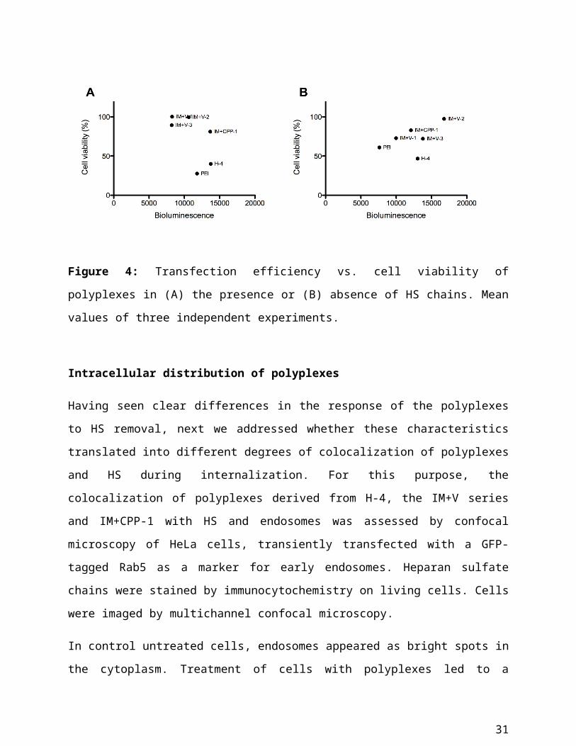
Figure 5: (A) Confocal microscopy images of HeLa cells, incubated for 1 hour with polyplexes
(cyan, ON). Rab5 (green) was used as a marker for early endosomes; HS chains stained by
immunofluorescence are displayed in red. Scale bars = 20 m. (B) Colocalization of polyplexes
(ON) with early endosomes (Rab5) and HS-chains. Rcoloc: Colocalization coefficient (0: No
colocalization; 1: Perfect colocalization). Results are expressed as mean ± SEM (n=30). (C-D)
Transfection efficiency vs. Rcoloc plot, (C) early endosomes, (D) HS. Mean values of 3
independent experiments.
To verify whether there was a relationship between transfection efficiency and cellular
localization, the bioluminescence values were plotted versus the degree of colocalization with
the endosomal marker and HS (Figure 5C-D and Supplement: Colocalization studies).
Consistent with expectations, polyplexes with the lowest endosomal colocalization showed the
22
highest transfection efficiency while there was no clear-cut correlation for the colocalization with
HS. Moreover, we observed that the highest degree of transfection was observed for those
polyplexes that showed low endosomal colocalization and a concomitant high HS colocalization
(H-4, IM+CPP-1).
Dose dependence of endosomal perturbation
As observed above, all the tested polyplexes entered cells after 2 hours of incubation at 100
g/mL. Nevertheless, at this concentration the uptake was always accompanied by a profound
perturbation of Rab5-positive endosomal structures. To assess whether this effect depended on
the concentration of the polyplexes, cells were incubated with IM+V-1- and IM+CPP-1-derived
polyplexes for 2 hours at the concentrations of 50 g/mL and 100 g/mL. These two polymers
were chosen because they clearly differed in their degree of colocalization with Rab5. The
concentrations were chosen based on pilot experiments, which had shown that below 50 g/mL
polyplexes did not show transfection efficiency and that the fluorescence of the ON was barely
detectable in confocal microscopy.
At 50 g/mL, incubation with IM+V-1 polyplexes caused the same perturbation of endosomal
structures as observed before. Although the structural changes were also prominent in cells
incubated with IM+CPP-1, at this concentration some defined endosomal structures could still be
observed (Figure S4). As before, incubation of cells with polyplexes at higher concentrations
caused massive endosomal perturbation (Figure S4). Even though we did not test lower
concentrations of IM+V-1 these data indicate that the polyplexes differ strongly in the dose
dependence of the structural changes of endosomes.
To understand the reason for the lack of toxicity in spite of the perturbation of endosomal
structures, we determined whether the observed changes were reversible. For this purpose, we
monitored the intracellular distribution of fluorescence for up to four hours after exposure of
cells to IM+CPP-1-based polyplexes, the polymer that showed the best risk/benefit ratio.
Indeed, after 1 hour recovery, punctate Rab5-positive structures reappeared and part of the ON-
associated fluorescence colocalized with these structures. However, a fraction of Cy5
fluorescence was located in the cytoplasm. After 2 hours recovery, Rab5-positive structures were
fully reconstituted, and, surprisingly, ON fluorescence was fully associated with them rather than
23
being homogenously distributed throughout the cytoplasm. Finally, after 4 hours recovery,
endosomes were still intact but ON-associated fluorescence was localized in the cytoplasm and
only partly in the endosomes (Figure 6). While endosomal release of ON was expected given the
observed activity of the delivered oligonucleotides, the initial dispersion of fluorescence was
more difficult to explain.
Figure 6: Reconstitution of endosomes (green) upon incubation with IM+CPP-1 polyplexes (100
g/mL, cyan, ON). Cells were allowed up to 4 hours recovery prior to imaging with confocal
microscopy at the indicated time points. Rec: Recovery. Scale bar = 20 m.
As a possible explanation, the polymer may overwhelm the endocytic machinery so that Rab5 is
associated with endocytic carriers that have not fused yet to form early endosomes. This idea was
supported by colocalisation experiments carried out with rhodamine-labelled dextran
(Supplement: Mechanism of endosomal perturbation).
24
DISCUSSION
In this study we aimed at establishing a mid-to-high-throughput screening strategy for drug
delivery polymers that would allow for a reliable selection of candidates for pre-clinical trials
and to understand molecular characteristics of active polymers. Current in vitro validation
strategies frequently lead to unsuccessful pre-clinical and clinical trials as they lack of critical
steps to predict the in vivo behavior of the tested compounds. High-throughput workflows for
gene delivery systems mainly focus on the optimization of transfection efficiency of polymers
synthesized in a high-throughput fashion [38, 39]. In some cases, the attention moves to the in
vivo biodistribution of gene delivery complexes [40]. However, a high-throughput method
should be employed for an early-stage screening of the crucial parameters that can limit the
application of a drug in clinical experimentation such as hemolysis and serum stability. A
parallel testing scheme with automated polyplex preparation and characterization, cytotoxicity
and transfection efficiency, was recently proposed [41]. The workflow presented here introduces
a logical order of testing steps starting with those that can be conducted robustly in high-
throughput and that have a high predictive power for pre-clinical safety (Scheme 2). Especially
for polymer collections of even higher complexity, the early stage elimination of candidates
based on clear cut-off criteria will be a prerequisite.
Of particular interest in the generation of a polymer collection, was the possibility to introduce
peptide-like functionalities into the polymer side chains. The inclusion of amines and peptide-
like functionalities during RAFT polymerization can be achieved thanks to the mild and
controlled conditions applied. In different reaction conditions, like the ones employed in anionic
or free radical polymerization [42], the presence of non-protected nucleophiles would lead to
unspecific reactions or chain termination and therefore require protecting group strategies with
subsequent deprotection (Scheme 1).
The inclusion of R4 showed an impact on the characteristic of the derived polyplexes, in terms of
both physicochemical and biological properties. In our approach, we conjugated the tetraarginine
via UV-mediated thiol-ene with a high yield. In spite of the high positive charge density of the
polymers, the inclusion of this class of molecules on the surface of drug delivery systems is
25
known to facilitate the cellular import of the latter, as shown for liposomes [43] and PEI [44].
Very likely this may be attributed to the specific characteristics of the guanidino group to engage
in interactions on the cell surface [18]. However, recently it has also been shown that a stretch of
arginines promotes uptake of polymers more efficiently than arginines dispersed over the
polymer [45]. Therefore, coupling of an oligoarginine should be superior to introduction of
guanidino functionalities directly into the polymer. The size of the complexes increased, while
there was no significant difference in their zeta-potentials when compared to that of polyplexes
generated from the non-functionalized polymers (Table S2). A similar zeta-potential suggests
that polyplexes would have a similar reactivity towards polyanions; however, their behaviors
towards heparin-mediated decomplexation differed strongly. This observation is in line with
what was described for CPP-siRNA oligoplexes [46]. The inclusion of the CPP functionality
promoted the formation of tighter complexes which showed high stability even at high
concentrations of heparin (Figure S3). The increase of particle size up to 600 nm in the presence
of serum proteins, may lead to undesired uptake by RES cells, as well limit the chance of taking
advantage of the enhanced permeation and retention effect [47, 48]. However, uptake by RES
cells, and particular macrophages, is mainly determined by the surface charge of the
particles, which mediates their coating by opsonines and the subsequent phagocytosis [49-
51]. The almost-neutral zeta-potential of the polyplexes should reduce the risk of opsonisation.
Moreover, the increase in size is possibly due to the formation of a protein corona around the
particles, which act as a protective coating and appear to be beneficial in terms of biodistribution
and cellular uptake [52, 53].
Remarkably, there was a strong correlation between polyanion sensitivity and impact of HS
removal on delivery efficiency. Polyplexes that showed a high sensitivity to heparin
decomplexation showed an increase in activity upon HS removal, while the transfection
efficiency of heparin-stable polyplexes was not affected by the presence of negatively charged
sugars (Figure 3). Moreover, the intracellular trafficking, and especially the colocalization with
HS during endocytosis, further supports this observation (Figure 5). Polyplexes which were
stable to the presence of heparin entered the cells as complexes with HS, while the colocalization
of heparin-sensitive polyplexes with HS was lost. These observations suggest that the evaluation
of decomplexation by heparin can be a good predictor for the activity of polyplexes and should
be therefore included into a screening workflow. Furthermore, there is still debate on the role of
26
HS on the cellular uptake of polyplexes [54]; our results demonstrate that this needs to be
determined for each indivdual case. Again, the induction of decomplexation is a molecular
mechanism that has not received sufficient attention so far.
The presence of R4 increased transfection efficiency and had an effect on the intracellular
trafficking of polyplexes. In particular, CPP-bearing polyplexes were poorly colocalized with
endosomes, whereas the complexes derived from CPP-free parent polymer were confined in
endocytic vesicles (Figure 5 and S5). For the CPP-bearing polyplexes, a clear, distinctive
trafficking was observed; upon endocytosis, first a highly disperse distribution of fluorescence
was observed. Analysis of the time dependence of uptake and counterstaining with dextran
revealed that this diffusive staining did not correspond to material released into the cytoplasm
but instead to highly dispersed endosomes (Figure S5). Only when the polymer was washed off,
clearly distinguishable endosomal structures reorganized from which endosomal release of
oligonucleotides occurred. The effect was dose dependent, as at lower concentration no
perturbation of the endosomes was observed. This sequence of events is different from the one
described by Rehman and coworkers for PEI polyplexes [55]. Here, uptake via clearly
distinguishable endosomes was followed by burst release of oligonucleotides. However, as also
shown here, PEI shows a stronger membrane-disruptive behaviour than the PMA polyplexes.
In establishing our workflow we limited ourselves to an N/P ratio of 3:1 in analogy to what
has been reported for PEI polyplexes, before. In particular when forming polyplexes with
ON of a different nature such as siRNA and plasmid DNA adaptations will be required as
polyplex formation depends on the type of ON. For plasmid DNA higher N/P ratios were
required to form polyplexes stable to serum (not shown).
CONCLUSION
In this study, we have applied a mid-to-high throughput approach to show that it is possible to
synthesize polymers suitable as gene delivery systems and to identify the optimal characteristics
to guarantee safety and effectiveness. Despite of the fact that the work was conducted on a
mid-throughput scale, the workflow can be considered as highly suited for “high-
throughput” as high-throughput-accessible read-outs were conducted in the initial phases
27
of the screening. The inclusion of side chains in the methacrylate polymer backbone that
facilitate the import of the polyplexes and mediate their endosomal release did not only result in
a considerable degree of transfection efficiency, but also contributed to a better toxicological
profile. However, the findings show that characteristics of the polyplexes do not follow
structure-activity relationships that can be interpreted in a straight-forward manner. The
proposed trial and error method can identify promising polymer samples out of polymer libraries
for detailed future in vitro and in vivo studies.
ACKNOWLEDGMENT
The authors thank the Dutch Polymer Institute (DPI, Technology area HTE, Project #730) for
financial support and Anne Spang and Turgay Yildirim for helpful discussions.
SUPPORTING INFORMATION
Supporting information is attached as a separate file.
REFERENCES
[1] X. Guo, L. Huang, Recent advances in nonviral vectors for gene delivery, Acc Chem Res, 45
(2012) 971-979.
[2] Z. Liu, M. Zheng, F. Meng, Z. Zhong, Non-viral gene transfection in vitro using endosomal
pH-sensitive reversibly hydrophobilized polyethylenimine, Biomaterials, 32 (2011) 9109-9119.
[3] F. Martello, M. Piest, J.F.J. Engbersen, P. Ferruti, Effects of branched or linear architecture
of bioreducible poly(amido amine)s on their in vitro gene delivery properties, J Control Release,
164 (2012) 372-379.
[4] D.W. Pack, A.S. Hoffman, S. Pun, P.S. Stayton, Design and development of polymers for
gene delivery, Nat Rev Drug Discov, 4 (2005) 581-593.
[5] A.K. Varkouhi, T. Lammers, R.M. Schiffelers, M.J. van Steenbergen, W.E. Hennink, G.
Storm, Gene silencing activity of siRNA polyplexes based on biodegradable polymers, Eur J
Pharm Biopharm, 77 (2011) 450-457.
[6] D.F. Baban, L.W. Seymour, Control of tumour vascular permeability, Advanced drug
delivery reviews, 34 (1998) 109-119.
[7] A.L. Koch, What size should a bacterium be? A question of scale, Annu Rev Microbiol, 50
(1996) 317-348.
28
[8] M. Longmire, P.L. Choyke, H. Kobayashi, Clearance properties of nano-sized particles and
molecules as imaging agents: considerations and caveats, Nanomedicine (Lond), 3 (2008) 703-
717.
[9] K.A. Mislick, J.D. Baldeschwieler, Evidence for the role of proteoglycans in cation-mediated
gene transfer, Proc Natl Acad Sci U S A, 93 (1996) 12349-12354.
[10] C.K. Payne, S.A. Jones, C. Chen, X. Zhuang, Internalization and trafficking of cell surface
proteoglycans and proteoglycan-binding ligands, Traffic, 8 (2007) 389-401.
[11] M. Ruponen, S. Ronkko, P. Honkakoski, J. Pelkonen, M. Tammi, A. Urtti, Extracellular
glycosaminoglycans modify cellular trafficking of lipoplexes and polyplexes, J Biol Chem, 276
(2001) 33875-33880.
[12] H. Karanth, R.S.R. Murthy, pH-sensitive liposomes--principle and application in cancer
therapy, J Pharm Pharmacol, 59 (2007) 469-483.
[13] J.S. Rossman, G.P. Leser, R.A. Lamb, Filamentous influenza virus enters cells via
macropinocytosis, J Virol, 86 (2012) 10950-10960.
[14] A.M. Funhoff, C.F. van Nostrum, M.C. Lok, J.A.W. Kruijtzer, D.J.A. Crommelin, W.E.
Hennink, Cationic polymethacrylates with covalently linked membrane destabilizing peptides as
gene delivery vectors, J Control Release, 101 (2005) 233-246.
[15] M. Koping-Hoggard, K.M. Varum, M. Issa, S. Danielsen, B.E. Christensen, B.T. Stokke, P.
Artursson, Improved chitosan-mediated gene delivery based on easily dissociated chitosan
polyplexes of highly defined chitosan oligomers, Gene Ther, 11 (2004) 1441-1452.
[16] H.Y. Nam, J. Kim, S. Kim, J.W. Yockman, S.W. Kim, D.A. Bull, Cell penetrating peptide
conjugated bioreducible polymer for siRNA delivery, Biomaterials, 32 (2011) 5213-5222.
[17] F. Milletti, Cell-penetrating peptides: classes, origin, and current landscape, Drug Discov
Today, 17 (2012) 850-860.
[18] N. Sakai, S. Matile, Anion-mediated transfer of polyarginine across liquid and bilayer
membranes, J Am Chem Soc, 125 (2003) 14348-14356.
[19] E. Lai, J.H. van Zanten, Monitoring DNA/poly-L-lysine polyplex formation with time-
resolved multiangle laser light scattering, Biophys J, 80 (2001) 864-873.
[20] K.-L. Chang, Y. Higuchi, S. Kawakami, F. Yamashita, M. Hashida, Efficient gene
transfection by histidine-modified chitosan through enhancement of endosomal escape,
Bioconjug Chem, 21 (2010) 1087-1095.
29
[21] D.J. Schibli, R.F. Epand, H.J. Vogel, R.M. Epand, Tryptophan-rich antimicrobial peptides:
comparative properties and membrane interactions, Biochem Cell Biol, 80 (2002) 667-677.
[22] C. Cheng, A.J. Convertine, P.S. Stayton, J.D. Bryers, Multifunctional triblock copolymers
for intracellular messenger RNA delivery, Biomaterials, 33 (2012) 6868-6876.
[23] N. Van Overstraeten-Schlogel, Y.-H. Shim, V. Tevel, G. Piel, J. Piette, P. Dubois, M. Raes,
Assessment of new biocompatible poly(N-(morpholino)ethyl methacrylate)-based copolymers by
transfection of immortalized keratinocytes, Drug Deliv, 19 (2012) 112-122.
[24] Y. Hu, D. Zhou, C. Li, H. Zhou, J. Chen, Z. Zhang, T. Guo, Gene delivery of PEI
incorporating with functional block copolymer via non-covalent assembly strategy, Acta
Biomater, 9 (2013) 5003-5012.
[25] A. Nouri, R. Castro, V. Kairys, J.L. Santos, J. Rodrigues, Y. Li, H. Tomas, Insight into the
role of N,N-dimethylaminoethyl methacrylate (DMAEMA) conjugation onto
poly(ethylenimine): cell viability and gene transfection studies, J Mater Sci Mater Med, 23
(2012) 2967-2980.
[26] Y. Qian, Y. Zha, B. Feng, Z. Pang, B. Zhang, X. Sun, J. Ren, C. Zhang, X. Shao, Q. Zhang,
X. Jiang, PEGylated poly(2-(dimethylamino) ethyl methacrylate)/DNA polyplex micelles
decorated with phage-displayed TGN peptide for brain-targeted gene delivery, Biomaterials, 34
(2013) 2117-2129.
[27] R.N. Palumbo, X. Zhong, D. Panus, W. Han, W. Ji, C. Wang, Transgene expression and
local tissue distribution of naked and polymer-condensed plasmid DNA after intradermal
administration in mice, J Control Release, 159 (2012) 232-239.
[28] J. Chiefari, Y.K. Chong, F. Ercole, J. Krstina, J. Jeffery, T.P.T. Le, R.T.A. Mayadunne, G.F.
Meijs, C.L. Moad, G. Moad, E. Rizzardo, S.H. Thang, Living free-radical polymerization by
reversible addition-fragmentation chain transfer: The RAFT process, Macromolecules, 31 (1998)
5559-5562.
[29] M.J. Manganiello, C. Cheng, A.J. Convertine, J.D. Bryers, P.S. Stayton, Diblock
copolymers with tunable pH transitions for gene delivery, Biomaterials, 33 (2012) 2301-2309.
[30] A. Mathew, H. Cao, E. Collin, W. Wang, A. Pandit, Hyperbranched PEGmethacrylate linear
pDMAEMA block copolymer as an efficient non-viral gene delivery vector, Int J Pharm, 434
(2012) 99-105.
30
[31] S.H. Kang, M.J. Cho, R. Kole, Up-regulation of luciferase gene expression with antisense
oligonucleotides: implications and applications in functional assay development, Biochemistry,
37 (1998) 6235-6239.
[32] M.M. Bradford, A rapid and sensitive method for the quantitation of microgram quantities
of protein utilizing the principle of protein-dye binding, Anal Biochem, 72 (1976) 248-254.
[33] T.H. van Kuppevelt, M.A. Dennissen, W.J. van Venrooij, R.M. Hoet, J.H. Veerkamp,
Generation and application of type-specific anti-heparan sulfate antibodies using phage display
technology. Further evidence for heparan sulfate heterogeneity in the kidney, J Biol Chem, 273
(1998) 12960-12966.
[34] K. Kempe, R. Hoogenboom, U.S. Schubert, A Green Approach for the Synthesis and Thiol-
ene Modification of Alkene Functionalized Poly(2-oxazoline)s, Macromol Rapid Comm, 32
(2011) 1484-1489.
[35] C.R. Becer, S. Hahn, M.W.M. Fijten, H.M.L. Thijs, R. Hoogenboom, U.S. Schubert,
Libraries of Methacrylic Acid and Oligo(ethylene glycol) Methacrylate Copolymers with LCST
Behavior, J Polym Sci Pol Chem, 46 (2008) 7138-7147.
[36] T.M. Eggenhuisen, C.R. Becer, M.W.M. Fijten, R. Eckardt, R. Hoogenboom, U.S.
Schubert, Libraries of statistical hydroxypropyl acrylate containing copolymers with LCST
properties prepared by NMP, Macromolecules, 41 (2008) 5132-5140.
[37] X.-h. Luo, F.-w. Huang, S.-y. Qin, H.-f. Wang, J. Feng, X.-z. Zhang, R.-x. Zhuo, A strategy
to improve serum-tolerant transfection activity of polycation vectors by surface hydroxylation,
Biomaterials, 32 (2011) 9925-9939.
[38] S.E. How, B. Yingyongnarongkul, M.A. Fara, J.J. Diaz-Mochon, S. Mittoo, M. Bradley,
Polyplexes and lipoplexes for mammalian gene delivery: from traditional to microarray
screening, Comb Chem High Throughput Screen, 7 (2004) 423-430.
[39] L. Li, F. Wang, Y. Wu, G. Davidson, P.A. Levkin, Combinatorial synthesis and high-
throughput screening of alkyl amines for nonviral gene delivery, Bioconjug Chem, 24 (2013)
1543-1551.
[40] I.J. Hildebrandt, M. Iyer, E. Wagner, S.S. Gambhir, Optical imaging of transferrin targeted
PEI/DNA complexes in living subjects, Gene Ther, 10 (2003) 758-764.
[41] A.C. Rinkenauer, A. Vollrath, A. Schallon, L. Tauhardt, K. Kempe, S. Schubert, D. Fischer,
U.S. Schubert, Parallel high-throughput screening of polymer vectors for nonviral gene delivery:
31
evaluation of structure-property relationships of transfection, ACS Comb Sci, 15 (2013) 475-
482.
[42] T. Janoschka, A. Teichler, A. Krieg, M.D. Hager, U.S. Schubert, Polymerization of free
secondary amine bearing monomers by RAFT polymerization and other controlled radical
techniques, J Polym Sci Pol Chem, 50 (2012) 1394-1407.
[43] D. Cai, W. Gao, B. He, W. Dai, H. Zhang, X. Wang, J. Wang, X. Zhang, Q. Zhang,
Hydrophobic penetrating peptide PFVYLI-modified stealth liposomes for doxorubicin delivery
in breast cancer therapy, Biomaterials, 35 (2014) 2283-2294.
[44] Y. Hu, B. Xu, Q. Ji, D. Shou, X. Sun, J. Xu, J. Gao, W. Liang, A mannosylated cell-
penetrating peptide-graft-polyethylenimine as a gene delivery vector, Biomaterials, (2014).
[45] K. Koschek, M. Dathe, J. Rademann, Effects of charge and charge distribution on the
cellular uptake of multivalent arginine-containing peptide-polymer conjugates, Chembiochem,
14 (2013) 1982-1990.
[46] A.H. van Asbeck, A. Beyerle, H. McNeill, P.H. Bovee-Geurts, S. Lindberg, W.P.
Verdurmen, M. Hallbrink, U. Langel, O. Heidenreich, R. Brock, Molecular parameters of
siRNA--cell penetrating peptide nanocomplexes for efficient cellular delivery, ACS nano, 7
(2013) 3797-3807.
[47] H. Maeda, J. Wu, T. Sawa, Y. Matsumura, K. Hori, Tumor vascular permeability and the
EPR effect in macromolecular therapeutics: a review, J Control Release, 65 (2000) 271-284.
[48] L. Novo, L.Y. Rizzo, S.K. Golombek, G.R. Dakwar, B. Lou, K. Remaut, E. Mastrobattista,
C.F. van Nostrum, W. Jahnen-Dechent, F. Kiessling, K. Braeckmans, T. Lammers, W.E.
Hennink, Decationized polyplexes as stable and safe carrier systems for improved biodistribution
in systemic gene therapy, J Control Release, 195 (2014) 162-175.
[49] D. Finsinger, J.S. Remy, P. Erbacher, C. Koch, C. Plank, Protective copolymers for nonviral
gene vectors: synthesis, vector characterization and application in gene delivery, Gene Ther, 7
(2000) 1183-1192.
[50] M. Ogris, S. Brunner, S. Schuller, R. Kircheis, E. Wagner, PEGylated DNA/transferrin-PEI
complexes: reduced interaction with blood components, extended circulation in blood and
potential for systemic gene delivery, Gene Ther, 6 (1999) 595-605.
32
[51] C. Plank, K. Mechtler, F.C. Szoka, Jr., E. Wagner, Activation of the complement system by
synthetic DNA complexes: a potential barrier for intravenous gene delivery, Human gene
therapy, 7 (1996) 1437-1446.
[52] J. Rejman, V. Oberle, I.S. Zuhorn, D. Hoekstra, Size-dependent internalization of particles
via the pathways of clathrin- and caveolae-mediated endocytosis, The Biochemical journal, 377
(2004) 159-169.
[53] G. Caracciolo, L. Callipo, S.C. De Sanctis, C. Cavaliere, D. Pozzi, A. Lagana, Surface
adsorption of protein corona controls the cell internalization mechanism of
DC-Chol-DOPE/DNA lipoplexes in serum, Biochimica et biophysica acta, 1798 (2010) 536-543.
[54] M.E. Favretto, R. Wallbrecher, S. Schmidt, R. van de Putte, R. Brock, Glycosaminoglycans
in the cellular uptake of drug delivery vectors - Bystanders or active players?, J Control Release,
180C (2014) 81-90.
[55] Z. ur Rehman, D. Hoekstra, I.S. Zuhorn, Mechanism of polyplex- and lipoplex-mediated
delivery of nucleic acids: real-time visualization of transient membrane destabilization without
endosomal lysis, ACS nano, 7 (2013) 3767-3777.
Supporting Information
33
Multifunctional poly(methacrylate) polyplexe libraries: A platform for gene delivery inspired by natureMarco Emanuele Favretto, Andreas Krieg, Stephanie Schubert, Ulrich S. Schubert, Roland Brock*
Table S1: Composition of methacrylate polymers represented as molar (%) fraction of each monomer (determined by 1 H NMR) with the exception of the CPP and the corresponding polymer characteristics (molar masses and polydispersity indexes as determined by SEC).
Polymer DMAEMA HEMA OEGMA MMA ImEtMA BEMA CPP Mn
(g/mol)
PDI
H-1 50 50 19,800 1.24
H-2 50 40 10 16,000 1.29
H-3 50 30 20 18,000 1.26
H-4 50 10 40 16,000 1.25
O-1 50 50 33,300 1.21
IM-1 65 21 13 30,600 1.19
IM-2 61 15 24 24,400 1.27
IM-3 57 9 34 23,800 1.23
IM-4 53 4 43 34,400 1.34
IM+V-1 62 20 12 6 30,800 1.28
IM+V-2 57 14 23 6 30,800 1.29
IM+V-3 51 4 40 5 24,000 1.35
IM+CPP-1 62 20 12 6 R4
IM+CPP-2 57 14 23 6 R4
IM+CPP-3 51 4 40 5 R4
34
Table S2: Size and polydispersity index of polyplexes in water or in RPMI+10% fetal calf serum. Zeta-potential of serum-stable polyplexes. [sd]: standard deviation, n.d.: no complex was detected
Polymer Size in H2O (nm) [sd]
PDI in H2O
Size in serum (nm) [sd]
PDI in serum
Zeta-potential (mV) [sd]
O-1 n.d. n.d.
H-1 238 [6] 0.118 n.d.
H-2 203 [8] 0.131 n.d.
H-3 n.d. n.d.
H-4 128 [8] 0.126 299 [10] 0.398 4.1 [0.8]
IM-1 262 [17] 0.213 886 [26] 0.389
IM-2 312 [14] 0.184 1106 [20] 0.114
IM-3 343 [22] 0.239 1257 [77] 0.498
IM-4 403 [40] 0.256 1245 [147] 0.527
IM+V-1 183 [14] 0.222 354 [17] 0.227 3.2 [1.2]
IM+V-2 191 [18] 0.254 387 [17] 0.242 4.6 [0.1]
IM+V-3 228 [21] 0.246 360 [21] 0.205 5.3 [1.0]
IM+CPP-1 323 [14] 0.160 572 [56] 0.342 4.2 [0.6]
IM+CPP-2 327 [12] 0.176 495 [22] 0.248 4.5 [0.6]
IM+CPP-3 219 [15] 0.184 347 [26] 0.293 5.7 [0.9]
Branched PEI 197 [13] 0.156 228 [28] 0.286 3.6 [0.7]
35
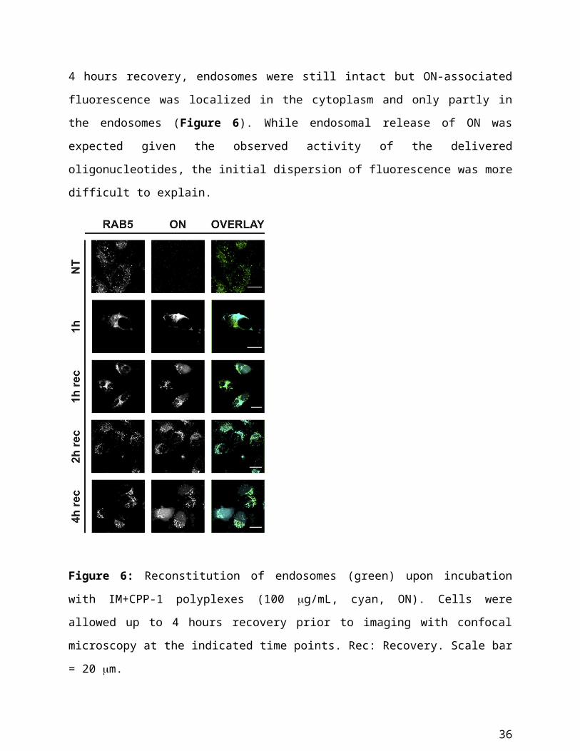
Figure S1: 1 H NMR spectrum of IM+V-2 in CDCl3 including assignments of the peaks
36
Figure S2: Bioluminescence after incubation with polyplexes for 2 hours at 10, 25 and 50 g/mL. Samples were normalized to the total protein content (10 g/mL). Cell lysis was performed 24 hours post-exposure incubation. Results are expressed as mean ± SEM (n=3)
Figure S3: DLS volume graphs of (A) H-4, (B) IM+V-1, (C) IM+V-2, (D) IM+V-3 and (E) IM+CPP-1 polyplexes, in the presence of 10% FCS and different heparin/ON molar ratios. One representative experiment is shown for each condition. (F) Size of polyplexes incubated in serum at increasing concentrations of heparin. Results are expressed as mean ± SEM (n=3).
Colocalization studies
Colocalization of polyplexes with HS and endosomes was assessed by image analysis. In particular, colocalization coefficients (Rcoloc) were calculated on at least 30 cells per condition. There was no clear-cut relationship between polymer structure/composition and localization with cell compartments: H-4, which lacks imidazoles that can trigger endosomal escape, showed lower colocalization with Rab5 than IM+V-3, in which the molar fraction of imidazoles is approximately 40%. Moreover, polyplexes with identical imidazole content (IM+V-1 and IM+CPP-1) showed a different degree of colocalization with Rab5 (Figure 5A). However, due to the diffuse staining of Rab5, it was difficult to interpret these various degrees of colocalization with respect to degrees of endosomal localization.
Consistent with endosomal release as a requirement for activity bioluminescence was higher for those polyplexes (H-4, IM+V-2 and IM+CPP-1), which showed the lowest colocalization with Rab5. On the other hand, transfection efficiency did not correlate with the colocalization with HS (Figure 5C and 5D). However, there seemed to be a correlation between colocalization with HS and the impact of HS removal on uptake of individual polyplexes. For polyplexes, which showed
37
high or moderate colocalization with HS (H-4, IM+V-1 and IM+CPP-1), the removal of HS did not have a major impact on transfection efficiency. On the other hand, polyplexes that were poorly colocalized with HS promoted a higher gene correction upon HS removal and were decomplexed even at low concentrations of heparin.
Dose dependence of endosomal perturbation
Figure S4: Confocal microscopy images of HeLa cells, incubated with polyplexes (cyan, ON), for 2 hours at the concentrations of 50 g/mL and 100 g/mL. Rab5-GFP (green) was used as a marker for early endosomes. Scale bar= 20 m.
Mechanism of endosomal perturbation
To test the hypothesis that the homogenous distribution of fluorescence was a result of a dispersion of endosomal vesicles rather than membrane rupture, we investigated whether other endocytic mechanisms were also affected, including fluid phase uptake. Rhodamine B-labeled
38
10,000 g/mol dextran as a marker for macropinocytosis was co-incubated (1 mg/mL) with the polyplexes derived from IM+CPP-1 (100 g/mL) on HeLa cells transfected with Rab5-GFP. After incubation for 1 hour, the distribution of fluorescence was imaged by confocal microscopy straight after incubation and after 1, 2 and 3 hours of recovery. In the absence of polyplexes, dextran-positive macropinosomes were only partly colocalized with Rab5-positive early endosomes. Consistent with a dispersion of endosomal structures, most of the dextran fluorescence was localized homogenously throughout the cytoplasm and the nucleus. Also, fluorescence of polyplexes was distributed homogeneously throughout the cytoplasm and some moderate nuclear staining could be observed. Cellular distribution changed over time. After 1 hour recovery, distinct endocytic structures reappeared, as shown by the more punctuated staining for Rab5 and dextran. Both dextran and polyplexes were highly colocalized with these vesicles; however, their fluorescence was also detected in the cytoplasm and nucleus. After a 2 hour recovery, endocytic vesicles were clearly detectable and contained both dextran and polyplexes, as demonstrated by the high degree of colocalization. Finally, after 3 hours of recovery, next to the endosomal structures Cy5-fluorescence was again visible in the cytoplasm and nucleus (Figure S4). Remarkably, this endosomal escape was not observed for the dextran-associated fluorescence. These results support the idea that endosomes were not properly formed due to the saturation of endocytic capacity of the cells and rather were in a constant sorting state. Cells require at least 1 hour recovery in the absence of polyplexes to regulate this process and allow the formation of typical endocytic vesicles. However, ultimately endosomal release of oligonucleotides occurs.
39
Figure S5: Effect of polyplexes on endosomal uptake of dextran. Rab5-GFP (green) expressing HeLa cells, incubated with polyplexes (100 g/mL, cyan, ON) and rhodamine-labeled dextran (1 mg/mL, red) were imaged with confocal microscopy at the indicated time points. Rec: recovery. Scale bar = 20 m.
40