Embed Size (px)
Citation preview

128
CASE REPORT
This is an open-access article distributed under the terms of the Creative Commons Attribution Non-Commercial License (http://creativecommons.org/licenses/by-nc/4.0/), which permits unrestricted non-commercial use, distribution, and reproduction in any medium, provided the original work is properly cited.
CC
Temporomandibular joint reconstruction with costochondral graft: case series study
Moon Gi Choi
Department of Oral and Maxillofacial Surgery, College of Dentistry, Wonkwang University, Iksan, Korea
Abstract (J Korean Assoc Oral Maxillofac Surg 2021;47:128-134)
Various techniques have been used to reconstruct the temporomandibular joints, including autogenous transplants and alloplastic implants. Among au-togenous grafts, costochondral grafts have mainly been used. A costochondral graft has many advantages over other autogenous grafts and alloplastic implants. Harvest is easy and has minimal impact on patients. The graft can bear functional load well and biocompatibility is excellent. A costochon-dral graft obviates foreign body reactions and further surgery for revision of alloplastic replacements if the graft takes well. Although long-term prog-nosis remains unclear, it appears that for autogenous condylar reconstruction, costochondral grafts can be used with few complications and acceptable results. This article describes cases and discusses surgical techniques and considerations related to costochondral grafts.
Key words: Costochondral graft, Temporomandibular joint, Autogenous graft[paper submitted 2020. 9. 8 / revised 2020. 10. 26 / accepted 2020. 11. 16]
Copyright © 2021 The Korean Association of Oral and Maxillofacial Surgeons. All rights reserved.
https://doi.org/10.5125/jkaoms.2021.47.2.128pISSN 2234-7550 · eISSN 2234-5930
I. Introduction
The indications for reconstruction of the temporomandibu-lar joint (TMJ) include ankylosis, end-stage osteoarthritis, rheumatoid arthritis, tumor of condyle, traumatic deficit, and congenital pathology1.
Various methods have been used to reconstruct the TMJ including autogenous transplants and alloplastic implants. Among the autogenous grafts, fibular, metatarsal, sterno-clavicular, iliac, and costochondral grafts have been used. Among the alloplastic grafts, total TMJ system is most wide-ly used1.
Costochondral transplantation has many advantages over other autogenous transplants. Harvesting is easy and has minimal impact on patients. The transplant bears functional loading well and has excellent biocompatibility. A costochon-
dral graft in children has the potential for growth. Despite these advantages, there are many potential problems. Costo-chondral grafts can be fractured and ankylosis can occur after transplantation. In addition, such a graft can show various growth behaviors. Although donor site morbidity is minimal, the wound can be bothersome to patients2,3.
II. Cases Report
Here, 4 patients with 5 operated joints were retrospectively reviewed. All patients were adults and received a costochon-dral graft at Wonkwang Dental Hospital due to diverse causes from 2005 to 2020. Issues regarding function, pain, long-term results, and adverse outcomes are presented.
1. Surgical techniques
The surgical field was opened through preauricular and/or submandibular incision. To obtain a definite surgical field, we used both preauricular and submandibular approaches in the first few cases. However, as our experience increased, only one of the approaches was used.
Costochondral graft was taken from the right chest at the 5th, 6th, and 7th rib bones through inframammary incision. The reason for collecting graft from the right side was to dis-
Moon Gi ChoiDepartment of Oral and Maxillofacial Surgery, College of Dentistry, Wonkwang University, 460 Iksan-daero, Iksan 54538, KoreaTEL: +82-63-859-2921E-mail: [email protected]: https://orcid.org/0000-0003-3502-7652

TMJ reconstruction with costochondral graft
129
tinguish postoperative pain from cardiac pain. If two grafts were harvested for bicondylar reconstruction, tissue was col-lected from separate rib bones to prevent sinking of the tho-
racic cage, leaving intact rib bone between them. Such a graft was generally about 5 cm in length, and the cartilage portion was trimmed to 2-3 mm in thickness.
Only if the TMJ disc was degenerated was it removed. Graft was pushed upward to contact the disc or articular sur-face loosely in centric occlusion and fixed to the ramus with a washer technique using a miniplate and bicortical screws. The minimum number of screws was three, and the graft had to be fixed with no change in occlusion. Condylar movement and occlusion were measured at mouth closed and open posi-tions. The mouth opening in the operation room had to be 3.0-3.5 cm in length after reconstruction.
As mouth opening increased, the end portion of the cos-tochondral graft might move along the articular surface and eminence. If mouth opening was not improved, unilateral or bilateral condylectomy was performed to secure the ideal opening.
In our first cases, intermaxillary fixation for bony consoli-dation was used for three weeks. As our experience increased, we ceased use of intermaxillary fixation. Instead, active opening exercises were scheduled for several months.
9 cm
Fig. 1. On computed tomography, bilateral intracapsular condylar fractures were observed.Moon Gi Choi: Temporomandibular joint reconstruction with costochondral graft: case series study. J Korean Assoc Oral Maxillofac Surg 2021
A B
Fig. 2. A. Trimmed end portion of the costochondral graft loosely contacted the temporomandibular joint in the centric occlusion state. B. Trimmed end portion of the costochondral graft moved along the articular fossa as mouth opening increased.Moon Gi Choi: Temporomandibular joint reconstruc-tion with costochondral graft: case series study. J Korean Assoc Oral Maxillofac Surg 2021
9 cm
A B C
9 cm 7 cm
Fig. 3. A, B. On three-dimensional computed tomography, the costochondral graft was fixed in a good position. C. On coronal plane view, costochondral grafts were positioned on the lateral surface of the articular fossa.Moon Gi Choi: Temporomandibular joint reconstruction with costochondral graft: case series study. J Korean Assoc Oral Maxillofac Surg 2021

J Korean Assoc Oral Maxillofac Surg 2021;47:128-134
130
2. Case 1
A 48-year-old female patient presented with bilateral con-dylar fractures due to a fall. Both fractures were within the capsule (level II fractures).(Fig. 1)
Initial mouth opening was limited due to pain. She was partially edentulous in both jaws and was wearing a partial denture, which was broken during the fall. She showed loss of vertical dimension. Because the fractured fragments were too small to fix, we planned their removal and bilateral con-dylar reconstruction using costochondral grafts. Temporary denture was fabricated for proper occlusion and vertical dimension. Two costochondral grafts were harvested from the 5th and 7th rib bones of the right chest through inframa-mmary incision. The cartilage portion was trimmed to 2-3 mm in thickness. Both TMJs were approached through the preauricular route. Both TMJ discs were intact without tear-ing or perforation. Each graft was pushed upward to contact the disc slightly and fixed with the washer technique with a temporary denture. Condylar movement and occlusion were measured on mouth closing and at open positions.(Fig. 2)
Intermaxillary fixation for bony consolidation was used for three weeks following surgery, and active opening exercises were prescribed for the three months after that. On three-dimensional computed tomography (CT), grafts were con-tacting a lateral surface of the articular fossa.(Fig. 3)
Shortly after operation, the patient complained of an un-comfortable itching sensation at the donor site stimulated by the bra strap.
The patient was followed for 10 years. At last examination, she was free of TMJ pain and donor site complications and showed near normal mandibular excursion and protrusion. Maximum mouth opening was 40 mm, right and left excur-
sion was about 7 mm, and protrusive movement was about 6 mm.(Fig. 4) There was no facial asymmetry. Though Frey’s syndrome had developed, she refused treatment. She showed no sensory nerve damage around the TMJ.
3. Case 2
A 48-year-old male patient visited the emergency room due to painful swelling on the right hemiface. On CT, there was abscess formation and bony destruction around the right con-dyle and ramus area.(Fig. 5) The patient was diagnosed with submasticatory space abscess and acute suppurative osteomy-elitis on the right condyle and ramus.
At first operation, incision and drainage and surgical debridement were performed through a submandibular ap-proach. After three weeks, the infection was controlled. A second operation was performed for condyle and ramus re-construction with a right 6th rib costochondral graft.
Submandibular incision was used without preauricular ap-proach. The disc of the TMJ was degenerated and removed. Costal cartilage was trimmed to 2-3 mm in thickness, and the graft was fixed with plate and screw fixation and wiring.
Intermaxillary fixation was used for three weeks after sur-gery. After releasing the intermaxillary fixation, active mouth opening exercise with therabite was performed for three months. The patient was followed up for six months, at which maximum mouth opening was 35 mm, protrusion was 7 mm,
Fig. 4. At the last visit, the patient showed near normal mandibu-lar excursion and protrusion.Moon Gi Choi: Temporomandibular joint reconstruction with costochondral graft: case series study. J Korean Assoc Oral Maxillofac Surg 2021
10 cm
Fig. 5. On computed tomography, submasticatory space abscess and bony destruction were observed around the condyle and ra-mus.Moon Gi Choi: Temporomandibular joint reconstruction with costochondral graft: case series study. J Korean Assoc Oral Maxillofac Surg 2021
TMJ reconstruction with costochondral graft
131
left excursion was 1 mm, and right excursion was 7 mm.On opening, the mandible deviated to the right side due to
limited movement of the right neo-condyle, but centric occlu-sion was obtained.
The patient was free of pain and donor site complications. He died of unrelated vascular accident eight months after op-eration.
4. Case 3
A 17-year-old male patient suffered a facial bone fracture in a car accident. The diagnosis was bilateral intracapsular condylar fractures and symphysis fracture.(Fig. 6)
The right condyle was intact enough to be repaired, but the left condyle was fractured into multiple pieces. Our plan was open reduction and fixation of the right condyle and symphy-sis and reconstruction with a costochondral graft for the left
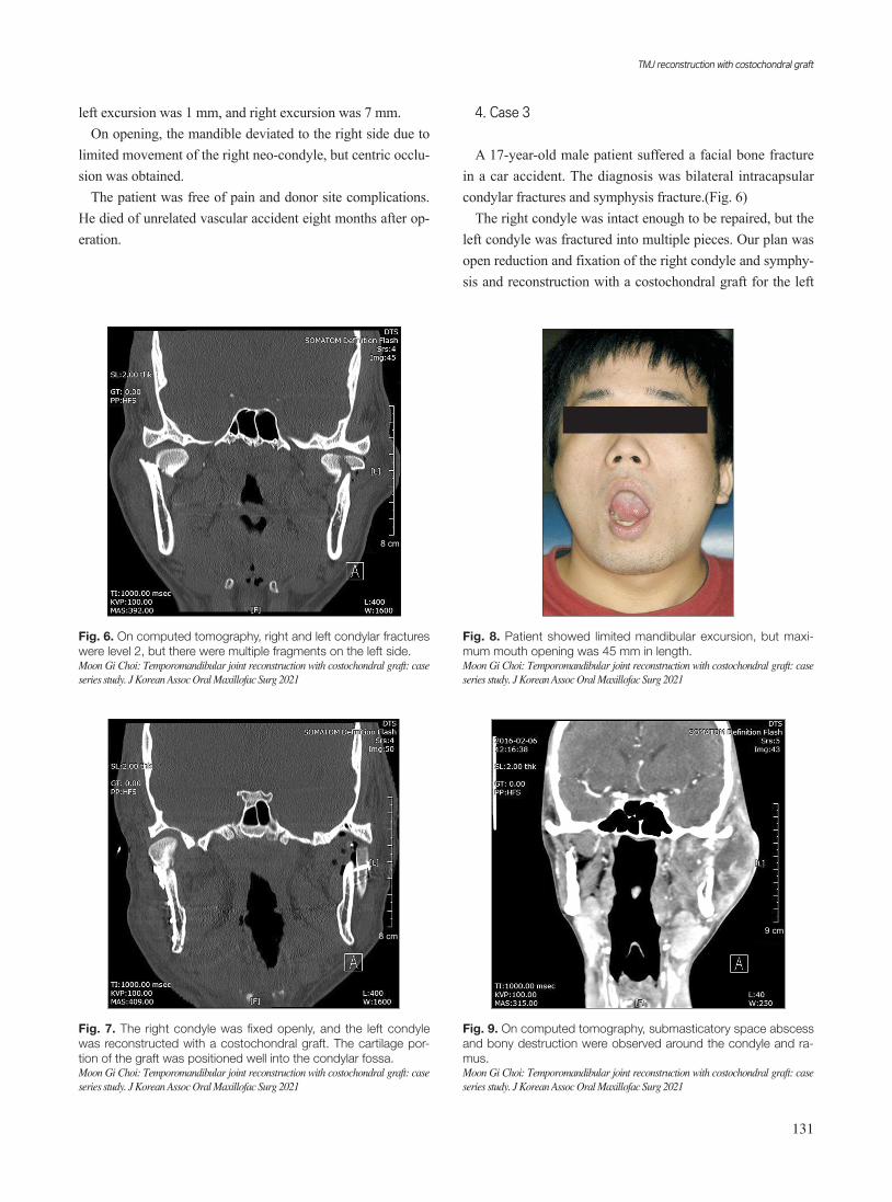
Fig. 8. Patient showed limited mandibular excursion, but maxi-mum mouth opening was 45 mm in length.Moon Gi Choi: Temporomandibular joint reconstruction with costochondral graft: case series study. J Korean Assoc Oral Maxillofac Surg 2021
9 cm
Fig. 9. On computed tomography, submasticatory space abscess and bony destruction were observed around the condyle and ra-mus.Moon Gi Choi: Temporomandibular joint reconstruction with costochondral graft: case series study. J Korean Assoc Oral Maxillofac Surg 2021
8 cm
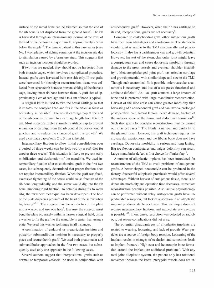
Fig. 7. The right condyle was fixed openly, and the left condyle was reconstructed with a costochondral graft. The cartilage por-tion of the graft was positioned well into the condylar fossa.Moon Gi Choi: Temporomandibular joint reconstruction with costochondral graft: case series study. J Korean Assoc Oral Maxillofac Surg 2021
8 cm
Fig. 6. On computed tomography, right and left condylar fractures were level 2, but there were multiple fragments on the left side.Moon Gi Choi: Temporomandibular joint reconstruction with costochondral graft: case series study. J Korean Assoc Oral Maxillofac Surg 2021

J Korean Assoc Oral Maxillofac Surg 2021;47:128-134
132
condyle. A 5 cm length of costochondral graft was harvested from the right chest, and cartilage was trimmed to 2-3 mm in thickness. Surgical approach was both preauricular and submandibular. The graft was fixed with a plate and screws using a washer technique.(Fig. 7)
The patient was followed up for 4 years 3 months. Maxi-mum mouth opening was 45 mm with 5 mm left deviation, protrusion was 4 mm with 2 mm left deviation, left excursion was 2 mm, and right excursion was 4 mm.(Fig. 8)
On opening, the mandible deviated to the right side, but centric occlusion was obtained. The patient complained of intermittent pain and swelling on the left TMJ. An anti-inflammatory drug reduced these problems.
5. Case 4
A 48-year-old male patient visited the emergency room due to painful swelling on the left hemiface. On CT, there was an abscess formation in the submasticatory space and bony destruction around the left condyle and ramus area.(Fig. 9) The patient was diagnosed with submasticatory space abscess and acute suppurative osteomyelitis of the left condyle and ramus. The patient was partially edentulous.
After infection was controlled, the left condyle and ramus were to be reconstructed with a costochondral graft after re-moval of the destroyed bony mass. To maintain the vertical dimension, upper and lower partial dentures were fabricated on an articulator. The surgical approach was submandibular and extended preauricular. Despite severe infection, the disc was intact. The washer technique was used for graft fixation. During the operation, neocondylar movement was good.
The patient was followed up for two months. Maximum mouth opening was 50 mm without deviation, and protrusion was 4 mm without deviation. Left excursion was 1 mm, and right excursion was 5 mm.
The vertical dimension was maintained without a den-ture. The patient was free of pain but produced audible TMJ sound. Donor site complication was negligible. After extrac-
tion of all remaining teeth, complete dentures were installed.The four cases are summarized in Table 1.
III. Discussion
Through reconstruction of the TMJ, we can restore the form and function of the mandible, reduce disability and pain, and prevent progression of disease.
The most widely accepted autogenous technique is cos-tochondral graft. Costochondral transplantation has many advantages over other autogenous transplants. Harvesting is easy and has minimal impact on patients. The graft bears functional loading well and show excellent biocompatibility because the costal cartilaginous cap is composed of hyaline cartilage1,4. A costochondral graft has potential for growth in children. Despite these advantages, there are many potential problems including fracture of the graft, further ankylosis, poor rib bone quality, possibility of separation of cartilage from rib bone, variable growth behavior of the graft, and donor-site morbidity2,3. Although donor site morbidity such as pleural tear, pneumothorax, pleural effusion, atelectasis, and emphy-sema are minimal, it can be bothersome to the patients1-4.
Because ribs from the ipsilateral side do not match well to the angulation of the neocondyle, the contralateral rib is pre-ferred for joint reconstruction. The 5th, 6th, and 7th ribs are commonly used1,3. Irrespective of the case, the rib tissue was harvested from the right thoracic cage to distinguish postop-erative pain from cardiac pain.
When a vascularized free rib graft is needed, it is collected from the fourth rib5. Reconstruction of the infratemporal fossa and condyle using vascularized costochondral and cal-varial bone grafts was reported by Jang et al.6. When wide reconstruction including the condyle is not indicated, non-vascularized costochondral graft can be sufficient.
If the rib is harvested from the ipsilateral side, it requires additional contouring. Because rib bone has flexibility, it can be bent according to a contoured plate. When angulation of the rib bone is irregular and hard to modify, the superolateral
Table 1. Summary of the four cases
Case No.
EtiologyF/U
periodOperation
siteMaximum
opening (mm)Rt. excursion
(mm)Lt. excursion
(mm)Protrusion
(mm)Complications
1 Trauma 10 yr Both 40 6 7 6 Frey’s syndrome2 Osteomyelitis 6 mo Rt. 35 7 1 7 No3 Trauma 4.5 yr Lt. 45 4 2 4 Intermittent pain
& swelling4 Osteomyelitis 2 mo Lt. 50 5 1 4 TMJ sound
(F/U: follow-up, Rt.: right, Lt.: left, TMJ: temporomandibular joint)Moon Gi Choi: Temporomandibular joint reconstruction with costochondral graft: case series study. J Korean Assoc Oral Maxillofac Surg 2021
TMJ reconstruction with costochondral graft
133
surface of the ramal bone can be trimmed so that the end of the rib bone is not displaced from the glenoid fossa4. The rib is harvested through an inframammary incision at the level of the end of the pectoralis major muscle, approximately 2.5 cm below the nipple1,7. The female patient in this case series (case No. 1) complained of itching sensation at the incision site due to stimulation caused by a brassiere strap. This suggests that such an incision location should be avoided.
If two ribs are needed, they should not be harvested from both thoracic cages, which involves a complicated procedure. Instead, grafts were harvested from one side only. If two grafts were harvested for bicondylar reconstruction, tissue was col-lected from separate rib bones to prevent sinking of the thoracic cage, leaving intact rib bone between them. A graft size of ap-proximately 1 cm of cartilage and 3 to 4 cm of bone is typical.
A surgical knife is used to trim the costal cartilage so that it imitates the condylar head and fits in the articular fossa as accurately as possible4. The costal cartilage cap at the end of the rib bone is trimmed to a cartilage length from 0.4 to 2 cm. Most surgeons prefer a smaller cartilage cap to prevent separation of cartilage from the rib bone at the costochondral junction and to reduce the chance of graft overgrowth8. We used a cartilage cap of only 2 to 3 mm in height.
Intermaxillary fixation to allow initial consolidation over a period of three weeks can be followed by a soft diet for another three weeks9. This situation is likely to prevent early mobilization and dysfunction of the mandible. We used in-termaxillary fixation after costochondral graft in the first two cases, but subsequently determined that proper fixation does not require intermaxillary fixation. When the graft was fixed, excessive tightening of the screw could cause fracture of the rib bone longitudinally, and the screw would dig into the rib bone, hindering rigid fixation. To obtain a strong fix to weak ribs, the “washer” technique has been developed. The hole of the plate disperses pressure of the head of the screw when tightening4,9-11. The surgeon has the option to cut the plate into a washer and use one hole7. Because the surgeon must bend the plate accurately within a narrow surgical field, using a washer to fix the graft to the mandible is easier than using a plate. We used this washer technique in all instances.
A combination of endaural or preauricular incision and posterior submandibular incision is necessary to properly place and secure the rib graft4. We used both preauricular and submandibular approaches in the first two cases, but subse-quently used only one approach in the following cases.
Several authors suggest that interpositional grafts such as dermal or temporomyofascial be used in conjunction with
costochondral graft4. However, when the rib has cartilage on its end, interpositional grafts are not necessary5.
Compared to costochondral graft, other autogenous grafts have their own advantages and shortcomings. The sternocla-vicular joint is similar to the TMJ anatomically and physio-logically. It also has a cartilaginous cap and growth potential. However, harvest of the sternoclavicular joint might leave a conspicuous scar and cause donor-site morbidity through damage to the great vessels and eventual shoulder instabil-ity2.3. Metatarsophalangeal joint graft has articular cartilage and growth potential, with similar shape and size to the TMJ. Though such anatomical fit is possible, microvascular anas-tomosis is necessary, and loss of a toe poses functional and aesthetic deficits2,3. An iliac graft contains a large amount of bone and is preferred with large mandibular reconstruction. Harvest of the iliac crest can cause greater morbidity than harvesting of a costochondral graft and can involve prolonged postoperative pain, lateral femoral nerve damage, fracture of the anterior spine of the ilium, and abdominal herniation2,3. Such iliac grafts for condylar reconstruction must be carried out in select cases12. The fibula is narrow and easily fit to the glenoid fossa. However, this graft technique requires mi-crovascular anastomosis, and the fibular bone does not have cartilage. Donor-site morbidity is serious and long lasting. Big toe flexion contractures and valgus deformity can result. Large mandibular defect is first choice for fibular flap2,3.
A number of alloplastic implants has been introduced for reconstruction of the TMJ to avoid problems of autogenous grafts. A better shaped neocondyle can be produced in the factory. Successful alloplastic prosthesis would offer several advantages. Without harvest of autogenous tissue, there is no donor site morbidity and operation time decreases. Immediate reconstruction becomes possible. Also, active physiotherapy can be performed without delay. Autogenous grafts have un-predictable resorption, but lack of absorption in an alloplastic implant produces stable occlusion. This technique does not require intermaxillary fixation, and immediate jaw exercise is possible1,13. In our cases, resorption was detected on radiol-ogy, but severe complications did not arise.
The potential disadvantages of alloplastic implants are related to wearing, loosening, and lack of growth. Wear par-ticles are a source of foreign body reaction. Loosening of the implant results in changes of occlusion and sometimes leads to implant fracture1. High cost and heterotopic bone forma-tion around the implant are additional problems1. With any total joint alloplastic system, the patient only has rotational movement because the lateral pterygoid muscle does not in-

J Korean Assoc Oral Maxillofac Surg 2021;47:128-134
134
sert onto the condyle. Only a 32-35 mm intraincisal opening can be expected4.
All the above cases were treated with autogenous tissue costochondral grafts because insurance companies did not cover TMJ alloplastic systems at that time and the equipment was too expensive.
Although the alloplastic implant is now an accepted treat-ment and has produced successful results, autogenous recon-struction of TMJ remains one of the treatments of choice. If the costochondral graft takes well, revisional surgery that is sometimes required for alloplastic implants is obviated. Costochondral grafts are always preferred for patients with growth potential because alloplastic implants do not provide for growth of the condyle. Although of limited dimension, costochondral grafts provide better mandibular movements. Resulting mouth opening is excellent, and excursion is lim-ited, neither of which can be provided by alloplastic joints.
In the case 1 bilateral costochondral graft, mandibular movement was excellent, the patient was free of pain, and there was no re-ankylosis caused by ectopic bone forma-tion. The other cases also showed good mandibular move-ment with limited excursion and were free of pain. No cases showed facial asymmetry or ankyloses.
Although the definitive prognosis is yet to be determined, we think that a costochondral graft can be used with minimal complications and acceptable results for autogenous condylar reconstruction.
Author’s Contributions
The data collection and manuscript was performed by M.G.C.
Acknowledgements
This paper was supported by Wonkwang University in 2019.
Consent for Publishing Photographs
Written informed consent was obtained from the patient for publication of this article and accompanying images.
Conflict of Interest
No potential conflict of interest relevant to this article was reported.
References
1. Saeed N, Hensher R, McLeod N, Kent J. Reconstruction of the temporomandibular joint autogenous compared with alloplastic. Br J Oral Maxillofac Surg 2002;40:296-9. https://doi.org/10.1016/s0266-4356(02)00139-0
2. Khadka A, Hu J. Autogenous grafts for condylar reconstruction in treatment of TMJ ankylosis: current concepts and considerations for the future. Int J Oral Maxillofac Surg 2012;41:94-102. https://doi.org/10.1016/j.ijom.2011.10.018
3. Mohan MC, Prasad BR, Bhat S, Bhat SS. Reconstruction of con-dyle following surgical correction of temporomandibular joint an-kylosis: current concepts and considerations for the future. NUJHS 2014;4:39-46.
4. Quinn PD. Color atlas of temporomandibular joint surgery. St. Louis: Mosby; 1998.
5. Bayat M, Badri A, Moharamnejad N. Treatment of temporoman-dibular joint ankylosis: gap and interpositional arthroplasty with temporalis muscle flap. Oral Maxillofac Surg 2009;13:207-12. https://doi.org/10.1007/s10006-009-0174-4
6. Jang HW, Kim NK, Lee WS, Kim HJ, Cha IH, Nam W. Mandibu-lar condyle and infratemporal fossa reconstruction using vascu-larized costochondral and calvarial bone grafts. J Korean Assoc Oral Maxillofac Surg 2014;40:83-6. https://doi.org/10.5125/jka-oms.2014.40.2.83
7. Fernandes R, Fattahi T, Steinberg B. Costochondral rib grafts in mandibular reconstruction. Atlas Oral Maxillofac Surg Clin North Am 2006;14:179-83. https://doi.org/10.1016/j.cxom.2006.05.007
8. Chen CT, Lai JP, Chen YR. Costochondral graft in acute mandibu-lar condylar fracture. Plast Reconstr Surg 1997;100:1234-9. https://doi.org/10.1097/00006534-199710000-00024
9. Mosby EL, Hiatt WR. A technique of fixation of costochondral grafts for reconstruction of the temporomandibular joint. J Oral Maxillofac Surg 1989;47:209-11. https://doi.org/10.1016/s0278-2391(89)80122-3
10. Shetty KP, Mehta R, Mokal N. An innovative technique for fixing costochondral grafts. Plast Reconstr Surg 2000;106:1658-9. https://doi.org/10.1097/00006534-200012000-00048
11. Kaban LB, Troulis MJ. Pediatric oral and maxillofacial surgery. Philadelphia: W.B. Saunders; 2004:351.
12. Matukas VJ, Szymela VF, Schmidt JF. Surgical treatment of bony ankylosis in a child using a composite cartilage-bone iliac crest graft. J Oral Surg 1980;38:903-5.
13. Kummoona R. Chondro-osseous iliac crest graft for one stage re-construction of the ankylosed TMJ in children. J Maxillofac Surg 1986;14:215-20. https://doi.org/10.1016/s0301-0503(86)80292-2
How to cite this article: Choi MG. Temporomandibular joint
reconstruction with costochondral graft: case series study. J Ko-
rean Assoc Oral Maxillofac Surg 2021;47:128-134. https://doi.
org/10.5125/jkaoms.2021.47.2.128