Embed Size (px)
Citation preview
Temporomandibular Joint
(TMJ)
One of the most frequently used
joints in the body
Objectives
Identify bones related to the
temporomandibular joint
Identify the motions that occur at the TMJ
Cite the muscular interactions involved with
closing and opening the mouth
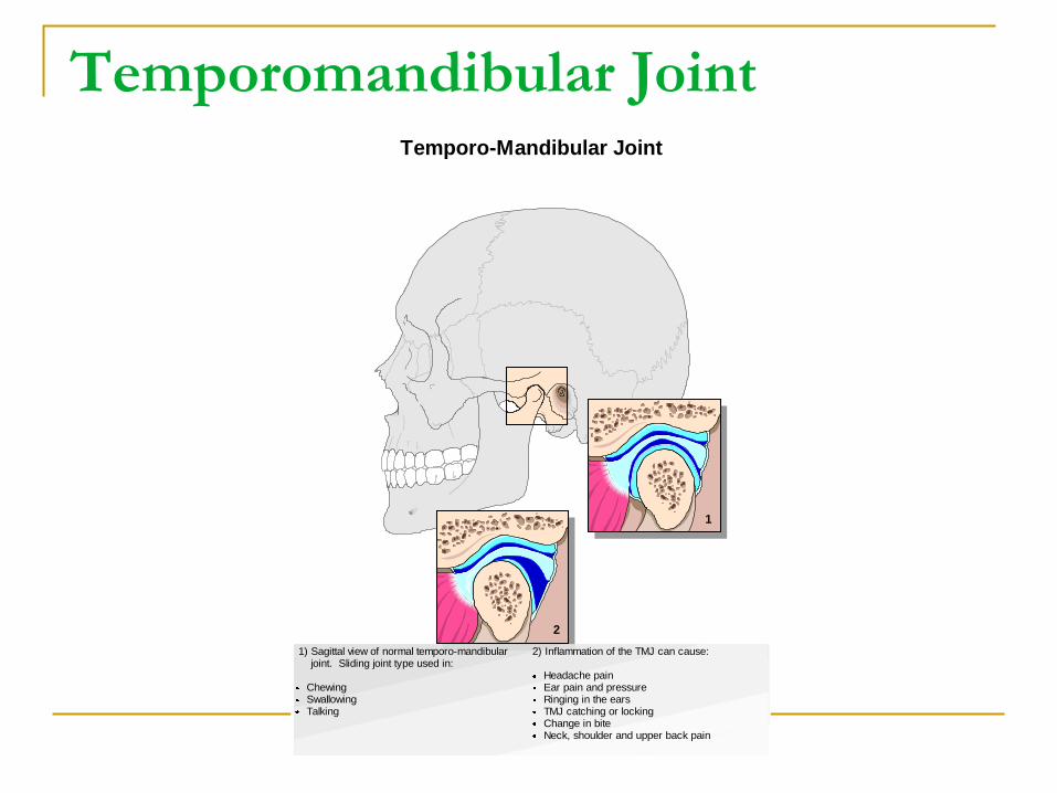
Temporomandibular Joint Temporo-Mandibular Joint
1) Sagittal view of normal temporo-mandibular joint. Sliding joint type used in:
Chewing Swallowing Talking
2) Inflammation of the TMJ can cause:
Headache pain Ear pain and pressure Ringing in the ears TMJ catching or locking Change in bite Neck, shoulder and upper back pain
1
2
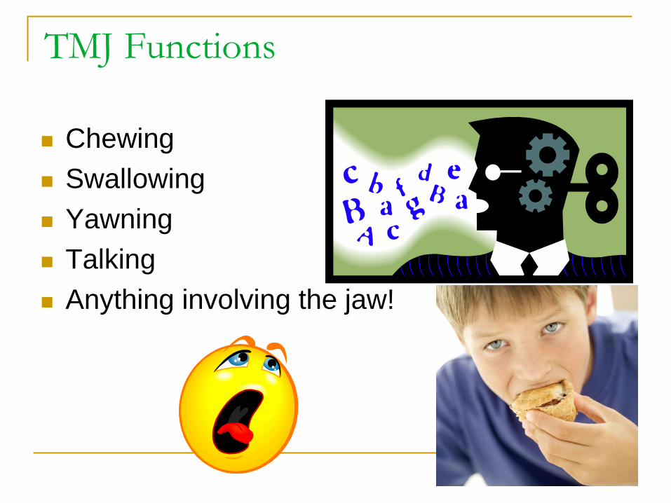
TMJ Functions
Chewing
Swallowing
Yawning
Talking
Anything involving the jaw!
Joint Structure & Motions
Made up of:
2 bones,
a disc that divides the joint into 2 joint spaces,
a joint capsule,
4 ligaments,
4 main muscles that create 5 motions.
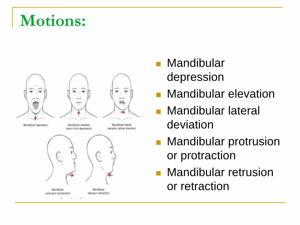
Motions:
Mandibular
depression
Mandibular elevation
Mandibular lateral
deviation
Mandibular protrusion
or protraction
Mandibular retrusion
or retraction
Resting position of the mandible:
The condyle of the mandible is seated in the
mandibular fossa of the temporal bone.
The lips would be closed and teeth would be
several millimeters apart.
Resting position of the mandible:
This would be maintained by low levels of activity of the
temporalis muscles
You should be able to open your mouth enough to fit 2-3
finger widths between the front upper and lower teeth.
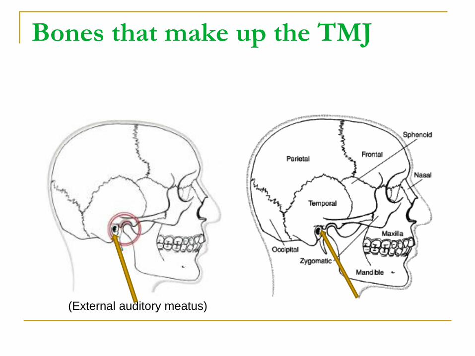
Bones that make up the TMJ
(External auditory meatus)
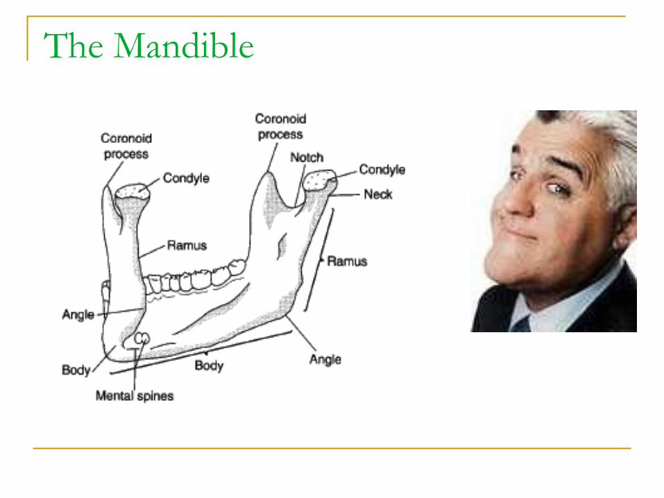
The Mandible
The Mandible
One bone, rests dependent upon muscle
relaxation and forms 2 identical joints with a
temporal bone on either side of the face
Makes up the inferior part of the face
The “jaw”
Bony landmarks
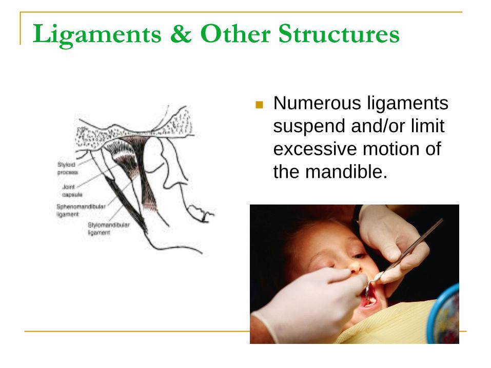
Ligaments & Other Structures
Numerous ligaments
suspend and/or limit
excessive motion of
the mandible.
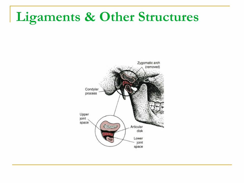
Ligaments & Other Structures
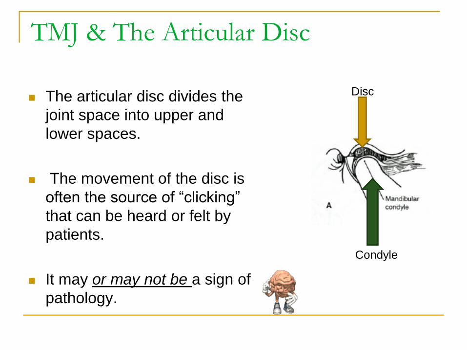
TMJ & The Articular Disc
The articular disc divides the
joint space into upper and
lower spaces.
The movement of the disc is
often the source of “clicking”
that can be heard or felt by
patients.
It may or may not be a sign of
pathology.
Disc
Condyle
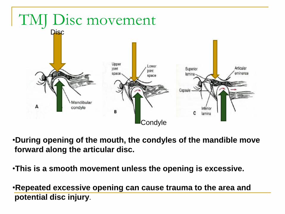
TMJ Disc movement
•During opening of the mouth, the condyles of the mandible move
forward along the articular disc.
•This is a smooth movement unless the opening is excessive.
•Repeated excessive opening can cause trauma to the area and
potential disc injury.
Disc
Condyle
Muscles of the TMJ
Temporalis
Masseter
Medial & Lateral Pterygoids
Muscle Names, Locations, Actions are the goal!
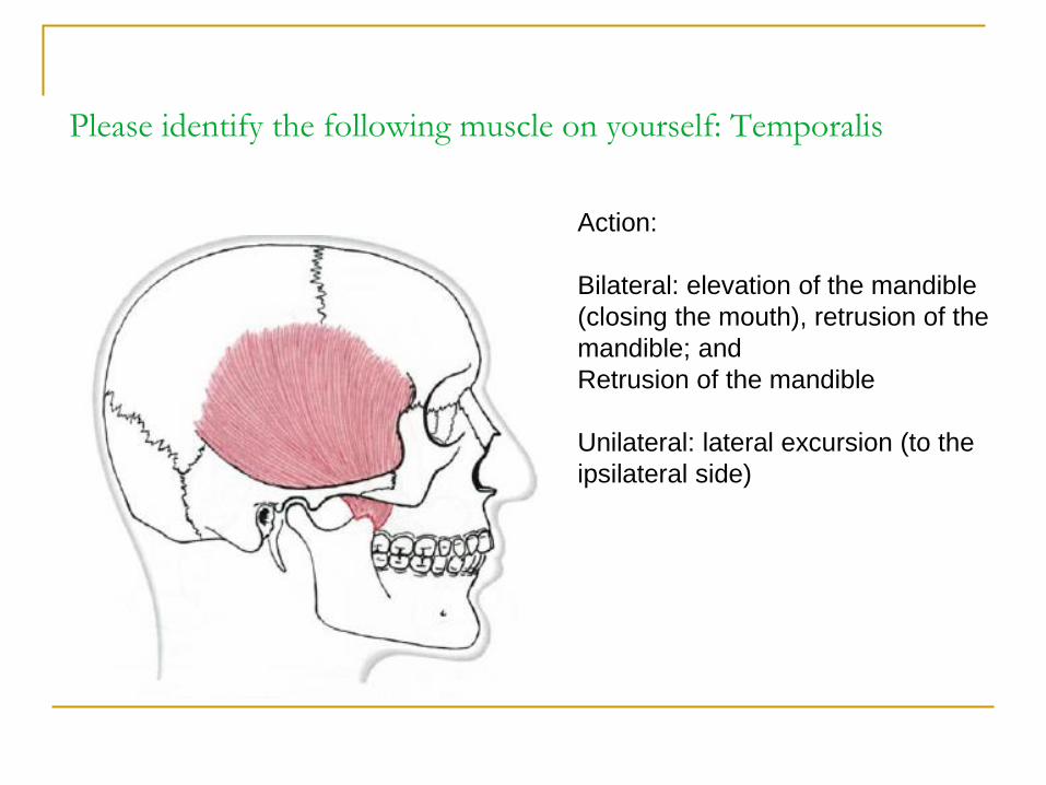
Please identify the following muscle on yourself: Temporalis
Action:
Bilateral: elevation of the mandible
(closing the mouth), retrusion of the
mandible; and
Retrusion of the mandible
Unilateral: lateral excursion (to the
ipsilateral side)
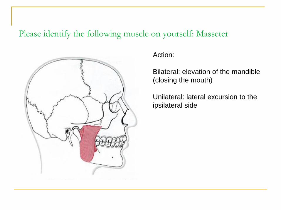
Please identify the following muscle on yourself: Masseter
Action:
Bilateral: elevation of the mandible
(closing the mouth)
Unilateral: lateral excursion to the
ipsilateral side
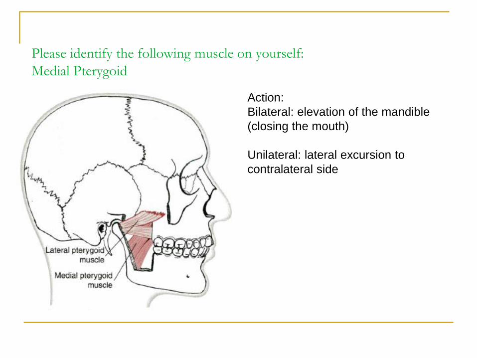
Please identify the following muscle on yourself:
Medial Pterygoid
Action:
Bilateral: elevation of the mandible
(closing the mouth)
Unilateral: lateral excursion to
contralateral side
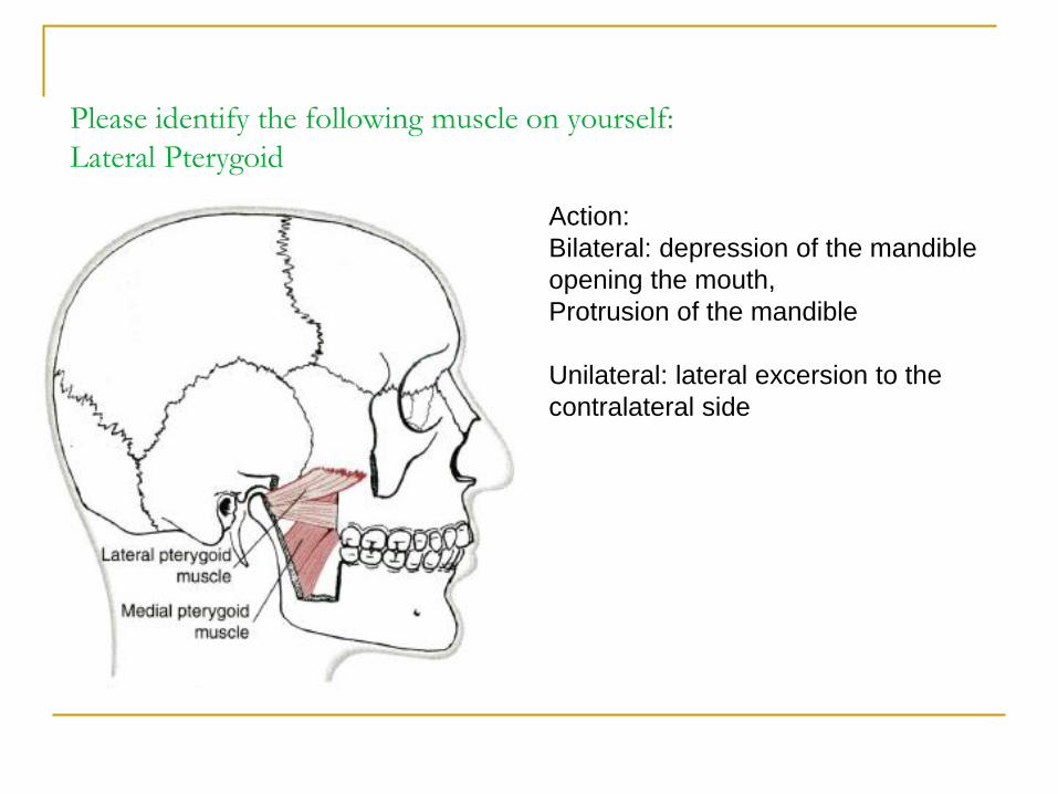
Please identify the following muscle on yourself:
Lateral Pterygoid
Action:
Bilateral: depression of the mandible
opening the mouth,
Protrusion of the mandible
Unilateral: lateral excersion to the
contralateral side
What do I need to know?
You should be able to palpate the masseter
and the temporalis on a classmate.
What do I need to know?
You should also be able to determine
whether or not there is any asymmetry in the
TMJ upon opening or closing when observing
a classmate.
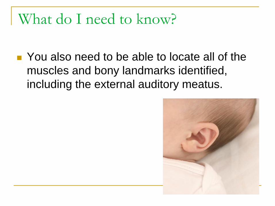
What do I need to know?
You also need to be able to locate all of the
muscles and bony landmarks identified,
including the external auditory meatus.
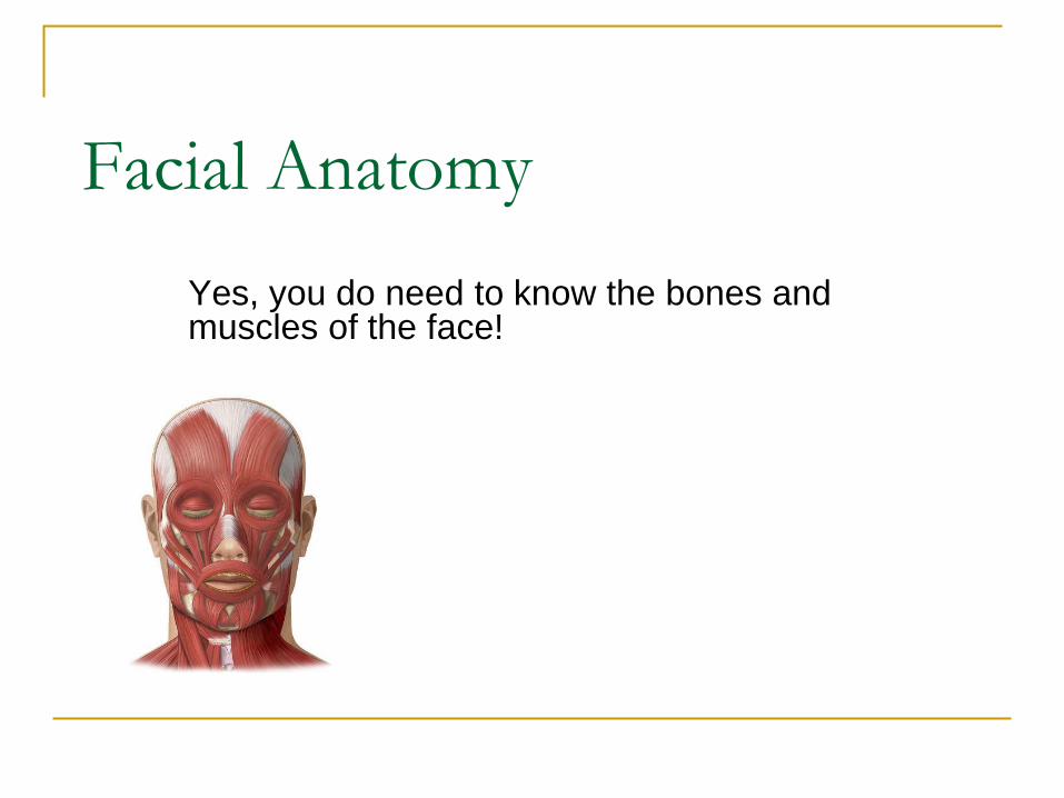
Facial Anatomy
Yes, you do need to know the bones and muscles of the face!
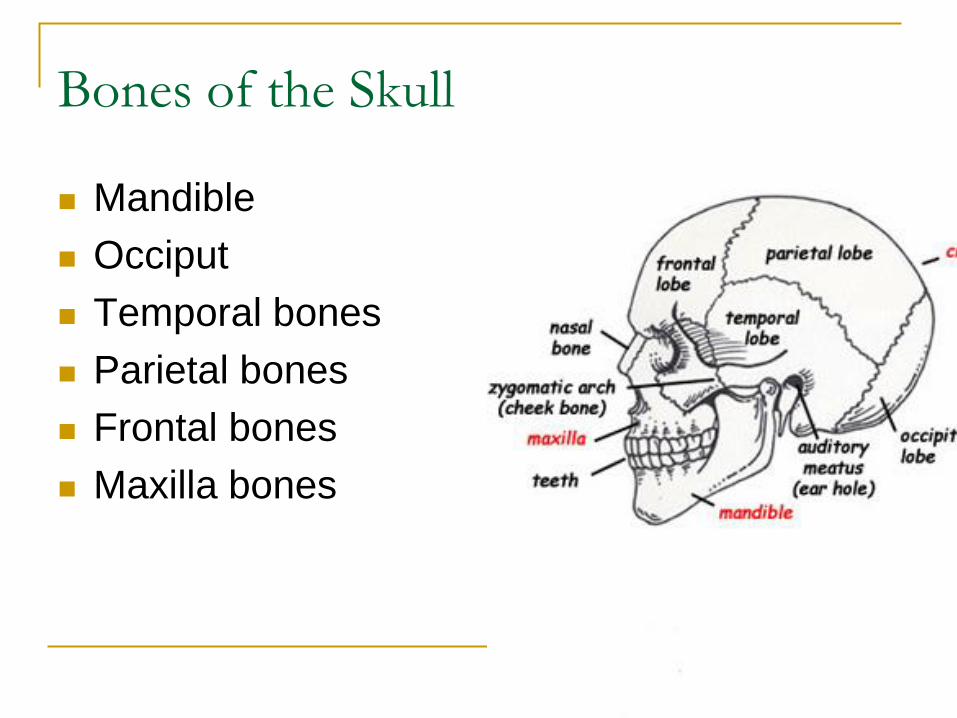
Bones of the Skull
Mandible
Occiput
Temporal bones
Parietal bones
Frontal bones
Maxilla bones
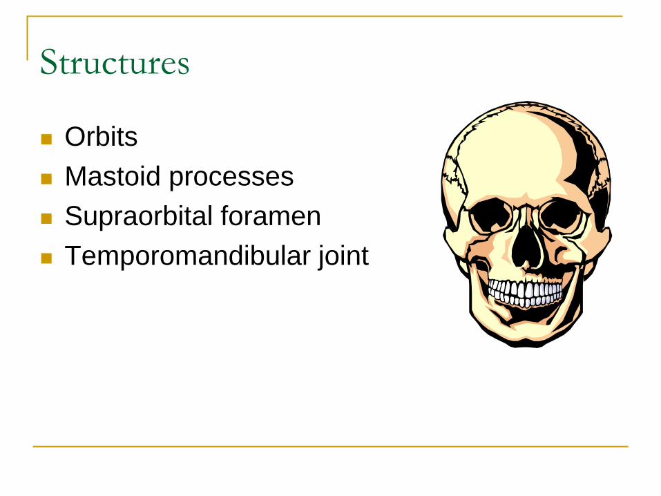
Structures
Orbits
Mastoid processes
Supraorbital foramen
Temporomandibular joint
Muscles of Mastication
Muscles of Mastication
Temporalis
Masseter
Medial Pterygoid
Lateral Pterygoid
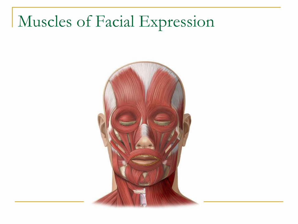
Muscles of Facial Expression
Muscles of Facial Expression
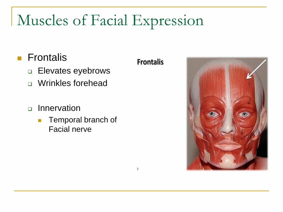
Frontalis
Elevates eyebrows
Wrinkles forehead
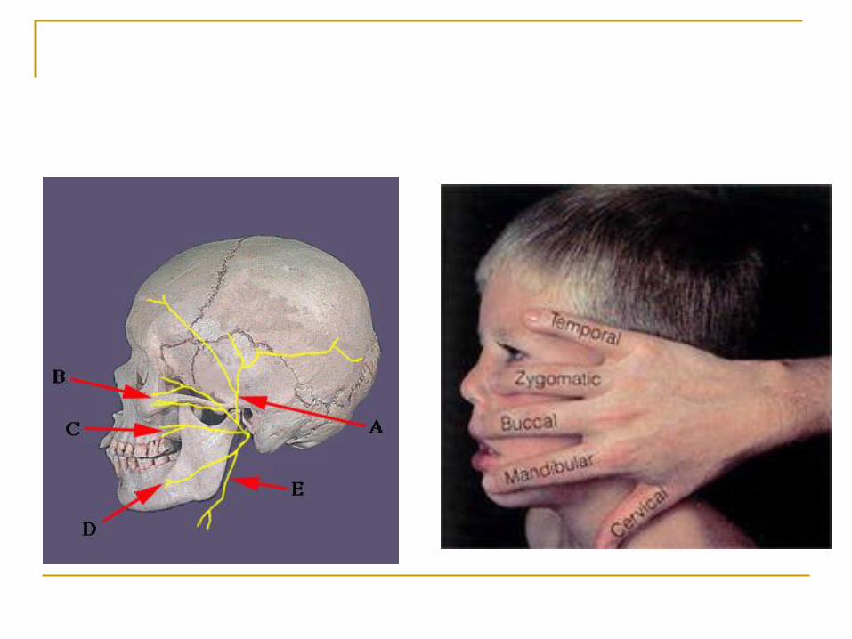
Innervation
Temporal branch of
Facial nerve
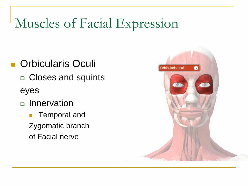
Muscles of Facial Expression
Orbicularis Oculi
Closes and squints
eyes
Innervation
Temporal and
Zygomatic branch
of Facial nerve
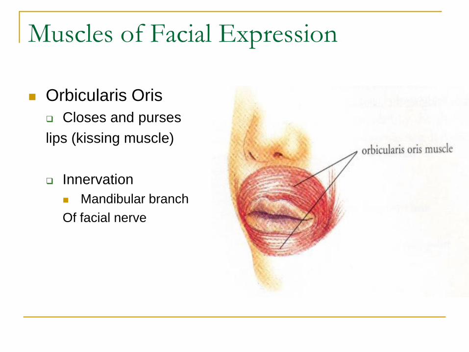
Muscles of Facial Expression
Orbicularis Oris
Closes and purses
lips (kissing muscle)
Innervation
Mandibular branch
Of facial nerve
Muscles of Facial Expression
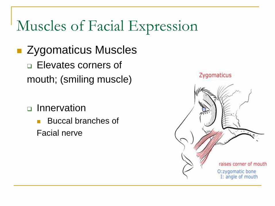
Zygomaticus Muscles
Elevates corners of
mouth; (smiling muscle)
Innervation
Buccal branches of
Facial nerve
Muscles of Facial Expression
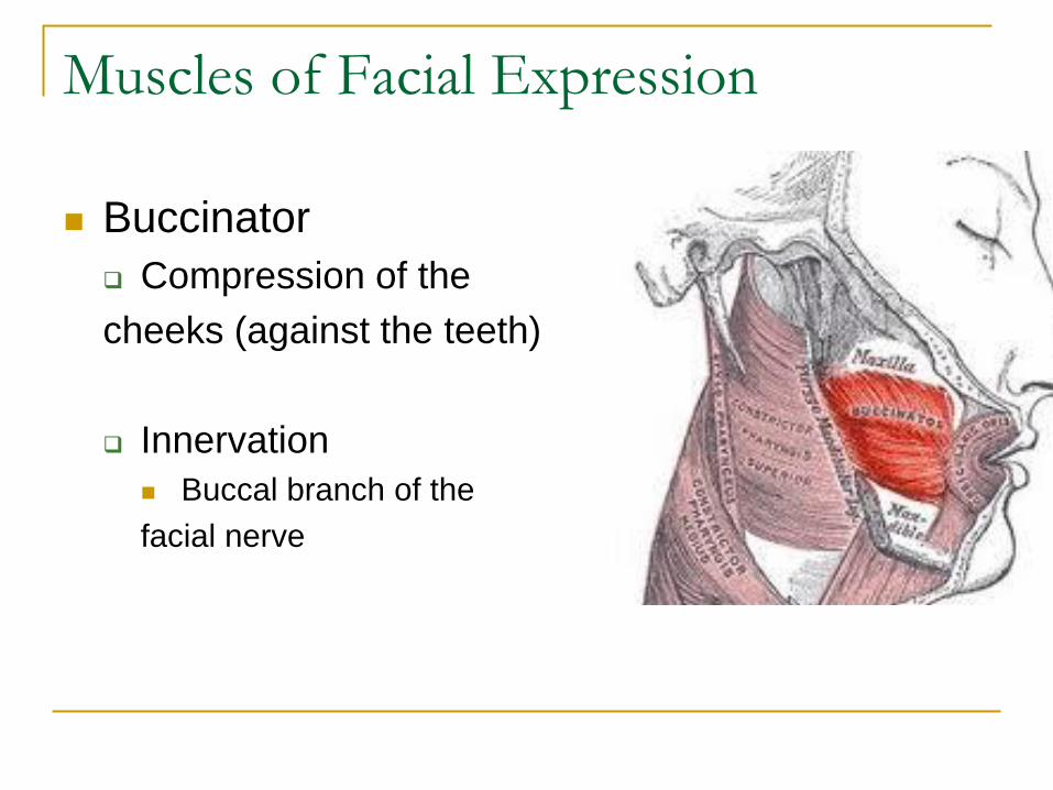
Buccinator
Compression of the
cheeks (against the teeth)
Innervation
Buccal branch of the
facial nerve
Muscles of Facial Expression
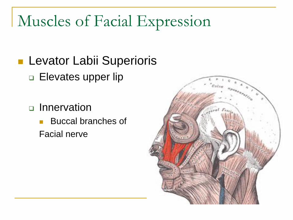
Levator Labii Superioris
Elevates upper lip
Innervation
Buccal branches of
Facial nerve
Muscles of Facial Expression
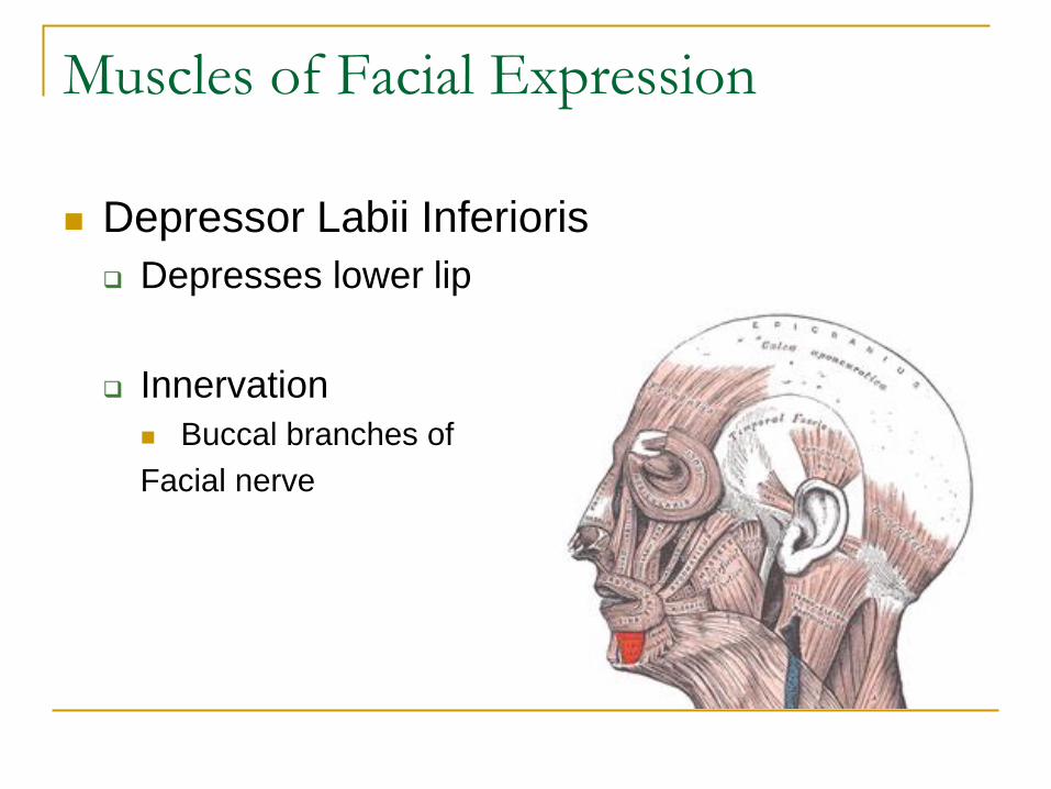
Depressor Labii Inferioris
Depresses lower lip
Innervation
Buccal branches of
Facial nerve
Muscles of Facial Expression
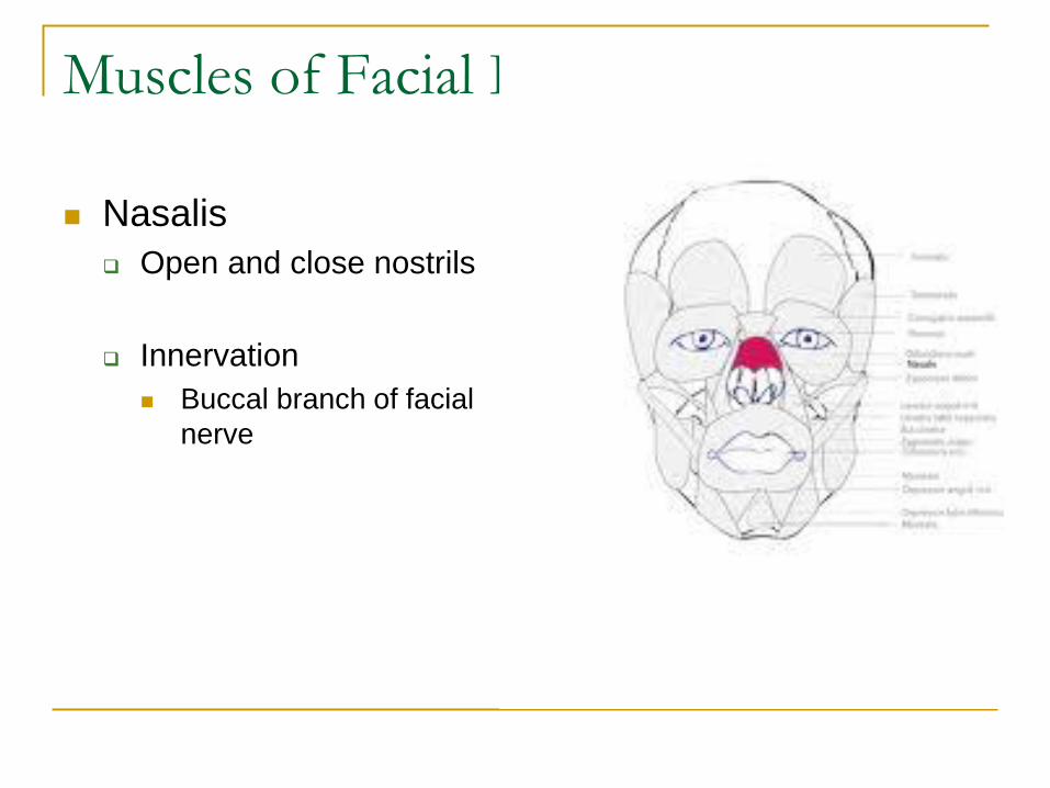
Nasalis
Open and close nostrils
Innervation
Buccal branch of facial
nerve
Muscles of Facial Expression
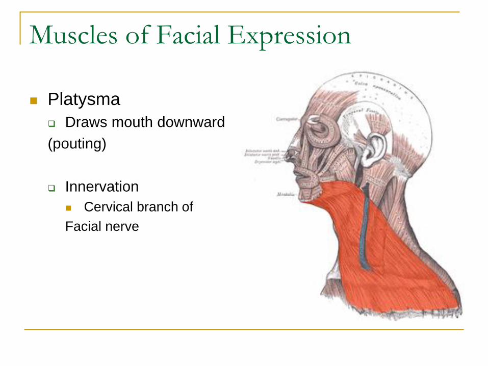
Platysma
Draws mouth downward
(pouting)
Innervation
Cervical branch of
Facial nerve
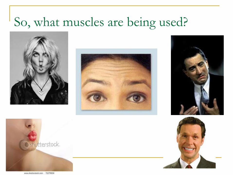
So, what muscles are being used?
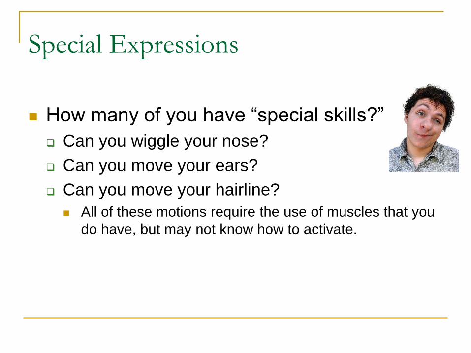
Special Expressions
How many of you have “special skills?”
Can you wiggle your nose?
Can you move your ears?
Can you move your hairline?
All of these motions require the use of muscles that you
do have, but may not know how to activate.
Last Exam Review
Ankle
Origins, insertions, actions, innervations of all
Describe position of ankle via pictures
Types of ankle sprains and ligaments involved
Names of joints at ankle and what motions occur
there
Muscle names by groups, i.e. Post Superficial
TMJ
Location of palpable muscles
Bony landmarks of mandible
Normative values of mandibular motions
Motions occuring at TMJ
Relevance of disc
Ventilation
Muscles of inspiration and expiration
Quiet and forced (accessory)
Origin, Insertion, Action, Innervation of diaphragm,
scalenes, and intercostals
Facial
Names of muscles and actions