Embed Size (px)
Citation preview
BIOPRO CLINICAL OUTCOME REPORT
MKT03 00
1
Tendon Anchor System: A promising new device in soft tissue attachment. Introduction: Several factors affect outcomes of tendon repair, with repair strength being the most important.1 Historically there have been numerous methods available for soft tissue reattachment; initial techniques utilized staples, screws, washers and plates. Today the most common involve suture anchors which are available in several variations of design, configuration, and material type. While these anchors follow the same basic principles, the techniques are often altered depending on surgeon preference and surgical application. Suture anchor techniques can be time-consuming and tedious. To improve strength, many techniques require multiple anchors and woven suturing methods. A typical post-operative protocol for a tendon transfer with a suture anchor technique suggests that a patient be splinted or casted immediately after surgery and allowed to begin weight bearing activities six weeks post op2. Contrary to this recommendation, research has shown that early motion (10 days) after tendon repair, as opposed to long-term immobilization, results in good return of strength, power and endurance.3 While comparing reattachment methods, it has been exhibited that spiked devices with multiple teeth provide for the strongest pull-out strength.4 Although common drawbacks of poor vascularity and irritation limited the use of these devices.4 When reviewing the histological data, it was discovered that these drawbacks were only seen in plastic and flat devices, while rigid
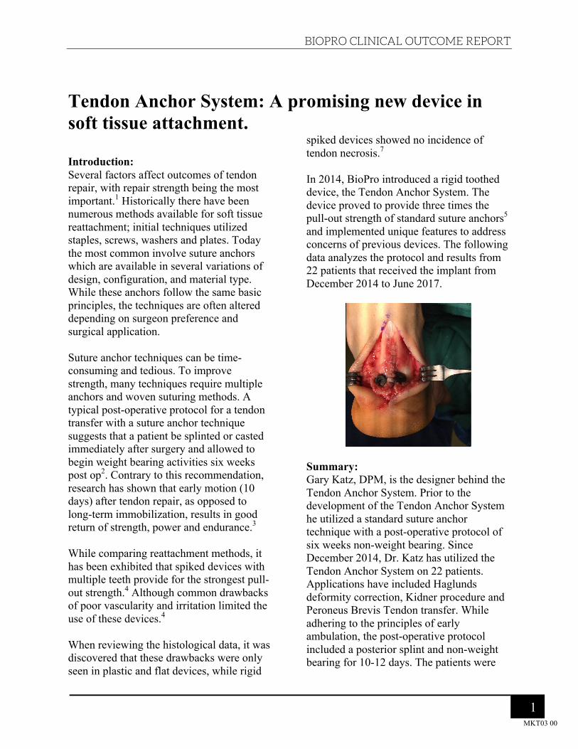
spiked devices showed no incidence of tendon necrosis.7 In 2014, BioPro introduced a rigid toothed device, the Tendon Anchor System. The device proved to provide three times the pull-out strength of standard suture anchors5 and implemented unique features to address concerns of previous devices. The following data analyzes the protocol and results from 22 patients that received the implant from December 2014 to June 2017.
Summary: Gary Katz, DPM, is the designer behind the Tendon Anchor System. Prior to the development of the Tendon Anchor System he utilized a standard suture anchor technique with a post-operative protocol of six weeks non-weight bearing. Since December 2014, Dr. Katz has utilized the Tendon Anchor System on 22 patients. Applications have included Haglunds deformity correction, Kidner procedure and Peroneus Brevis Tendon transfer. While adhering to the principles of early ambulation, the post-operative protocol included a posterior splint and non-weight bearing for 10-12 days. The patients were
BIOPRO CLINICAL OUTCOME REPORT
MKT03 00
2
then placed in a walking boot for four to six weeks. Although a limited patient population and follow-up, the results are extremely promising. In Dr. Katz’s patients, the Tendon Anchor System has not shown any soft tissue irritation, post-operative pain, or tendon necrosis. One incidence of anchor pull-out has occurred due to an unrelated traumatic injury at one week post-op. The patient underwent an additional surgery to reattach the tendon utilizing multiple 1-hole anchors, which provided a successful outcome. Conclusion: The initial follow up of 22 patients has shown no incidence of soft tissue irritation, post-operative pain, or tendon necrosis. Further research is currently being conducted. About the surgeon: Gary E. Katz, DPM graduated from The Ohio College of Podiatric Medicine (Kent State University College of Podiatric Medicine) in 1996, completed a residency at University Hospital in Cleveland, OH in 1998 and has been in practice for 20 years. Dr. Katz is a practicing podiatrist who specializes in foot and ankle surgery in St. Petersburg, FL.
References:
1. Schuind F, Garcia-Elias M, Cooney WP, An KN. Flexor tendon forces: in vivo measurements. J Hand Surg Am. 1992;17(2):291–298.
2. Tendon Transfers. American Orthopaedic Foot & Ankle Society. (2017, May 16). Retrieved from www.aofas.org/footcaremd/treatments/Pages/Tendon-Transfers.aspx
3. Randal L. Troop, M.D., Gary M. Losse, M.D., John G. Lane, M.D., Daniel B. Robertson, M.D., Pamela S. Hastings, M.S., P.T., Mark E. Howard, A.T.C., O.T.C., O.P.A. "Early Motion After Repair of Achilles Tendon Ruptures." SAGE JOURNALS. Foot & Ankle International, 1 Nov. 1995. Web. 16 May 2017.
4. Christopher B. Straight, MS, E. Paul France,* PhD, Lonnie E. Paulos, MD, Thomas D. Rosenberg, MD, and Jeffrey A. Weiss, MS. "Soft Tissue Fixation to Bone A Biomechanical Analysis of Spiked Washers." The American Journal of Sports Medicine22.3 (1994): 339-43. Web. 15 May 2017.
5. Jill S. Kawalec-Carroll, PhD. Evaluation of a New Device for the Reattachment of the Achilles Tendon. Ohio College of Podiatric Medicine, 12 Apr. 2012. Unpublished raw data.
6. Schuind F, Garcia-Elias M, Cooney WP, An KN. Flexor tendon forces: in vivo measurements. J Hand Surg Am. 1992;17(2):291–298.
7. Straight, C. B., France, E. P., Paulos, L. E., Rosenbert, T. D., Weiss, J. A. Soft tissue fixation to bone: a biomechanical analysis of spiked washers. Am. J. Sport s Med. 22(3):339-343, 1994.