Embed Size (px)
Citation preview

Terbium-mediated Footprinting Probes a CatalyticConformational Switch in the Antigenomic HepatitisDelta Virus Ribozyme
Dinari A. Harris, Rebecca A. Tinsley and Nils G. Walter*
Department of ChemistryThe University of Michigan930 N. University, Ann ArborMI 48109-1055, USA
The two forms of the hepatitis delta virus ribozyme are derived from thegenomic and antigenomic RNA strands of the human hepatitis deltavirus (HDV), where they serve a crucial role in pathogen replication bycatalyzing site-specific self-cleavage reactions. The HDV ribozymerequires divalent metal ions for formation of its tertiary structure, consist-ing of a tight double-nested pseudoknot, and for efficient self- (or cis-)cleavage. Comparison of recently solved crystal structures of the cleavageprecursor and 30 product indicates that a significant conformational switchis required for catalysis by the genomic HDV ribozyme. Here, we haveused the lanthanide metal ion terbium(III) to footprint the precursor andproduct solution structures of the cis-acting antigenomic HDV ribozyme.Inhibitory Tb3þ binds with high affinity to similar sites on RNA as Mg2þ
and subsequently promotes slow backbone scission. We find subtle, yetsignificant differences in the terbium(III) footprinting pattern betweenthe precursor and product forms of the antigenomic HDV ribozyme, con-sistent with differences in conformation as observed in the crystal struc-tures of the genomic ribozyme. In addition, UV melting profiles provideevidence for a less tight tertiary structure in the precursor. In both theprecursor and product we observe high-affinity terbium(III) binding sitesin joining sequence J4/2 (Tb1/2 < 4 mM) and loop L3, which are key struc-tural components forming the catalytic core of the HDV ribozyme, as wellas in several single-stranded regions such as J1/2 and the L4 tetraloop(Tb1/2 < 50 mM). Sensitized luminescence spectroscopy confirms thatthere are at least two affinity classes of Tb3þ binding sites. Our resultsthus demonstrate that a significant conformational change accompaniescatalysis in the antigenomic HDV ribozyme in solution, similar to thecatalytic conformational switch observed in crystals of the genomic form,and that structural and perhaps catalytic metal ions bind close to thecatalytic core.
q 2004 Elsevier Ltd. All rights reserved.
Keywords: catalytic RNA folding; melting curve; metal ion binding site;reaction mechanism; terbium luminescence*Corresponding author
Introduction
RNA enzymes or ribozymes require multivalentmetal ions for optimal activity. Structural and bio-chemical studies have revealed that particularly
divalent metal ions play a role in promoting fold-ing of a ribozyme and often directly participate incatalysis. Since structure and dynamics dictatefunction, the structural and catalytic roles of metalions have proven to be difficult to dissect,especially since the vast majority of divalent metalions are bound diffusely, thus facilitating tertiarystructure formation by neutralizing and bridgingthe negatively charged phosphoribose backbone.Recently, there have been major advances in ourunderstanding of the intimate link between
0022-2836/$ - see front matter q 2004 Elsevier Ltd. All rights reserved.
E-mail address of the corresponding author:[email protected]
Abbreviations used: HDV, hepatitis delta virus; NMR,nuclear magnetic resonance; rNTPs, ribonucleosidetriphosphates; Tm, melting temperature.
doi:10.1016/j.jmb.2004.05.074 J. Mol. Biol. (2004) 341, 389–403

metal ion binding and RNA function.1 – 5 Many ofthese studies have utilized the class of “small”ribozymes, which includes the hammerhead, hair-pin, hepatitis delta virus (HDV), and NeurosporaVarkud satellite (VS) ribozymes. These ribozymesare classified by their relatively small size (lessthan 200 nucleotides) and common reactionchemistry; however, they are structurally diverseand have different metal ion requirements (orspecificities). Particularly, in the past few years theHDV ribozyme has received considerable attentionand has redefined our understanding of how RNAenzymes work.
The hepatitis delta virus is a satellite RNA of thehuman hepatitis B virus (HBV).6 Co-infection ofpatients with both HBV and HDV results in moresevere forms of hepatitis, including liver cirrhosis,and can lead to death. During viral replication,both the ,1700 nucleotides long genomic RNAand its complement, the antigenomic RNA,undergo self-cleavage reactions catalyzed byinternal ribozyme motifs that are absolutely essen-tial for propagation of the virus in vivo.7,8 Theseself-cleavage activities reside within contiguous,85 nucleotide sequence elements that formsimilar double-nested pseudoknots, the genomicand antigenomic forms of the HDV ribozyme.9 Inthe presence of divalent cations, the HDV ribo-zyme undergoes self-cleavage in vitro, duringwhich deprotonation of a specific 20-OH groupand nucleophilic attack on the adjacent scissilephosphate result in formation of 20,30-cyclic phos-phate and 50-OH termini. The crystal structure ofthe self-cleaved genomic form revealed for thefirst time that the ribozyme adopts a tight tertiarystructure by forming the two base-pair helix P1.1between loop L3 and the joiner sequence J1/4,creating a pair of nested pseudoknots, whichburies the active site in a deep cleft.10 Thus, thecleavage site resides in close proximity to thecatalytically important cytosine 75/76 (C75 inthe genomic ribozyme, C76 in the antigenomicribozyme) (Figure 1). This observation inspiredbiochemical studies that found C75/76 to bedirectly involved in reaction chemistry as a generalbase or acid, thereby opening our eyes to the factthat RNA is not solely dependent on the functionalcapabilities of divalent metal ions to performcatalysis.11 – 14
Refinement of the crystal structure of the self-cleaved genomic HDV ribozyme located 12 diva-lent metal ions throughout the structure, yet noneof them was found in proximity to the proposedcatalytic core.15 A possible explanation for thislack of metal ions in the active site is that thecrystallized sequence lacks the scissile phosphateand the nucleophilic 20-OH group, which bothcould serve as metal binding ligands. This obser-vation is consistent with biochemical datasuggesting that the 50 sequence upstream of thecleavage site, although not obviously interactingwith the 30 product sequence, has a significantimpact on catalytic activity. For example, kinetic
studies of a trans-acting HDV ribozyme havesuggested that components of the substrateimmediately 50 to the cleavage site destabilizeground-state substrate binding and thereby lowerthe activation free energy barrier of cleavage.16
This result, in conjunction with the observationthat the active site cleft in the product crystal struc-ture is deep and tightly packed, thus not obviouslyproviding a trajectory for substrate exit, has led tothe proposal that the 50 sequence may be accommo-dated by straining (bending) of the substrate and/or loosening of the catalytic core.9,17 In support ofthis notion, fluorescence spectroscopy studieshave recently shown that a trans-acting HDV ribo-zyme undergoes both substantial global and localconformational rearrangements upon catalysis.17 –19
In addition, NMR spectroscopy has providedfurther evidence for structural differences betweenthe precursor and product forms of trans-actingHDV ribozymes.20,21 Most recently, Doudna andco-workers have succeeded in crystallizing severalforms of the genomic cis-acting precursorribozyme.22 Comparison with the product structurereveals that an important conformational changeaccompanies and, in fact, appears to facilitateHDV ribozyme cleavage. Consistent with thebiochemical data, the precursor crystal structuredepicts an outer-sphere coordinated divalent
Figure 1. Secondary structure of the cis-acting anti-genomic HDV ribozyme used in this study. The non-cleavable precursor form was generated by modifyingthe 20-OH group of the underlined nucleotide 50 to thecleavage site (open arrow) to 20-methoxy during chemi-cal synthesis. In order to maintain the previously definedantigenomic numbering system in our P4-truncated ribo-zyme, we renumbered the nucleotides downstream ofthe L4 loop. The boxed cytosine residue at the bottom ofP4 is defined as residue 69. Outlined are the five nucleo-tides for which we observe the largest (. twofold)change in normalized scission intensity between the pre-cursor and product forms of the antigenomic ribozyme.
390 Conformational Switch in Antigenomic HDV Ribozyme

metal ion in an active site pocket formed by loopL3. This metal ion is lost from the active sitetogether with the 50 sequence as the catalyticconformational switch occurs, which prevents theproduct form of the genomic HDV ribozyme fromcatalyzing the reverse ligation reaction.22
To address the questions of whether a similarconformational switch accompanies catalysis inthe antigenomic HDV ribozyme and what form ittakes outside of a crystal lattice, we have utilizedterbium(III)-mediated footprinting as a probe ofsecondary and tertiary structure as well as metalbinding properties in cis-acting precursor and pro-duct forms in solution. The lanthanide metal ionterbium(III) has previously been found to be apowerful probe of RNA tertiary structure atnucleotide resolution, as well as of metal ion bind-ing sites in RNA. Tb3þ binds to similar sites on thepolyanionic RNA as Mg2þ, with an affinity two tofour orders of magnitude higher.23 – 25 The inhibi-tory terbium(III) has the ability to substitute formagnesium(II) because Tb3þ has an ionic radius(0.92 A) similar to that of Mg2þ (0.72 A), and theyshare the same preference for coordination to oxy-gen ligands.23,24,26 Once bound to RNA, the transi-ently deprotonated aqua complex Tb(OH)(aq)2þ
abstracts protons from nearby 20-OH groups,which allows for nucleophilic attack of the result-ing 20 oxyanion on the juxtaposed phosphodiester,leading to slow local strand scission 30 to a givennucleotide.23,24,27,28 Tb3þ can effectively catalyzescission of an RNA backbone at physiologic pHdue to its lower pKa (,7.9) compared to that ofMg2þ (11.4).23,24,29 By systematically varying theTb3þ concentration, it is possible to obtain a foot-print of secondary and tertiary structure (at milli-molar [Tb3þ]) and a map of metal-binding sites (atmicromolar [Tb3þ]).23,24 Our terbium(III) footprint-ing patterns indicate that the precursor andproduct forms of the cis-acting antigenomic HDVribozyme are similar in overall conformation butindeed show distinctions in several key regions,consistent with the differences observed in thecrystal structures of the genomic ribozyme. Inaddition, UV melting profiles provide evidencefor a less tight tertiary structure in the precursor.We also map high-affinity terbium(III) bindingsites to J4/2 (Tb1/2 < 4 mM), the joiner sequencethat contains the catalytic C76, and to loop L3,found in the precursor crystal structure to bindthe potentially catalytic metal ion. Sensitized lumi-nescence spectroscopy confirms that there are atleast two different affinity classes of Tb3þ bindingsites. Our data are thus consistent with the notion
that a significant conformational change leads tocatalysis in the cis-cleaving antigenomic HDV ribo-zyme in solution, similar to that proposed for thegenomic ribozyme, and that structural and perhapscatalytic metal ions bind close to the catalytic core.
Results
Terbium(III) inhibits the HDV ribozyme,probably by competing with cations thatsupport catalysis
The lanthanide metal terbium(III) has proven tobe a versatile probe of both metal binding as wellas secondary and tertiary structure of RNA. Inaddition, the luminescent properties of Tb3þ pro-vide for a facile means of measuring metal-bindingaffinity. It has long been known that Tb3þ replacesMg2þ as an RNA ligand of much higher affinity,which as a result usually inhibits catalysis, forexample in the hammerhead30 and hairpinribozymes.23 In previous work we found that clea-vage in a trans-acting HDV ribozyme was signifi-cantly inhibited in the presence of terbium(III),19
so that we felt compelled to investigate the impactthat terbium(III) has on catalysis in a cis-actingHDV ribozyme. The HDV ribozyme used here(Figure 1) is slightly modified from a previouslydescribed antigenomic HDV ribozyme.13 Our con-struct differs from the latter wild-type antigenomicHDV sequence in that the sequence upstream of U-3 was modified to 50-GGGA-30 for optimal in vitrotranscription of the self-cleaving RNA, and in thatP4 was truncated and capped with a stable UUCGtetraloop to limit the total length to 80 nucleotidesfor chemical synthesis and introduction of a20-methoxy modification at the cleavage site tostructurally study a non-cleavable precursor.
First, we tested the single-turnover cleavageactivity of our cis-acting antigenomic HDV ribo-zyme in the presence and absence of 5 mM TbCl3,which is the highest concentration used in theterbium(III)-mediated footprinting assays. Asexpected, we found terbium(III) to be an inhibitorof catalysis. Specifically, self-cleavage of the pre-annealed precursor ribozyme under standard con-ditions (25 mM acetic acid, 25 mM Mes, 50 mMTris–HCl (pH 7.5), at 37 8C) was initiated by theaddition of Mg2þ to a final concentration of 11 mM(Materials and Methods), showing that the conver-sion of precursor to product follows first-orderkinetics, with an observed rate constant kobs of43.0(^4.6) min21 and a 72(^2)% extent of cleavage(Table 1). This finding is in good agreement withreported values of kobs ¼ 31 min21 and 77% clea-vage extent for the wild-type antigenomic HDVribozyme sequence,31 indicating that our sequencemodifications do not interfere with catalysis.When the reaction was initiated by the addition ofa mixture of Mg2þ and Tb3þ (final concentrations11 mM and 5 mM, respectively), kobs was unaf-fected (44.4(^5.9) min21); however, the extent of
Table 1. Self-cleavage activity of the antigenomic HDVribozyme used in this study
Metal ion conditionsCleavage rate(kobs) (min21)
Cleavageextent (%)
11 mM Mg2þ, 0 mM Tb3þ 43.0 ^ 4.6 72 ^ 211 mM Mg2þ, 5 mM Tb3þ 44.4 ^ 5.9 27 ^ 2
Conformational Switch in Antigenomic HDV Ribozyme 391

cleavage was significantly lowered to 27(^2)%.Such a lowered cleavage amplitude in the presenceof terbium(III) was also observed with the hairpinribozyme, but the mechanism of this type ofinhibition is not well understood.23,30
Terbium(III) footprinting reveals subtlestructural differences between the precursorand product at nucleotide resolution
High (millimolar) concentrations of terbium(III)have previously been used to slowly cut an RNAphosphodiester backbone in a largely sequence-independent manner, preferentially in single-stranded or non-Watson–Crick base-pairedregions, thus generating a footprint of the RNA’ssecondary and tertiary structure at nucleotideresolution.19,23 – 26,29,32 – 37 In addition, due to thestrongly electrostatic interactions of the highlycharged Tb3þ with the polyanionic RNA backbone,scission is particularly enhanced on RNA surfacesof high local charge density, revealing potentialmetal binding sites at low (micromolar) concen-tration of terbium(III).19,24,27,37 Because of its near-physiologic aqueous pKa of 7.9 and its Mg2þ and
Ca2þ-like coordination properties with a preferencefor oxygen ligands, terbium(III) has recentlybecome popular as an alternative to Mg2þ andPb2þ-induced footprinting.24,25 We therefore set outto utilize Tb3þ to probe and compare the structuresof the precursor and product forms of our anti-genomic HDV ribozyme in solution.
To this end, trace amounts of the radiolabelednon-cleavable precursor or product form of ourantigenomic HDV ribozyme were prefolded understandard conditions in the presence of 11 mMMg2þ, as described in Materials and Methods,followed by addition of increasing concentrationsof Tb3þ (from 1 mM to 5 mM) to initiate slow back-bone scission at 37 8C over one hour (under theseconditions, only a small fraction of RNA is cleaved,avoiding secondary hits on an already cut RNAmolecule). The footprinting patterns of the precur-sor and product (Figures 2 and 3) show similaritieswith respect to their protected and susceptibleregions, which is consistent with the fact that theprecursor and product forms of the antigenomicHDV ribozyme share an overall similar confor-mation. In particular, both scission patterns areconsistent with the expected secondary structure
Figure 2. Terbium(III)-mediated footprinting of 50-32P-labeled antigenomic HDV ribozyme. A, Precursor footprintafter incubation for one hour in 25 mM acetic acid, 25 mM Mes, 50 mM Tris–HCl (pH 7.5), 11 mM MgCl2, at 37 8C(see Materials and Methods). From left to right: lane 1, radiolabeling precursor without incubation; lanes 2–10,samples incubated with increasing Tb3þ concentrations (0, 0.001, 0.005, 0.01, 0.05, 0.1, 0.5, 1, and 5 mM), as indicated;lanes 11 and 12, G-specific RNase T1 and alkaline hydrolysis (OH2) ladders, respectively. Note that significant scissionoccurs in J4/2, L3, J1/2, and P4 at low micromolar Tb3þ concentrations, which is indicative of high-affinity metal bind-ing. As the Tb3þ concentration is increased, backbone scission becomes more intense. Scission occurs 30 to the indicatednucleotide. Asterisks indicate background scission in 50-UpA-30 and 50-CpA-30 steps. B, Product footprint under identi-cal conditions and with a similar lane arrangement as in A.
392 Conformational Switch in Antigenomic HDV Ribozyme

of the HDV ribozyme; relative protection isobserved in all five Watson–Crick base-pairedstems, P1–P4 and P1.1; while strong scission isobserved in the backbone of loops L3 and L4 aswell as in joiners J1/2 and J4/2 (Figures 2 and 3).
Interestingly, the precursor (as well as product)consistently show significant backbone scission,even in the absence of Tb3þ, 30 to C8 (J1/2), C13, C19
(P250), C43 (P450), and U77 (J4/2) (marked by asterisksin Figure 2A). Further inspection revealed thatthese nucleotides represent the five sequenceelements of the type 50-CpA-30 or 50-UpA-30 presentin our antigenomic HDV ribozyme. Previousstudies have shown that the phosphodiesterbonds of 50-UpA-30 and 50-CpA-30 sequenceelements are .100-fold and up to 35-fold morereactive, respectively, at physiologic pH and tem-perature than those of other dinucleotide steps.38
A possible explanation is a distinct hydration pat-tern around the scissile phosphate, attacking20-OH and neighboring bases that, by hydrogenbonding, lowers the transition state barrier for scis-sion in these sequence elements.38,39 In our quanti-tative analysis, we therefore normalized againstthe no-Tb3þ background (Materials and Methods),thus correcting for this background scission inthe absence of Tb3þ. Still, terbium(III) inducesadditional phosphodiester bond scission 30 to C8
and U77, as detailed in the following.Figure 4A summarizes the relative percentage
of RNA cut at each resolved nucleotide of the
precursor and product forms at 5 mM Tb3þ (topand bottom, respectively). These patterns wereconsistently observed on altogether 11 sequencinggels resolving either the 50 or 30 ends of the pre-cursor and 16 gels resolving either the 50 or 30
ends of the product. Interestingly, under all con-centrations of terbium(III) tested, the histogramplots of the precursor reveal more even scissionintensities than those of the product. By compari-son, the histogram plots of the product appearmore rugged. There are two potential explanationsfor this effect: (1) a technical one in that the foot-printing gels of the precursor appeared to typicallyhave more of a smear to them, somewhat eveningout the intensities; (2) a structural one in that theprecursor HDV ribozyme structure may be con-siderably more dynamic than the product struc-ture, so that terbium(III) footprinting results in anoverall averaging of a broader variety of structures.This latter explanation is consistent with the ideathat the precursor structure must be more dynamic(loose) to accommodate the 50 sequence extensionand is further supported by differences in theprecursor and product melting profiles (see below).
Footprinting at high Tb3þ concentrations gener-ates a direct map of secondary and tertiary struc-ture, as scission in Watson–Crick base-pairedregions such as P3 is suppressed relative to that insingle-stranded regions. A concentration of 5 mMTb3þ in the precursor induces strong scission at(or 30 to) U4-C5 (P150), A9 (J1/2), U10-C18 (P250),
Figure 3. Terbium(III)-mediated footprinting, resolving the 30-end of the antigenomic HDV ribozyme. A, The 30-end-labeled precursor after incubation for one hour in 25 mM acetic acid, 25 mM Mes, 50 mM Tris–HCl (pH 7.5), 11 mMMgCl2, at 37 8C (see Materials and Methods). From left to right: lane 1, radiolabeling precursor without incubation;lanes 2–10, samples incubated with increasing Tb3þ concentrations (0, 0.001, 0.005, 0.01, 0.05, 0.1, 0.5, 1, and 5 mM),as indicated; lanes 11 and 12, G-specific RNase T1 and alkaline hydrolysis (OH2) ladders, respectively. Note that scis-sion occurs 50 to the indicated nucleotide. B, Footprint of 50-end-labeled product, in which the gel was run longerthan in Figure 2B to better resolve the 30 end. Conditions and lane arrangement were similar as in A, but scissionoccurs 30 to the indicated nucleotide.
Conformational Switch in Antigenomic HDV Ribozyme 393

C21-C22 (P350), U23-U26 (L3), C51-G52 (L4), andC69 (P430). The product in the presence of 5 mMTb3þ shows strong scission at C8-A9 (J1/2), U10,C13 (P250), C29 (L3) and moderate scission at G12,A14 (P250), C24-G28 (L3), U50-G52 (L4), and C69(P430). In Figure 4A we have highlighted by arrowsthose five nucleotides, C8, U17, C29, G80, andG81, that show a more than twofold change innormalized scission intensity between the precur-sor and product; Figure 4B shows the ratio of pre-cursor over product scission intensity for allpositions. Please note that C8 and C29 (gray)increase in scission intensity after cleavage, whileU17, G80, and G81 decrease (black). The terbium-(III)-mediated footprinting patterns of the precur-sor and product HDV ribozyme thus are subtly,but noticeably distinct, consistent with the ideathat their global structures are not identical.Our results therefore support previous evidencefrom fluorescence resonance energy transfer,17
2-aminopurine fluorescence quenching,18 NMRspectroscopy,20,21 and terbium(III)-mediatedfootprinting19 that have revealed structural differ-ences between the precursor and product formsof trans-acting HDV ribozymes. Here, we havederived specifics of structural rearrangements inthe cis-acting antigenomic HDV ribozyme atnucleotide resolution that can be compared withrecent crystal structures of precursor and productforms of the genomic HDV ribozyme, which pro-vide evidence for a significant and catalyticallyessential conformational switch in the active site.22
High-affinity terbium binding sites are locatednear the catalytic core of the HDV ribozyme
One of the most striking observations in theterbium(III)-mediated scission patterns of boththe precursor and product antigenomic HDV ribo-zyme is the significant scission at micromolar
Figure 4. Quantification ofterbium(III)-induced backbonescission of the antigenomic HDVribozyme. A, Histogram plots ofprecursor (Pre; striped bars; top)and product (Pro; open bars;bottom) showing the relative per-cent scission 30 to each resolvednucleotide when incubating theRNA with 5 mM Tb3þ. The relativepercentage cut was calculated asdetailed in Materials and Methodsand additionally normalized to themost intensely hit nucleotide, C8 ofthe product at 5 mM Tb3þ, for arelative comparison. Gray arrowsindicate an increase in scissionintensity by . twofold from pre-cursor to product, while blackarrows indicate a decrease inscission intensity by .twofold inthe same direction. B, Histogramplot showing the relative change inscission intensity from precursor toproduct, expressed as ratio betweenthe normalized intensities in A.Continuous horizontal line, nochange (ratio ¼ 1); broken horizon-tal lines, twofold change; gray bars,increase in scission .twofold fromprecursor to product; black bars,decrease in scission intensity by. twofold in the same direction.
394 Conformational Switch in Antigenomic HDV Ribozyme

concentrations of Tb3þ, indicative of high-affinityterbium(III) binding sites, in joining sequence J4/2and loop L3, which are key structural componentsforming the catalytic core of the HDV ribozyme,as well as in other single-stranded regions such asJ1/2 and the L4 tetraloop (Figures 2 and 3). Par-ticularly scission in J4/2, comprising nucleotidesG75, the catalytic C76, and U77, starts to becomevisible at only 5 mM Tb3þ, indicating a high-affinity,apparently non-specific outer-sphere metal ionbinding site in this region. By comparison, thehigh-affinity Tb3þ binding site involving the L4tetraloop (including U49-G52 and C69; see Figure1) seems to be of slightly lower affinity than thesite in J4/2 (Figures 2 and 3).
To more quantitatively compare these two high-affinity Tb3þ binding sites, the relative percentageof scission in these regions was summed,normalized (Materials and Methods), and plottedagainst the Tb3þ concentration (Figure 5). Thetitration curves in the J4/2 region of the precursorand product forms of our antigenomic HDV ribo-zyme have an interesting characteristic. As theconcentration of Tb3þ increases, the intensity ofscission increases up to 50 mM Tb3þ, while furthertitration results in a significant decrease in relative
scission intensity. This relative “protection” fromterbium(III) scission may be due to a terbium(III)-induced structural perturbation, as it coincideswith a shift in relative scission intensity fromU77 . C76 . G75 to C76 . U77 . G75 (see, forexample, Figure 3B). A similar “protection” effectwas observed above 100 mM Tb3þ in the footprint-ing of tRNALys,3..32 The concentration of Tb3þ
required for half-maximal scission intensity inJ4/2 (Tb1/2) was calculated based on the initial riseand gave similar values of 5 mM and 3 mM for theprecursor and product forms, respectively, of ourcis-acting antigenomic HDV ribozyme (Figure 5Aand B). (It should be noted that these values arelower estimates for Tb1/2, due to overlap withthe decrease at higher Tb3þ concentration.) Such ahigh affinity of the joiner sequence J4/2 forterbium(III) is consistent with calculations of thesurface charge potential19,40 and with the obser-vation of a metal ion binding site in J4/2, alongwith several peripheral binding sites in J1/2 andat the bottom of P2, in the refined product crystalstructure of the genomic HDV ribozyme.15
The other region of high affinity for Tb3þ forwhich we were able to derive quantitativeinformation is the L4 tetraloop region (comprising
Figure 5. Tb3þ affinity of joiner J4/2 and tetraloop L4 in the antigenomic HDV ribozyme. A, Tb3þ concentrationdependence of terbium(III)-induced backbone scission of joiner J4/2, comprising nucleotides G75, C76, and U77(Figure 1), in the precursor. The relative percentage cut was calculated as detailed in Materials and Methods, averagedfor four separate gels, and additionally normalized to the highest scission intensity (here reached at 50 mM Tb3þ). Con-tinuous line, fit to a Hill equation, yielding the indicated apparent terbium(III) dissociation constant. Broken line, aTb3þ-induced structural change at higher Tb3þ concentrations leads to a relative decrease in terbium(III)-inducedscission intensity. B, Tb3þ concentration dependence of terbium(III)-induced backbone scission of joiner J4/2 in the pro-duct, analyzed as described in A. C, Tb3þ concentration dependence of terbium(III)-induced backbone scission of tetra-loop L4, comprising nucleotides U49-G52 and C69 (Figure 1), in the precursor, analyzed as described for A. D, Tb3þ
concentration dependence of terbium(III)-induced backbone scission of tetraloop L4 in the product, analyzed asdescribed for A. Increasing the Mg2þ concentration from 11 mM (B) to 110 mM (A) significantly reduces the Tb3þ
affinity (i.e. increases the apparent terbium(III) dissociation constant), as indicated.
Conformational Switch in Antigenomic HDV Ribozyme 395
U49-G52 and C69). As shown in Figure 4C and D,a continuous rise in backbone scission withincreasing Tb3þ concentration is observed in boththe precursor and product, respectively. We foundTb1/2 to be 55 mM and 45 mM for the precursorand the product, respectively, indicating an, tenfold lower Tb3þ affinity than that of J4/2.Interestingly, scission at nucleotides U49 and G52in both precursor and product at 5 mM Tb3þ
(where secondary structure formation is enhanced)is slightly weaker than that of nucleotides U50,C51, and C69 (see Figures 3 and 4B), consistentwith NMR and crystal structures that depict thesetwo nucleotides engaged in a trans-wobble G-Ubase-pair.41 – 43 However, neither the NMR norcrystal structures resolve any bound multivalents,perhaps due to the fact that the NMR structurewas determined in low ionic strength (50 mMNaCl) devoid of multivalents, while the crystalstructure was solved in high monovalent (2 MNH4
þ, 50 mM Kþ), possibly preventing binding ofthe low concentration of divalent present (1.5 mMMg2þ). To confirm that our terbium(III) scissionsite in the UUCG tetraloop indeed represents ahigh-affinity, low-specificity multivalent metal ionbinding site, we performed the Tb3þ-mediated foot-printing in the presence of 110 mM instead of11 mM Mg2þ. While the terbium(III) scission pat-terns of precursor and product at 110 mM Mg2þ
were nearly identical with those at 11 mM Mg2þ
(data not shown), Tb1/2 for loop L4 increased from45 mM to 160 mM upon raising the [Mg2þ] tenfoldfrom 11 mM to 110 mM (Figure 4D). This changecompares surprisingly well with the fivefoldincrease in Tb1/2 observed in terbium(III) footprint-ing of the suspected Mg2þ binding region of the P4region in RNase P, when raising the [Mg2þ] tenfoldfrom 15 mM to 150 mM Mg2þ.36
The cis-acting HDV ribozyme binds terbium(III)with higher affinity than a trans-actingHDV ribozyme
In addition to the use of Tb3þ as a probe of metalbinding and secondary/tertiary structure, its dis-tinctive spectroscopic properties allow this lantha-nide ion to be used to investigate metal binding ofthe entire RNA structure. Although terbium has asmall extinction coefficient of direct absorption,upon binding proximal to chromophores it canemit sensitized luminescence. Such a case occurswhen terbium(III) ions bind to RNA where nearbyexcited guanine bases transfer their energy to thelanthanide ion. Using this unique property, themetal ion binding characteristics of the hammer-head and hairpin ribozymes have recently beenprobed.23,44 Here, we set out to examine, by sensi-tized luminescence spectroscopy, the bindingaffinity of terbium(III) to the product form of ourcis-acting antigenomic HDV ribozyme and com-pare this affinity to that of the previously charac-terized trans-acting HDV ribozyme.19 (Due to therelatively high RNA concentrations needed for
these measurements (1 mM) we were unable to per-form Tb3þ titrations of the synthetic non-cleavablecis-acting precursor form.)
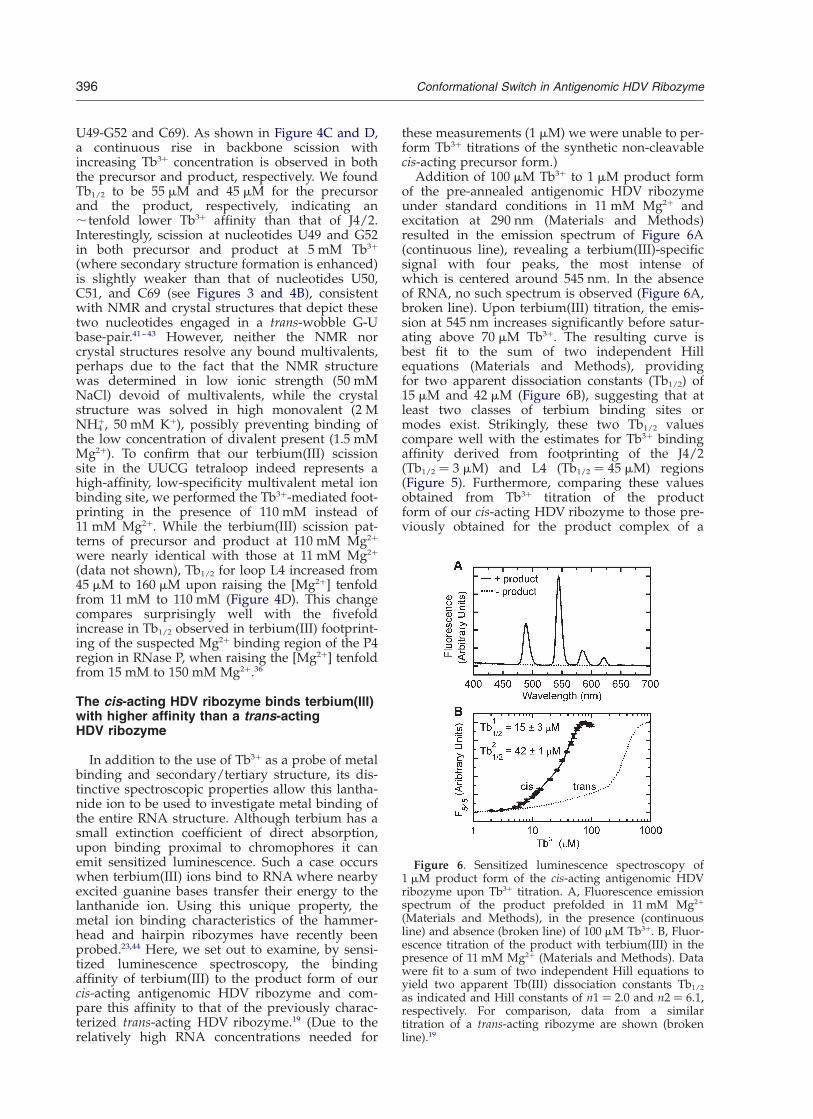
Addition of 100 mM Tb3þ to 1 mM product formof the pre-annealed antigenomic HDV ribozymeunder standard conditions in 11 mM Mg2þ andexcitation at 290 nm (Materials and Methods)resulted in the emission spectrum of Figure 6A(continuous line), revealing a terbium(III)-specificsignal with four peaks, the most intense ofwhich is centered around 545 nm. In the absenceof RNA, no such spectrum is observed (Figure 6A,broken line). Upon terbium(III) titration, the emis-sion at 545 nm increases significantly before satur-ating above 70 mM Tb3þ. The resulting curve isbest fit to the sum of two independent Hillequations (Materials and Methods), providingfor two apparent dissociation constants (Tb1/2) of15 mM and 42 mM (Figure 6B), suggesting that atleast two classes of terbium binding sites ormodes exist. Strikingly, these two Tb1/2 valuescompare well with the estimates for Tb3þ bindingaffinity derived from footprinting of the J4/2(Tb1/2 ¼ 3 mM) and L4 (Tb1/2 ¼ 45 mM) regions(Figure 5). Furthermore, comparing these valuesobtained from Tb3þ titration of the productform of our cis-acting HDV ribozyme to those pre-viously obtained for the product complex of a
Figure 6. Sensitized luminescence spectroscopy of1 mM product form of the cis-acting antigenomic HDVribozyme upon Tb3þ titration. A, Fluorescence emissionspectrum of the product prefolded in 11 mM Mg2þ
(Materials and Methods), in the presence (continuousline) and absence (broken line) of 100 mM Tb3þ. B, Fluor-escence titration of the product with terbium(III) in thepresence of 11 mM Mg2þ (Materials and Methods). Datawere fit to a sum of two independent Hill equations toyield two apparent Tb(III) dissociation constants Tb1/2
as indicated and Hill constants of n1 ¼ 2.0 and n2 ¼ 6.1,respectively. For comparison, data from a similartitration of a trans-acting ribozyme are shown (brokenline).19
396 Conformational Switch in Antigenomic HDV Ribozyme

trans-cleaving ribozyme, 70 mM and 340 mM,19 wefind an ,five- to eightfold higher terbium(III)binding affinity in the cis-acting ribozyme (Figure6B). This higher affinity for multivalents is possiblylinked to the ,30-fold faster cleavage rate constantof our cis-acting ribozyme compared to the trans-acting one under similar conditions (43 min21
(Table 1) versus 1.34 min21 18 in 11 mM Mg2þ (pH7.5), at 37 8C), which is consistently observedwhen severing the J1/2 and/or L4 connections(Figure 1) to obtain a trans-acting construct.9
Thermal melting curves confirm tertiarystructural differences between the precursorand product forms
To complement our terbium(III)-mediated foot-printing data with global structural information,thermal UV-melting profiles were acquired understandard conditions (11 mM Mg2þ, pH 7.5) for thenon-cleavable precursor and product forms of thecis-acting antigenomic HDV ribozyme (Materialsand Methods). Both the precursor and productexhibit two distinct folding transitions that havenearly the same melting temperatures (Tm) for thetwo RNAs. In the precursor the first transition(Tm1) occurs at 62 8C and the second transition(Tm2) at 84 8C, while in the product the transitionsoccur at 64 8C (Tm1) and 83 8C (Tm2) (Figure 7). Thepartial unfolding around Tm1 is most likely due toloss of some tertiary interactions, while the highermelting temperature, Tm2, most likely characterizesloss of secondary structure. Strikingly, while theoverall behavior of the precursor and product isquite similar, particularly the lower-temperaturetransitions have first derivatives of remarkablydifferent full-widths at half-maximum. From thisdifference we estimate a considerably lower tran-sition enthalpy of DH ¼ 224.6 kcal/mol for theprecursor compared to DH ¼ 290.1 kcal/mol inthe product (Figure 7). These observations suggestthat the precursor form of the cis-acting anti-genomic HDV ribozyme has a significantly moredynamic, less cooperative tertiary structure thanthe product form, consistent with our terbium(III)-mediated footprinting results.
Discussion
For over 20 years, since the discovery of RNAenzymes, it has been clear that RNA is not just apassive carrier of genetic information. In fact,RNA plays a key role in almost every aspect ofcellular metabolism, including protein transport,RNA splicing, peptide bond formation, and regu-lation of translation.45 – 47 Metal ions, particularlydivalents, are essential cofactors that aid in form-ing a fully structured and functional RNAmolecule.1 – 5 Ribozymes are no exception, as theyrequire metal ions to properly position reactivegroups such that they are poised for catalysis, andin some cases to directly participate in reaction
chemistry. Deciphering the exact role of thesemetals has proven quite challenging. The HDVribozyme is one example of a catalytic RNA thatappears to utilize both structural and catalyticmetal ions to carry out catalysis. Bevilacqua andco-workers have estimated that the overall rateenhancement by divalent metal ions is ,3000-fold, of which the catalytic contribution is ,25-fold and the remaining larger contribution ischiefly structural in nature.48 To complement theexisting biochemical data, we have usedterbium(III)-mediated footprinting to probe theHDV ribozyme’s metal ion binding properties aswell as secondary and tertiary structure.
Recently, Doudna and co-workers have solvedseveral precursor crystal structures of the genomicHDV ribozyme and have proposed, by comparisonwith the product crystal structure, that a significantconformational switch controls catalysis by ejectinga catalytically critical divalent metal ion from theactive site, thus preventing the 30 product fromcatalyzing re-ligation.22 Using terbium(III)-foot-printing in solution, we have observed subtle, yetsignificant structural differences between the pre-cursor and 30 product of the other natural HDVribozyme, the antigenomic form. While the pre-cursor and product are similar in global architec-ture and share helices P1–P4 and P1.1, they show
Figure 7. Thermal denaturation of the antigenomicHDV ribozyme in 11 mM Mg2þ. A, Melting profile of theprecursor. The relative absorbance of the RNA at260 nm (X, left axis) and a bimodal gaussian distributionfit to the smoothed first derivative (color, right axis) areshown. The melting temperatures Tm and associatedtransition enthalpies DH (in parentheses) for melting ofthe tertiary and secondary structure of the RNA weredetermined from the red and blue curves, respectively(Materials and Methods). B, Melting profile of theproduct, analyzed as described for A.
Conformational Switch in Antigenomic HDV Ribozyme 397

structural differences consistent with a confor-mational switch. Figure 8 identifies the five nucleo-tides in the genomic precursor and product crystalstructures that correspond to those nucleotides forwhich we observe the largest (. twofold) differ-ence in relative terbium(III)-induced scission inten-sity in the antigenomic sequence (arrows in Figure4A; please note that gA8, gC14, and gC26 in thegenomic crystal structure have their equivalents inC8, U17, and C29, respectively, in our antigenomicribozyme; g is used in the following to distinguishgenomic from antigenomic numbering). Impor-tantly, these five nucleotides divide into two differ-ent classes (Figure 4A): (1) gC14/U17, G80, andG81 (both G80 and G81 are common to both geno-mic and antigenomic), which are all located atthe bottom of helix P2 (Figures 1 and 8) and forwhich scission intensity decreases . twofold from
precursor to product; and (2) gA8/C8, in joinerJ1/2, and gC26/C29, in loop L3, for which scissionintensity increases . twofold from precursor toproduct. The first class of terbium(III)-inducedscission sites is located at the exact position, at thebottom of helix P2, where an earlier study found aMg2þ-induced scission site that is correlated withenzymatic activity of a shortened trans-actingHDV ribozyme.49 Combined with the fact that weobserve a decrease in terbium(III)-induced scissionat this site in the cleavage product, we proposethat this is a non-specific metal ion binding sitethat becomes significantly less populated uponcleavage. Given that it is distant from the cleavagesite by .20 A we further propose that this is animportant structural, rather than catalytic, metalbinding site in the antigenomic HDV ribozyme.
The second class of terbium(III) scission sites isenhanced in the product over the precursor struc-ture and is found in two separate locations thatare both implicated in the catalytic conformationalswitch of the genomic HDV ribozyme (Figure 8).22
gC26/C29 is part of loop L3 (Figure 1) and is oneof the nucleotides that undergo the most dramaticrearrangements upon cleavage in the genomicribozyme (Figure 8). These rearrangements in L3inevitably lead to changes in local electrostaticsand metal binding properties as well as backbonegeometry, thus providing for a plausible expla-nation for enhanced terbium(III)-induced scission.While gC26/C29 shows the most pronouncedenhancement within L3, the loop is generally cutquite strongly and its overall scission pattern exhi-bits among the most significant changes betweenprecursor and product (Figure 4). Similar obser-vations were made using terbium(III)-mediatedfootprinting on a trans-acting HDV ribozyme.19
These findings are striking since the available crys-tallographic evidence implicates L3 in binding of acatalytic divalent metal ion.22 We therefore proposethat L3 provides for a negative electrostatic surfacethat non-specifically localizes metal ions close tothe catalytic site; in case of hydrated divalents,such as Mg2þ and Ca2þ, this leads to catalysis,while trivalent metal ions such as Tb3þ and theCo(NH3)6
3þ complex bind tightly but are notcompatible with catalysis.
gA8/C8 in joiner J1/2 also shows enhanced ter-bium(III)-mediated scission in the product com-pared to the precursor, yet essentially does notrearrange upon cleavage (Figure 8). To understandthis behavior, it is instructive to consider the struc-tural context of this nucleotide; it is part of thestrand crossover between helices P1 and P2 thatjoins the two parallel stacks of the ribozyme, con-sisting of P1–P1.1–P4 and P2–P3 (Figure 1). gA8/C8 as well as its neighbor A9 (gU9 in the genomicsequence) stack on P1, while U10 (gG10) stacks onP2, leading to a sharp backbone turn betweenpositions 9 and 10.10,22 While this backbone geome-try does not change significantly upon cleavage,the conformational switch accompanying cleavageentails movement of the P1–P1.1–P4 and P2–P3
Figure 8. Backbone ribbon representations of the pre-cursor (red, PDB ID 1SJ3) and 30 product (silver, PDB ID1CX0) forms of the genomic HDV ribozyme crystal struc-tures. The indicated nucleotides represent the residuescorresponding to those in our antigenomic HDV ribo-zyme with the largest (. twofold) changes in terbiu-m(III)-mediated scission intensity between precursorand product. Interestingly, these sites mark regions thateither represent a known metal ion binding site at thebottom of helix P2 (gC14, G80, G81; highlighted in blackto indicate, as in Figure 4, a decrease in terbium(III)scission intensity by . twofold from precursor to pro-duct) or are implicated in conformational changesaccompanying and essential for catalysis (gA8 andgC26; highlighted in gray to indicate, as in Figure 4, anincrease in terbium(III) scission intensity by .twofoldfrom precursor to product). Please note that gA8, gC14,and gC26 in the genomic crystal structure have theirequivalents in C8, U17, and C29, respectively, in our anti-genomic ribozyme (parentheses), while G80 and G81 areshared between the two forms. This Figure was gener-ated using InsightII.
398 Conformational Switch in Antigenomic HDV Ribozyme
stacks towards each other as the “wedge” pre-sented by the sequence 50 to the cleavage site isremoved (Figure 8). A similar, yet more pro-nounced global conformational change has beenobserved by fluorescence resonance energy trans-fer (FRET) in a trans-acting ribozyme.17 – 19,50,51 It isfurther supported in our cis-acting antigenomicHDV ribozyme by the significantly less cooperativetertiary structure melting transition of the pre-cursor compared to the product, from which anearly 66 kcal/mol less favorable transitionenthalpy derives (Figure 7). This global structuralchange may well alter the structural dynamics orstrain around the joining J1/2 sequence in the cis-acting ribozyme, thus leading to the observedenhanced susceptibility toward terbium(III)scission after catalysis.
Another region that shows strong susceptibilitytowards terbium(III)-mediated backbone scission,especially in the product at low Tb3þ concen-trations, is the joiner J4/2 sequence that containsthe catalytic C76 residue. Again, similar obser-vations were made using terbium(III)-mediatedfootprinting on a trans-acting HDV ribozyme.19 Inthe cis-acting antigenomic ribozyme, the terbium-(III) affinity of the J4/2 region is in fact one of thehighest reported so far for an RNA,19,23,26,29,32 – 37
with Tb1/2 ¼ 5 mM and 3 mM in the precursor andproduct, respectively, in a background of 11 mMMg2þ (Figure 5A and B). A similar Tb1/2 of 4.2 mMwas observed in hammerhead ribozyme HH16under similar ionic conditions using luminescencetitration of Tb(III).44 Our findings are consistentwith Pb2þ-induced scission of the genomic clea-vage product, which occurs predominantly atC75/76 in J4/2 in the presence of low concen-trations of Mg2þ and which is suppressed byhigher Mg2þ concentrations.52 Furthermore, underbasic conditions (pH 9.0) specific cleavage isextended throughout the J4/2 joining region sothat cleavage is observed at G74, C75 (major clea-vage site) and G76 with Mg2þ, Mn2þ, or Ca2þ (corre-sponding to our antigenomic G75, C76, U77,respectively).52 Both the precursor and productcrystal structures depict the J4/2 region as a tighttrefoil turn with a sharp double-twist in the phos-phodiester backbone.10,22 This unique structuralmotif projects the catalytic C75/76 deeply into thecatalytic core so that it approaches the scissilephosphate, making the trefoil turn a key structuralfeature essential for formation of the catalyticpocket. In the refined genomic product crystalstructure, a Mg2þ is resolved that appears to beouter-sphere (i.e. mostly electrostatically) bound tothe phosphate of G76 (corresponding to U77 inour antigenomic ribozyme);15 if this metal ionwere replaced by Tb3þ, the transiently deproto-nated Tb(OH)(aq)2þ species may be close enoughto deprotonate the 20-OH of C75/76 (the metal–20O distance is 4.3 A10), leading to subsequent back-bone scission 30 to C75/76. This is consistentwith our observation of terbium(III)-mediatedbackbone scission with relative intensities of
C76 . U77 . G75 in the antigenomic HDV ribo-zyme (Figure 4A). Since the crystallographic J4/2-bound Mg2þ is at a distance of ,9 A from the scis-sile phosphate, its major function is likely thestructural organization of the essential trefoilmotif, not participation in catalysis. It should benoted, however, that inhibition of catalysis by Tb3þ
or Co(NH3)63þ may not necessarily relate to dis-
placement of a catalytically involved divalent bythe higher-affinity trivalent metal ion (complex);it is also conceivable that substitution of Tb3þ orCo(NH3)6
3þ for an essential structural metal ion,such as those bound to P2 or to joiner J4/2, mayresult in sufficient perturbation of the sugar-phos-phate backbone or base-stacking geometry torender a ribozyme molecule inactive. Such a possi-bility is supported by the fact that previous struc-ture probing by Pb2þ revealed that the formationof P2 directs the overall fold of the ribozyme,specifically the correct formation of L3 and J4/2.52
This latter finding parallels our data, whichsuggest significant cooperativity between bindingof Tb3þ at the bottom of P2 and in J4/2 (Figure 3),which then may render the structure and electro-statics of L3 such that the loop attracts a catalyticmetal ion. Also in support of the notion of an effectof terbium(III) on structural rather than catalyticmetal ions in RNA, two of the other small ribo-zymes, the hairpin and hammerhead, are bothcatalytically highly active in the absence of diva-lents, thus are not obligatory metallo-enzymes, yetare still strongly inhibited by Tb3þ.23,30,53
We also observe significant Tb3þ-induced scis-sion in the L4 tetraloop of both the precursor andproduct of our cis-acting antigenomic HDV ribo-zyme, which is suppressed by higher Mg2þ concen-trations (Figure 5C and D). To our knowledge, thisis the first evidence that the ubiquitous UUCG tet-raloop, a member of the most frequent and thermo-dynamically stable UNCG family of tetraloopsfound in natural RNAs,54 does in fact bind metalions with moderate affinity (Tb1/2 < 50 mM in abackground of 11 mM Mg2þ). High-resolutionstructures from NMR spectroscopy and X-ray crys-tallography did not reveal a well-coordinatedmetal binding site;41 – 43 however, metal ion bindingmay help explain the unusual stability of this tetra-loop motif. Our findings complement a recentreport on binding of europium(III), another lantha-nide metal ion, to the other stable family of tetra-loops of type GNRA.55 It is also interesting to notethat the Tb3þ affinity of ,50 mM that we find forthe UUCG tetraloop is similar to that of the lower-affinity class of Tb3þ binding sites observed by sen-sitized terbium(III) luminescence titration, whichappears to represent the largest fraction of terbiu-m(III) binding sites in the cis-acting HDV ribozyme(Figure 6).
In summary, the data presented here not onlyreveal that the cis-acting antigenomic HDV ribo-zyme undergoes a similar conformational switchin solution as has been proposed, based on crystal-lographic evidence, to enable catalysis in the
Conformational Switch in Antigenomic HDV Ribozyme 399

genomic form, but they also shed light on locationsof metal ions likely to be relevant to proper struc-ture formation and catalysis of the HDV ribozyme.Future studies are needed to address how exactlystructural and catalytic metal ions exert theirinfluence on catalysis.
Materials and Methods
Preparation of RNA
The non-cleavable precursor of our antigenomic HDVribozyme, modified with a 20-methoxy group at thecleavage site (Figure 1), was chemically synthesized byDharmacon Research, Inc., and the 20-orthoester protec-tion groups were removed following the manufacturer’srecommendations.56 Deprotected RNA was purified byC8 reverse-phase HPLC chromatography with a linearacetonitrile gradient in triethylammonium acetate asdescribed.57,58 The unmodified self-cleaved product formof the antigenomic HDV ribozyme was generated byrun-off transcription from a double-stranded, PCR-amplified template that encoded an upstream T7 promo-ter. Transcription reactions contained 40 mM Tris–HCl(pH 7.5), 15 mM MgCl2, 5 mM dithiothreitol, 2 mMspermidine, 4 mM each rNTP, five units/ml of inorganicpyrophosphatase, and 0.1 mg/ml of T7 RNA polymeraseand were incubated at 37 8C overnight (,16 hours). Theself-cleaved full-length transcript was isolated afterdenaturing, 8 M urea, 10% (w/v) polyacrylamide gelelectrophoresis by UV shadowing, diffusion elution ofsmall gel slices, and ethanol-precipitation.
For cleavage reactions, the radiolabeled precursorHDV ribozyme was transcribed as described above,except that 0.04 mCi of [a-32P]GTP and 0.125 mg/ml ofa DNA oligonucleotide were added to the reaction mix-ture. This DNA oligonucleotide has a sequence fullycomplementary to the 15 nucleotides at the 50 end of theprecursor (G27 to C8) and was used to increase the yieldof the uncleaved precursor RNA.59 The transcriptionreaction was incubated for one hour at 37 8C and theRNA fractionated by electrophoresis on an 8 M urea,10% (w/v) polyacrylamide denaturing gel. Uncleavedprecursor RNA was located by autoradiography, excised,eluted into 0.1 mM EDTA, and recovered by ethanol-precipitation. The radiolabeled RNA was stored in0.1 mM EDTA at 220 8C.
Terbium stock solutions
The highest purity terbium(III) chloride (99.9%) waspurchased from Sigma-Aldrich. TbCl3 stock solutions at100 mM were prepared in 5 mM cacodylate (pH 5.5)and stored in small aliquots at 220 8C to preventformation of insoluble hydroxide species.
Cleavage assays
Radiolabeled precursor RNA (5–50 nM) was heated to90 8C for two minutes in a buffer containing 5 mM Tris–HCl (pH 7.5), 0.5 mM spermidine. The precursor wasthen pre-incubated at 37 8C for ten minutes, after whichthe reactions were adjusted to the final pH with a buffercontaining 25 mM acetic acid, 25 mM Mes, 50 mM Tris–HCl (pH 7.5). These mixtures were incubated for anadditional five minutes at 37 8C, followed by addition of
0.25 volumes of a solution (also at 37 8C) containing55 mM MgCl2 and 0.5 mM spermidine to start thereaction.13 For cleavage assays performed in the presenceof 5 mM Tb3þ, the latter solution was supplemented with25 mM TbCl3. Cleavage kinetics were followed byremoving aliquots (5 ml) at specified times and quench-ing them with 10 ml of 80% (v/v) formamide, 0.025%(w/v) xylene cyanol, 0.025% (w/v) bromophenol blue,and 50 mM EDTA. The reaction product was separatedfrom the precursor by gel electrophoresis under denatur-ing conditions (8 M urea, 6% (w/v) polyacrylamidegels), quantified, and normalized to the sum of the pre-cursor and product bands using a PhosphorImagerStorm 840 with ImageQuant software (MolecularDynamics). Time traces of product formation were fitto the single-exponential first-order rate equationy ¼ y0 þ A1(1 2 e2t/t1), employing Marquardt–Leven-berg non-linear least-squares regression (Microcal Origin7.0), where A1 is the amplitude and t21 the pseudo first-order rate constant kobs.
Terbium(III)-mediated footprinting
To observe the slow backbone scission mediated byTb(OH)(aq)2þ, purified non-cleavable precursor and self-cleaved product forms of the antigenomic HDV ribo-zyme were (50-32P)-phosphorylated with T4 polynucleo-tide kinase and [g-32P]ATP and re-purified bydenaturing 8 M urea, 10% polyacrylamide gel electro-phoresis, followed by diffusion elution, and ethanol-pre-cipitation, as described.19,23,24 To resolve its 30 end regionthe precursor had to be 30 end labeled using [32P]pCpand T4 RNA ligase, since (50-32P)-labeling of the precur-sor did not resolve this region well due to smearing inthe gel. The radiolabeled RNA (250,000 cpm per 10 mlreaction volume) was pre-annealed in buffer (5 mMTris–HCl (pH 7.5), 0.5 mM spermidine), denatured at90 8C for two minutes, and incubated at 37 8C for tenminutes. To fold the tertiary structure of the RNA, Mg2þ
was added five minutes prior to the addition of variousTb3þ concentrations (1 mM to 5 mM final concentration)and incubated for an additional one hour at 37 8C. Thescission reaction was stopped by adding EDTA (pH 8.0),to a final concentration of 50 mM and ethanol-precipi-tation at 220 8C. The precipitated RNA was redissolvedin urea loading buffer (80% formamide, 0.025% xylenecyanol, 0.025% bromophenol blue, 50 mM EDTA, 9 Murea) and analyzed on an 8 M urea, wedged 15% poly-acrylamide sequencing gel, alongside sequencing lad-ders from partial digestion with G-specific RNase T1
and alkaline hydrolysis as described.19,23,24 Productbands were either directly visualized using autoradio-graphy or quantified, using a PhosphorImager Storm840 with ImageQuant software (Molecular Dynamics),and normalized by calculating a relative extent of scis-sion (P) from the equation:
P ¼
band intensity at nucleotide xXi
band intensity at nucleotide i
0BB@
1CCA
y ½Tb3þ�
band intensity at nucleotide xXi
band intensity at nucleotide i
0BB@
1CCA
0 mM½Tb3þ�
where y is the terbium(III) concentration in a particularscission reaction and x the analyzed nucleotide position
400 Conformational Switch in Antigenomic HDV Ribozyme

of the RNA. 0 mM [Tb3þ] signifies a control reactionincubated in the same fashion as the terbium(III)-con-taining ones, except that no terbium(III) was added.Normalizing in this way the intensities at the variousterbium(III) concentrations to the samples without ter-bium(III) corrects for any non-specific backgrounddegradation. To derive a Tb3þ affinity the relative percen-tage cut was plotted over varying terbium(III) concen-trations ([Tb3þ]) and fit to a Hill equation, essentially asdescribed:19,23
y ¼ ymax½Tb3þ�n
½Tb3þ�n þ Tbn1=2
to yield an apparent terbium(III) dissociation constantTb1/2 and a cooperativity or Hill constant n.
Terbium(III) luminescence measurements
Steady-state luminescence spectra of terbium(III)bound to the pre-annealed and equilibrated productform of the antigenomic HDV ribozyme (1 mM) in stan-dard reaction buffer (25 mM acetic acid, 25 mM Mes,50 mM Tris–HCl (pH 7.5), 11 mM MgCl2) at 37 8C weremeasured on an Aminco-Bowman Series 2 (AB2)spectrofluorometer (Thermo Spectronic), while slowlyincreasing the Tb3þ concentration. The latter was accom-plished by adding small aliquots of an identical solutionof 1 mM product in 25 mM acetic acid, 25 mM Mes,50 mM Tris–HCl (pH 7.5), 11 mM MgCl2, but sup-plemented with 0.4 mM Tb3þ, followed by mixing andremoval of a solution volume equivalent to that of theadded aliquot. This procedure ensures that neither theconcentrations of RNA and buffer nor the total solutionvolume change over the course of the titration. Aftereach titration step, the solution was equilibrated for fiveminutes until the signal had stabilized, before an emis-sion spectrum was recorded. Excitation was at 290 nm(slit width of 4 nm), while the steady-state emission wasscanned with a slit width of 8 nm. To extract the lumines-cence intensity of the major peak at 545 nm, each peakwas fit between 535 nm and 555 nm with a gaussiandistribution function:
y ¼ y0 þA
Wffiffiffiffiffiffiffiffip=2
p e22ðx2x0Þ2=w2
to yield the peak height as the pre-exponential factor,from which the background value in the absence of Tb3þ
was subtracted. These signals were plotted over varyingterbium(III) concentrations and were fit to a Hillequation, essentially as described above, to yield anapparent terbium(III) dissociation constant Tb1/2 and acooperativity or Hill constant n. For a fit over the com-plete terbium(III) titration range, a sum of two inde-pendent Hill equations produced the best result, asjudged by the x2 deviation and the residuals.
Thermal denaturation
Thermal denaturation of 1 mM precursor or productform of the cis-acting antigenomic HDV ribozyme wascarried out using a Beckman DU-640B UV–Vis spectro-photometer with High Performance Temperature Con-troller and Micro Auto 6 Tm cell holder. RNA samples(,300 ml) were prepared as described for the cleavagereaction and degassed for five minutes prior to obtainingUV melting curves. Absorbance readings at 260 nm werecollected every 1.0 deg.C as the sample was heated from
35 8C to 99 8C at a rate of 0.2 deg.C/minute. Each UVmelting curve represents data collected from two meltsof freshly prepared RNA. After normalization, the firstderivative was determined, smoothed by adjacentaveraging, and two melting temperatures were obtainedby fitting to two gaussian distributions using MicroCalOrigin 7.0. The full-widths at half-maximum of the gaus-sian distributions yield transition enthalpies from:
DH0 ¼B
1
T12
1
T2
� �
where B is 27.00 cal/mol K for this unimolecular melt-ing transition and T1 and T2 are the temperatures athalf-maximum of the first derivative curve.60
Acknowledgements
We thank Jennifer Doudna and Ailong Ke forsharing crystal structure data and a manuscriptprior to publication, Stephen Scaringe for synthe-sizing the 80-mer precursor RNA, and all themembers of the Walter laboratory for stimulatingdiscussions and thoughtful suggestions. This workwas supported by NIH grant GM62357 to N.G.W.,a University of Michigan Rackham Merit predoc-toral fellowship to R.A.T. and D.A.H., a pre-doc-toral Merck/UNCF fellowship to D.A.H., and anNIH supplement to grant GM62357 to R.A.T.
References
1. Hanna, R. & Doudna, J. A. (2000). Metal ions inribozyme folding and catalysis. Curr. Opin. Chem.Biol. 4, 166–170.
2. Pyle, A. M. (2002). Metal ions in the structure andfunction of RNA. J. Biol. Inorg. Chem. 7, 679–690.
3. DeRose, V. (2002). Two decades of RNA catalysis.Chem. Biol. 9, 961–969.
4. Bokinsky, G., Rueda, D., Misra, V. K., Rhodes, M. M.,Gordus, A., Babcock, H. P. et al. (2003). Single-molecule transition-state analysis of RNA folding.Proc. Natl Acad. Sci. USA, 100, 9302–9307.
5. Draper, D. E. (2004). A guide to ions and RNA struc-ture. RNA, 10, 335–343.
6. Hadziyannis, S. J. (1997). Review: hepatitis delta.J. Gastroenterol. Hepatol. 12, 289–298.
7. Lai, M. M. (1995). The molecular biology of hepatitisdelta virus. Annu. Rev. Biochem. 64, 259–286.
8. Macnaughton, T. B., Shi, S. T., Modahl, L. E. & Lai,M. M. (2002). Rolling circle replication of hepatitisdelta virus RNA is carried out by two different cellu-lar RNA polymerases. J. Virol. 76, 3920–3927.
9. Shih, I. H. & Been, M. D. (2002). Catalytic strategiesof the hepatitis delta virus ribozymes. Annu. Rev. Bio-chem. 71, 887–917.
10. Ferre-D’Amare, A. R., Zhou, K. & Doudna, J. A.(1998). Crystal structure of a hepatitis delta virusribozyme. Nature, 395, 567–574.
11. Perrotta, A. T., Shih, I. & Been, M. D. (1999). Imida-zole rescue of a cytosine mutation in a self-cleavingribozyme. Science, 286, 123–126.
12. Nakano, S., Chadalavada, D. M. & Bevilacqua, P. C.
Conformational Switch in Antigenomic HDV Ribozyme 401

(2000). General acid–base catalysis in the mechanismof a hepatitis delta virus ribozyme. Science, 287,1493–1497.
13. Wadkins, T. S., Shih, I., Perrotta, A. T. & Been, M. D.(2001). A pH-sensitive RNA tertiary interactionaffects self-cleavage activity of the HDV ribozymesin the absence of added divalent metal ion. J. Mol.Biol. 305, 1045–1055.
14. Oyelere, A. K., Kardon, J. R. & Strobel, S. A. (2002).pK(a) perturbation in genomic hepatitis delta virusribozyme catalysis evidenced by nucleotide analogueinterference mapping. Biochemistry, 41, 3667–3675.
15. Ferre-D’Amare, A. R. & Doudna, J. A. (2000).Crystallization and structure determination of ahepatitis delta virus ribozyme: use of the RNA-bind-ing protein U1A as a crystallization module. J. Mol.Biol. 295, 541–556.
16. Shih, I. & Been, M. D. (2001). Energetic contributionof non-essential 50 sequence to catalysis in a hepatitisdelta virus ribozyme. EMBO J. 20, 4884–4891.
17. Pereira, M. J., Harris, D. A., Rueda, D. & Walter, N. G.(2002). Reaction pathway of the trans-acting hepatitisdelta virus ribozyme: a conformational changeaccompanies catalysis. Biochemistry, 41, 730–740.
18. Harris, D. A., Rueda, D. & Walter, N. G. (2002). Localconformational changes in the catalytic core of thetrans-acting hepatitis delta virus ribozyme accom-pany catalysis. Biochemistry, 41, 12051–12061.
19. Jeong, S., Sefcikova, J., Tinsley, R. A., Rueda, D. &Walter, N. G. (2003). Trans-acting hepatitis deltavirus ribozyme: catalytic core and global structureare dependent on the 50 substrate sequence. Bio-chemistry, 42, 7727–7740.
20. Luptak, A., Ferre-D’Amare, A. R., Zhou, K., Zilm,K. W. & Doudna, J. A. (2001). Direct pK(a) measure-ment of the active-site cytosine in a genomichepatitis delta virus ribozyme. J. Am. Chem. Soc. 123,8447–8452.
21. Tanaka, Y., Tagaya, M., Hori, T., Sakamoto, T.,Kurihara, Y., Katahira, M. & Uesugi, S. (2002). Clea-vage reaction of HDV ribozymes in the presence ofMg2 þ is accompanied by a conformational change.Genes Cells, 7, 567–579.
22. Ke, A., Zhou, K., Ding, F., Cate, J. H. & Doudna, J. A.(2004). A conformational switch controls hepatitisdelta virus ribozyme catalysis. Nature, 429, 201–205.
23. Walter, N. G., Yang, N. & Burke, J. M. (2000). Probingnon-selective cation binding in the hairpin ribozymewith Tb(III). J. Mol. Biol. 298, 539–555.
24. Harris, D. A. & Walter, N. G. (2003). Probing RNAstructure and metal-binding sites using terbiumfootprinting. Curr. Protocols Nucl. Acid Chem. 6.8,6.8.1–6.8.8.
25. Sigel, R. K. & Pyle, A. M. (2003). Lanthanide ions asprobes for metal ions in the structure and catalyticmechanism of ribozymes. Met. Ions Biol. Syst. 40,477–512.
26. Saito, H. & Suga, H. (2002). Outersphere and inner-sphere coordinated metal ions in an aminoacyl-tRNA synthetase ribozyme. Nucl. Acids Res. 30,5151–5159.
27. Ciesiolka, J., Marciniec, T. & Krzyzosiak, W. (1989).Probing the environment of lanthanide binding sitesin yeast tRNA(Phe) by specific metal-ion-promotedcleavages. Eur. J. Biochem. 182, 445–450.
28. Matsumura, K. & Komiyama, M. (1997). Enormouslyfast RNA hydrolysis by lanthanide(III) ions underphysiological conditions: eminent candidates fornovel tools of biotechnology. J. Biochem. 122, 387–394.
29. Sigel, R. K., Vaidya, A. & Pyle, A. M. (2000). Metalion binding sites in a group II intron core. NatureStruct. Biol. 7, 1111–1116.
30. Feig, A. L., Scott, W. G. & Uhlenbeck, O. C. (1998).Inhibition of the hammerhead ribozyme cleavagereaction by site-specific binding of Tb. Science, 279,81–84.
31. Wadkins, T. S., Perrotta, A. T., Ferre-D’Amare, A. R.,Doudna, J. A. & Been, M. D. (1999). A nested doublepseudoknot is required for self-cleavage activity ofboth the genomic and antigenomic hepatitis deltavirus ribozymes. RNA, 5, 720–727.
32. Hargittai, M. R. & Musier-Forsyth, K. (2000). Use ofterbium as a probe of tRNA tertiary structure andfolding. RNA, 6, 1672–1680.
33. Hargittai, M. R., Mangla, A. T., Gorelick, R. J. &Musier-Forsyth, K. (2001). HIV-1 nucleocapsid pro-tein zinc finger structures induce tRNA(Lys,3) struc-tural changes but are not critical for primer/template annealing. J. Mol. Biol. 312, 985–997.
34. Flynn-Charlebois, A., Lee, N. & Suga, H. (2001).A single metal ion plays structural and chemicalroles in an aminoacyl-transferase ribozyme. Biochem-istry, 40, 13623–13632.
35. Vaidya, A. & Suga, H. (2001). Diverse roles of metalions in acyl-transferase ribozymes. Biochemistry, 40,7200–7210.
36. Kaye, N. M., Zahler, N. H., Christian, E. L. & Harris,M. E. (2002). Conservation of helical structure con-tributes to functional metal ion interactions in thecatalytic domain of ribonuclease P RNA. J. Mol. Biol.324, 429–442.
37. Saito, H. & Suga, H. (2002). Outersphere and inner-sphere coordinated metal ions in an aminoacyl-tRNA synthetase ribozyme. Nucl. Acids Res. 30,5151–5159.
38. Kaukinen, U., Lyytikainen, S., Mikkola, S. &Lonnberg, H. (2002). The reactivity of phospho-diester bonds within linear single-stranded oligo-ribonucleotides is strongly dependent on the basesequence. Nucl. Acids Res. 30, 468–474.
39. Bibillo, A., Figlerowicz, M., Ziomek, K. & Kierzek, R.(2000). The nonenzymatic hydrolysis of oligoribo-nucleotides. VII. Structural elements affectinghydrolysis. Nucleosides Nucleotides Nucl. Acids, 19,977–994.
40. Nakano, S., Proctor, D. J. & Bevilacqua, P. C. (2001).Mechanistic characterization of the HDV genomicribozyme: assessing the catalytic and structuralcontributions of divalent metal ions within a multi-channel reaction mechanism. Biochemistry, 40,12022–12038.
41. Cheong, C., Varani, G. & Tinoco, I., Jr (1990). Solutionstructure of an unusually stable RNA hairpin,50GGAC(UUCG)GUCC. Nature, 346, 680–682.
42. Allain, F. H. & Varani, G. (1995). Structure of the P1helix from group I self-splicing introns. J. Mol. Biol.250, 333–353.
43. Ennifar, E., Nikulin, A., Tishchenko, S., Serganov, A.,Nevskaya, N., Garber, M. et al. (2000). The crystalstructure of UUCG tetraloop. J. Mol. Biol. 304, 35–42.
44. Feig, A. L., Panek, M., Horrocks, W. D., Jr &Uhlenbeck, O. C. (1999). Probing the binding ofTb(III) and Eu(III) to the hammerhead ribozymeusing luminescence spectroscopy. Chem. Biol. 6,801–810.
45. Doudna, J. A. & Cech, T. R. (2002). The chemicalrepertoire of natural ribozymes. Nature, 418,222–228.
402 Conformational Switch in Antigenomic HDV Ribozyme

46. Moore, P. B. & Steitz, T. A. (2002). The involvement ofRNA in ribosome function. Nature, 418, 229–235.
47. Hannon, G. J. (2002). RNA interference. Nature, 418,244–251.
48. Nakano, S., Cerrone, A. L. & Bevilacqua, P. C. (2003).Mechanistic characterization of the HDV genomicribozyme: classifying the catalytic and structuralmetal ion sites within a multichannel reactionmechanism. Biochemistry, 42, 2982–2994.
49. Lafontaine, D. A., Ananvoranich, S. & Perreault, J. P.(1999). Presence of a coordinated metal ion in atrans-acting antigenomic delta ribozyme. Nucl. AcidsRes. 27, 3236–3243.
50. Tinsley, R. A., Harris, D. A. & Walter, N. G. (2003).Significant kinetic solvent isotope effects in foldingof the catalytic RNA from the hepatitis delta virus.J. Am. Chem. Soc. 125, 13972–13973.
51. Tinsley, 5.1., Harris, R. A., Walter, D. A. & Magnesium,N. G. (2004). dependence of the amplified confor-mational switch in the trans-acting hepatitis deltavirus ribozyme. Biochemistry, in the press.
52. Matysiak, M., Wrzesinski, J. & Ciesiolka, J. (1999).Sequential folding of the genomic ribozyme of thehepatitis delta virus: structural analysis of RNA tran-scription intermediates. J. Mol. Biol. 291, 283–294.
53. Murray, J. B., Seyhan, A. A., Walter, N. G., Burke,J. M. & Scott, W. G. (1998). The hammerhead, hairpin
and VS ribozymes are catalytically proficient inmonovalent cations alone. Chem. Biol. 5, 587–595.
54. Antao, V. P., Lai, S. Y. & Tinoco, I., Jr (1991). A ther-modynamic study of unusually stable RNA andDNA hairpins. Nucl. Acids Res. 19, 5901–5905.
55. Mundoma, C. & Greenbaum, N. L. (2002). Sequester-ing of Eu(III) by a GAAA RNA tetraloop. J. Am.Chem. Soc. 124, 3525–3532.
56. Scaringe, S. A. (2001). RNA oligonucleotide synthesisvia 50-silyl-20-orthoester chemistry. Methods, 23,206–217.
57. Walter, N. G. (2001). Structural dynamics of catalyticRNA highlighted by fluorescence resonance energytransfer. Methods, 25, 19–30.
58. Walter, N. G. (2002). Probing RNA structuraldynamics and function by fluorescence resonanceenergy transfer (FRET). Curr. Protocols Nucl. AcidChem. 11.10, 11.10.1–11.10.23.
59. Wadkins, T. S. & Been, M. D. (1997). Core-associatednon-duplex sequences distinguishing the genomicand antigenomic self-cleaving RNAs of hepatitisdelta virus. Nucl. Acids Res. 25, 4085–4092.
60. Marky, L. A. & Breslauer, K. J. (1987). Calculatingthermodynamic data for transitions of any molecu-larity from equilibrium melting curves. Biopolymers,26, 1601–1620.
Edited by J. Doudna
(Received 1 April 2004; received in revised form 17 May 2004; accepted 19 May 2004)
Conformational Switch in Antigenomic HDV Ribozyme 403