Embed Size (px)
Citation preview

TEST BANK FOR ORAL PATHOLOGY FOR THE DENTAL HYGIENIST 6TH EDITION BY IBSEN
CLICK HERE TO ACCESS FULL TEST BANK
TEST BANK

N U R S I N G T B . C O M
Chapter 02: Inflammation and Repair
Test Bank
MULTIPLE CHOICE
1. A decrease in the size and function of a cell, a tissue, an organ, or the whole body is referred
to as: A. emigration. B. atrophy. C. hyperplasia. D. phagocytosis.
ANS: B
Feedback
A Emigration is the passage of white blood cells through the endothelium and wall
of the microcirculation into the injured tissue. B Correct! Atrophy is defined as a decrease in the size and function of a cell, a
tissue, an organ, or the whole body. C Hyperplasia is the enlargement of a tissue or organ resulting from an increase in
the number of normal cells. D Phagocytosis is the process of ingestion and digestion of particulate material by
cells.
REF: Reactive Tissue Responses, pages 43-44 OBJ: 8
2. The first response of the body to injury is: A. anaphylaxis. B. erythema. C. fever. D. inflammation.
ANS: D
Feedback
A Anaphylaxis is a severe type of hypersensitivity or allergic reaction in which
there is an exaggerated immunologic reaction resulting from the release of
vasoactive substances such as histamine. B Erythema is redness of the skin or mucosa and is a local sign of inflammation. C Fever is the elevation of the normal body temperature and is a systemic sign of
inflammation. D Correct! The inflammatory response is the first reaction to injury, and it involves
a series of microscopic events.
REF: Inflammation, page 34 OBJ: 1
3. Which type of inflammation occurs when the injury is minimal and brief and its source is
removed from the tissue? A. Acute
TEST BANK FOR ORAL PATHOLOGY FOR THE DENTAL HYGIENIST 6TH EDITION BY IBSEN
NURSINGTB.COM

N U R S I N G T B . C O M
B. Chronic
C. Local D. Systemic
ANS: A
Feedback
A Correct! Acute inflammation occurs when the injury is minimal and brief. B Chronic inflammation occurs when the inflammatory response lasts for longer
periods, even indefinitely. C Local is a term used to describe a specific area of inflammation. D Systemic factors such as fever, leukocytosis, and lymphadenopathy occur when
the injury is extensive.
REF: Inflammation, page 34 OBJ: 2
4. Which type of cell is the first to arrive at the site of injury and is the primary cell type
involved in acute inflammation? A. Macrophage
B. Neutrophil C. Plasma cell D. Mast cell
ANS: B
Feedback
A The macrophage is the second cell type to participate in the inflammatory
response. B Correct! The neutrophil is the first cell to arrive at the site of injury and is the
primary cell type involved in acute inflammation. C The plasma cell is involved in chronic inflammation. D The mast cell participates in both the inflammatory and immune responses.
REF: White Blood Cells in the Inflammatory Response, page 38
OBJ: 4
5. Which one of the following is not a classic local sign of inflammation? A. Redness
B. Swelling
C. Leukocytosis
D. Loss of normal tissue function
ANS: C
Feedback
A Redness is a local clinical change at the site of injury and is one of the classic
local signs of inflammation. B Swelling is a local clinical change observed at the site of injury and is one of the
classic local signs of inflammation. C Correct! Leukocytosis is an increase in the number of white blood cells and is a
sign of systemic inflammation.
TEST BANK FOR ORAL PATHOLOGY FOR THE DENTAL HYGIENIST 6TH EDITION BY IBSEN
NURSINGTB.COM

N U R S I N G T B . C O M
D Loss of normal tissue function at the site of injury is a classic local sign of
inflammation.
REF: Leukocytosis, page 41 OBJ: 5
6. Healing of an injury in which there is little loss of tissue, such as a surgical incision, is
referred to as healing by: A. tertiary intention. B. keloid formation. C. secondary intention. D. primary intention.
ANS: D
Feedback
A Healing by tertiary intention occurs when an infection develops at the site of a
surgical incision that is healing by primary intention. Healing by secondary
intention may ensue. B Keloid formation is excessive scar tissue development that can occur in healing
by secondary intention when there is a significant loss of tissue. C Healing by secondary intention occurs when the injury involves significant loss
of tissue and the edges of the injury cannot be joined during healing. A large clot
forms, resulting in an increase in granulation tissue. D Correct! Healing by primary intention occurs when there is very little loss of
tissue. The clean edges of the surgical incision are joined with sutures, and very
little granulation tissue forms.
REF: Healing by Primary Intention, page 45 OBJ: 11
7. The wearing away of tooth structure during mastication is called: A. attrition. B. erosion. C. abrasion. D. abfraction.
ANS: A
Feedback
A Correct! Attrition is defined as the wearing away of tooth structure during
mastication. B Erosion is the loss of tooth structure from chemical action. C Abrasion is a pathologic wearing of tooth structure resulting from a repetitive
mechanical habit. D Abfraction is the result of biomechanical forces on the teeth.
REF: Attrition, page 46 OBJ: 14
8. The loss of tooth structure seen in bulimia is caused by: A. anorexia. B. erosion.
TEST BANK FOR ORAL PATHOLOGY FOR THE DENTAL HYGIENIST 6TH EDITION BY IBSEN
NURSINGTB.COM

N U R S I N G T B . C O M
C. attrition. D. bruxism.
ANS: B
Feedback
A Patients with anorexia nervosa do not vomit after eating. B Correct! Generalized erosion, especially on the lingual surfaces of maxillary
anterior teeth, is caused by frequent vomiting in patients with bulimia. C Attrition is the wearing away of tooth structure during mastication. D Bruxism occurs when there is nonfunctional grinding or clenching of the teeth.
REF: Erosion, page 49 OBJ: 14
9. A patient comes to the office for an emergency visit. The patient complains of a toothache in
the left mandibular posterior area. On clinical examination you notice a gray-to-white patch
on the left posterior buccal mucosa. On questioning, the patient tells you that this area is also
painful. After reviewing the patient’s medical history, you question the patient regarding his
recent use of:
A. hydrogen peroxide. B. aspirin. C. antibiotics. D. mouthwash.
ANS: B
Feedback
A A chemical burn from the use of hydrogen peroxide would be more diffuse,
probably bilateral, and not a white plaque. B Correct! This is a classic case of aspirin burn caused by the misuse of aspirin.
The patient placed aspirin near the tooth that was aching; thus necrosis of the
mucosa occurred, resulting in the painful white patch on the buccal mucosa. C Antibiotics would be taken systemically and most likely swallowed. D Commercial mouthwashes would not cause a localized lesion.
REF: Aspirin Burn, page 50 OBJ: 17
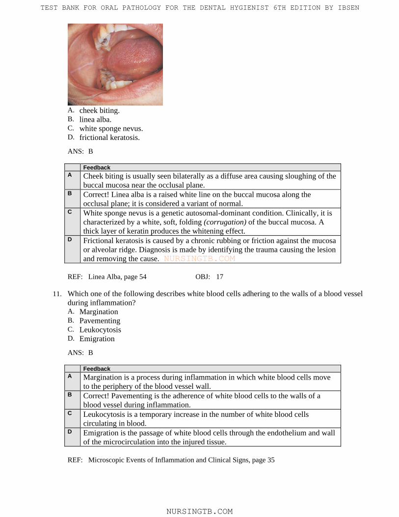
10. This white raised line observed on the buccal mucosa along the occlusal plane of the teeth is:
TEST BANK FOR ORAL PATHOLOGY FOR THE DENTAL HYGIENIST 6TH EDITION BY IBSEN
NURSINGTB.COM
N U R S I N G T B . C O M
A. cheek biting. B. linea alba. C. white sponge nevus. D. frictional keratosis.
ANS: B
Feedback
A Cheek biting is usually seen bilaterally as a diffuse area causing sloughing of the
buccal mucosa near the occlusal plane. B Correct! Linea alba is a raised white line on the buccal mucosa along the
occlusal plane; it is considered a variant of normal. C White sponge nevus is a genetic autosomal-dominant condition. Clinically, it is
characterized by a white, soft, folding (corrugation) of the buccal mucosa. A
thick layer of keratin produces the whitening effect. D Frictional keratosis is caused by a chronic rubbing or friction against the mucosa
or alveolar ridge. Diagnosis is made by identifying the trauma causing the lesion
and removing the cause.
REF: Linea Alba, page 54 OBJ: 17
11. Which one of the following describes white blood cells adhering to the walls of a blood vessel
during inflammation? A. Margination
B. Pavementing
C. Leukocytosis
D. Emigration
ANS: B
Feedback
A Margination is a process during inflammation in which white blood cells move
to the periphery of the blood vessel wall. B Correct! Pavementing is the adherence of white blood cells to the walls of a
blood vessel during inflammation. C Leukocytosis is a temporary increase in the number of white blood cells
circulating in blood. D Emigration is the passage of white blood cells through the endothelium and wall
of the microcirculation into the injured tissue.
REF: Microscopic Events of Inflammation and Clinical Signs, page 35
TEST BANK FOR ORAL PATHOLOGY FOR THE DENTAL HYGIENIST 6TH EDITION BY IBSEN
NURSINGTB.COM

N U R S I N G T B . C O M
OBJ: 1
12. Which one of the following is a systemic sign of inflammation? A. Redness
B. Pain
C. Loss of normal tissue function
D. Fever
ANS: D
Feedback
A Redness is a local sign of inflammation. B Pain is a local sign of inflammation caused by pressure on nerves by exudate
formation. C Loss of normal tissue function is a local sign of inflammation associated with
local swelling and pain. D Correct! Fever is a systemic sign of inflammation.
REF: Microscopic Events of Inflammation and Clinical Signs, Table 2-1, page 35
OBJ: 3
13. The enlargement of lymph nodes is called: A. atrophy. B. lymphadenopathy. C. hyperplasia. D. leukocytosis.
ANS: B
Feedback
A Atrophy is a decrease in size and function of a cell, a tissue, an organ, or the
whole body. B Correct! Lymphadenopathy occurs when lymph nodes become enlarged and
palpable. C Hyperplasia is an enlargement of a tissue or an organ resulting from an increase
in the number of normal cells. D Leukocytosis is a temporary increase in the number of white blood cells.
REF: Lymphadenopathy, page 41 OBJ: 1
14. The first microscopic event in the inflammatory response is: A. decreased blood flow. B. constriction of the microvasculature. C. phagocytosis. D. dilation of microvasculature.
ANS: B
Feedback
A Decreased blood flow occurs after exudate formation. B Correct! After injury to the tissue, the first microscopic event is constriction of
TEST BANK FOR ORAL PATHOLOGY FOR THE DENTAL HYGIENIST 6TH EDITION BY IBSEN
NURSINGTB.COM

N U R S I N G T B . C O M
the microvasculature. C Phagocytosis occurs when the white blood cells remove foreign substances from
the site by ingestion and digestion; these substances must be removed for the
inflammation to resolve. D Dilation of the microvasculature is the second microcirculation event to occur
after injury.
REF: Microscopic Events of Inflammation and Clinical Signs, page 35
OBJ: 4
15. Serous exudate is composed of: A. tissue debris and many white blood cells. B. suppuration. C. plasma fluids and proteins with a few white blood cells. D. plasma fluids and red blood cells.
ANS: C
Feedback
A Purulent exudate contains tissue debris and many white blood cells. B Suppuration is the formation and discharge of pus, as seen in purulent exudate. C Correct! Serous exudate is composed of plasma fluids and proteins with a few
white blood cells. D Serous describes the watery consistency of plasma. Red blood cells are not a
component of serous fluid.
REF: Microscopic Events of Inflammation and Clinical Signs, pages 36-37
OBJ: 5
16. When formation of exudate is excessive, a drainage tract may develop through the injured
tissue. This channel is often called: A. a fistula. B. leukocytosis. C. erythema. D. emigration.
ANS: A
Feedback
A Correct! A fistula is the channel through which excessive exudate passes to drain
to the outside. B Leukocytosis is a temporary increase in white blood cells. C Erythema is redness of the skin or mucosa. D Emigration occurs when white blood cells pass through the endothelium and
wall of the microcirculation into the injured tissue.
REF: Microscopic Events of Inflammation and Clinical Signs, page 37
OBJ: 5
17. Neutrophils constitute how much of the entire white blood cell population? A. 5%
TEST BANK FOR ORAL PATHOLOGY FOR THE DENTAL HYGIENIST 6TH EDITION BY IBSEN
NURSINGTB.COM

N U R S I N G T B . C O M
B. 20%
C. 65%
D. 90%
ANS: C
Feedback
A There are significantly more than 5% of neutrophils in the entire white blood cell
count. B There are significantly more than 20% of neutrophils in the entire white blood
cell count. C Correct! Neutrophils make up 60% to 70% of all white blood cells. D There are fewer than 90% of neutrophils in the entire white blood cell count.
REF: Neutrophils, page 39 OBJ: 6
18. All of the following are true concerning the neutrophil except that the neutrophil is: A. the first cell at the site of injury. B. the primary cell in acute inflammation. C. the primary cell in chronic inflammation. D. a phagocyte.
ANS: C
Feedback
A Neutrophils are the first cells at the site of injury. B Neutrophils are the primary cells in acute inflammation. C Correct! In chronic inflammation, the primary cells are macrophages,
lymphocytes, and plasma cells. D The main function of the neutrophil is phagocytosis.
REF: White Blood Cells in the Inflammatory Response, page 38
OBJ: 6
19. Which system in the blood mediates inflammation by causing increased dilation of the blood
vessels at the site of injury and increases the permeability of local blood vessels? A. Kinin system
B. Clotting system
C. Complement system
D. Lysosomal enzymes
ANS: A
Feedback
A Correct! The kinin system mediates inflammation by causing increased dilation
of the blood vessels at the site of injury and increases the permeability of local
blood vessels. B The clotting mechanism functions primarily in the clotting of blood. C The complement system involves the production of a sequential cascade of
plasma proteins that function in inflammation and immunity. D Lysosomal enzymes are released from granules in the white blood cells; they act
TEST BANK FOR ORAL PATHOLOGY FOR THE DENTAL HYGIENIST 6TH EDITION BY IBSEN
NURSINGTB.COM
N U R S I N G T B . C O M
as chemotactic factors and can cause damage to connective tissues and the clot
that has formed at the site of injury.
REF: Kinin System, page 40 OBJ: 4
20. Which one of the following is a steroidal antiinflammatory drug? A. Aspirin
B. Prednisone
C. Ibuprofen
D. Motrin
ANS: B
Feedback
A Aspirin is a nonsteroidal antiinflammatory agent. B Correct! Prednisone is a steroidal antiinflammatory drug. C Ibuprofen is a nonsteroidal antiinflammatory agent. D Motrin is ibuprofen, a nonsteroidal antiinflammatory drug.
REF: Antiinflammatory Drugs, page 43 OBJ: 6
21. Which of the following is defined as an increase in the number of cells in a tissue or organ? A. Hypertrophy
B. Atrophy
C. Hyperplasia
D. Repair
ANS: C
Feedback
A Hypertrophy is an increase in the size of an organ or tissue but not in the number
of cells. B Atrophy is a decrease in the size and function of a cell, a tissue, or an organ. C Correct! Hyperplasia is an increase in the number of cells in a tissue or organ. D Repair is the restoration of damaged or diseased tissue.
REF: Reactive Tissue Responses, page 43 OBJ: 1
22. Excessive scarring in skin is called: A. a keloid. B. healing by primary intention. C. a hematoma. D. healing by tertiary intention.
ANS: A
Feedback
A Correct! A keloid occurs when there is excessive scarring in the skin. B In healing by primary intention, very little granulation tissue forms. C A hematoma occurs when there is hemorrhage into the tissue. This may impair
healing.
TEST BANK FOR ORAL PATHOLOGY FOR THE DENTAL HYGIENIST 6TH EDITION BY IBSEN
NURSINGTB.COM

N U R S I N G T B . C O M
D Healing by tertiary intention occurs after the infection has been resolved and
surgical tissue repair has been performed.
REF: Healing by Secondary Intention, page 46 OBJ: 9
23. The first sign of attrition is: A. open contacts. B. disappearance of mamelons on incisors. C. temporomandibular joint dysfunction. D. biomechanical forces on the teeth.
ANS: B
Feedback
A Open contacts are associated with erosion. B Correct! The first sign of attrition is the disappearance of mamelons on incisors. C Temporomandibular joint dysfunction problems are more likely associated with
bruxism. Excessive attrition, muscle pain, and wear facets are present in
bruxism. D Abfraction results from biomechanical forces on the teeth.
REF: Attrition, page 46 OBJ: 14
24. Which one of the following is not a cause of abrasion? A. Pipe placement by pipe smokers
B. Playing wind instruments
C. Holding needles or pins with the teeth
D. Frequent sucking on lemons
ANS: D
Feedback
A Abrasion can be caused by pipe placement by pipe smokers. B Abrasion may be caused by playing wind instruments. C Abrasion can be caused by needles and pins held between the teeth. D Correct! Erosion, not abrasion, can be caused by the frequent sucking of lemons.
REF: Erosion, pages 48-49 OBJ: 14
25. This type of erosion is classically associated with:
A. anorexia nervosa. B. bulimia. C. sucking lemons.
TEST BANK FOR ORAL PATHOLOGY FOR THE DENTAL HYGIENIST 6TH EDITION BY IBSEN
NURSINGTB.COM

N U R S I N G T B . C O M
D. abrasive toothpaste.
ANS: B
Feedback
A Anorexia nervosa is an eating disorder, but it is not associated with erosion
because vomiting after eating is not a component of the disorder. B Correct! Bulimia is an eating disorder characterized by food binges followed by
self-induced vomiting that causes erosion to the lingual aspects of the teeth. C Sucking lemons would cause erosion to the facial aspects of the teeth. D Abrasive toothpastes are more responsible for contributing to abrasion.
REF: Erosion, Figure 2-25, page 49 OBJ: 14
26. Aspirin burn on the oral mucosa: A. is caused by ingestion of too many aspirin tablets. B. is caused by placing the aspirin on the tooth with the toothache, causing the
surrounding mucosa to become necrotic. C. is painless. D. requires a biopsy for diagnosis.
ANS: B
Feedback
A Aspirin burn is caused by a topical misuse of aspirin; it is not systemic. B Correct! Aspirin burn is caused when aspirin is placed on the tooth with the
toothache, causing the surrounding mucosa to become necrotic. C Aspirin burn is very painful and slow to heal. D Aspirin burn is usually diagnosed by questioning the patient to reveal the cause
of the lesion.
REF: Aspirin Burn, page 50 OBJ: 17
27. Electric burns in the oral area are usually seen in: A. electricians. B. infants and young children. C. the elderly. D. individuals involved in an electrical fire.
ANS: B
Feedback
A Electricians do not usually have electric burns in the oral area. B Correct! Electric burns in the oral area are most often seen in infants and young
children who have bitten or chewed a live electrical cord. C The elderly do not usually have electric burns in the oral area. D Individuals in an electrical fire do not usually have electric burns in the oral area.
REF: Electric Burn, page 51 OBJ: 17
28. The diagnosis of a traumatic ulcer is usually based on:
TEST BANK FOR ORAL PATHOLOGY FOR THE DENTAL HYGIENIST 6TH EDITION BY IBSEN
NURSINGTB.COM

N U R S I N G T B . C O M
A. history of the lesion. B. scalpel biopsy. C. therapeutic procedures. D. laboratory tests.
ANS: A
Feedback
A Correct! Traumatic ulcers are usually diagnosed on the basis of the relationship
of the history to the lesion. B Scalpel biopsy is not used in the diagnosis of traumatic ulcers. However, if the
trauma persists and the ulcer lasts 14 days, a biopsy may be performed. C Therapeutic measures are not used to diagnose traumatic ulcers. D Laboratory tests are not used to diagnose traumatic ulcers.
REF: Traumatic Ulcer, page 53 OBJ: 17
29. The major cause of a mucocele is: A. a sialolith. B. salivary duct obstruction. C. trauma to a minor duct. D. allergic reaction.
ANS: C
Feedback
A A sialolith is a salivary gland stone. B Dilated salivary gland ducts are believed to develop as a result of salivary duct
obstruction. C Correct! The major cause of a mucocele is trauma to a minor duct. The mucous
salivary gland secretion spills into the adjacent connective tissue. D A mucocele is not caused by an allergic reaction.
REF: Mucous Retention Lesions, page 57 OBJ: 19
30. Necrotizing sialometaplasia is thought to result from: A. lack of blood supply to the affected salivary gland. B. a sialolith. C. trauma to the floor of the mouth. D. pleomorphic adenoma.
ANS: A
Feedback
A Correct! Necrotizing sialometaplasia results from lack of blood supply to the
affected salivary gland. B A sialolith is a salivary gland stone that causes an obstruction in the salivary
gland. C Necrotizing sialometaplasia occurs on the hard palate, not the floor of the mouth. D Pleomorphic adenoma is a benign salivary gland tumor found unilaterally on the
posterior palate.
TEST BANK FOR ORAL PATHOLOGY FOR THE DENTAL HYGIENIST 6TH EDITION BY IBSEN
NURSINGTB.COM

N U R S I N G T B . C O M
REF: Necrotizing Sialometaplasia, page 59 OBJ: 18
31. Which of the following is most likely to result in frictional keratosis? A. High-fiber diet B. Chewing on an edentulous ridge
C. Malignancy
D. Daily use of mouthwash
ANS: B
Feedback
A A high-fiber diet does not cause frictional keratosis. B Correct! Frictional keratosis results from chronic chewing on an edentulous
ridge. C Frictional keratosis is not associated with malignancy. D Mouthwashes do not cause frictional keratosis.
REF: Frictional Keratosis, page 53 OBJ: 17
32. This lesion on the palate is typically associated with heavy pipe and cigar smoking and is
called:
A. tobacco pouch keratosis. B. necrotizing sialometaplasia. C. nicotine stomatitis. D. frictional keratosis.
ANS: C
Feedback
A Tobacco pouch keratosis occurs in the mucobuccal fold and is caused by
chewing/spitting tobacco. B Necrotizing sialometaplasia is caused by lack of blood supply to a specific area
of the palate. An ulcer is often present. C Correct! Nicotine stomatitis is a benign lesion of the hard palate typically
associated with heavy pipe and cigar smoking. D Frictional keratosis results from chronic chewing on an edentulous alveolar
ridge.
REF: Nictotine Stomatitis, page 54 OBJ: 18
33. Traumatic neuroma is a lesion caused by injury to:
TEST BANK FOR ORAL PATHOLOGY FOR THE DENTAL HYGIENIST 6TH EDITION BY IBSEN
NURSINGTB.COM

N U R S I N G T B . C O M
A. The epithelium
B. A peripheral nerve
C. A salivary gland
D. Striated muscle
ANS: B
Feedback
A The traumatic neuroma does not result from epithelial injury. B Correct! The traumatic neuroma is a lesion caused by injury to a peripheral
nerve. The mental foramen is the most common location. C The traumatic neuroma does not result from injury to a salivary gland. D The traumatic neuroma does not result from injury to striated muscle.
REF: Traumatic Neuroma, page 54 OBJ: 18
34. Which of the following is a lesion that occurs on the gingiva or alveolar process and contains
many multinucleated giant cells, red blood cells, and chronic inflammatory cells? A. Ranula
B. Central giant cell granuloma
C. Fibroma
D. Peripheral giant cell granuloma
ANS: D
Feedback
A The ranula is found on the floor of the mouth. B The central giant cell granuloma is found within bone. C The fibroma occurs most frequently on the buccal mucosa and is composed of
dense scarlike connective tissue containing few blood vessels. D Correct! The peripheral giant cell granuloma occurs on the gingiva or alveolar
process; originates from the periodontal ligament; is thought to be a response to
injury; and histologically is characterized by many multinucleated giant cells,
red blood cells, and inflammatory cells.
REF: Peripheral Giant Cell Granuloma, page 60 OBJ: 18
35. Epulis fissuratum is caused by: A. Denture adhesive products
B. Poor suction in the palatal vault C. Poor denture hygiene
D. An ill-fitting denture flange
ANS: D
Feedback
A Denture adhesive products do not cause epulis fissuratum. B Poor suction in the palatal vault causes papillary hyperplasia of the palate. C Poor denture hygiene does not cause epulis fissuratum. D Correct! Epulis fissuratum is caused by an ill-fitting denture flange.
TEST BANK FOR ORAL PATHOLOGY FOR THE DENTAL HYGIENIST 6TH EDITION BY IBSEN
NURSINGTB.COM

N U R S I N G T B . C O M
REF: Denture-induced Fibrous Hyperplasia, page 61 OBJ: 18
36. This granular, erythematous papillary surface of the palatal vault was caused by:
A. Poor oral hygiene
B. An ill-fitting suction area of a maxillary denture
C. The denture flange
D. Soaking the denture in caustic rinses
ANS: B
Feedback
A Poor oral hygiene does not cause papillary hyperplasia. It may contribute to the
inflammatory response of the area. B Correct! Papillary hyperplasia is caused by the palatal suction of an ill-fitting
maxillary denture. C The ill-fitting denture flange causes epulis fissuratum. D Soaking the denture in caustic rinses may contribute to inflammation but not
papillary hyperplasia.
REF: Inflammatory Papillary Hyperplasia of the Palate, page 62
OBJ: 17
37. The most common site for the pulp polyp is: A. the occlusal surface of a large open carious tooth. B. the apex of the tooth. C. the gingival margin of the tooth. D. deep in the pulp canal.
ANS: A
Feedback
A Correct! The most common site for the pulp polyp is in the occlusal surface of
large open carious teeth. It is seen as a red or pink nodule that fills the occlusal
surface. It is an excessive proliferation of chronically inflamed dental pulp
tissue. B Pulp polyps are not seen at the apex of teeth. C Pulp polyps are not seen on the gingival margin of teeth. D Pulp polyps are not seen deep in the pulp canal.
TEST BANK FOR ORAL PATHOLOGY FOR THE DENTAL HYGIENIST 6TH EDITION BY IBSEN
NURSINGTB.COM

N U R S I N G T B . C O M
REF: Chronic Hyperplastic Pulpitis, page 63 OBJ: 18
38. Which one of the following does not cause gingival enlargement? A. Hormonal changes
B. Calcium channel blockers
C. Hereditary factors
D. Nitroglycerin
ANS: D
Feedback
A Hormonal changes do contribute to gingival enlargement. B Calcium channel blockers do cause gingival enlargement. C Certain hereditary factors do cause gingival enlargement. D Correct! Nitroglycerin is prescribed for angina and does not cause gingival
enlargement.
REF: Gingival Enlargement, pages 62-63 OBJ: 8
39. Which one of the following inflammatory periapical lesions is most painful? A. Periapical abscess
B. Periapical granuloma
C. Radicular cyst D. Residual cyst
ANS: A
Feedback
A Correct! The periapical abscess is associated with severe pain caused by the
inflammation. B Periapical granuloma is most often asymptomatic. C The radicular cyst is often asymptomatic and discovered on radiographic
examination. D The residual cyst forms when the radicular cyst is incompletely removed and left
behind at the extraction site.
REF: Periapical Abscess, page 63 OBJ: 22
40. Resorption of tooth structure from outside the tooth is called: A. internal resorption. B. external resorption. C. idiopathic tooth resorption. D. condensing osteitis.
ANS: B
Feedback
A Internal resorption begins inside the pulpal area. B Correct! External resorption begins outside the tooth. C Idiopathic tooth resorption can involve the crown or roots of impacted teeth, and
the cause cannot be identified.
TEST BANK FOR ORAL PATHOLOGY FOR THE DENTAL HYGIENIST 6TH EDITION BY IBSEN
NURSINGTB.COM

N U R S I N G T B . C O M
D Condensing osteitis is a change in the bone near the apices of teeth that is
thought to be a reaction to a low-grade infection. The mandibular first molar is
most commonly involved, and the area is seen radiographically as a radiopacity
below the root apex of the involved tooth.
REF: Tooth Resorption, page 64 OBJ: 13
41. A process during inflammation in which white blood cells move to the blood vessel wall is
referred to as: A. chemotaxis. B. margination. C. leukocytosis. D. transudate.
ANS: B
Feedback
A Chemotaxis is the directed movement of white blood cells to the area of injury
by biochemical mediators. B Correct! Margination is defined as a process during inflammation in which white
blood cells move to the blood vessel wall. C Leukocytosis is a temporary increase in the number of white blood cells
circulating in blood. D Transudate is the fluid component of blood that normally passes through the
endothelial walls of the microvasculature.
REF: Microscopic Events of Inflammation and Clinical Signs, page 35
OBJ: 1
42. An example of an irreversible cellular response that occurs during tissue injury is: A. atrophy. B. hypertrophy. C. hyperplasia. D. necrosis.
ANS: D
Feedback
A Atrophy is an example of a reversible cellular response. B Hypertrophy is an example of a reversible cellular response. C Hyperplasia is an example of a reversible cellular response. D Correct! Necrosis is the pathologic death of one or more cells or a portion of the
tissue or an organ that results from irreversible damage to cells.
REF: Injury, page 34 OBJ: 2
43. The inflammatory response is a dynamic process, continually changing in response to injury
and repair. Repair of tissue occurs only if the persistent source of injury is removed. A. Both statements are true. B. Both statements are false. C. The first statement is true; the second is false.
TEST BANK FOR ORAL PATHOLOGY FOR THE DENTAL HYGIENIST 6TH EDITION BY IBSEN
NURSINGTB.COM

N U R S I N G T B . C O M
D. The first statement is false; the second is true.
ANS: A
Feedback
A Correct! Both statements are true. B Both statements are true. C Both statements are true. D Both statements are true.
REF: Inflammation, page 34 OBJ: 3
44. Hyperemia is responsible for which two clinical signs of inflammation? A. Emigration and pain
B. Heat and erythema
C. Transudation and redness
D. Swelling and chemotaxis
ANS: B
Feedback
A Emigration is the process by which the white blood cells escape from the blood
vessels and is not a clinical sign of inflammation. B Correct! Heat and erythema are caused by hyperemia. C Transudation is the process of plasma cells passing between the endothelial cells
and entering the tissue and is not a clinical sign of inflammation. D Chemotaxis is the directed movement of white blood cells toward the site of the
injury and is not a clinical sign of inflammation.
REF: Microscopic Events of Inflammation and Clinical Signs, page 35
OBJ: 4
45. During the microscopic event of inflammation, pain may be caused by which of the
following? A. Phagocytosis
B. Leukocytosis
C. Exudate formation
D. Anaphylaxis
ANS: C
Feedback
A Phagocytosis is the ingestion and digestion of foreign substances and is not a
cause of pain. B Leukocytosis is an increase in the number of white blood cells and is not a cause
of pain. C Correct! Exudate presses on sensory nerves and may cause pain. D Anaphylaxis is a type of hypersensitivity or allergic reaction and is not a direct
cause of pain.
REF: Microscopic Events of Inflammation and Clinical Signs, page 35
TEST BANK FOR ORAL PATHOLOGY FOR THE DENTAL HYGIENIST 6TH EDITION BY IBSEN
NURSINGTB.COM

N U R S I N G T B . C O M
OBJ: 5
46. During the acute inflammatory process, the second type of white blood cell to emigrate from
the blood vessel into the injured tissue is the: A. macrophage. B. neutrophil. C. plasma cell. D. lymphocyte.
ANS: A
Feedback
A Correct! The macrophage is the second cell to participate in the inflammatory
response. B The neutrophil is the first cell to arrive at the site of injury and is the primary cell
involved in acute inflammation. C The plasma cell is involved in chronic inflammation. D The lymphocyte is involved in chronic inflammation.
REF: White Blood Cells in the Inflammatory Response, page 38
OBJ: 6
47. Each of the following statements regarding the atrophy of tissue cells is true except one.
Which one is the exception? A. Atrophied cells are capable of returning to their normal size after stress is
removed. B. Atrophy can occur with changes in cellular growth, malnutrition, ischemia, or
hormonal changes. C. Atrophy can be present in the muscular wasting that occurs in some chronic
diseases that do not allow for mobility. D. Atrophy occurs in the smooth muscles of the uterus and the mammary glands in
response to pregnancy.
ANS: D
Feedback
A This statement is true. B This statement is true. C This statement is true. D Correct! Hypertrophy occurs in the smooth muscles of the uterus and the
mammary glands in response to pregnancy.
REF: Reactive Tissue Response, page 43 OBJ: 8
48. If the source of injury has been completely removed, the inflammation and immune responses
in the tissues are completed in approximately what time frame? A. Day after removal of injury
B. Two days after removal of injury
C. Seven days after removal of injury
D. Two weeks after removal of injury
TEST BANK FOR ORAL PATHOLOGY FOR THE DENTAL HYGIENIST 6TH EDITION BY IBSEN
NURSINGTB.COM

N U R S I N G T B . C O M
ANS: C
Feedback
A The day after injury removal, acute inflammation takes place in the area of
future repair. B Two days after removal of injury, fibroplasia, angiogenesis, the formation of
granulation tissue, and epithelialization occur. C Correct! If the source of injury has been completely removed, the inflammation
and immune responses in the tissues are completed in approximately 7 days. D Two weeks after removal of injury, matured fibrous connective tissue or scar
tissue occurs.
REF: Seven Days After Injury, page 45 OBJ: 9
49. Repair of bone injury is similar to the process that takes place in fibrous connective tissue
except that it involves the creation of bone tissue. The removal of osteoblast-producing tissues
and excessive movement of the bone promote bone healing. A. Both statements are true. B. Both statements are false. C. The first statement is true; the second is false. D. The first statement is false; the second is true.
ANS: C
Feedback
A The first statement is true and the second is false. B The first statement is true and the second is false. C Correct! The removal of osteoblast-producing tissues and excessive movement
of the bone can interrupt healing. D The first statement is true and the second is false.
REF: Bone Tissue Repair, page 46 OBJ: 10
50. In cases of healing, if an infected injury is left open and the edges are not surgically joined
until the infection is controlled, this is referred to as _____ intention. A. primary
B. secondary
C. tertiary
ANS: C
Feedback
A Healing by primary intention refers to the healing of an injury in which little loss
of tissue takes place. B Healing by secondary intention involves injury in which tissue is lost; thus the
edges of the injury cannot be joined by healing. C Correct! Healing by tertiary intention occurs when an infected injury is left open
and the edges are not surgically joined until the infection is controlled.
REF: Healing by Tertiary Intention, page 46 OBJ: 11
TEST BANK FOR ORAL PATHOLOGY FOR THE DENTAL HYGIENIST 6TH EDITION BY IBSEN
NURSINGTB.COM

N U R S I N G T B . C O M
51. Each of the following is a factor that may impair healing except one. Which one is the
exception? A. Tobacco use
B. Staphylococcus
C. Nutritional supplements
D. Renal failure
ANS: C
Feedback
A Tobacco use has been shown to impair healing. B Staphylococcus infection has been shown to impair healing. C Correct! The use of nutritional supplements has not been shown to impair
healing. D Renal failure has been shown to impair healing.
REF: Factors that Impair Healing, page 46 OBJ: 12
52. A tooth must be extracted if internal root resorption is present and a perforation occurs. A. Both the statement and reason are correct and related. B. Both the statement and reason are correct but not related. C. The statement is correct, but the reason is not. D. The statement is not correct, but the reason is correct. E. Neither the statement nor the reason is correct.
ANS: A
Feedback
A Correct! A tooth must be extracted if internal root resorption is present and a
perforation occurs. B Both the statement and reason are correct and related. C Both the statement and reason are correct and related. D Both the statement and reason are correct and related. E Both the statement and reason are correct and related.
REF: Tooth Resorption, page 64 OBJ: 13
53. A wedge-shaped defect at the cervical area of a tooth, the cause of which is related to
microfracture of the tooth structure in areas of concentration of stress, is called: A. attrition. B. erosion. C. abrasion. D. abfraction.
ANS: D
Feedback
A Attrition is defined as the wearing away of tooth structure during mastication. B Erosion is the loss of tooth structure from chemical action. C Abrasion is a pathologic wearing of tooth structure resulting from a repetitive
mechanical habit.
TEST BANK FOR ORAL PATHOLOGY FOR THE DENTAL HYGIENIST 6TH EDITION BY IBSEN
NURSINGTB.COM

N U R S I N G T B . C O M
D Correct! Abfraction is the result of biomechanical forces on the teeth.
REF: Abfraction, page 48 OBJ: 14
54. Which of the following is characteristic of erosion? A. It is a pathologic wearing away of tooth structure that results from a repetitive
mechanical habit. B. It is caused by local factors such as occlusal interferences in combination with
stress and tension. C. If tooth structure is lost around a restoration, the restoration will appear raised
from the surrounding demineralized tooth structure. D. Its first clinical sign is the disappearance of mamelons on the anterior teeth and the
flattening of occlusal cusps on the molar teeth.
ANS: C
Feedback
A This is the definition of abrasion. B This describes bruxism. C Correct! This phenomenon is seen in erosion and is not seen in abrasion,
bruxism, or attrition. D This describes attrition.
REF: Erosion, pages 48-49 OBJ: 15
55. Aspirin burn to the oral mucosa appears as: A. white. B. pigmented. C. bulbous. D. papillary.
ANS: A
Feedback
A Correct! Aspirin burn causes the tissue to become necrotic and appears white. B Aspirin burn does not appear pigmented. C Aspirin burn does not appear bulbous. D Aspirin burn does not appear papillary.
REF: Aspirin Burn, page 50 OBJ: 17
56. The most likely cause of a ranula is: A. inflammation of gland tissue. B. blockage of blood supply. C. trauma to a minor duct. D. salivary duct obstruction.
ANS: D
Feedback
A Inflammation of salivary gland tissue is referred to as sialadenitis.
TEST BANK FOR ORAL PATHOLOGY FOR THE DENTAL HYGIENIST 6TH EDITION BY IBSEN
NURSINGTB.COM

N U R S I N G T B . C O M
B Necrotizing sialometaplasia results from blockage of the blood supply. C The major cause of a mucocele is trauma to a minor duct. D Correct! Salivary gland obstruction is the most likely cause of a ranula.
REF: Mucous Retention Lesions, page 58 OBJ: 19
57. Each of the following is most likely to result in frictional keratosis except one. Which one is
the exception? A. Chronic cheek biting
B. Chewing on an edentulous ridge
C. Cigarette smoking
D. Tongue chewing
ANS: C
Feedback
A Chronic cheek biting can result in frictional keratosis. B Frictional keratosis results from chronic chewing on an edentulous ridge. C Correct! Frictional keratosis is not associated with cigarette smoking. D Tongue chewing can result in frictional keratosis.
REF: Frictional Keratosis, page 53 OBJ: 17
58. This sessile-based lesion is on the gingiva of a 13-year-old female. It is soft to palpation
and bleeds easily.
The accurate diagnosis for this lesion is: A. peripheral giant cell granuloma. B. pyogenic granuloma. C. traumatic neuroma. D. irritation fibroma.
ANS: B
Feedback
A Peripheral giant cell granuloma does not exhibit these characteristics. B Correct! Pyogenic granulomas are commonly found in teenagers and exhibit a
lesion that is soft to palpation and bleeds easily. C Traumatic neuroma does not exhibit these characteristics. D Irritation fibroma does not exhibit these characteristics.
REF: Pyogenic Granuloma, page 59 OBJ: 19
59. These elongated folds of tissue are a result of irritation from an ill-fitting denture.
TEST BANK FOR ORAL PATHOLOGY FOR THE DENTAL HYGIENIST 6TH EDITION BY IBSEN
NURSINGTB.COM

N U R S I N G T B . C O M
The accurate diagnosis for this lesion is: A. palatal papillomatosis. B. gingival hyperplasia. C. chronic hyperplastic pulpitis. D. epulis fissuratum.
ANS: D
Feedback
A Palatal papillomatosis is seen on the palate. B Gingival hyperplasia is an enlargement of the gingiva. C Chronic hyperplastic pulpitis is an excessive proliferation of chronically
inflamed dental pulp tissue. D Correct! Epulis fissuratum (denture-induced fibrous hyperplasia) consists of
elongated folds of tissue as a result of irritation from an ill-fitting denture.
REF: Denture-induced Fibrous Hyperplasia, page 61 OBJ: 18
TEST BANK FOR ORAL PATHOLOGY FOR THE DENTAL HYGIENIST 6TH EDITION BY IBSEN
NURSINGTB.COM