Embed Size (px)
Citation preview
Testing the nematophagous biological control strainPaecilomyces lilacinus 251 for paecilotoxin production
Alamgir Khan a;�, Keith Williams a, Helena Nevalainen b
a Proteome Systems Ltd., 1/35-41 Waterloo Road, North Ryde, NSW 2113, Australiab Department of Biological Sciences, Macquarie University, Sydney, NSW 2109, Australia
Received 9 May 2003; received in revised form 17 July 2003; accepted 20 August 2003
First published online 12 September 2003
Abstract
Paecilomyces lilacinus is a nematophagous fungus currently developed as a biological control agent. In order to evaluate potential toxinproduction, culture extract and concentrated culture supernatant of P. lilacinus strain 251 were tested against Gram-negative and Gram-positive bacteria. High-performance liquid chromatography analysis was carried out to compare the chromatograms of P. lilacinus strain251 with the chromatogram of known paecilotoxin. It was found that the 251 strain of P. lilacinus did not produce detectable levels ofpaecilotoxin or other toxins with antimicrobial activity.1 2003 Published by Elsevier B.V. on behalf of the Federation of European Microbiological Societies.
Keywords: Paecilotoxin; Antibacterial ; Paecilomyces lilacinus
1. Introduction
The use of fungi in biological control of pests has beenadopted recently in agriculture. Paecilomyces lilacinus, acommon soil hyphomycete is well known as an egg para-site of plant parasitic nematodes [1^3] and is currentlydeveloped as biocontrol agent [4]. In addition to infectingplant parasitic nematodes, P. lilacinus has been reported toinfect humans [5,6] and animals [7]. Therefore, potentialproduction of toxins such as paecilotoxin by isolates ap-plied for biological control presents a safety risk andshould be assessed carefully. Even though not compulsoryfor product registration, testing for toxin production isrecommended by regulatory authorities in some countriessuch as Australia.
Toxins produced by microorganisms are typically sec-ondary metabolites featuring peptides, polypeptides andnon-peptide antibiotics [8^10]. Mycotoxin production
and toxic e¡ects vary according to the fungal strain, cul-ture medium and target organism. For example, culture¢ltrates of P. lilacinus grown on a medium containingmalt, tested against 17 species of nematodes, were shownto be toxic only against Meloidogyne and Heterodera spp.The toxic e¡ect of the unknown toxic metabolite in nem-atodes was neurotropic [11] ; however, it is not knownwhether the culture ¢ltrates had any antibacterial activity.
Several P. lilacinus isolates from Japan, some of whichare of clinical origin, have been shown to produce paeci-lotoxins, known as leucinostatins [12]. These paecilotoxinsare neutral straight peptides that contain an unsaturatedfatty acid and an amine residue in their N-terminus andC-terminus, respectively. They exhibited uncoupling activ-ity against rat liver mitochondria [13] and antimicrobialactivity against Gram-positive bacteria [12]. It was alsodemonstrated that the paecilotoxins caused oral toxicityand formation of mycoses in humans [13] and in the in-fection of insects and nematodes [12]. Since P. lilacinuskills nematodes by invading nematode eggs, paecilotoxinsmay not be involved in the infection process and thereforetheir absence is not likely to a¡ect the overall ability of thefungus to control nematodes.Paecilomyces marquandii (Massee) has been reported to
produce di¡erent forms of paecilotoxins [14], which havebeen found to be identical with paecilotoxins produced by
0378-1097 / 03 / $22.00 1 2003 Published by Elsevier B.V. on behalf of the Federation of European Microbiological Societies.doi :10.1016/S0378-1097(03)00654-2
* Corresponding author. Tel. : +61 (2) 8877 8905;Fax: +61 (2) 9889 1805.E-mail address: [email protected] (A. Khan).
FEMSLE 11204 30-9-03
FEMS Microbiology Letters 227 (2003) 107^111
www.fems-microbiology.org

P. lilacinus [12]. Apart from paecilotoxins, there are noreports describing characterization of other toxic metabo-lites from Paecilomyces spp.
The Japanese ¢nding that paecilotoxin isolated from thedi¡erent strains of P. lilacinus showed antimicrobial activ-ity against Gram-positive bacteria [12] would provide asimple test for the screening of other strains of Paecilomy-ces spp. for paecilotoxin production. The aim of this studywas to assess the ability of P. lilacinus strain 251, a poten-tial biological agent against plant parasitic nematodes, toproduce paecilotoxin. This strain was selected for furtherdevelopment from 15 other P. lilacinus strains based on itsnematophagous ability and resistance to UV [14]. The test-ing is part of the registration process and addresses anoccupational health risk to workers exposed to the strain.
2. Materials and methods
2.1. Fungal strain and cultivation conditions
Two liquid culture media were used to induce toxinproduction by P. lilacinus strain 251 (deposited at Austra-lian Government Analytical Laboratory, accession num-ber 89/030550). The ¢rst medium (CM1) contained sucrose5% (w/v), bactopeptone (Oxoid) 0.5% (w/v), yeast extract(Difco) 0.5% (w/v), Na2CO3 1% (w/v) K2HPO4 0.1%(w/v), and MgSO4W7H2O, 0.02% (w/v) at pH 8.5 [12].The second medium (CM2) was a standard potato dex-trose broth (Difco). Spores of P. lilacinus were harvestedin sterile water (about 10 ml per plate) from a potatodextrose agar (PDA) plate grown for 10 days and 150 mlof medium in 500 ml Erlenmeyer £asks was inoculatedwith the spores at 2.75U106 ml31. The £asks were incu-bated at 27‡C for 2 weeks on an orbital shaker at 125 rpm.
2.2. Extraction of secreted metabolites
Potential toxins were extracted using the method de-scribed earlier [12] with minor modi¢cations. Culturesupernatants (100 ml) were adjusted to pH 3.0 with 1 NHCl and extracted with the same volume of ethyl acetate.The extracts were washed with 5% (w/v) NaHCO3 andvacuum-concentrated. Crude metabolite fraction preparedin this way was dissolved in a small amount (300^400 Wl)of methanol. Each sample was passed through a 0.2-Wmpore size te£on ¢lter (Advantec MFS, CA, USA). Theresulting extracted potential toxin (EPT) was analyzedby high-performance liquid chromatography (HPLC)and assayed against Gram-positive and -negative bacteriato reveal possible antibacterial activities. Concentratedculture supernatant (CCS) was also tested in the assay.
2.3. Detection of antimicrobial activity against bacteria
Two Gram-positive bacteria, Bacillus subtilis (strain
CL062) and Micrococcus luteus [15], and the Gram-nega-tive Escherichia coli (strain JM109) were prepared as fol-lows. Standard medium (SM) plates (1.2% (w/v) agar, 1%(w/v) bactopeptone, 1% (w/v) glucose, 0.1% (w/v) yeastextract, 0.1% (w/v) MgSO4W7H2O, 0.22% (w/v) KH2PO4)were inoculated with 100 Wl of a culture previously grownovernight in axenic broth [16] at 37‡C.
Antibacterial activity was tested with the followingmethods. Method 1: a 3-mm hole was cut aseptically inthe center of each 24-h-old culture plate of B. subtilis andE. coli and 10 Wl of either EPT or CCS was pipetted intoeach well with four replications. Plates were then incuba-ted at either at 33 or 37‡C up to 48 h. Method 2: 7.5Wlof an overnight culture of E. coli, B. subtilis or M. luteuswas spread on SM plates with three replications. The bac-teria were allowed to dry on the plate, after which 100 Wlof either EPT or CCS was added on top and the plateswere incubated as above. Antimicrobial activity was as-sessed by counting the number of colony forming units(cfu) and observing the diameter of bacterial colonies byeye. Method 3: 6-h-old liquid cultures of E. coli, B. subtilisand M. luteus were mixed with CCS from fungal cultiva-tion (bacterial culture:CCS ratio was 5:2) for 18 h at37‡C. After incubation, 100 Wl aliquots of the mixtureswere plated on SM plates. Bacterial growth inhibi-tion was assessed by counting the number of cfu andcomparing the numbers to those from the treatment with-out CCS.
2.4. Comparison of antimicrobial activity of EPT with acrystallized paecilotoxin
This method involved a culture of M. luteus and paperdisks. The crystallized toxin from P. lilacinus Odashimastrain [12] was kindly donated by Dr. Y. Mikami, Re-search Center for Pathogenic Fungi and Microbial Toxi-coses, Chiba University, Japan. Sterile paper disks (7 mm)were soaked either in EPT (T1) crystallized toxin in meth-anol (T2), chloramphenicol (5 Wg ml31) in 70% (v/v) meth-anol (T3), and methanol only (T4). The disks were thenair-dried and placed on a 1-day-old bacterial lawn on SM.The plates containing paper disks with three replicationswere incubated at 26‡C for 7 days. Toxic e¡ect was shownby formation of clearing zones around the disks on thebacterial lawn.
2.5. HPLC analysis
HPLC was employed to compare the chromatograms ofEPT from P. lilacinus strain 251 and the crystallized pae-cilotoxin from Odashima strain [12]. All chromatogramswere performed using a C8 column (Pharmacia, Sweden).The solvent used was methanol:2-propanol:water :acetoni-trile :diethylamine in a ratio 40:30:20:10:0.1. The £owrate was 1 ml min31 and total running time was 20 minfor each run.
FEMSLE 11204 30-9-03
A. Khan et al. / FEMS Microbiology Letters 227 (2003) 107^111108

3. Results and discussion
3.1. E¡ects of EPT and CCS on plate assays
The EPT and CCS of P. lilacinus 251 applied in a holeof a B. subtilis plate (method 1) showed almost no e¡ecton the growth of bacteria (Table 1). A very small e¡ect onthe growth was observed by the application of CCS at33‡C, showing a 1-mm clearing zone around the hole.However, the clearing zone disappeared under bacterialgrowth when the plates were checked 48 h after incuba-tion. There was no e¡ect against E. coli.
EPT and CCS applied on the dried E. coli, B. subtilisand M. luteus growth on SM plates (method 2) had littleor no e¡ect on the growth of bacteria (data not shown). Aminor e¡ect by CCS was observed 24 h after incubationon B. subtilis and M. luteus but this e¡ect was minimalafter 48 h. When CCS of P. lilacinus was mixed with 6-h-old liquid cultures of E. coli, B. subtilis and M. luteus andthe mixture incubated for 18 h prior to plating on SMplates (method 3), no signi¢cant inhibition of bacterialgrowth was observed based on the cfu numbers (datanot shown).
The negative results obtained suggest that P. lilacinusstrain 251 does not produce detectable levels of antibacte-rial toxins, paecilotoxin included, or other metabolites thatmay be toxic to Gram-positive and Gram-negative bacte-ria. Using a similar test with B. subtilis, it was found that19 of the 20 P. lilacinus isolates obtained mainly fromhumans but also including isolates from nematodes, in-sects and soil, produced toxins [12]. The toxic e¡ect wasobserved only in Gram-positive bacteria.
Production of toxins seems to depend on a particularfungal isolate. This has shown to be true with a range offungal species, for example, Aspergillus £avus in relation toproduction of a£atoxins, cycleopiazonic acid and aftatrem[17]. Also, it has been demonstrated that 28 of 42 isolatesof Fusarium graminearum produced deoxynivalenol(DON) [18]. DON is one of the most common contami-nating mycotoxins in food.
3.2. Production of potential paecilotoxin
In order to verify the production or non-production ofpaecilotoxin by the strain P. lilacinus 251, paper diskssoaked with EPT (T1), crystallized paecilotoxin (T2) ob-
tained from Mikami and an antibiotic chloramphenicolwere placed on 1-day-old culture plates of M. luteus. Aclearing zone around the bacterial culture indicating inhi-bition was observed only when using crystallized paecilo-toxin (T2) and chloramphenicol (T3). No inhibition ofM. luteus was observed by the EPT of P. lilacinus strain251 (T1) or methanol only (T4) after incubation of theplates for 7 days (Fig. 1). In the case of T2 and T3, theclearing zone was signi¢cantly visible from the second dayof incubation onwards. Inhibition of bacterial growth wasnot observed either using EPT (T1 in Fig. 1) or CSS ofP. lilacinus in the same conditions (data not shown).Again, the results indicate that P. lilacinus strain 251does not produce metabolites that are toxic to Gram-pos-itive bacteria. Mikami et al. [12] observed toxic activityagainst another Gram-positive bacterium, B. subtilis,with the same crystallized paecilotoxin used in this experi-ment.
Table 1E¡ect of EPT and CCS from P. lilacinus on the growth of B. subtilis on an agar plate
Incubation temperature (‡C) Extract 24 h after incubation 48 h after incubation
33 EPT no e¡ect no e¡ect33 CCS 1 mm clearing around hole no e¡ect37 EPT no e¡ect no e¡ect37 CCS no e¡ect no e¡ect
EPT or CCS were applied on a central hole cut in agar.
Fig. 1. E¡ect of EPT of Paecilomyces lilacinus 251 (T1), crystallized tox-in from Odashima strain by Mikami (T2), chloramphenicol (T3) andmethanol (T4) on growth of M. luteus. Paper disks containing a particu-lar compound are seen as white in the middle of the photograph. Plateswere incubated at 26‡C for 7 days. Clearing zone, a dark circle aroundthe paper disk is seen only in T2 and T3. Shadow of the paper disk ap-pears on T1 and T4.
FEMSLE 11204 30-9-03
A. Khan et al. / FEMS Microbiology Letters 227 (2003) 107^111 109

3.3. HPLC analysis for evidence of toxin produced byP. lilacinus
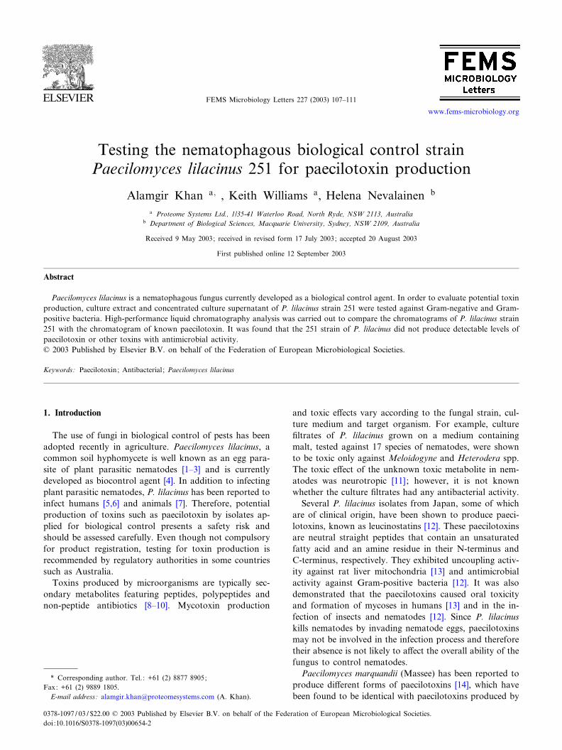
HPLC chromatograms of the EPT of P. lilacinus strain251 were compared with the chromatogram published forthe crystallized paecilotoxin from the Odashima strain[12]. The chromatograms presented in Fig. 2B,C were ob-tained from the EPT of P. lilacinus strain 251 grown usingconditions known to promote toxin production [12] andfrom a culture in PD broth, extracted according to thepublished method [12]. The chromatogram published forcrystallized paecilotoxin is presented in Fig. 2A for com-parison. The two large peaks appearing at 2 min after thestart on all three chromatograms provide a cross-referenceto the work of Mikami et al. [12], who showed that thesepeaks did not represent toxins. The three paecilotoxinpeaks, labeled as a, b and c (Fig. 2A) appearing between3.75 and 7.0 min, were absent from the chromatogramsobtained from the P. lilacinus strain 251 using two di¡er-ent culture media (Fig. 2B,C). This indicates that P. lila-cinus strain 251 does not produce detectable levels of pae-cilotoxin.
On the basis of the evidence obtained in this study,P. lilacinus strain 251 does not produce paecilotoxin thatwould be detectable by HPLC analysis and bioassays. Thecrude extract (EPT) or CCS had no or only a minimale¡ect on growth of the Gram-negative E. coli and theGram-positive bacteria B. subtilis and M. luteus. Eventhough some clinical isolates of Paecilomyces have caused
oculomycosis disease in humans [5,12], it is unlikely thatthe strain 251 of P. lilacinus would cause a mycotic dis-ease, as this strain does not grow at 37‡C and does notsurvive more than 48 h at 37‡C (unpublished observa-tions). A study by Garcia [19] indicated that isolates ofP. lilacinus di¡er genetically with only those isolates capa-ble of growing at 37‡C were shown to be infectious tohumans. Therefore, it is reasonable to conclude thatP. lilacinus strain 251 can be applied as a biocontrol agentwithout any hazard to humans and without interfering toomuch with the growth of other microorganisms in the soil.
Acknowledgements
We thank Warren Kett, Macquarie University Centerfor Analytical Biochemistry (MUCAB), for helping withHPLC and Rita Holland for assistance in collecting data.This work was funded by an Australian Post-graduateAward to A.K. The work was carried out when A.K.and K.W. were at the Department of Biological Sciencesat Macquarie University in Sydney.
References
[1] Jatala, P., Kaltenback, R. and Bocangel, M. (1979) Biological controlof Meloidogyne incognita acrita and Globodera pallida on potatoes.J. Nematol. 11, 303 (Abstr).
[2] Lay, E.C., Lara, J., Jatala, P. and Gonzales, F. (1982) Preliminaryevaluation of Paecilomyces lilacinus as a biological control agent ofthe root-knot nematode, Meloidogyne incognita, in industrial toma-toes. Nematropica 12, 154.
[3] Morgan-Jones, G., White, J.F. and Rodriguez-Kabana, R. (1984)Phytonematode pathology: Ultrastructural studies II. Parasitism ofMeloidogyne arenaria eggs and larvae by Paecilomyces lilacinus. Nem-atropica 14, 57^71.
[4] Gunasekera, T.S., Holland, R.J., Gillings, M.R., Briscoe, D.A.,Neethling, D.C., Williams, K.L. and Nevalainen, K.M.H. (2000) Phe-notypic and genetic characterization of Paecilomyces lilacinus strainswith biocontrol activity against root-knot nematodes. Can. J. Micro-biol. 46, 775^783.
[5] Arai, M., Mikami, Y., Fukushima, K., Utsumi, T. and Yazawa, K.(1973) A new antibiotic, leucinostatin, derived from Penicillium lila-cinum. J. Antibiot. (Tokyo) 26, 157^161.
[6] Ono, N., Sato, K., Yokomise, H. and Tamura, K. (1999) Lung ab-scess caused by Paecilomyces lilacinus. Respiration 66, 85^87.
[7] Maslen, M., Whitehead, J., Forsyth, W.M., McCracken, H. andHocking, A.D. (1988) Systemic mycotic disease of captive crocodilehatching (Crocodylus porosus) caused by Paecilomyces lilacinus.J. Med. Vet. Mycol. 26, 219^225.
[8] Blondelle, S.E., Takahashi, E., Dinh, K.T. and Houghten, R.A.(1995) The antimicrobial activity of hexapeptides derived from syn-thetic combinatorial libraries. J. Appl. Bacteriol. 78, 39^46.
[9] Gilquin, B., Bourgoin, M., Menez, R., Le Du, M.H., Servent, D.,Zinn-Justin, S. and Menez, A. (2003) Motions and structural vari-ability within toxins: Implication for their use as sca¡olds for proteinengineering. Prot. Sci. 12, 266^277.
[10] Xaio, J.Z., Kumazawa, S., Yoshikawa, N., Mikawa, T. and Sato, Y.(1993) Dactylfungins, novel antifungal antibiotics produced by Dac-tylaria parvispora. J. Antibiot. (Tokyo) 46, 48^55.
Fig. 2. Chromatograms of the EPT of P. lilacinus strain 251 and crys-tallized paecilotoxin. A: Crystallized paecilotoxin from the Odashimastrain; B: EPT of P. lilacinus strain 251 cultured under conditions pro-moting paecilotoxin synthesis ; C: EPT of P. lilacinus strain 251 culturedin PD broth. Paecilotoxin peaks (a, b and c in panel A) are not pro-duced by P. lilacinus strain 251 under the culture conditions applied.
FEMSLE 11204 30-9-03
A. Khan et al. / FEMS Microbiology Letters 227 (2003) 107^111110

[11] Cayrol, J., Djian, C. and Pijarowski, L. (1989) Study of the nemati-cidal properties of the culture ¢ltrate of the nematophagous fungusPaecilomyces lilacinus. Rev. Nematol. 12, 331^336.
[12] Mikami, Y., Yazawa, K., Fukushima, K., Arai, T., Udagawa, S. andSamon, R.A. (1989) Paecilotoxin production in clinical or terrestrialisolates of Paecilomyces lilacinus strains. Mycopathologia 108, 195^199.
[13] Mikami, Y., Fukushima, K., Arai, T., Abe, F., Shibuya, H. andOmmura, Y. (1984) Leucinostatins, peptide mycotoxins producedby Paecilomyces lilacinus and their possible roles in fungal infection.Zbl. Bakt. Hyg. A 257, 275^283.
[14] Radics, L., Kajatar-Peredy, M., Gasinovi, C.G., Rossi, C., Ricc, M.and Tuttobello, L. (1987) Leucinostatin H and K, two novel peptideantibiotics with tertiary amine-oxide terminal group form Paecilomy-ces marquandii. Isolation, structure and biological activity. J. Anti-biot. 40, 714^716.
[15] Wilczynska, Z. and Fisher, P.R. (1994) Analysis of a complex plas-mid insertion in a phototaxis-de⁄cient transformant of Dictyosteliumdiscoideum selected on a Micrococcus luteus lawn. Plasmid 32, 182^194.
[16] Jung, E., Gooley, A.A., Packer, N.H., Slade, M.B., Williams, K.L.and Werner Dittrich (1997) An in vivo approach for the identi¢cationof acceptor sites for O-glycosyltransferases: motifs for the addition ofO-GlcNAc in Dictyostelium discoideum. Biochemistry 36, 4034^4040.
[17] Richard, J.L. and Gallagher, R.T. (1979) Multiple toxin productionby an isolate of Aspergillus £avus. Mycopathologia 67, 161^163.
[18] Abramson, D., Clear, R.M., Gaba, D., Smith, D.M., Patrick, S.K.and Saydak, D. (2001) Trichothecene and moniliformin productionby Fusarium species from western Canadian wheat. J. Food Prot. 64,1220^1225.
[19] Garcia, G.T. (1991) Classi¢cation of Paecilomyces lilacinus. MAPP.Sc. Thesis, University of New South Wales, Sydney, Australia.
FEMSLE 11204 30-9-03
A. Khan et al. / FEMS Microbiology Letters 227 (2003) 107^111 111