Embed Size (px)
Citation preview

Novel Advances in the Treatment of Osteoporosis
Christopher KY Chan1, Alice Mason1, Cyrus Cooper2, Elaine Dennison2
1University Hospital Southampton NHS Foundation Trust, Southampton, UK
2MRC Lifecourse Epidemiology Unit, University of Southampton, Southampton, UK
Corresponding Author:
Professor Elaine Dennison, MRC Lifecourse Epidemiology Unit, University of
Southampton, Southampton General Hospital, Southampton. SO16 6YD
Tel: 023 8076 4032; Fax: 023 8070 4021
Email: [email protected]
Abstract
Introduction
Osteoporosis is a significant public health issue affecting over half of women aged over
50. With an aging population its importance is set to increase further over time.
Prevention of fragility fractures avoids significant mortality and morbidity as well as
saving significant direct and indirect costs to the economy. In this review, we discuss
existing treatments to contextualise the treatment landscape, and demonstrate how our
understanding of bone pathophysiology has led to novel therapies – in the form of
combinations and altered durations of existing treatments, as well as newer drug
therapies.
Sources of data
Pubmed and Embase were searched for randomised controlled trials of new therapies
for osteoporosis. These searches were supplemented with material presented in abstract
form at international meetings
Areas of agreement
New drugs that appear promising in the treatment of osteoporosis include the cathepsin
K inhibitor, monoclonal antibodies against sclerostin, and parathyroid hormone related
peptide.
Areas of controversy
Separate to the development of novel drug therapies is the issue of how best to use
agents that are currently available to us; specifically which agent to choose, alone or in
combination; duration of therapy; how best to identify patients at highest risk of
fracture, and to ensure the highest possible adherence to medication. Many of these

issues have been addressed in other excellent review papers, and will not be considered
in detail here.
Growing points
As with all new treatments, we await results of long term use, and experience in ‘real
life’ patient populations
Areas timely for developing research
As alluded to above, data are urgently required regarding the optimal duration of
therapy; use of combination therapy; ordering of therapies for best therapeutic effect. As
stratified medicine becomes more strongly considered in all areas of therapy, its merits
in osteoporosis as in other musculoskeletal conditions, is timely and valuable.

Introduction
Definition of Osteoporosis
Osteoporosis is a systemic disorder characterised as the depletion of bone mass with
structural deterioration of bone tissue [1]. This results in a decrease in bone mineral
density (BMD) and a predisposition to fragility fractures. Dual-energy x-ray
absorptiometry (DXA) is currently the criterion standard for the evaluation of BMD.
DXA is used to measure BMD at the hip, neck of femur, vertebrae and wrist. DXA
provides the patient’s T-score, which is the BMD value compared with that of control
subjects who are young, healthy adults at the peak of their BMD. The World Health
Organisation define osteoporosis in postmenopausal women as a BMD value at least 2.5
standard deviations below the average value in healthy young women (T-score) [2].
Fragility fractures are fractures which result from low energy trauma which would not
usually occur in normal bone. The most common sites for fragility fractures are the
vertebrae, proximal femur and distal radius. In this review, we discuss existing
osteoporosis treatments to contextualise the treatment landscape, and demonstrate how
our understanding of bone pathophysiology has led to novel therapies – in the form of
combinations and altered durations of existing treatments, as well as newer drug
therapies.
Epidemiology of Osteoporosis and Current Guidance
Osteoporosis affects approximately 30% of all postmenopausal women in the United
States and in Europe [3]. With an aging population osteoporosis is becoming hugely
relevant to healthcare in the UK. We know that fragility fractures carry with them

significant mortality, morbidity and financial implications. Every year over 300,000
patients present with fragility fractures to hospitals in the UK [4]. In the UK those who
present with hip fracture have a 30 day mortality of 8.2% [5] with permanent disability
resulting in 50% of those affected [6]. Direct medical costs from fragility fractures to
the UK healthcare economy were estimated at £1.8 billion in 2000, with the potential to
increase to £2.2 billion by 2025, and with most of these costs relating to hip fracture
care [7]. There are likely to be significant extra costs to society in days lost from the
workplace by the patient and carers.
Decreasing BMD is part of the normal aging process with osteoclast activity becoming
greater than osteoblast activity. The process is accelerated in females after menopause,
males also tend to have a greater peak in BMD, contributing to the increased incidence
in fracture presenting in older females. Risk factors are outlined in table 1.
UK guidance suggests that we consider assessing fracture risk in women over 65 and
men over 75, or those over 50 with the presence of risk factors as summarised in table 1.
In practice the most commonly used tool is the fracture risk assessment tool (FRAX),
which can be used with or without BMD score [8]. It gives a predicted risk of major
osteoporotic and hip fracture over 10 years as a percentage.
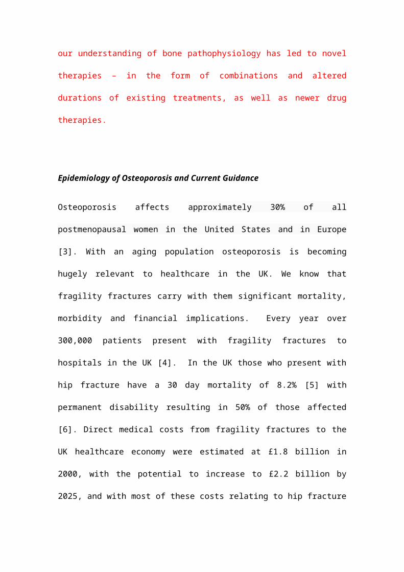
Risks independent of bone mineral density Risks dependent on bone mineral density
Age
Previous history of fragility fracture
Parental history of hip fracture
Smoking
Alcohol intake of 4 or more units per day
Steroid use
Rheumatoid arthritis
Body mass index < 19 kg/m2
Low sunlight exposure
Falls
Drugs (glucocorticoids, aromatase inhibitors,
androgen deprivation therapy, heparin therapy,
proton pump inhibitors)
Malabsorption
Conditions resulting in prolonged immobility
Untreated premature menopause, untreated
hypogonadism
Endocrine disease e.g. hyperthyroidism
Chronic liver disease
Chronic renal disease
Chronic obstructive pulmonary disease
Table 1: Risk factors for decreased bone mineral density. Adapted with permission from
Curtis et al. [9]
The National Osteoporosis Guideline Group (NOGG) issued updated guidance for
diagnosis and management of osteoporosis in postmenopausal women and men over 50
in 2014 [10]. This guidance can advise the clinician on assessing fracture risk in
different populations, use of FRAX and anti-osteoporotic drug choice and duration.
Ultimately, however, treatment decision is based on individual clinician and patient
choice.

Existing Pharmacological Treatments
Vitamin D and Calcium Supplementation
There has been huge debate regarding the role of vitamin D supplementation in many
fields of medicine. The majority of randomised controlled trials use calcium and
vitamin D supplementation, although this has been at varying doses (500mg – 1000mg
calcium; 250-1200 IU vitamin D daily) therefore it is difficult to establish efficacy of
treatment. The literature reviewing the benefits of calcium and vitamin D
supplementation has been very conflicting. However, it is generally recommended that
patients should ensure good calcium and vitamin D intake in their diet [11]. Vitamin D
and calcium supplementation alone is less effective as compared to other osteoporosis
treatments discussed [12].
Drugs that Inhibit Bone Resorption
Bisphosphonates
The majority of primary and secondary osteoporosis treatment involves the use of
bisphosphonate therapy, and most randomised controlled trials of osteoporosis therapy
have used these agents. The nitrogen containing bisphosphonates alendronate,
risedronate, ibandronate and zoledronate are analogues of inorganic pyrophosphate and
inhibit farnesyl pyrophosphate synthase in the mevalonate pathway, which results in
apoptosis of the osteoclasts. This leads to inhibition of bone resorption and increases in
BMD. There is data to suggest inhibition can continue following cessation of the drug,
with markers of bone turnover 50 percent lower 5 years after discontinuation [13].
The most commonly prescribed is alendronate, given orally once weekly. The drugs
must be given on an empty stomach because food and liquids interfere with
gastrointestinal absorption. Suboptimal administration can result in oseophageal
irritation. Poor adherence is associated with increased fracture rates [14]. If oral
bisphosphates cannot be tolerated, intravenous zoledronate can be used as an alternative
at a dose of 5mg yearly for 3 years. The concerning side effect of osteonecrosis of the
jaw (ONJ) has been predominantly seen in larger doses of bisphosphonates given to
oncology patients, and the risk of those being treated for osteroporosis is very low [15].
The American Society for Bone and Mineral Research (ASBMR) recommends that
patients should be informed that there is a low risk of development of ONJ and that
health providers should encourage patients to practice good oral hygiene and have
regular dental visits [16]. Atypical fracture is now a recognised potential complication
of prolonged use of bisphosphonates and patient reassessment after 3-5 years is now
recommended, as outlined in figure 1.
Figure 1: Algorithm for treatment monitoring with the use of bisphosphonates from the
National Osteoporosis Guidelines Group (NOGG)
Strontium ranelate
Strontium ranelate is a compound made up of 2 atoms of the element strontium bound
with ranelic acid. It is below calcium on the periodic table and is incorporated into bone
at the same rate. Recently the use of strontium has been restricted following concern
regarding cardiovascular risks. In March 2014 the European Medicines Agency
concluded its review of the risks and benefits of stronium ranelate and advised that
strontium ranelate should be restricted to only people with severe osteoporosis for

whom there are no alternate treatments for osteoporosis [17]. Consequently there has
been a large decline in its use.
Denosumab
Denosumab is a fully human RANKL (receptor activator of nuclear factor kappaB
(RANK) ligand) antibody. Precursors to osteoclasts, called pre-osteoclasts, express
surface receptors called RANK. RANK is activated by RANKL, which exists as cell
surface molecules on osteoblasts. Activation of RANK by RANKL promotes the
maturation of pre-osteoclasts into osteoclasts. Denosumab inhibits this maturation of
osteoclasts by binding to and inhibiting RANKL. This mimics the natural action of
osteoprotegerin, an endogenous RANKL inhibitor. This decreases bone resorption and
increases BMD. It is administered via a 6 monthly subcutaneous injection. Side effects
are uncommon but include transient hypocalcaemia, particularly if vitamin D deficient,
and cellulitis often away from the injection site [18].
Selective oestrogen receptor modulators (SERMs)
Raloxifene is a partial oestrogen agonist that acts as an agonist in bone, but an
antagonist in other areas of the body such as the uterus and breast. It therefore has
positive oestrogen effects in the bones, without other unwanted oestrogen effects
elsewhere. It has been shown to reduce deteriorating BMD and vertebral fractures in
post menopausal women [19]. However, there is an increased risk of venous thrombo-
embolic events and patients should be assessed prior to commencing treatment; active
or past history of venous thromboembolic events (VTE), including deep vein
thrombosis, pulmonary embolism and retinal vein thrombosis is a contraindication to
use.
Drugs that Promote Bone Formation
Teriparatide
Teriparatide is a recombinant form of parathyroid hormone (PTH)(1-34). It is currently
the only widely available anabolic agent used for osteoporosis. Teriparatide increases
renal re-absorption of calcium and enhances intestinal calcium absorption via its effect
on one hydroxylation of 25(OH)D3. Intermittent administration of PTH increases the
number of bone forming osteoblasts whereas continuous administration increases the
number of bone resorbing osteoclasts. Therefore, for an anabolic effect, it is given daily
as a low dose subcutaneous injection of 20μg for a period of 18-24 months. The high
cost of teriparatide restricts its use to those with high fracture risk who have failed other
therapies.
Combination Therapy
In recent times there has been interest in looking at combination therapies, either
treatment given at the same time or following on from each other. Most trials data have
assessed the combination of anabolic therapy (teriparatide) with an antiresorptive
therapy (a bisphosphonate or denosumab). Although there are no trials sufficiently
powered to show a difference in fracture outcome, some trials have been able to show a
difference in BMD particularly at the hip: a greater BMD at the hip as compared to the
spine has been a consistent finding across many trials [20]. The DATA extension study
looked specifically at using two years of concomitant teriparatide and denosumab
therapy and found increases in BMD more than therapy with either medication alone,
and more than has been reported with any current therapy [21]. However, to date no
trial looking at combination therapy has shown a reduction in fracture rate.

Bone Turnover and Pathways
Remodelling of bone is a continuous process with two phases that are normally coupled
and balanced: bone resorption mediated by osteoclasts followed by bone formation
mediated by osteoblasts, within a bone remodelling unit. Osteocytes, a third bone cell
type, play an important role in regulating activities of osteoclasts and osteoblasts. An
imbalance in this process with either an increase in bone resorption or decrease in bone
formation leads to a progressive loss of bone mass and impaired bone microarchitecture.
In post-menopausal osteoporosis this balance shifts to bone resorption over formation.
Drugs should counteract this balance, going towards increased formation or decreased
resorption (or both).
However, the challenge is that inhibiting osteoclasts may also result in decreased bone
formation, as they are coupled processes. The coupling is thought to be via osteogenic
factors released by osteoclasts. Inhibiting both processes may also lead to a decrease in
the repair of microbreaks from normal activities and lead to defective microarchitecture
[22][23]. This is likely to be an inherent problem of prolonged use of bisphosphonates
and denosumab as they inhibit osteoclastogenesis, with the reduced number of
osteoclasts leading to low bone turnover and hence less coupled activity of osteoblasts
for bone formation. The potential consequent defective microarchitecture could increase
the risk (albeit a low one) for atypical subtrochanteric fractures. Nevertheless, the
association between atypical fractures and the use of bisphosphonates remains
contentious, and is based on case reports and retrospective case reviews;
bisphosphonates are not thought to be the sole risk factor for atypical fractures [24].
Microdamage accumulation is only one hypothesised mechanism of atypical fracture;

others include reduced heterogeneity of mineralisation and cortical hardness resistant to
plastic deformation [25].
With the possible link between reduced bone remodelling and atypical fractures, a drug
that preserves osteoclast numbers to enable the coupled activity of bone formation by
osteoblasts, but inhibits the activity of the osteoclasts could be beneficial. Recent studies
suggest a hypothesis of a cell layer lining the bone marrow and forming a canopy over
the whole remodelling surface, as a source of osteoblast progenitors, and that this is
induced by osteoclastic factors to favour the initiation of bone formation [26].
Bone resorption is made up of the removal of the predominant constituents: inorganic
bone mineral and organic bone matrix. The former is removed by acid, and the latter by
cathepsin K (under acidic conditions). Therefore targeting cathepsin K rather than
osteoclastogenesis should allow continued signals to osteoblasts and consequent bone
formation. This has been found to be the case in ovariectomized rabbits where histology
has shown that odanacatib (a cathepsin K inhibitor) induces a shorter reversal phase,
higher osteoblast recruitment and an increase in osteoclast surface, whereas those
treated with alendronic acid did not show these responses [27]. Accordingly odanacatib
had a positive effect on bone formation rate at the same time as decreasing bone
resorption, whereas alendronic acid decreased bone formation rate [28]. Whilst bone
formation rate is increased at both trabecular and cortical sites in CatK knockout mice
and odanacatib treated ovariectomized rabbits, in ovariectomized adult rhesus monkeys
bone formation rate was site dependent (reduced at trabecular and intracortical bone but
unchanged at endocortical surface and increased at the periosteal surface). However the
authors suggested that odanacatib brought trabecular bone formation rate in estrogen-
deficient animals to the level of intact animals [29].
Loss of function mutations in the cathepsin K gene is described in a naturally occurring
disease called pycnodysostosis. It results in the loss of bone resorption and subsequent
increased bone mass. It is a rare autosomal recessive syndrome of skeletal dysplasia
associated with brachycephaly, wide cranial sutures, short stature, osteosclerosis,
fragility fractures and high bone mineral density. Impairment of bone remodelling is
reflected by a reduction in bone resorption markers (N-telopeptides of type 1 collagen
(NTx) and C-telopeptides of type 1 collagen (CTx)). In cathepsin K deficient mice they
show an osteopetrotic skeleton with increased trabecular and cortical bone. This
histology shows a normal rate of bone mineralisation with normal or increased
osteoclast numbers but a decrease in the bone matrix resorption ability of osteoclasts
[30].
Alternatively we may wish to target osteoblasts for its role in bone formation. Existing
treatments such as oestrogens and teriparatide promote the activity of osteoblasts. The
leading agents in this field relate to the Wnt signalling pathway, via an inhibitor of
sclerostin (e.g. Romosozumab), and separately a PTH related peptide (PTHrp) (e.g.
Abaloparatide).
In skeletal cells, activation of the canonical Wnt/β-catenin signaling pathway
induces osteoblast cell differentiation [31]. It is the translocation of
unphosphorylated β-catenin to the nucleus that activates these Wnt target genes.
However, β-catenin is phosphorylated and degraded in the proteasome when Wnt
receptor binding interactions are absent: it is the binding of Wnt to frizzled
receptors and to the low-density lipoprotein receptor–related protein (LRP) 5 and
6 co-receptors that leads to the stabilization of β-catenin and its translocation to
the nucleus. Through theses interactions, Wnt thereby induces osteoblastogenesis
and bone formation. The activity of Wnt is modulated by extracellular antagonists that
act by binding Wnt itself or by preventing its interactions with its receptor or
coreceptors. Sclerostin and dickkopf 1 (Dkk-1) bind to LRP5 and LRP6. Mutations in
LRP5 and LRP6, preventing the association of sclerostin or Dkk-1 with LRP5, result in
increased bone mass as the Wnt pathway is thereby unimpeded. Therefore, Wnt
signaling is a suitable target for the development of new anabolic therapies.
Sclerostin, encoded by the SOST gene, is an osteocyte-secreted glycoprotein that
normally inhibits the Wnt signalling pathway on the cell membranes of osteoblasts, and
thereby inhibits osteoblast proliferation and function. Blocking the action of sclerostin
should therefore yield bone formation. Indeed patients with a genetic deficiency of
sclerostin (van Buchem disease) have high bone mass and correspondingly increased
bone strength that is resistant to fractures [32]. These patients tend to lead a normal life
with nerve entrapment being the only problem of the high skeletal BMD.
PTHrp shares a similar structural organization to PTH that interacts with the same
receptor as PTH, but it performs a different role in bone, with an important paracrine
action on committed osteoblast precursors in order to enhance their differentiation and
reduce osteoblast apoptosis [33]. Mice with an osteoblast-specific targeted disruption of
PTHrp had marked osteoporosis characterized by impaired bone formation, even in the
presence of normal levels of circulating PTH, because of the loss of the paracrine action
of PTHrp [34].
Novel Therapies
Cathepsin K Inhibitor
Cathepsin K is a lysosomal cysteine proteinase expressed by osteoclasts, and is one of
the enzymes that degrade type I collagen, a major component (90%) of bone matrix.
Other cathepsins B, L and S degrade collagen in other tissues such as skin and lung, so a
Cathepsin K inhibitor has to be selective over these other types in order to avoid side
effects such as morphea-like skin reactions and respiratory abnormalities. Cathepsin K
has also been found to be expressed in other cells which may explain some of the off-
target effects seen in clinical trials, including for example the macrophages and smooth
muscle cells of atherosclerotic lesions [35].
Odanacatib is a highly selective cathepsin K inhibitor. In phase I clinical trials, a half
life of 66-93 hours was observed, allowing for once weekly dosing [36]. A phase II
randomized, multicenter, placebo-controlled trial enrolled 399 postmenopausal women
with low BMD (T-score ≤-2.0 and ≥-3.5) allocated to one of the following weekly
odanacatib treatments: 3, 10, 25 and 50 mg. The trial was designed as a 12-month study
with a 12-month extension period [37]. In this study population of postmenopausal
women with low bone density, odanacatib treatment at doses of 10, 25, and 50 mg once
weekly generally resulted in dose-dependent increases, compared with placebo, in
lumbar spine, total-hip, femoral neck, trochanter, and one-third radius BMD. Substantial
further increases in BMD were seen in the second year. Dose dependent decreases in
levels of bone resorption markers urinary CTx/creatinine ratio and serum NTx with the
three higher doses were consistent with an antiresorptive effect. Nevertheless, because
of the direct role of cathepsin K in the production of collagen fragments, interpretation
of these bone resorption markers may be different compared to other antiresorptive
drugs.
Decreases in markers of bone formation were modest and transient compared with those
seen with other antiresorptive therapies (e.g., alendronate and risedronate) and were
consistent with the non-significant decreases in bone-formation rate and mineralizing
surface in the biopsy samples. This would be consistent with the hypothesised coupling
of bone remodelling described earlier, which is enabled by the targeting of the enzyme
produced by the osteoclast rather than reducing the number of osteoclasts. Indeed,
when treated with odanacatib, TRAP5b (an index of osteoclast metabolic activity and
cell number) was found to initially decrease but then recover to, or reach levels slightly
higher than, that seen in the placebo group. This differs dramatically from the large
decreases in TRAP5b seen with other antiresorptive agents [37].
The phase II study was subsequently extended by a year to look at further efficacy and
safety as well as the effects of discontinuation [38]. After 2 years, patients (n = 189)
were re-randomized to odanacatib 50 mg weekly or placebo for an additional year.
Endpoints included BMD at the lumbar spine (primary), total hip, and hip sub-regions;
levels of bone turnover markers; and safety assessments. Continued treatment with 50
mg of odanacatib for 3 years produced significant increases from baseline and from year
2 in BMD at the spine (7.9% and 2.3%) and total hip (5.8% and 2.4%). The cumulative
gains in BMD seen with odanacatib are similar to those seen with alendronate and
zoledronic acid, although BMD increases with odanacatib did not plateau over time, as
has been observed with other antiresorptive therapies. Urine cross-linked N-telopeptide
of type I collagen (NTx) remained suppressed by 50% over the 3 year period compared
to a lowering of 17% in the placebo group. There were no differences in skin adverse
effects or upper respiratory tract infections between the odanacatib and placebo groups,
except an increase in cases reported and treated as urinary tract infections in the
odanacatib group (n = 12 vs. 3), that did not lead to drug discontinuation. Because
odanacatib does not persist in bone, it is not surprising that after its discontinuation,
much of the bone density that had been gained in the initial 2 years was lost during the
following year. In those that switched from active treatment to placebo, there was an
initial rapid loss in BMD over 6 months, eventually levelling off to near-baseline levels.
These data are more akin to the findings with hormone-replacement therapy,
denosumab, and parathyroid hormone than the bisphosphonates [38].

In a further extension to 5 years of this phase II trial, further monitoring of bone mineral
density showed an ongoing almost linear increase from baseline at multiple sites with
continuous treatment of odanacatib 50mg weekly. It also continued to show return of
BMD to baseline or just below upon discontinuation of the odanacatib to placebo.
Safety and adverse effects were not studied, as there was a crossover. This extension
does contain small numbers (n = 13 to 14) in each arm but does show consistent results
over measured sites and over time as described above (also see figure 2) [39].
Figure 2 Percentage change from baseline in BMD over 5 years in continuous
odanacatib 50mg weekly, versus discontinuation at month 24, versus placebo from
months 0 to 36 [39]. – permission granted

Whilst BMD is conventionally an areal measure using DXA, volumetric information
using quantitative computed tomography (QCT) scans are being used to provide further
information on effects on cortical and trabecular bone. A 2-year international,
randomized, double‐blind, placebo‐controlled phase 3 trial enrolled 214
postmenopausal women (mean age 64 years) with low areal BMD (T-score ≤-1.5 and ≥-
3.5). Subjects were randomized to odanacatib 50 mg weekly or placebo, and all
participants received calcium and vitamin D. Hip QCT scans at 24 months were
available for 158 women (odanacatib: n=78; placebo: n=80). The cortical, subcortical,
and trabecular volumetric BMD and bone mineral content of the proximal femur
increased in postmenopausal women treated with odanacatib for 24 months compared to
placebo [40].
The above data is promising, and an ongoing phase III fracture trial will try and
translate this into the real world through reduction in fractures. LOFT is a randomized,
double-blind, placebo-controlled, event-driven trial, including a pre-planned, blinded
placebo-controlled extension study. The trial enrolled 16,713 women, 65 years of age or
older, diagnosed with osteoporosis, who have been postmenopausal for five years or
more. Patients were randomized to receive odanacatib 50 mg/week (n=8,357) or
placebo (n=8,356). All patients received vitamin D (5600 IU/week) and calcium up to
1200 mg/day, if required. The three primary outcomes were radiologically determined
vertebral, hip, and clinical non-vertebral fractures. Secondary end points included
clinical vertebral fractures, BMD, bone turnover markers, and safety and tolerability,
including bone histology [41].
The drug company producing Odanacatib, Merck, have released data from LOFT
indicating that compared to patients receiving placebo, patients who received
odanacatib had a: 54% relative risk reduction of new and worsening morphometric
(radiographically-assessed) vertebral fractures, 47% relative risk reduction of clinical
hip fractures, 23% relative risk reduction of clinical non-vertebral fractures, and 72%
relative risk reduction of clinical vertebral fractures (all p<0.001). Adjudicated events of
morphea-like skin lesions and atypical femoral fractures occurred more often in the
odanacatib group than in the placebo group. Adjudicated major adverse cardiovascular
events were generally balanced overall between the treatment groups. There were
numerically more adjudicated stroke events with odanacatib than with placebo [42].
Anti Sclerostin Antibody
Romosozumab is a humanized monoclonal antibody that blocks sclerostin from
inhibiting osteoblast maturation and function. A phase I multicenter, randomized,
double-blind, placebo-controlled, ascending-dose study looked at the safety and
tolerability of multiples doses of romosozumab as its primary study endpoints. For the
16 healthy men and 32 healthy postmenopausal women with low bone mass studied,
multiple doses of romosozumab were well tolerated and associated with significant
improvements in BMD of the lumbar spine in every dose cohort, with maximum
increases from baseline that ranged from 4% to 7%. These improvements persisted from
the end of treatment at 12 week, through to the end of follow up at 24 weeks after
initiation of study treatment. Interestingly, in the pharmacodynamic analyses, multiple
doses of romosozumab increased bone formation markers such as PINP, but decreased
the bone resorption marker serum CTx, suggesting an uncoupling of remodeling [43].
Wnt activation resulting from the inhibition of sclerostin has been associated with
decreased bone resorption both in humans and in animal models, probably owing to
direct or indirect actions on osteoclasts through the Wnt pathway. The mechanism by
which the sclerostin pathway interacts with bone resorption may involve the OPG-
RANKL axis because the osteoclast inhibitor OPG is considered a downstream target of
Wnt/-catenin signalling. However, in vivo data demonstrating an effect of Scl-Ab on
serum OPG levels is limited [44].
A phase II, multicenter, international, randomised, placebo-controlled, parallel-
group, eight-group study, in which the primary end point was the percentage
change from baseline in BMD at the lumbar spine after 12 months treatment has
reported. The eight groups were split into five varying doses of romosozumab, oral
alendronate, subcutaneous teriparatide, or placebo injections. The 419 participants were
postmenopausal females with a low BMD (T-score ≤-2.0 and ≥-3.5) and no prior
fragility or vertebral fracture. Romosozumab was associated with a significant mean
change in lumbar BMD at month 12 of +11.3% in the 210 mg monthly dose compared
with a decrease of 0.1% in the placebo group and increases of 4.1% with alendronate
and 7.1% with teriparatide. Assessment of bone turnover markers again showed the
uncoupling of remodelling. Circulating markers of bone formation increased rapidly
with romosozumab but returned to baseline values despite continued administration:
increases were noted 1 week after the initial dose was administered and were greatest at
month 1. Levels of bone formation marker returned to baseline values or fell below
baseline values between months 2 and 9, depending on the dose and the marker. By
comparison, a decrease in a circulating marker of bone resorption was maintained over
the 12-month dosing period. The reason for the transitory nature of the effect on bone
formation is unclear. It may be due to changes in counter-regulatory signaling pathways
in the control of bone remodeling. The overall incidence of adverse events was balanced
between groups, with the exception of the increased frequency of injection-site reactions
in the romosozumab groups as compared with the other groups [45]. Phase III studies
are currently underway.

Blosozumab is another anti-sclerostin antibody but remains in phase II development and
has not advanced to phase III studies.
Parathyroid Hormone Related Protein (PTHrp)
PTHrP has been shown to be well tolerated, even at larger doses compared to that for
teriparatide, the latter having issues with adverse effects and mild hypercalcemia at
higher doses. Initial studies also showed that there were rapid increases in spine BMD
of 6-8% in 3 months, and that it appeared to be a pure skeletal anabolic agent with no
associated increased in bone resorption markers seen [46].
Abaloparatide is a novel synthetic peptide analog of PTHrP. There has been a multi-
center, multi-national, double-blind placebo controlled trial in which postmenopausal
women were randomly assigned to receive 24 weeks of treatment with daily
subcutaneous injections of placebo, abaloparatide, 20, 40, or 80 μg, or teriparatide, 20
μg. At 24 weeks, lumbar spine BMD increased by 2.9, 5.2, and 6.7% in the
abaloparatide, 20-, 40-, and 80-μg groups, respectively, and 5.5% in the teriparatide
group. The increases in the 40- and 80-μg abaloparatide groups and the teriparatide
group were significantly greater than placebo (1.6%). Femoral neck BMD increased by
2.7, 2.2, and 3.1% in abaloparatide, 20-, 40-, and 80-μg groups, respectively, and 1.1%
in the teriparatide group. The increase in femoral neck BMD with abaloparatide, 80 μg
was significantly greater than placebo (0.8%). Total hip BMD increased by 1.4, 2.0, and
2.6% in the abaloparatide, 20-, 40-, and 80-μg groups, respectively. The total hip
increases in the 40- and 80-μg abaloparatide groups were greater than both placebo
(0.4%) and teriparatide (0.5%) [47]. Phase III trials are underway but not yet published
in a peer-reviewed journal. The Phase III ACTIVE study has been reported to show a
similar 80% reduction in vertebral fracture rate with abaloparatide compared to
teriparatide, but a statistically significant reduction in non-vertebral fractures with
abaloparatide compared with placebo, and this was not seen with teriparatide compared
with placebo [48].
Conclusions
The range of treatments available to us to manage osteoporosis is expanding as we gain
a better understanding of the pathophysiology of bone remodelling and bone fragility.
Interestingly an uncoupling of bone formation and resorption has been found in the
newer drugs in development that favours increased bone mineral density. As yet the
long-term effects of our new drugs are obviously uncertain, just as the atypical fractures
with the use of bisphosphonates over long periods have become apparent with time.
Although the newer drugs all show significant increases in lumbar bone mineral density,
we do not yet have evidence that these newer treatments are any better than existing
treatments at reducing the risk of non-vertebral fractures. Newer treatments (odanacatib,
romosozumab, and abaloparatide) are likely to be used as second or third line treatments
after an initial period of 3 to 5 years on a bisphosphonate, to try and increase BMD and
counter concerns about continuing suppression of bone remodelling and possible
implications for atypical fracture. Guidance in many countries including the UK dictates
the first choice of agent used in osteoporosis; there is a current evidence gap around
effectiveness of agents used in different sequence, although, for example, observational
data have suggested that the effectiveness of teriparatide therapy is not removed by
prior treatment with bisphosphonate treatment. A well-tolerated drug with minimal
long-term adverse effects, that equally reduces risk of non-vertebral fractures as well as
vertebral fractures, has not yet been found but remains the holy grail. No drug is
effective if the patient fails to take it; a particular challenge is ensuring good
compliance.

Over the next five years we should have more long term phase III results to place these
newer treatments amongst our existing options. Current efforts have been geared
towards bone but neglect the link between muscle and bone, and an integrated approach
may be more effective in treating musculoskeletal aging to reduce falls and fractures.
For this reason we may expect new treatments to embrace both osteoporosis and
sarcopenia. Sarcopenia is the progressive and generalized loss of muscle mass that can
lead to a loss of strength or performance. Whilst it is more established that muscle and
bone interact anatomically and through biomechanical signals through “mechanostat
theory”, the paracrine and endocrine signals that co-ordinate their development is a
more recent research point. Studies suggest a bone-muscle unit where the skeletal
muscle secretome releases molecules that affect bone, which in turn produce osteokines
from osteoblasts and osteocytes that impact muscle cells. These molecular factors are
not yet fully identified. Newly emerging pathways involving these molecular factors
includes activin-signaling inhibitors such as myostatin-neutralising antibodies-
propeptides, recombinant follistatin, follistatin derivatives and soluble activin receptors
or myokines. More central regulation of both bone and muscle could also be targeted
and includes possible targets of growth hormone and growth hormone secretagogues,
androgens, and selective androgen receptor modulators (SARMs) [49].
However, there have been safety concerns with these agents related to off-target effects
of manipulating widespread signalling pathways. For instance, activin pathway
inhibitors have been reported to cause telangiectasia, bleeding and gonadotropin
suppression. SARMs have been specifically targeted to muscle and bone to try and
avoid the undesirable effects of androgens. One example of a SARM is ostarine and
phase I-III clinical trials have shown an increase in lean mass and physical function in
elderly men, post-menopausal women and cancer patients. However, effects on BMD
have not been demonstrated, possibly due to short study periods of up to 3 months [50].
Beyond five years, in the long term, these agents may supplant existing treatments for
patients either over post-menopausal age with low BMD or an equivalent to be defined
state of sarcopenia.
Genomics may also potentially help identify potential novel osteoporosis drug targets.
Sclerostin has already been identified as a Wnt signalling target, but other potential
targets include LRP4, LRP5/LRP6, SFRP4, WNT16 and NOTUM. NOTUM is a lipase
that inactivates WNTs by removing the palmitoleic acid group needed to bind to
frizzled receptors. Notum KO mice have been found to have elevated cortical bone
thickness and strength [51].
In conclusion, a greater understanding of bone pathophysiology is offering us
newer treatments in the forms of cathepsin K inhibitor, monoclonal antibodies
against sclerostin, and parathyroid hormone related peptide, all with promising
data on BMD, and positive preliminary reports on reduced fracture rates for
abaloparatide and odanacatib.

References
1. AW Friedman. Important determinants of bone strength: beyond bone mineral
density. J Clin Rheumatol, 12 (2006), pp. 70–77