Embed Size (px)
Citation preview
Original Article
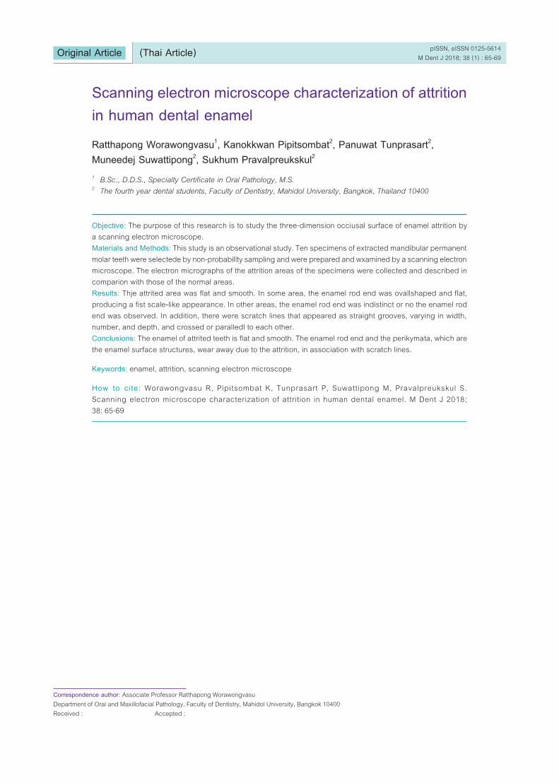
Scanning electron microscope characterization of attrition in human dental enamel
Objective: The purpose of this research is to study the three-dimension occiusal surface of enamel attrition by a scanning electron microscope.Materials and Methods: This study is an observational study. Ten specimens of extracted mandibular permanent molar teeth were selectede by non-probabillty sampling and were prepared and wxamined by a scanning electron microscope. The electron micrographs of the attrition areas of the specimens were collected and described in comparion with those of the normal areas.Results: Thje attrited area was flat and smooth. In some area, the enamel rod end was ovallshaped and flat, producing a fist scale-like appearance. In other areas, the enamel rod end was indistinct or no the enamel rod end was observed. In addition, there were scratch lines that appeared as straight grooves, varying in width, number, and depth, and crossed or paralledl to each other.Conclusions: The enamel of attrited teeth is flat and smooth. The enamel rod end and the perikymata, which are the enamel surface structures, wear away due to the attrition, in association with scratch lines.
Keywords: enamel, attrition, scanning electron microscope
How to ci te: Worawongvasu R, Pipitsombat K, Tunprasart P, Suwattipong M, Pravalpreukskul S. Scanning electron microscope characterization of attrition in human dental enamel. M Dent J 2018; 38: 65-69
Ratthapong Worawongvasu1, Kanokkwan Pipitsombat2, Panuwat Tunprasart2, Muneedej Suwattipong2, Sukhum Pravalpreukskul2
1 B.Sc., D.D.S., Specialty Certificate in Oral Pathology, M.S.2 The fourth year dental students, Faculty of Dentistry, Mahidol University, Bangkok, Thailand 10400
Correspondence author: Associate Professor Ratthapong WorawongvasuDepartment of Oral and Maxillofacial Pathology, Faculty of Dentistry, Mahidol University, Bangkok 10400Received : Accepted :
pISSN, eISSN 0125-5614M Dent J 2018; 38 (1) : 65-69(Thai Article)
pISSN, eISSN 0125-5614M Dent J 2017; 38 (1) : 65-69Original Article
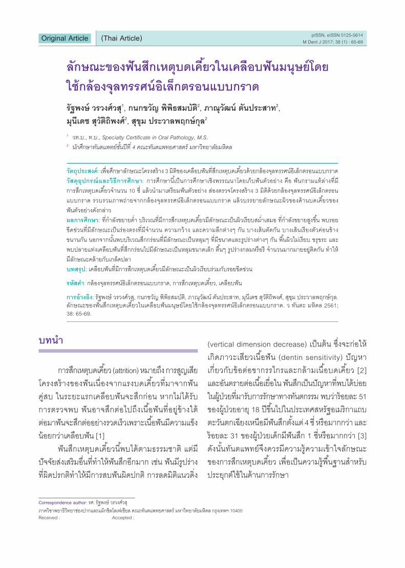
ลกษณะของฟนสกเหตบดเคยวในเคลอบฟนมนษยโดย
ใชกลองจลทรรศนอเลกตรอนแบบกราด
วตถประสงค: เพอศกษาลกษณะโครงสราง 3 มตของเคลอบฟนทสกเหตบดเคยวดวยกลองจลทรรศนอเลกตรอนแบบกราด
วสดอปกรณและวธการศกษา: การศกษานเปนการศกษาเชงพรรณนาโดยเกบฟนตวอยาง คอ ฟนกรามแทลางทม
การสกเหตบดเคยวจ�านวน 10 ซ แลวน�ามาเตรยมฟนตวอยาง สองตรวจโครงสราง 3 มตดวยกลองจลทรรศนอเลกตรอน
แบบกราด รวบรวมภาพถายจากกลองจลทรรศนอเลกตรอนแบบกราด แลวบรรยายลกษณะผวของดานบดเคยวของ
ฟนตวอยางดงกลาว
ผลการศกษา: ทก�าลงขยายต�า บรเวณทมการสกเหตบดเคยวมลกษณะเปนผวเรยบสม�าเสมอ ทก�าลงขยายสงขน พบรอย
ขดขวนทมลกษณะเปนรองตรงทมจ�านวน ความกวาง และความลกตางๆ กน บางเสนตดกน บางเสนเรยงตวคอนขาง
ขนานกน นอกจากนนพบบรเวณสกกรอนทมลกษณะเปนหลมๆ ทมขนาดและรปรางตางๆ กน พนผวไมเรยบ ขรขระ และ
พบปลายแทงเคลอบฟนทสกกรอนไปมลกษณะเปนหลมขนาดเลก ตนๆ รปรางกลมหรอร จ�านวนมากมายอยตดกน ท�าให
มลกษณะคลายกบเกลดปลา
บทสรป: เคลอบฟนทมการสกเหตบดเคยวมลกษณะเปนผวเรยบรวมกบรอยขดขวน
รหสค�า: กลองจลทรรศนอเลกตรอนแบบกราด, การสกเหตบดเคยว, เคลอบฟน
การอางอง: รฐพงษ วรวงศวส, กนกขวญ พพธสมบต, ภาณวฒน ตนประสาท, มนเดช สวตถพงศ, สขม ประวาลพฤกษกล. ลกษณะของฟนสกเหตบดเคยวในเคลอบฟนมนษยโดยใชกลองจลทรรศนอเลกตรอนแบบกราด. ว ทนตะ มหดล 2561; 38: 65-69.
รฐพงษ วรวงศวส1, กนกขวญ พพธสมบต2, ภาณวฒน ตนประสาท2,
มนเดช สวตถพงศ2, สขม ประวาลพฤกษกล2
1 วท.บ., ท.บ., Specialty Certificate in Oral Pathology, M.S. 2 นกศกษาทนตแพทยชนปท 4 คณะทนตแพทยศาสตร มหาวทยาลยมหดล
Correspondence author: รศ. รฐพงษ วรวงศวส
ภาควชาพยาธวทยาชองปากและแมกซลโลเฟเชยล คณะทนตแพทยศาสตร มหาวทยาลยมหดล กรงเทพฯ 10400Received : Accepted :
(Thai Article)
บทน�า
การสกเหตบดเคยว (attrition) หมายถง การสญเสย
โครงสรางของฟนเนองจากแรงบดเคยวทมาจากฟน
คสบ ในระยะแรกเคลอบฟนจะสกกอน หากไมไดรบ
การตรวจพบ ฟนอาจสกตอไปถงเนอฟนทอยขางใต
ตอมาฟนจะสกตออยางรวดเรวเพราะเนอฟนมความแขง
นอยกวาเคลอบฟน [1]
ฟนสกเหตบดเคยวนพบไดตามธรรมชาต แตม
ปจจยสงเสรมอนทท�าใหฟนสกอกมาก เชน ฟนมรปราง
ทผดปรกตท�าใหมการสบฟนผดปกต การลดมตแนวดง
(vertical dimension decrease) เปนตน ซงจะกอให
เกดภาวะเสยวเนอฟน (dentin sensitivity) ปญหา
เกยวกบขอตอขากรรไกรและกลามเนอบดเคยว [2]
และอนตรายตอเนอเยอใน ฟนสกเปนปญหาทพบไดบอย
ในผปวยทมารบการรกษาทางทนตกรรม พบวารอยละ 51
ของผ ปวยอาย 18 ปขนไปในประเทศสหรฐอเมรกาแถบ
ตะวนตกเฉยงเหนอมฟนสกตงแต 4 ซ หรอมากกวา และ
รอยละ 31 ของผ ปวยเดกมฟนสก 1 ซหรอมากกวา [3]
ดงนนทนตแพทยจงควรมความรความเขาใจลกษณะ
ของการสกเหตบดเคยว เพอเปนความรพนฐานส�าหรบ
ประยกตใชในดานการรกษา
Scanning electron microscope characterization of attrition in human dental enamel
http://www.dt.mahidol.ac.th/division/th_Academic_Journal_Unit 67
ในป พ.ศ. 2516 Tronstad [4] และในป พ.ศ. 2522
Mendis และ Darling [5] ไดศกษาเนอฟนทสกเหตบดเคยว
โดยใชกลองจลทรรศนอเลกตรอนแบบกราด ตางกพบวา
หลอดฝอยเนอฟน (dentinal tubule) อดตนดวยผลก
รปรางตางๆ
ในป พ.ศ. 2523 Brännström และ Garberoglio [6]
ไดศกษาหลอดฝอยเนอฟนของเนอฟนในบรเวณทม
การสกเหตบดเคยวทลกประมาณ 1.5 มลลเมตรจากผว
ของบรเวณทสก พบวาหลอดฝอยเนอฟนอดตนดวยวสด
คลายกบทพบในเนอฟนรอบหลอดฝอย (peritubular
dentin)
รายงานการวจยทศกษาลกษณะของฟนสกเหต
บดเคยวในเคลอบฟนมนษยโดยใชกลองจลทรรศน
อเลกตรอนแบบกราดทตพมพในวารสารภาษาองกฤษ
ยงมนอย
การศกษานมวตถประสงคเพอศกษาลกษณะของ
การสกเหตบดเคยวโดยใชกลองจลทรรศนอเลกตรอน
แบบกราด (scanning electron microscope, SEM)
วสดอปกรณและวธการศกษา
ฟนตวอยางทใชในงานวจยน คอ ฟนกรามแทลาง
จ�านวน 10 ซทถกถอนจากผ ปวยชวงอาย 20-60 ป
โดยฟนทใชจะตองมการสกเหตบดเคยว ไมม
การบรณะฟน และไมมการแตกราวของตวฟน รากฟน
หรอทงสองต�าแหนง จากคลนกทนตกรรมและ
โรงพยาบาลในจงหวดกรงเทพมหานคร แลวแชฟน
ในสารละลายฟอรมาลนความเขมขนรอยละ 10 (10%
formalin) ทนทเพอคงสภาพไมใหเนาเปอย หลงจากนน
ลางตวอยางฟนดวยน�าสะอาด ใช carborundum disc
ตดบรเวณของฟนทตองการใหเปนรปสเหลยมกวาง
2 มลลเมตร ยาว 2 มลลเมตร และหนา 2 มลลเมตร แลว
แชฟนในโซเดยมไฮโพคลอไรตความเขมขนรอยละ 5.25
(5.25% sodium hypochlorite) เปนระยะเวลา 24 ชวโมง
ทอณหภมหอง เพอก�าจดสารอนทรยทเกาะอยบนผวฟน
ออกหมด แลวลางฟนตวอยางในน� ากลนเพอไมให
โซเดยมไฮโพคลอไรตตกคาง หลงจากนนน�าฟนตวอยาง
ไปผานกระบวนการขจดน�า (dehydration) โดยแชใน
เอทลแอลกอฮอล (ethyl alcohol) ทมความเขมขนจาก
นอยไปมาก คอ 50%, 60%, 70%, 85%, 95%, 100%
ตามล�าดบโดยแชทแตละความเขมขนครงละ 15 นาท
จ�านวน 2 ครง แลวจงเปลยนความเขมขนสงขน ตอมา
น�าฟนตวอยางไปตากใหแหงทอณหภมหองเปนระยะเวลา
24 ชวโมง แลวน�าฟนตวอยางไปเคลอบดวยทองหนา
100-300 องสตรอมดวยเครองฉาบทองและคารบอน
(ion sputter coater) รน SPI แลวสองตรวจดดวย
กลองจลทรรศนอเลกตรอนแบบกราดรน JSM-5410
LV บรษท JEOL ประเทศญป น รวบรวมภาพถายจาก
กลองจลทรรศนอเลกตรอนแบบกราด บรรยายลกษณะผว
ของดานบดเคยวของฟนกรามแทลางทสกและทไมสก
ผลการศกษา
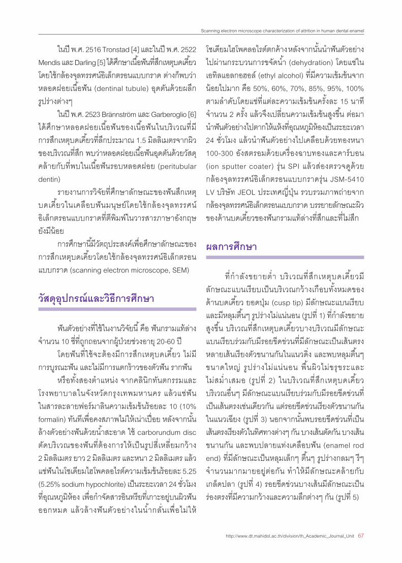
ทก�าลงขยายต�า บรเวณทสกเหตบดเคยวม
ลกษณะแบนเรยบเปนบรเวณกวางเกอบทงหมดของ
ดานบดเคยว ยอดป ม (cusp tip) มลกษณะแบนเรยบ
และมหลมตนๆ รปรางไมแนนอน (รปท 1) ทก�าลงขยาย
สงขน บรเวณทสกเหตบดเคยวบางบรเวณมลกษณะ
แบนเรยบรวมกบมรอยขดขวนทมลกษณะเปนเสนตรง
หลายเสนเรยงตวขนานกนในแนวดง และพบหลมตนๆ
ขนาดใหญ รปรางไมแนนอน พนผวไมขรขระและ
ไมสม�าเสมอ (รปท 2) ในบรเวณทสกเหตบดเคยว
บรเวณอนๆ มลกษณะแบนเรยบรวมกบมรอยขดขวนท
เปนเสนตรงเชนเดยวกน แตรอยขดขวนเรยงตวขนานกน
ในแนวเฉยง (รปท 3) นอกจากนนพบรอยขดขวนทเปน
เสนตรงเรยงตวในทศทางตางๆ กน บางเสนตดกน บางเสน
ขนานกน และพบปลายแทงเคลอบฟน (enamel rod
end) ทมลกษณะเปนหลมเลกๆ ตนๆ รปรางกลมๆ รๆ
จ�านวนมากมายอยตอกน ท�าใหมลกษณะคลายกบ
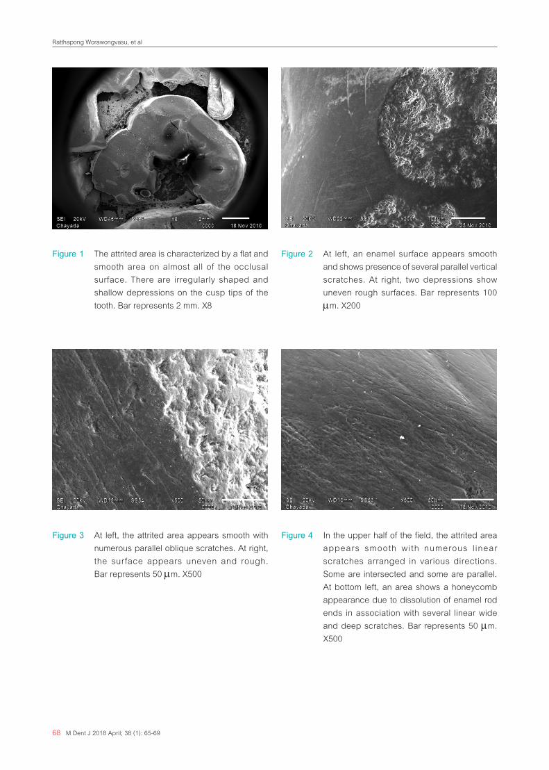
เกลดปลา (รปท 4) รอยขดขวนบางเสนมลกษณะเปน
รองตรงทมความกวางและความลกตางๆ กน (รปท 5)
68 M Dent J 2018 April; 38 (1): 65-69
Ratthapong Worawongvasu, et al
Figure 1 The attrited area is characterized by a flat and smooth area on almost all of the occlusal surface. There are irregularly shaped and shallow depressions on the cusp tips of the tooth. Bar represents 2 mm. X8
Figure 2 At left, an enamel surface appears smooth and shows presence of several parallel vertical scratches. At right, two depressions show uneven rough surfaces. Bar represents 100 µm. X200
Figure 3 At left, the attrited area appears smooth with numerous parallel oblique scratches. At right, the surface appears uneven and rough. Bar represents 50 µm. X500
Figure 4 In the upper half of the field, the attrited area appears smooth with numerous l inear scratches arranged in various directions. Some are intersected and some are parallel. At bottom left, an area shows a honeycomb appearance due to dissolution of enamel rod ends in association with several linear wide and deep scratches. Bar represents 50 µm. X500
Scanning electron microscope characterization of attrition in human dental enamel
http://www.dt.mahidol.ac.th/division/th_Academic_Journal_Unit 69
Figure 5 At higher magnification, the bottom left area of figure 4 shows a few oblique scratches which are characterized by deep grooves. Numerous small shallow round to ovoid depressions are also seen in this area. Bar represents 5 µm. X3,000
บทวจารณ
จากการศกษาน พบวาบรเวณเคลอบฟนท
มการสกเหตบดเคยวมลกษณะเปนผวเรยบ และ
พบรอยขดขวน ท มลกษณะเปนเ สนตรงเ รยงตว
ในทศทางตางๆ กน บางเสนขนานกน บางเสนตดกน
และบางเสนมลกษณะเปนรองตรงทมความกวาง
และความลกตางๆ กน นอกจากนนพบวาในบางบรเวณ
เหนปลายแทงเคลอบฟนรปรางกลมๆ รๆ เรยบแบน
จ�านวนมากมายอยตอกน ท�าใหมลกษณะคลายกบ
เกลดปลา และพบ บรเวณเคลอบฟนทมลกษณะ
เปนหลมๆ ทมขนาดและรปรางตางๆ กน พนผวไมเรยบ
ขรขระ บรเวณเหลาน คอ บรเวณทมการสกกรอน
(erosion) ของเคลอบฟน
Funding: Faculty of Dentistry, Mahidol University
Competing interests: None declared
Ethical approval: The Mahidol University Institutional
Review Board with Protocal No. MU-DT/PY-IRB
2010/037.0710.
กตตกรรมประกาศ
ขอขอบพระคณ ผศ.ทพ.รฐพงษ วรวงศวส
ทกรณาใหขอเสนอแนะและค�าวจารณงานวจยและ
ขอขอบคณ นางสาวชญาดา เทยนไชย ผ ควบคม
กลองจลทรรศอเลกตรอนแบบกราด ทชวยเหลอ
อ�านวยความสะดวกในการเตรยมชนตวอยางฟน และ
ชวยควบคมการถายภาพจากกลองจลทรรศนอเลกตรอน
แบบกราด
ขอขอบพระคณ คณะทนตแพทยศาสต ร
มหาวทยาลยมหดล ทใหเงนอดหนนงานวจย
เอกสารอางอง
1. Neville BW, Damm DD, Allen CM, Bouquot JE.
Oral & maxillofacial pathology. 2nd ed. Philadelphia:
WB Sanunders; 2002: 56.
2. Seligman DA, Pullinger AG. Dental attrition models
predicting temporomandibular joint disease or
masticatory muscle pain versus asymptomatic
controls. J Oral Rehabil 2006; 33: 789-99.
3. Cunha-Cruz J, Pashova H, Packard JD, Zhou L,
Hilton TJ; for Northwest PRECEDENT. Tooth wear:
prevalence and associated factors in general practice
patients. Community Dent Oral Epidemiol 2010;
38: 228-34.
4. Tronstad L. Scanning electron microscopy of
attrited dentinal surfaces and subjacent dentin in
human teeth Scand J Dent Res 1973; 81: 112-22.
5. Mendis BR, Darling AI. A scanning electron
microscope and microradiographic study of
closure of human coronal dentinal tubules related
to occlusal attrition and caries. Arch Oral Bio 1979;
24: 725-33.
6. Brännström M, Garberoglio R. Occlusion of dentinal
tubules under superficial attrited dentine. Swed
Dent J 1980; 4: 87-91.