Embed Size (px)
Citation preview
The world of β‐glucans – a review of biological roles, applications and potential areas of
research
Thesis for the requirement of Master of Science – Medical Biology
From:
Syed Haris Ali
Institute of Medical Biology, Faculty of Medicine University of Tromsø, Norway [email protected]
Under the kind supervision of:
Lill‐Tove Busund, MD, PhD and Kjetil Elvevold, PhD
Dept. of Pathology, University of Tromsø / University Hospital of Northern Norway
May, 2009

2

3
The effort herein is dedicated to my darling brother Wajid, the sweetest little brother in the whole wide world, a true charm ☺

4
5
INDEX:
Title Page Number
ABSTRACT 6
INTRODUCTION 7
To Innate immunity 7
To Βeta‐glucans 13
To receptors in innate immunity 19
DISCUSSION
Receptors implicated in Glucan research 24
Future prospects of Glucan research 31
The Glucan Group in Tromsø 38
CONCLUSION 40
REFERENCES 41
6
ABSTRACT
Among many known and tested immunomodulators, polysaccharides isolated from various natural sources
occupy a prominent position. An important group of these polysaccharides is represented by the
homopolymers of β‐glucose, called β‐glucans. Their very low‐to‐negligible toxicity and immunomodulating
effects show the promise it has in the therapy of a variety of infectious and cancer illnesses. Nowadays, the
popularity of Glucans as food additive and a disease‐modifying agent is increasing. Here, a review of the
various biological roles, applications and potential areas of β‐glucan research is presented. Also, a short
introduction to current work by the Glucan group in Tromsø is provided. The goal has remained to understand
and aptly present the diverse roles of β‐glucans and pin‐pointing the prospective areas of research, both on
the receptor / biochemical level, and in clinical research. It seems that with the tide of modern medical
research, gradually, β‐glucans will take the position they deserve in diagnostic and preventive medicine.
7
INTRODUCTION
INNATE IMMUNITY:
Immunity, why and how: With Immunity’s definition as body’s resistance to an agent (a microbe, a cancerous
cell, etc.) that may cause a derangement in body’s homeostasis, many questions arise in one’s mind. What
gives the immune system the capability and mechanistic adaptability to drive a network of specialized cells
and organs toward defense against infectious agents and cancerous cells? And how its malfunction could
unleash a torrent of diseases, from allergy to arthritis to cancer to AIDS?
One of immune system’s remarkable characteristics is distinguishing between "self" and "non‐self". The
Journal of Clinical Investigation defines [1] “self” as, “the antigens expressed on the surface of normal human
cells, which are ineffective at triggering immune responses against themselves.” In this context, the “non‐self”
could be defined as mutated self‐antigens (as in cancer), antigens expressed on the surface of infectious
agents (bacteria, virus, fungi, parasites, etc.), and chemicals deemed hazardous by the immune system.
Indeed, immune system is remarkable in the specificity it exercises in recognizing many millions of distinctive
non‐self molecules.
The two major divisions of human immune system, the innate and the adaptive, provide a comprehensive
coverage of microorganisms and cancer cells. The “innate” immune system is our first line of defense, and the
“adaptive” system affords protection against re‐exposure to the same pathogen. While both systems have
cellular and humoral components, the innate immune system also possesses anatomical barriers to infection
described later. And they complement each other too: the innate being the capturer and processor of foreign
antigens, and the adaptive taking heed from the former, spearheading the development of antibodies against
an antigen the body has already encountered.
Fig. 1: The types of immunity and the methods to boost them [*1]
8
Litt om “Innate” immunity:
The innate immunity has the job of immediate recognition of antigenic molecules that are prevalent and
conserved throughout a particular invading species or compound. Such molecules are uniform structures
essentially embedded in the invader’s architecture, unaltered by mutations or selection, and can come from
any foreign invader such as viruses, bacteria, fungi, parasites, pollens, foods, drugs, etc. The antigens could be
proteins or oligosaccharides, and act as markers on microbial surface to help identify the cell as self or non‐
self, identify the type of cell and stimulate immune cell responses (T cells) and the production of antibodies.
Male et al [*2] divide innate immune system in three components: the anatomical barriers, the humoral
component, and the cellular component.
The anatomical barriers are the first line of defense and provide mechanical hindrance to invading hazardous
agents. They include the skin and internal epithelial layers, the peristaltic intestinal movements and the
oscillation of bronchopulmonary cilia. Associated with these protective surfaces are chemical and biological
agents. The Chemical factors in the anatomical barriers include fatty acids (in sweat), lysozyme and
phospholipase (tears), saliva and nasal secretions, low pH gastric secretions, Defensins (lung & gastrointestinal
tract), and surfactants in the lung acting as opsonins (promoting phagocytosis). The Biological factors are the
normal flora of the skin and the gastrointestinal tract (GIT), which secrete toxic substances to prevent the
colonization by pathogenic bacteria or by competing against them for nutrients.
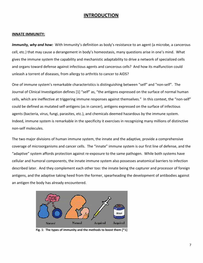
The Humoral barriers to infection come into play when anatomical barriers are breached. The humoral
response includes ‘secretory’ factors found in serum or at the site of infection and include: complement
(proteins) system, the activation of which can lead to raised vascular permeability, recruitment of phagocytic
cells, and lysis and opsonization of bacteria; coagulation system, the contribution of which lies in its ability to
increase vascular permeability, to act as chemotactic agents for phagocytic cells, and having a direct
antimicrobial action in some of its products (e.g., β‐lysin, a platelet protein); lactoferrin and transferring, which
devoid bacteria of an essential nutrient, iron; interferons, which limit viral replication; lysozyme, which breaks
down bacterial cell wall; and interleukins, especially IL‐1, which induces fever and promotes opsonization by
causing a rise in Acute Phase Protein levels.
9
Fig 2: Complement proteins in action swell & burst [2]
The Cellular barrier to infection are posed by White Blood Cells (WBCs), the major component of immune
system cells. Among the most prominent ones are: neutrophils, with a characteristic CD66 marker on their
surface, which cause phagocytosis and intracellular killing but may cause collateral tissue damage (‘pus’
formation); macrophages, with CD14 as their surface marker, which cause intra and extra‐cellular killing of
infected or altered‐self target cells plus act as antigen‐presenting cells; Natural Killer (NK) and Lymphokine
Activated Killer (LAK) cells, which non‐specifically kill virus‐infected or tumor cells; eosinophils, which cause
allergy responses and kill certain parasites. The recruitment of eosinophils and macrophages to the site of
infection is the main line of defense in the innate immune system.
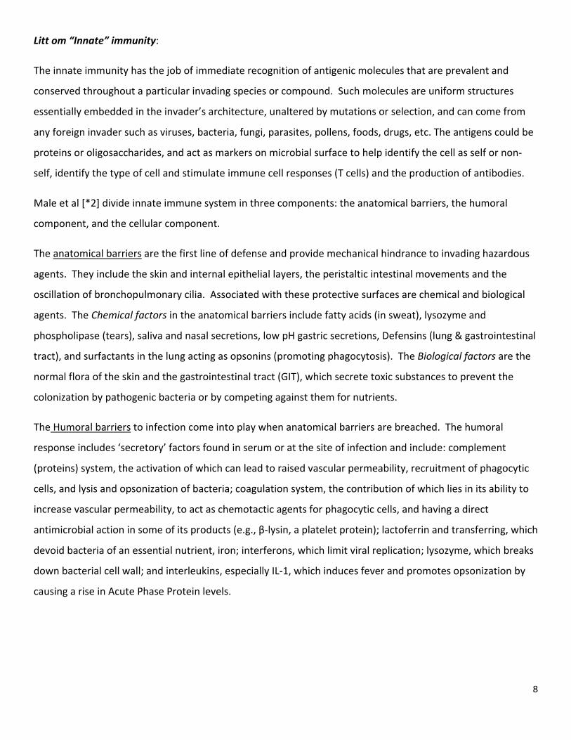
Also, the cellular barrier includes non‐specific killer cells such as NK, LAK and K cells. (NK) cells are a special
sub‐type of cytotoxic lymphocytes identified by presence of CD56 and CD16 and lack of CD3 cell surface
markers. They kill virus‐infected and malignant target cells (Fig 6), and are more potent when stimulated with
IL‐2 and IFN‐γ, which transforms them to lymphokine‐activated killer (LAK) cells. NK and LAK attach killer
activating ligand (KAL) present on the diseased cell’s surface to mark it as diseased and ‘destined to be killed’.
On the other hand, if an inhibitory ligand (MHC‐class I molecule, a sign of normal cells) is present on the cell’s
surface, it binds the Killer Inhibitory Receptor (KIR) on NK and LAK cells and spares itself from destruction.
Killer (K) cells mediate antibody‐dependent cellular cytotoxicity (ADCC) in which an antibody acts as a link to
10
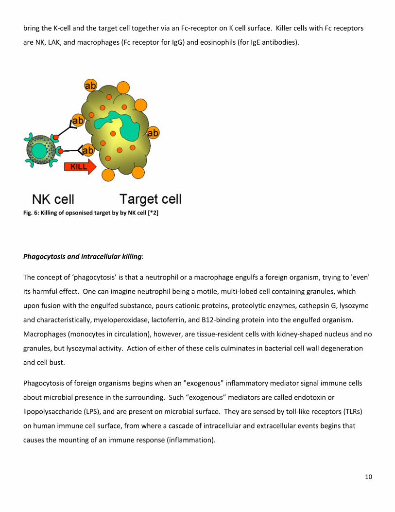
bring the K‐cell and the target cell together via an Fc‐receptor on K cell surface. Killer cells with Fc receptors
are NK, LAK, and macrophages (Fc receptor for IgG) and eosinophils (for IgE antibodies).
Fig. 6: Killing of opsonised target by by NK cell [*2]
Phagocytosis and intracellular killing:
The concept of ‘phagocytosis’ is that a neutrophil or a macrophage engulfs a foreign organism, trying to 'even'
its harmful effect. One can imagine neutrophil being a motile, multi‐lobed cell containing granules, which
upon fusion with the engulfed substance, pours cationic proteins, proteolytic enzymes, cathepsin G, lysozyme
and characteristically, myeloperoxidase, lactoferrin, and B12‐binding protein into the engulfed organism.
Macrophages (monocytes in circulation), however, are tissue‐resident cells with kidney‐shaped nucleus and no
granules, but lysozymal activity. Action of either of these cells culminates in bacterial cell wall degeneration
and cell bust.
Phagocytosis of foreign organisms begins when an "exogenous" inflammatory mediator signal immune cells
about microbial presence in the surrounding. Such “exogenous” mediators are called endotoxin or
lipopolysaccharide (LPS), and are present on microbial surface. They are sensed by toll‐like receptors (TLRs)
on human immune cell surface, from where a cascade of intracellular and extracellular events begins that
causes the mounting of an immune response (inflammation).
11
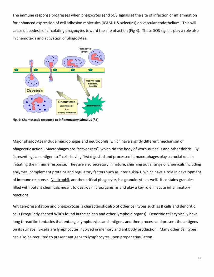
The immune response progresses when phagocytes send SOS signals at the site of infection or inflammation
for enhanced expression of cell adhesion molecules (ICAM‐1 & selectins) on vascular endothelium. This will
cause diapedesis of circulating phagocytes toward the site of action (Fig 4). These SOS signals play a role also
in chemotaxis and activation of phagocytes.
Fig. 4: Chemotactic response to inflammatory stimulus [*2]
Major phagocytes include macrophages and neutrophils, which have slightly different mechanism of
phagocytic action. Macrophages are “scavengers“, which rid the body of worn‐out cells and other debris. By
“presenting” an antigen to T cells having first digested and processed it, macrophages play a crucial role in
initiating the immune response. They are also secretory in nature, churning out a range of chemicals including
enzymes, complement proteins and regulatory factors such as interleukin‐1, which have a role in development
of immune response. Neutrophil, another critical phagocyte, is a granulocyte as well. It contains granules
filled with potent chemicals meant to destroy microorganisms and play a key role in acute inflammatory
reactions.
Antigen‐presentation and phagocytosis is characteristic also of other cell types such as B cells and dendritic
cells (irregularly shaped WBCs found in the spleen and other lymphoid organs). Dendritic cells typically have
long threadlike tentacles that entangle lymphocytes and antigens and then process and present the antigens
on its surface. B‐cells are lymphocytes involved in memory and antibody production. Many other cell types
can also be recruited to present antigens to lymphocytes upon proper stimulation.
12
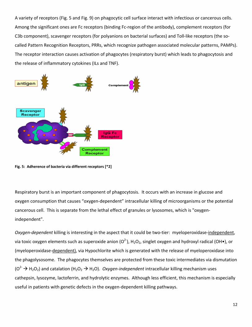
A variety of receptors (Fig. 5 and Fig. 9) on phagocytic cell surface interact with infectious or cancerous cells.
Among the significant ones are Fc receptors (binding Fc‐region of the antibody), complement receptors (for
C3b component), scavenger receptors (for polyanions on bacterial surfaces) and Toll‐like receptors (the so‐
called Pattern Recognition Receptors, PRRs, which recognize pathogen associated molecular patterns, PAMPs).
The receptor interaction causes activation of phagocytes (respiratory burst) which leads to phagocytosis and
the release of inflammatory cytokines (ILs and TNF).
Fig. 5: Adherence of bacteria via different receptors [*2]
Respiratory burst is an important component of phagocytosis. It occurs with an increase in glucose and
oxygen consumption that causes “oxygen‐dependent” intracellular killing of microorganisms or the potential
cancerous cell. This is separate from the lethal effect of granules or lysosomes, which is "oxygen‐
independent".
Oxygen‐dependent killing is interesting in the aspect that it could be two‐tier: myeloperoxidase‐independent,
via toxic oxygen elements such as superoxide anion (O2‐), H2O2, singlet oxygen and hydroxyl radical (OH•), or
(myeloperoxidase‐dependent), via Hypochlorite which is generated with the release of myeloperoxidase into
the phagolysosome. The phagocytes themselves are protected from these toxic intermediates via dismutation
(O2‐ H2O2) and catalation (H2O2 H2O). Oxygen‐independent intracellular killing mechanism uses
cathepsin, lysozyme, lactoferrin, and hydrolytic enzymes. Although less efficient, this mechanism is especially
useful in patients with genetic defects in the oxygen‐dependent killing pathways.
13
β‐GLUCANS:
Origin and Structure:
β‐glucans are glucose polymers, recognized as the effective ingredients in fungal and certain bacterial cell
walls. About half the mass of the fungal cell wall consists of β‐glucans. Natural products containing fungal β‐
glucans have been consumed for probably thousands of years, especially in China and Japan for their role in
improving general health. In recent years, β‐glucans have been noted as potent stimulators of mammalian
immune system, and now are used clinically in China and Japan.
Among the products with Β‐glucans, Zymosan is a very potent immunostimulator that has been widely used in
research. It is a mixture of proteins, lipids and polysaccharides isolated from the cell wall of Saccharomyces
cerevisiae and was first prepared and investigated in 1941 by Pillemer et al. Carrying on the work, Di Luzio
and coworkers in 1970s, pioneered the immunological research on the function of purified β‐glucans.
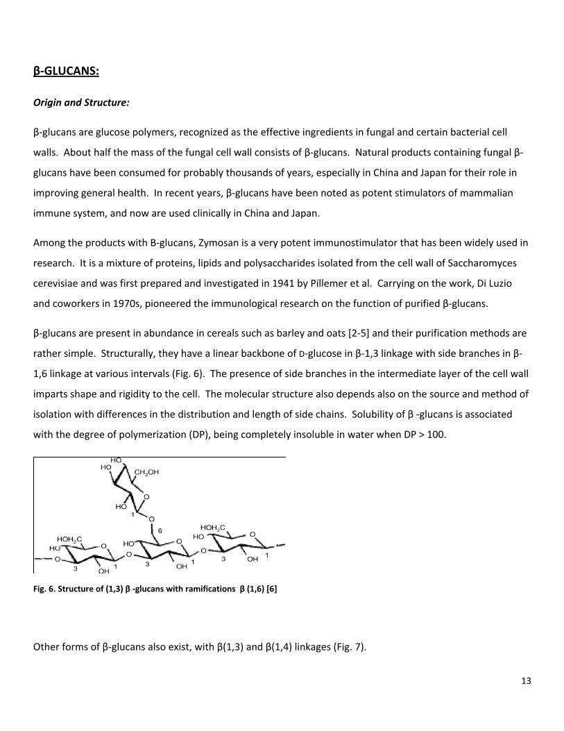
β‐glucans are present in abundance in cereals such as barley and oats [2‐5] and their purification methods are
rather simple. Structurally, they have a linear backbone of D‐glucose in β‐1,3 linkage with side branches in β‐
1,6 linkage at various intervals (Fig. 6). The presence of side branches in the intermediate layer of the cell wall
imparts shape and rigidity to the cell. The molecular structure also depends also on the source and method of
isolation with differences in the distribution and length of side chains. Solubility of β ‐glucans is associated
with the degree of polymerization (DP), being completely insoluble in water when DP > 100.
Fig. 6. Structure of (1,3) β ‐glucans with ramifications β (1,6) [6]
Other forms of β‐glucans also exist, with β(1,3) and β(1,4) linkages (Fig. 7).
14
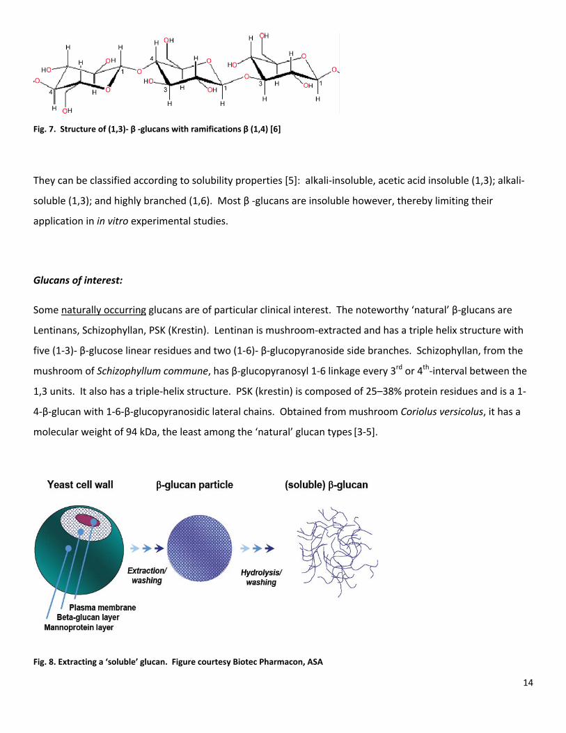
Fig. 7. Structure of (1,3)‐ β ‐glucans with ramifications β (1,4) [6]
They can be classified according to solubility properties [5]: alkali‐insoluble, acetic acid insoluble (1,3); alkali‐
soluble (1,3); and highly branched (1,6). Most β ‐glucans are insoluble however, thereby limiting their
application in in vitro experimental studies.
Glucans of interest:
Some naturally occurring glucans are of particular clinical interest. The noteworthy ‘natural’ β‐glucans are
Lentinans, Schizophyllan, PSK (Krestin). Lentinan is mushroom‐extracted and has a triple helix structure with
five (1‐3)‐ β‐glucose linear residues and two (1‐6)‐ β‐glucopyranoside side branches. Schizophyllan, from the
mushroom of Schizophyllum commune, has β‐glucopyranosyl 1‐6 linkage every 3rd or 4th‐interval between the
1,3 units. It also has a triple‐helix structure. PSK (krestin) is composed of 25–38% protein residues and is a 1‐
4‐β‐glucan with 1‐6‐β‐glucopyranosidic lateral chains. Obtained from mushroom Coriolus versicolus, it has a
molecular weight of 94 kDa, the least among the ‘natural’ glucan types [3‐5].
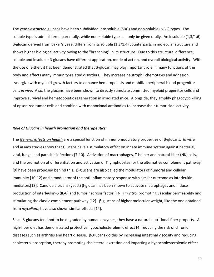
Fig. 8. Extracting a ‘soluble’ glucan. Figure courtesy Biotec Pharmacon, ASA
15
The yeast‐extracted glucans have been subdivided into soluble (SBG) and non‐soluble (NBG) types. The
soluble type is administered parentally, while non‐soluble type can only be given orally. An insoluble (1,3/1,6)
β‐glucan derived from baker's yeast differs from its soluble (1,3/1,4) counterparts in molecular structure and
shows higher biological activity owing to the "branching" in its structure. Due to this structural difference,
soluble and insoluble β‐glucans have different application, mode of action, and overall biological activity. With
the use of either, it has been demonstrated that β‐glucan may play important role in many functions of the
body and affects many immunity‐related disorders. They increase neutrophil chemotaxis and adhesion,
synergize with myeloid growth factors to enhance hematopoiesis and mobilize peripheral blood progenitor
cells in vivo. Also, the glucans have been shown to directly stimulate committed myeloid progenitor cells and
improve survival and hematopoietic regeneration in irradiated mice. Alongside, they amplify phagocytic killing
of opsonized tumor cells and combine with monoclonal antibodies to increase their tumoricidal activity.
Role of Glucans in health promotion and therapeutics:
The General effects on health are a special function of immunomodulatory properties of β‐glucans. In vitro
and in vivo studies show that Glucans have a stimulatory effect on innate immune system against bacterial,
viral, fungal and parasitic infections [7‐10]. Activation of macrophages, T‐helper and natural killer (NK) cells,
and the promotion of differentiation and activation of T lymphocytes for the alternative complement pathway
[9] have been proposed behind this. β‐glucans are also called the modulators of humoral and cellular
immunity [10‐12] and a modulator of the anti‐inflammatory response with similar outcome as interleukin
mediators[13]. Candida albicans (yeast) β‐glucan has been shown to activate macrophages and induce
production of interleukin‐6 (IL‐6) and tumor necrosis factor (TNF) in vitro, promoting vascular permeability and
stimulating the classic complement pathway [12]. β‐glucans of higher molecular weight, like the one obtained
from mycelium, have also shown similar effects [14].
Since β‐glucans tend not to be degraded by human enzymes, they have a natural nutritional fiber property. A
high‐fiber diet has demonstrated protective hypocholesterolemic effect [4] reducing the risk of chronic
diseases such as arthritis and heart disease. β‐glucans do this by increasing intestinal viscosity and reducing
cholesterol absorption, thereby promoting cholesterol excretion and imparting a hypocholesterolemic effect
16
[15]. This has prompted the U.S. Food and Drug Administration (FDA) to approve some patents of β‐glucans to
be sold over‐the‐counter, for treatment of hypercholesterolemia.
Therapeutic effects of β‐glucans are multifarious, ranging from general health benefits to specific therapeutic
benefits. Upon reductive amination, the glucans extracted from Poria cocos and Pleurotus tuber‐regiu show
antimicrobial and especially anti‐viral effect, as one study suggested [16]. Upon sulfation, the glucans act as
anticoagulant, especially the ones extracted from bacterium Alicaligenes faecalis, and a mushroom,
Parmotrema mantiqueirense [17, 18]. Comparing the cationic and native (untreated) β‐glucans, the latter was
shown to inhibit bacterial growth by ~35%, while the cationic one showed 80% inhibition. Such properties can
be attributed to increased solubility owing to increased ion density. This implies that after undergoing
commercial treatment (e.g, amination) antimicrobial effects of β‐glucan can be promoted.
Compound of potential genetic mutagenic nature could be antagonized by usage of β‐glucans, preventing the
hazards of developing many illnesses such as cancer. The barley β‐glucan shows protective effect against
methyl methanesulfonate (MMS)‐induced damage in the CHO‐K1 (hamster ovary) cell line, which prevents
abnormalities in drug metabolism [19]. It also has a protective effect against genotoxicity and cytotoxicity
from anti‐cancer drugs such as cyclophosphamide, adriamycin and cisplatin. β‐glucan, when administered
prior to Methotrexate (a cytotoxic anti‐rheumatic and anti‐cancer agent) prevents organ damage resulting
from Methotrexate‐mediated depletion of GSH enzyme in ileum, liver and kidney [20]. β‐glucans of different
origin are found to be potent anti‐oxidants as well, preventing damage by H2O2 and other reactive oxygen
species [21, 22].
Among the latest studies on the diagnostic and therapeutic potential of β‐glucans, a few are as follows.
In 2004, a group of researchers in Japan published a paper [23] on the potential use of β‐(1,3) glucan in the
diagnosis and treatment of keratomycosis, a fungal infectious disease of the cornea of the eye. Using animal
model of keratomycosis, they proposed a new method of detecting β‐(1,3) glucan in the tears of the
experimental animals. This species‐unique β‐glucan would be the component of fungal cell wall, and
therefore this technique hints at the causative infectious agent. The study also showed good efficacy of
topical application of Micafungin, an antifungal agent that inhibits the activity of β‐glucan synthase. A couple
of years later, Pazos et al. [24] published a clinically‐relevant study on the diagnostic potential of detection of
β‐(1,3) glucan and antibodies to Candida albicans germ in invasive candidiasis in neutropenic adult patients.
17
With regards to therapeutics, within the last couple of years two studies [25, 26] are especially of note. One of
them on the potential antioxidant activity of β‐(1,3) glucan and protein fractions from Saccharomyces
cerevisiae cell walls, and the other on therapeutic potential of various β‐glucan sources in conjunction with
antitumor monoclonal antibodies in cancer therapy. Both studies highlight the role β‐glucan can play, not just
in preventive medicine, but also in diagnostics and therapeutics of major illnesses such as infectious diseases
and cancer.
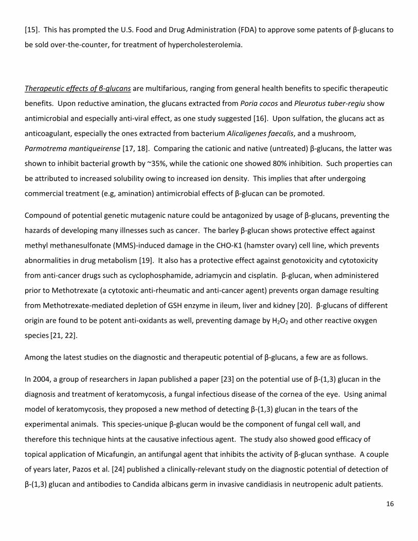
Table 1. Summary of various functions of β‐glucans, 1‐3, and 1‐4 and 1‐6 [6]
Current interest in Glucans:
Basic research on the mode of action of the different types of β (1,3‐1,6)‐glucans at cellular level has been the
foundation for scientific experiments with whole animals and clinical trials. Many studies have confirmed that
β‐glucan enhances overall disease resistance and improves health and performance, with per oral
administration either as feed formulations or as stand‐alone products onto mucous surfaces. These studies
have shown that pure β‐glucan is a non‐toxic modulator of various immune processes.
18
Insoluble and soluble β‐glucan and some clinical trials: Currently, there has been interest shown in the role
of β‐glucan in tumor regression and protection from various infections and to attenuation of ischemia
reperfusion injury [27, 28]. Also, the effectiveness of glucans obtained from different sources has been under
the spotlight. In a study on the efficacy of various β‐glucans, the bioavailability of three different soluble ones
was found to be as low as 0,5‐4,9% in circulation, whereas a water‐insoluble microparticulate β‐glucan was
not detected in the systemic circulation after oral ingestion [29]. On the other hand, some have reported
immunomodulating effects of soluble[30‐35] as well as microparticulate‐insoluble β‐glucan [36, 37] following
oral administration. In more experimental studies, oral β‐glucan has also been shown to protect against
lipopolysaccharide‐induced shock (from infectious agents) in rodents [30‐35] , and lower release of myocardial
enzymes in the postoperative period following coronary artery bypass grafting, indicating a possible cardio‐
protective effect [31]. Another study highlighted the role of microparticulate β‐glucan in protecting against
renal ischemia reperfusion injury [32].
In our experiments at UiT, we use the NBG and SBG samples provided by Biotec Pharmacon, a company of bio‐
products based in Norway. Their products have shown beneficial applications in general infection prophylaxis
especially HIV‐AIDS and tuberculosis, immune therapy of cancer and pre‐op and post‐op repair of damaged
heart tissue in cases of myocardial infarction. In collaboration with Biotec Pharmacon, the β‐glucan group at
UiT has some clinical development and R&D programmes with cooperating academic and scientific institutions
in Norway and abroad, particularly in South Africa and USA. Among other focuses, we want to investigate the
receptors involved in uptake of β‐glucan in the gut. Dectin‐1 has previously been described in leukocytes and
dendritic cells [38], but has not been extensively studied in epithelial cells. So for a while, one subgroup of the
UiT Glucan group has been working on epithelial cell lines to investigate potential receptors for β‐glucan.
19
RECEPTORS IN INNATE IMMUNITY:
Introduction: Characteristic microbial products are recognized by scavenger cells of innate immunity. This
recognition is via receptors and leads to intracellular signaling mechanisms to give an efficient cellular
response. Such responses range from phagocytosis and related intracellular processes essential for handling
ingested microbes to release of a broad range of mediators. The mediators are involved in the efferent arm of
the innate immune response and include cytokines, chemokines, antimicrobial peptides, lysozyme, BPI,
lactoferrin, proteases, lipases, glycosidases, superoxides, nitric oxide, and many others. But first, we will
elaborate a bit on the basis of receptor‐ligand interaction in innate immunity.
In order to recognize and respond to the antigens that are their specific targets, both B cells and T cells carry
special receptors on their surface. The B cell receptor is a homologue of the antibody secreted by that
particular B cell itself. When a B cell encounters a matching antigen, this antibody‐like receptor allows the B
cell to interact with it. The T cell receptor is more complex. It is made of a pair of chemically linked chains
with variable and constant regions and requires signaling and anchoring cell surface molecules (CD3) in order
to work. It cannot recognize antigen in its natural state. The antigen must first be broken down, and the
fragments bound to an Major Histocompatibility Complex (MHC) molecule, by an antigen‐presenting cell.
Helper T cells (CD4 cells) look for antigen bound to a class II MHC molecule on Antigen Presenting Cells (APCs)
like macrophages and B cells. The cytotoxic T cells (CD8), however, respond to antigen bound to MHC class I
molecules which can be found on almost all body cells. This is how a T‐cell receptor molecule forms a three‐
way complex with its specific foreign antigen and an MHC protein.
The major antigen receptor, called “α/β” for its two chains, is found on most CD4 and CD8 cells. Another type
is “γ/δ” found on a distinct subset of T‐cells, but with a yet‐undiscovered function. Both receptor types work
in conjunction with CD3, a signal transduction module made up of various chains.
20
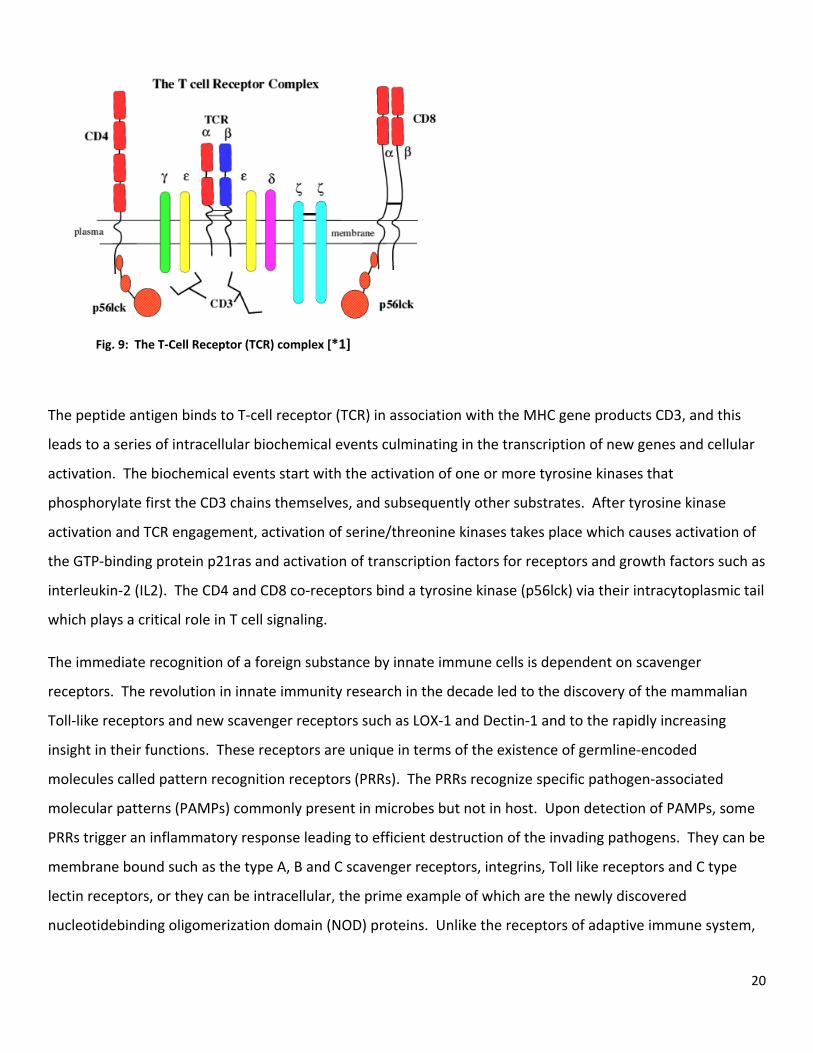
Fig. 9: The T‐Cell Receptor (TCR) complex [*1]
The peptide antigen binds to T‐cell receptor (TCR) in association with the MHC gene products CD3, and this
leads to a series of intracellular biochemical events culminating in the transcription of new genes and cellular
activation. The biochemical events start with the activation of one or more tyrosine kinases that
phosphorylate first the CD3 chains themselves, and subsequently other substrates. After tyrosine kinase
activation and TCR engagement, activation of serine/threonine kinases takes place which causes activation of
the GTP‐binding protein p21ras and activation of transcription factors for receptors and growth factors such as
interleukin‐2 (IL2). The CD4 and CD8 co‐receptors bind a tyrosine kinase (p56lck) via their intracytoplasmic tail
which plays a critical role in T cell signaling.
The immediate recognition of a foreign substance by innate immune cells is dependent on scavenger
receptors. The revolution in innate immunity research in the decade led to the discovery of the mammalian
Toll‐like receptors and new scavenger receptors such as LOX‐1 and Dectin‐1 and to the rapidly increasing
insight in their functions. These receptors are unique in terms of the existence of germline‐encoded
molecules called pattern recognition receptors (PRRs). The PRRs recognize specific pathogen‐associated
molecular patterns (PAMPs) commonly present in microbes but not in host. Upon detection of PAMPs, some
PRRs trigger an inflammatory response leading to efficient destruction of the invading pathogens. They can be
membrane bound such as the type A, B and C scavenger receptors, integrins, Toll like receptors and C type
lectin receptors, or they can be intracellular, the prime example of which are the newly discovered
nucleotidebinding oligomerization domain (NOD) proteins. Unlike the receptors of adaptive immune system,
21
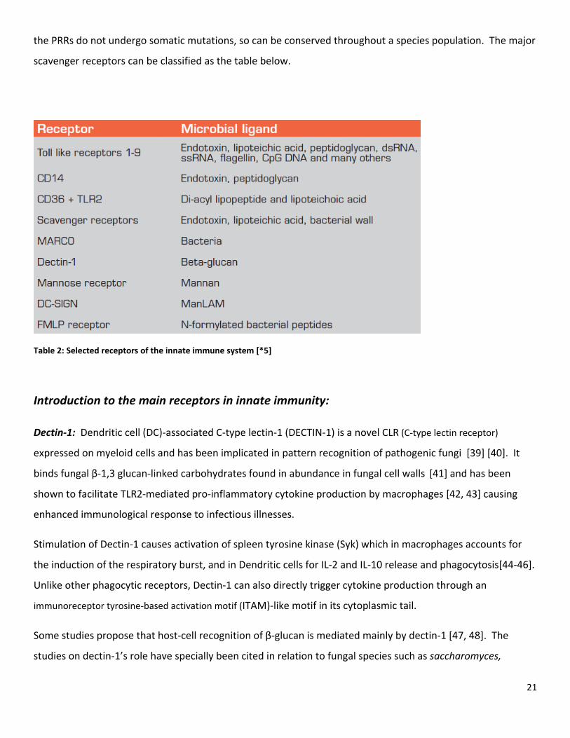
the PRRs do not undergo somatic mutations, so can be conserved throughout a species population. The major
scavenger receptors can be classified as the table below.
Table 2: Selected receptors of the innate immune system [*5]
Introduction to the main receptors in innate immunity:
Dectin‐1: Dendritic cell (DC)‐associated C‐type lectin‐1 (DECTIN‐1) is a novel CLR (C‐type lectin receptor)
expressed on myeloid cells and has been implicated in pattern recognition of pathogenic fungi [39] [40]. It
binds fungal β‐1,3 glucan‐linked carbohydrates found in abundance in fungal cell walls [41] and has been
shown to facilitate TLR2‐mediated pro‐inflammatory cytokine production by macrophages [42, 43] causing
enhanced immunological response to infectious illnesses.
Stimulation of Dectin‐1 causes activation of spleen tyrosine kinase (Syk) which in macrophages accounts for
the induction of the respiratory burst, and in Dendritic cells for IL‐2 and IL‐10 release and phagocytosis[44‐46].
Unlike other phagocytic receptors, Dectin‐1 can also directly trigger cytokine production through an
immunoreceptor tyrosine‐based activation motif (ITAM)‐like motif in its cytoplasmic tail.
Some studies propose that host‐cell recognition of β‐glucan is mediated mainly by dectin‐1 [47, 48]. The
studies on dectin‐1’s role have specially been cited in relation to fungal species such as saccharomyces,
22
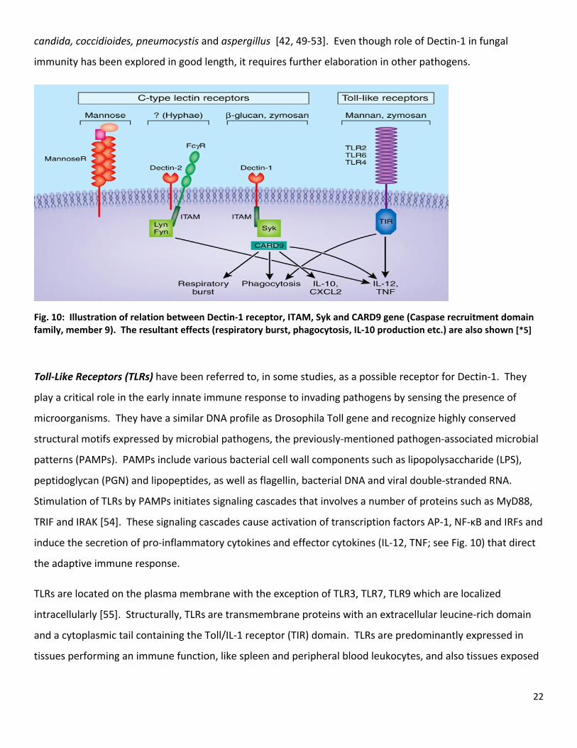
candida, coccidioides, pneumocystis and aspergillus [42, 49‐53]. Even though role of Dectin‐1 in fungal
immunity has been explored in good length, it requires further elaboration in other pathogens.
Fig. 10: Illustration of relation between Dectin‐1 receptor, ITAM, Syk and CARD9 gene (Caspase recruitment domain family, member 9). The resultant effects (respiratory burst, phagocytosis, IL‐10 production etc.) are also shown [*5]
Toll‐Like Receptors (TLRs) have been referred to, in some studies, as a possible receptor for Dectin‐1. They
play a critical role in the early innate immune response to invading pathogens by sensing the presence of
microorganisms. They have a similar DNA profile as Drosophila Toll gene and recognize highly conserved
structural motifs expressed by microbial pathogens, the previously‐mentioned pathogen‐associated microbial
patterns (PAMPs). PAMPs include various bacterial cell wall components such as lipopolysaccharide (LPS),
peptidoglycan (PGN) and lipopeptides, as well as flagellin, bacterial DNA and viral double‐stranded RNA.
Stimulation of TLRs by PAMPs initiates signaling cascades that involves a number of proteins such as MyD88,
TRIF and IRAK [54]. These signaling cascades cause activation of transcription factors AP‐1, NF‐κB and IRFs and
induce the secretion of pro‐inflammatory cytokines and effector cytokines (IL‐12, TNF; see Fig. 10) that direct
the adaptive immune response.
TLRs are located on the plasma membrane with the exception of TLR3, TLR7, TLR9 which are localized
intracellularly [55]. Structurally, TLRs are transmembrane proteins with an extracellular leucine‐rich domain
and a cytoplasmic tail containing the Toll/IL‐1 receptor (TIR) domain. TLRs are predominantly expressed in
tissues performing an immune function, like spleen and peripheral blood leukocytes, and also tissues exposed
23
to external environment such as lung and the gastrointestinal tract, but the receptor subtype and pattern of
expression varies among tissues and the cell types.
NOD‐Like Receptors (NLRs) constitute a recently identified family of intracellular PRRs, which contains more
than 20 members in mammals. Although the ligands and functions of many of these receptors are not known,
their primary role is to recognize cytoplasmic PAMPs and endogenous danger signals. They are characterized
by a tripartite‐domain with a conserved nucleotide binding oligomerization domain (NOD) and leucine‐rich
repeats (LRRs). The general domain structure consists of C‐terminal leucine‐rich repeats involved in microbial
sensing, a centrally located NOD domain and an N‐terminal effector region made up of a protein‐protein
interaction domain such as the CARD, Pyrin or BIR domain.
The NOD‐like receptors have been categorized into subfamilies on the basis of their effector domains: NODs,
NALPs, CIITA(MHC Class II transactivator), IPAF, and NAIPs. NODs and IPAF contain CARD (caspase recruitment
domain) effector domains, whereas NALPs and NAIPs contain pyrin (PYD) effector domains and three BIR
(baculovirus inhibitor of apoptosis protein repeat) domains, respectively. They have only been cited in few
studies in connection to possible β‐glucan binding and uptake.
Among the noteworthy NOD‐like receptors (NLRs) are NOD1 and NOD2, which are the first mammalian NLRs
reported to sense intracellular microbial PAMPs, which contain one and two N‐terminal CARD domains. They
recognize peptidoglycan (PGN), an essential constituent of the bacterial cell wall. NOD1 and NOD2 detect
specific motifs within the PGN. NOD1 senses the D‐γ‐glutamyl‐meso‐DAP dipeptide (iE‐DAP) which is found in
PGN of all Gram‐negative and certain Gram‐positive bacteria [56, 57] whereas NOD2 recognizes the muramyl
dipeptide (MDP) structure found in almost all bacteria [58]. Thus NOD2 acts as a general sensor of PGN and
NOD1 is involved in the recognition of a specific subset of bacteria. NALPs are a subfamily of NLRs that
consists of 14 members characterized by the presence of PYD effector domains. Although precise functions of
many NALPs are unknown, several have been reported to play a key role in the regulation of caspase‐1 by
forming a mutiprotein complex known as the ‘inflammasome’. Caspase‐1 participates in the processing and
subsequent release of proinflammatory cytokines, such as IL‐1β and IL‐18 [59]. IPAF and NAIP5 constitute
another set of NLRs. IPAF belongs to the CARD (caspase recruitment domain) subfamily whereas NAIP5 is a
member of the BIR subfamily. Both have been shown to respond to flagellin, the main component of the
bacterial flagellum, restricting the proliferation of intracellular bacteria such as Salmonella typhimurium,
Shigella flexneri and Legionella pneumophila [60, 61].
24
DISCUSSION
RECEPTORS IMPLICATED IN β‐GLUCAN RESEARCH:
Studies implicating Dectin‐1 in β‐glucan binding and uptake:
Various studies have been done to investigate the role of Dectin‐1 in binding and uptake of β‐glucans. Some
of the latest studies have been elaborated in the following:
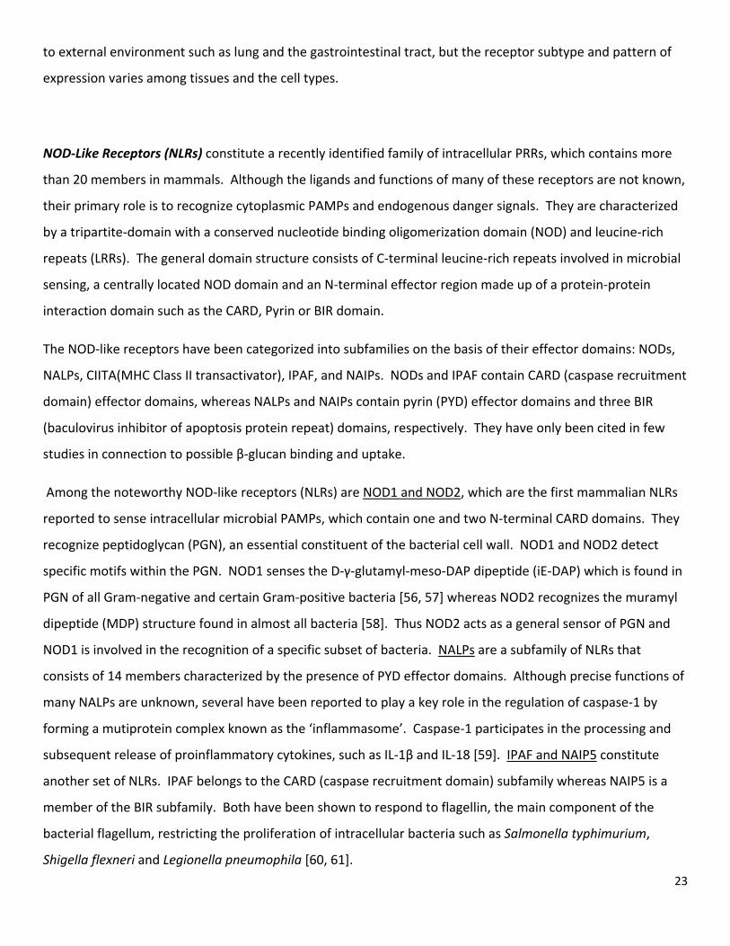
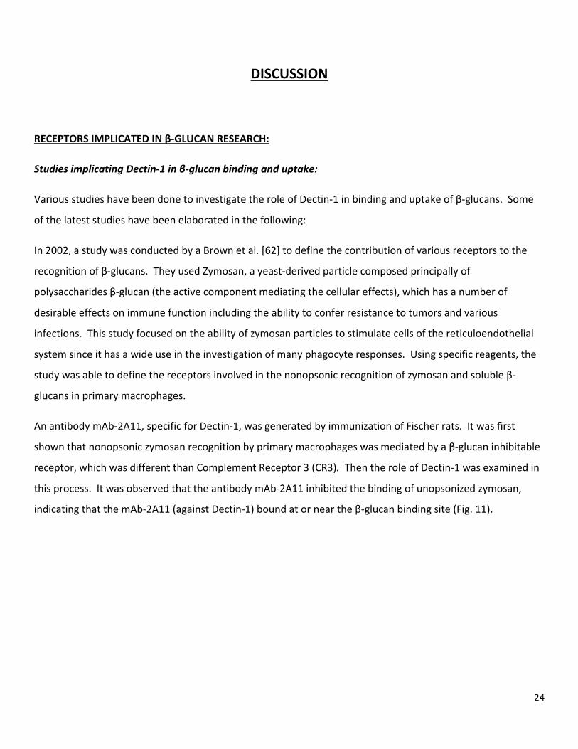
In 2002, a study was conducted by a Brown et al. [62] to define the contribution of various receptors to the
recognition of β‐glucans. They used Zymosan, a yeast‐derived particle composed principally of
polysaccharides β‐glucan (the active component mediating the cellular effects), which has a number of
desirable effects on immune function including the ability to confer resistance to tumors and various
infections. This study focused on the ability of zymosan particles to stimulate cells of the reticuloendothelial
system since it has a wide use in the investigation of many phagocyte responses. Using specific reagents, the
study was able to define the receptors involved in the nonopsonic recognition of zymosan and soluble β‐
glucans in primary macrophages.
An antibody mAb‐2A11, specific for Dectin‐1, was generated by immunization of Fischer rats. It was first
shown that nonopsonic zymosan recognition by primary macrophages was mediated by a β‐glucan inhibitable
receptor, which was different than Complement Receptor 3 (CR3). Then the role of Dectin‐1 was examined in
this process. It was observed that the antibody mAb‐2A11 inhibited the binding of unopsonized zymosan,
indicating that the mAb‐2A11 (against Dectin‐1) bound at or near the β‐glucan binding site (Fig. 11).
25
Figure 11. 2A11 (anti‐Dectin‐1), but not 5C6 (anti‐CR3), specifically inhibits the recognition of unopsonized FITC‐labeled zymosan by macrophages. Increased % of relative fluorescence indicates decreased binding and uptake of glucan particles. The level of inhibition with 2A11 is similar to glucan phosphate (GluP). Brown, G.D., et al. 2002; fig reproduced with permission
The individual levels of inhibition by mAb‐2A11 and 5C6 (the anti‐CR3 antibody) depended on the degree of
opsonization and the simultaneous addition of both antibodies, which has an synergistic effect. The study
indicates that Dectin‐1 mediates the β‐glucan–dependent recognition of opsonized zymosan by macrophages.
Further studies were conducted in 2004 and 2005 on the role of Dectin‐1 in the binding and uptake of β‐
glucans. Three noteworthy ones were from Reid et al., Adachi et al., and Willment et al.
Reid et al. [63] elaborated on the role of Dectin‐1 as a PRR on macrophages (Ms), neutrophils, and dendritic
cells (DCs). They study cited the mediation of the nonopsonic recognition of, and response to, soluble and
particulate yeast ß‐glucans by Dectin‐1‐carrying macrophages and bone marrow‐derived DCs.
Immunohistochemical detection of Dectin‐1 was optimized and its expression was demonstrated on
neutrophils, subpopulations of Ms in splenic red and white pulp, alveolar Ms, Kupffer cells, and Ms and DCs in
the lamina propria of gut villi. This shows the consistency of Dectin‐1's role in pathogen surveillance, the
hallmark of which is the recognition of β‐glucan component of microbial antigen.
26
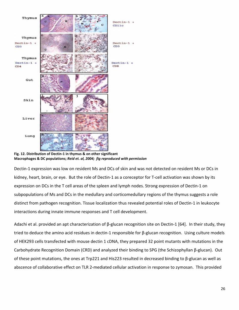
Fig. 12. Distribution of Dectin‐1 in thymus & on other significant Macrophages & DC populations; Reid et. al, 2004; fig reproduced with permission Dectin‐1 expression was low on resident Ms and DCs of skin and was not detected on resident Ms or DCs in
kidney, heart, brain, or eye. But the role of Dectin‐1 as a coreceptor for T‐cell activation was shown by its
expression on DCs in the T cell areas of the spleen and lymph nodes. Strong expression of Dectin‐1 on
subpopulations of Ms and DCs in the medullary and corticomedullary regions of the thymus suggests a role
distinct from pathogen recognition. Tissue localization thus revealed potential roles of Dectin‐1 in leukocyte
interactions during innate immune responses and T cell development.
Adachi et al. provided an apt characterization of β‐glucan recognition site on Dectin‐1 [64]. In their study, they
tried to deduce the amino acid residues in dectin‐1 responsible for β‐glucan recognition. Using culture models
of HEK293 cells transfected with mouse dectin 1 cDNA, they prepared 32 point mutants with mutations in the
Carbohydrate Recognition Domain (CRD) and analyzed their binding to SPG (the Schizophyllan β‐glucan). Out
of these point mutations, the ones at Trp221 and His223 resulted in decreased binding to β‐glucan as well as
abscence of collaborative effect on TLR 2‐mediated cellular activation in response to zymosan. This provided
27
further insight into the binding properties of β‐glucan and Dectin‐1, and showed the critical position of amino
acid sequence W221‐I222‐H223 in the CRD of Dectin‐1.
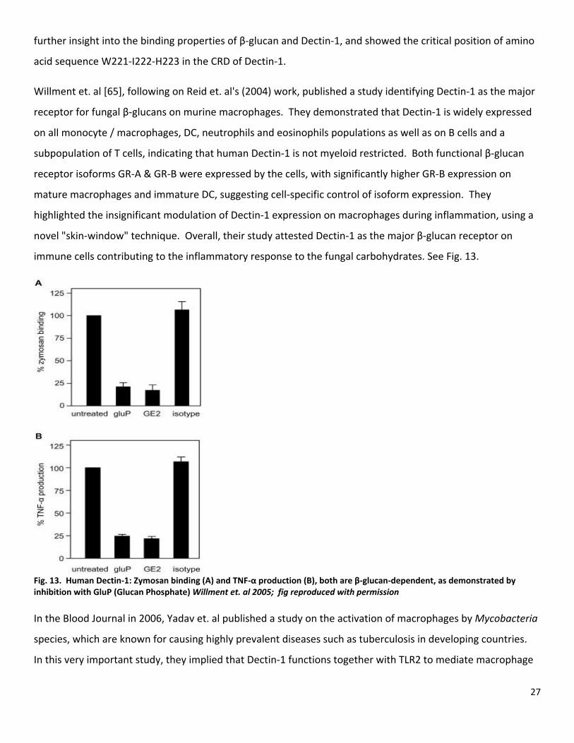
Willment et. al [65], following on Reid et. al's (2004) work, published a study identifying Dectin‐1 as the major
receptor for fungal β‐glucans on murine macrophages. They demonstrated that Dectin‐1 is widely expressed
on all monocyte / macrophages, DC, neutrophils and eosinophils populations as well as on B cells and a
subpopulation of T cells, indicating that human Dectin‐1 is not myeloid restricted. Both functional β‐glucan
receptor isoforms GR‐A & GR‐B were expressed by the cells, with significantly higher GR‐B expression on
mature macrophages and immature DC, suggesting cell‐specific control of isoform expression. They
highlighted the insignificant modulation of Dectin‐1 expression on macrophages during inflammation, using a
novel "skin‐window" technique. Overall, their study attested Dectin‐1 as the major β‐glucan receptor on
immune cells contributing to the inflammatory response to the fungal carbohydrates. See Fig. 13.
Fig. 13. Human Dectin‐1: Zymosan binding (A) and TNF‐α production (B), both are β‐glucan‐dependent, as demonstrated by inhibition with GluP (Glucan Phosphate) Willment et. al 2005; fig reproduced with permission In the Blood Journal in 2006, Yadav et. al published a study on the activation of macrophages by Mycobacteria
species, which are known for causing highly prevalent diseases such as tuberculosis in developing countries.
In this very important study, they implied that Dectin‐1 functions together with TLR2 to mediate macrophage
28
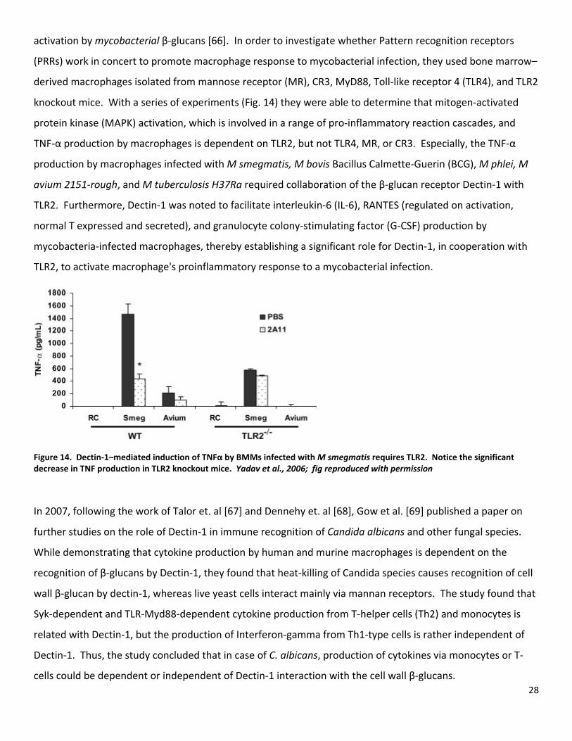
activation by mycobacterial β‐glucans [66]. In order to investigate whether Pattern recognition receptors
(PRRs) work in concert to promote macrophage response to mycobacterial infection, they used bone marrow–
derived macrophages isolated from mannose receptor (MR), CR3, MyD88, Toll‐like receptor 4 (TLR4), and TLR2
knockout mice. With a series of experiments (Fig. 14) they were able to determine that mitogen‐activated
protein kinase (MAPK) activation, which is involved in a range of pro‐inflammatory reaction cascades, and
TNF‐α production by macrophages is dependent on TLR2, but not TLR4, MR, or CR3. Especially, the TNF‐α
production by macrophages infected with M smegmatis, M bovis Bacillus Calmette‐Guerin (BCG), M phlei, M
avium 2151‐rough, and M tuberculosis H37Ra required collaboration of the β‐glucan receptor Dectin‐1 with
TLR2. Furthermore, Dectin‐1 was noted to facilitate interleukin‐6 (IL‐6), RANTES (regulated on activation,
normal T expressed and secreted), and granulocyte colony‐stimulating factor (G‐CSF) production by
mycobacteria‐infected macrophages, thereby establishing a significant role for Dectin‐1, in cooperation with
TLR2, to activate macrophage's proinflammatory response to a mycobacterial infection.
Figure 14. Dectin‐1–mediated induction of TNFα by BMMs infected with M smegmatis requires TLR2. Notice the significant decrease in TNF production in TLR2 knockout mice. Yadav et al., 2006; fig reproduced with permission
In 2007, following the work of Talor et. al [67] and Dennehy et. al [68], Gow et al. [69] published a paper on
further studies on the role of Dectin‐1 in immune recognition of Candida albicans and other fungal species.
While demonstrating that cytokine production by human and murine macrophages is dependent on the
recognition of β‐glucans by Dectin‐1, they found that heat‐killing of Candida species causes recognition of cell
wall β‐glucan by dectin‐1, whereas live yeast cells interact mainly via mannan receptors. The study found that
Syk‐dependent and TLR‐Myd88‐dependent cytokine production from T‐helper cells (Th2) and monocytes is
related with Dectin‐1, but the production of Interferon‐gamma from Th1‐type cells is rather independent of
Dectin‐1. Thus, the study concluded that in case of C. albicans, production of cytokines via monocytes or T‐
cells could be dependent or independent of Dectin‐1 interaction with the cell wall β‐glucans.
29
Among the studies from within the last two years, ones by Shah et al. (2008), Harada (2008) and Ujita (2009)
et al. are of note.
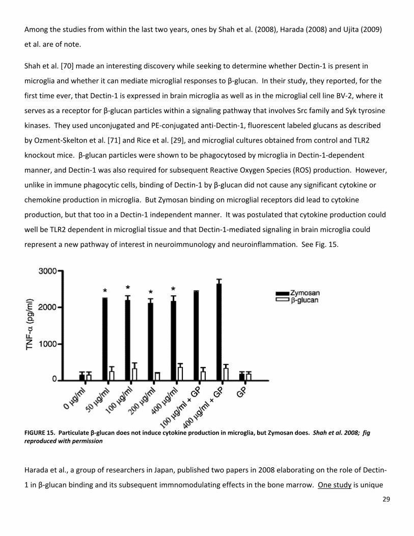
Shah et al. [70] made an interesting discovery while seeking to determine whether Dectin‐1 is present in
microglia and whether it can mediate microglial responses to β‐glucan. In their study, they reported, for the
first time ever, that Dectin‐1 is expressed in brain microglia as well as in the microglial cell line BV‐2, where it
serves as a receptor for β‐glucan particles within a signaling pathway that involves Src family and Syk tyrosine
kinases. They used unconjugated and PE‐conjugated anti‐Dectin‐1, fluorescent labeled glucans as described
by Ozment‐Skelton et al. [71] and Rice et al. [29], and microglial cultures obtained from control and TLR2
knockout mice. β‐glucan particles were shown to be phagocytosed by microglia in Dectin‐1‐dependent
manner, and Dectin‐1 was also required for subsequent Reactive Oxygen Species (ROS) production. However,
unlike in immune phagocytic cells, binding of Dectin‐1 by β‐glucan did not cause any significant cytokine or
chemokine production in microglia. But Zymosan binding on microglial receptors did lead to cytokine
production, but that too in a Dectin‐1 independent manner. It was postulated that cytokine production could
well be TLR2 dependent in microglial tissue and that Dectin‐1‐mediated signaling in brain microglia could
represent a new pathway of interest in neuroimmunology and neuroinflammation. See Fig. 15.
FIGURE 15. Particulate β‐glucan does not induce cytokine production in microglia, but Zymosan does. Shah et al. 2008; fig reproduced with permission Harada et al., a group of researchers in Japan, published two papers in 2008 elaborating on the role of Dectin‐
1 in β‐glucan binding and its subsequent immnomodulating effects in the bone marrow. One study is unique
30
in the sense that it describes Dectin‐1 receptor on “dendritic cells” of the marrow. In the paper published in
Journal of Interferon and Cytokine Research [72], they showed the result of their investigation on the effect of
Sparassis crispa β‐glucan (SCG) on bone marrow‐derived dendritic cells (BMDCs) in DBA/2 mice. Having
already proved earlier [73] that SCG has significant antitumor activity in DBA/1 and DBA/2 mice (inbred
strains, with 98% genetic match), they went on to show increased CD80, MHC‐I and MHC‐II molecules'
expression on the cell membrane of dendritic cells as well as increased interleukin‐12p70, IL‐6, and TNF‐α
production in mice of selected lineages. The magnitude of cytokine induction was higher in DBA/2 mice than
in mice of other lineage, and so was the expression level of Dectin‐1. Blocking Dectin‐1 remarkably inhibited
TNF‐α production suggesting that the bone marrow DCs from DBA/2 mice are highly sensitive to the induction
of cytokine production by the Sparassis β‐glucan (SCG) in vitro, and that this sensitivity is related to the
expression level of dectin‐1. In the other paper published in the International Immunopharmacology journal
[74], Harada et al. studied the contribution of dectin‐1 and granulocyte macrophage‐colony stimulating factor
(GM‐CSF) to immunomodulating actions of β‐glucan. They elaborated on the strain difference in the reactivity
of mice to SCG, showing that DBA/1 and DBA/2 are highly sensitive strains. Although complete abscence of
Dectin‐1 receptor would completely abolish the cytokine production in dendritic cells, as in the case of dectin‐
1 knockout mice, but controlling the level of endogenous GM‐CSF alongside dectin‐1 expression could regulate
the reactivity to β‐glucan. The point of focus was the role of GM‐CSF, and the study's result indicates that GM‐
CSF production and dectin‐1 expression are the key factors in the responsiveness to β‐glucan.
In 2009, more research has been published [75, 76] with regards to the receptors for fungal β‐glucans and
their immune‐stimulatory capability. But a stand‐out study by Ujita et al. [77] sheds some light on the
biological chemistry and carbohydrate binding specificity of human macrophage β‐glucan receptor dectin‐1.
Upon expressing Dectin‐1 as a fusion protein with an N‐terminal hexahistidine tag and glutathione S‐
transferase (His‐GST‐hDectin‐1) in an Escherichia coli cell‐free translation system, the researchers assayed the
recombinant protein for binding specificity with β‐glucan in comparison with human dectin‐1. The
recombinant dectin‐1 specifically bound to some β‐glucans, but not to other carbohydrates, showing its
relative specificity. The β‐glucan binding of recombinant dectin‐1 was inhibited by laminarin and
laminarioligosaccharides, the soluble β‐glucans, but not by other carbohydrates, again showing that dectin‐1 is
rather specific in its ligand‐binding. Overall, the study shows that the recombinant human dectin‐1, owing to
its strict binding specificty, has potential use as a probe in identifying ligands in humans and tonic foods.
31
FUTURE PROSPECTS OF Β‐GLUCAN RECEPTOR AND CLINICAL STUDIES
Studying the response of human immune system to β‐glucans extracted from various sources has been the
mainstay of glucan studies uptil now. What we need to focus on is the exact nature of receptor studies and
the intracellular pathways it activates.
Prospective research in production & purification of β‐(1,3) glucans: The sources of natural occurrence,
degree and character of polymerization, isolation protocols, degree of solubility of preparations… in fact all
the trial‐and‐error phases of the preparation of β‐glucan, especially the particulate one, point towards the fact
that each isolated batch is heterogeneous. As extraction and purification processes drastically influence the
glucan properties, numerous patents have been published on the subject. Processing may affect the
molecular (chemical structure and DP), structural (molecular interactions) and functional properties (viscosity,
water binding capacity and solubility), which may cause change in the physiological properties of glucans, and
so can shearing damage due to mechanical processing or excessive heat treatment of food products.
Decreased molecular weight and reduced viscosity can result from structural changes taking place during
commercial purification [78]. Endogenous β‐glucanases, if not deactivated through high temperatures, may
cause increased depolymerisation of the β‐glucans [79]. Even if in some cases the heterogeneity of β‐glucan
(different molecular size, branching, and crystalline or amorphous structure) does not principally change the in
vivo activities, it still represents substantial complications for pharmacological research and regulatory
authorities. Here, one could ask the question, how can we avoid such complications in preparing β‐glucan
supplements? The answer is “research”.
Emphasis should be put on reliable research techniques to allow the problems of heterogeneity and
nonexistent standards of various natural β‐glucans to be solved. Improved isolation techniques could be the
first step in this regard, so that products with closely defined chemical composition could be obtained and up‐
to‐date physicochemical methods could be used for identification and analyses of these products. Another
method could be to produce chemically‐modified β‐glucans, whose solubility could artificially be enhanced,
but this may cause decrease in the glucan’s biological activity. However, attempts have been made to
construct semi‐synthetic or synthetic probes suitable for immunological research. For example, in 2006,
Descroix et al. proposed that short oligomers of glucose containing β(1→3) and β(1→6) linkages could be
bound to a well‐defined polymer carrier, so that such a “synthetic” β‐glucan will interact with receptors of
immunocompetent cells and elicit analogous reactions as natural β‐glucan [80]. With the advent of synthetic
β‐glucan probes in immunopharmacological research, the shortcomings of natural glucans can be addressed.
32
Prospective areas of β‐glucan research in Food industry:
Owing to its polysaccharide nature, β‐(1,3) glucans have found a wide range of potential applications in food
industry, like in edible films, feed for domestic animals and low calorie food.
There is some scope in the area of research in glucan gelling properties. Curdlan, a neutral gel‐forming linear
β‐(1,3) glucan produced in Agrobacterium species, has been used as a biothickening and gelling agent in foods.
Apart from being tasteless, colorless and odorless, studies have shown that heating process can produce
different forms of curdlan gel with different textural qualities, physical stabilities and water‐holding capacities.
A prospective research area is to exploit these properties of Curdlan and other β‐glucans to synthesize gels of
differing strength for incorporation into gums for value‐added food products.
An area of research for glucans in food industry is its application as an adjuvant for delivery of active
compounds. Glucans can act as capsules or “tablet‐like” ingredients which can deliver their content further,
after ingestion. They are non‐protein, heat‐resistant capsule that could be filled with any product. A few
experiments by Ono et al. (1996) and Shiomi et al. (2005) have already shown the promise offered by glucans
in this regard.
When it comes to non‐fat products, cereal β‐glucan can be of use as soluble dietary fibre and as such can open
avenues of research in food industry as stabilizer in low‐fat products such as salad dressings, ice creams,
yoghurts and cheese. This way, scientists could explore how β‐glucans could help decrease the calorie content
of food, as well as have beneficial effects on their gelation and rheological characteristics. In milk
preparations, one could study how the gelling properties of β‐glucan solutions could reduce curd cutting time
and increases curd yields.
Prospective Medical and Pharmaceutical research areas:
Use of Β‐glucans in medical and pharmaceutical science is a potentially huge area of research. Cereal β‐
glucans have been studied to influence hyperglycemia and hypercholesterolemia, and the glucans obtained
from bacteria, yeasts and fungi have shown promise as immunopotentiating and antitumorogenic agents. This
paves way for research against serious infectious illnesses such as tuberculosis and AIDS.
β‐glucan administration: With the goal of treating patients with β‐glucans, parenteral (intravenous or
intraperitoneal administrations) need to be investigated further. Since insoluble or hardly soluble β‐glucans
33
cause significant adverse effects (granuloma formation, microembolization, inflammation, pain) when
administered by parenteral routes [81], research needs to be focused on how oral administration could be
optimized for quick delivery and higher bioavailability.
Hypercholesterolemia, Diabetes and Glycaemia: Cereal β‐glucans have been studied for potential use as
dietary fibre. Dietary fibre is a type of food substance mainly derived from plant material (cellulose,
hemicellulose, pectin, lignin, etc) that resists human digestive enzymes. Research has shown that insoluble
dietary fiber reduces bowel transit time [82], prevents constipation, reduces risk of colorectal cancer [83], and
helps production of short chain fatty acids [84] for better handling of hyperlipidemia. And soluble dietary
fibers, especially β‐(1,3)(1,4) glucans, help lower blood cholesterol [85], sugar and insulin level [86],
hypercholesterolemia [87], cardiovascular disease[88], cancer[89], and promotion of the growth of beneficial
gut microflora (as a prebiotic). With this wide range of benefits already related to the use of β‐glucan as
dietary fiber, further studies are needed to elaborate on such clinical and preventive health benefits.
With special regard to hypercholesterolemia, the cholesterol‐lowering potential of β‐glucans is considered to
result from effects manifest in the upper gastrointestinal tract via its ability to form a gel‐like network and
alter gastrointestinal viscosity. A few studies have implicated the amount and quality of fiber, increased
intrinsic viscosity of the food in combination with fluids, maintenance of physical integrity of the food material
and incomplete starch gelatinisation as the potential mechanism of action behind these effects [90] . It could
be because intestinal viscosity reduces the cholesterol level by stimulating the production of bile acid. This
could be via upregulation in the activity of cholesterol 7 α‐hydroxylase (CYP7A1), an enzyme associated with
the regulation of the pathway through which cholesterol is converted into bile acids. More research is
required to elucidate the effect of β‐glucan on enzyme and immune‐regulation. Another potential area of
cholesterol research could be to mix β‐glucan with a non‐digestible fat in one product for concomitant control
of both high cholesterol and sugar level. Such mixtures, if developed through further research, could then be
used as dietary supplement or as food additive when incorporated in products such as cereals, dairy products
such as yoghurt or nutritional beverages, bakery products and prepared meals.
With regards to diabetes, β‐glucan from different sources could be studied in a comparative fashion to
highlight the different responses they elicit. For example, one could see if barley or oat β‐glucans cause
different level of reduction in blood glucose levels and if one of them is more effective in the regulation of
glucose and insulin responses compared to the other. A study by Hallfrisch et al. published in the journal
Cereal Chemistry in 2003 could be useful as a template where they investigated the use of β‐glucans isolated
34
from barley and oats, and their corresponding effects upon plasma glucose and insulin responses in non‐
diabetic adults. But the extraction procedures, and factors such as dose, molecular weight and fine structure,
and rheological characteristics of extracted and native β‐glucans are important in this regard, as underscored
by Wood et al. in 1994 [91].
An interesting prospective area in studying the medical and pharmaceutical effects of glucans is the effect of
variation in viscosity of its preparations. One such study has already shown that the glycemic response of
fiber‐rich foods was inversely related to viscosity of the preparation [92]. Another group of researchers
(Tappy et al.) also found that inclusion of oat β‐glucan into breakfast cereals could reduce the postprandial
glycaemic response by up to 50% [93]. Further studies could be done on the combination of altered viscosity
and modified structure of β‐glucans, and noting its resultant neutraceutical effects.
Inclusion of β‐glucan could be made to a staple or regular‐use food and could be studied over longer periods.
For example, Yokoyama et al. (1997) and Knuckles et al. (1997) have done comparisons of blood glucose and
insulin responses of healthy individuals following the ingestion of different sources of β‐glucans. This way, the
ability of β‐glucans to influence the rate of starch degradation and hence the glycaemic index of foods could
be studied with regard to obesity and diabetes, and standards could be set with blood glucose levels of
diabetic and pre‐diabetic individuals with the use of β‐glucan rich foods.
Prospective research as Immunopotentiator: β‐glucan could have important application as an
immunopotentiator. Through earlier research, a wide variety of mushroom polysaccharides including
scleroglucan, lentinan, schizophyllan, and grifolan have been described as biological‐response modifiers [94]
acting through mechanisms mediated by the immune system, including its nonspecific stimulation. Such
polysaccharides obtained from bacterial, fungal or yeast sources have a beneficial effect on a variety of
infectious or cancerous disease processes. In this regard, the baker’s yeast cell wall preparation (Zymosan)
has been a milestone. Being the first defined pharmaceutical yeast product with immunostimulatory activity,
its active component was identified to be β‐d‐glucan and was reported to stimulate phagocytosis, cytotoxic
activity in macrophages, and other biological activities [95]. Further research could be done on the
mechanism behind these actions, especially the receptor studies of the innate immune system. One could
investigate the signaling cascades which regulate gene expression concerned with the secretion of co‐
stimulatory molecules and increased antigen‐presentation activity which help direct the adaptive immune
response.
35
Research in anti‐tumorigenic effects: Although, anticarcinogenic effect of many mushroom polysaccharides
such as lentinan, grifolan, scleroglucan and schizophyllan have been described in earlier studies, the
mechanism of their anti‐tumour action is still not completely clear. It remains to be seen whether these β‐
glucans appear to mediate their antitumour activity by activation or augmentation of the host’s immune
system, via activation of leukocytes and production of inflammatory cytokines [96], or may be a combination
of all of these. Although glucan‐specific receptors have been found on phagocytic cell membranes and their
role in phagocytosis and killing has been described, there tends to be a greater variation in activity against
various cancer types, such as sarcomas, breast cancer, adenocarcinoma, colon cancer and some leukemias. To
normalize this degree of variation, and to define the exact mechanism of anti‐tumorigenic effect of β‐glucans,
further research is needed.
Anti‐HIV & AIDS effects: The HIV has the ability to incorporate its DNA, multiply inside, and cause destruction
of the T‐helper cell subtype of lymphocytes, while dodging the normal scavenging leukocytes such as
macrophages. In the past, extracts of Lentinus edodes (lentinan) has been found to stimulate specific T‐helper
cells in healthy humans while stimulating lymphokine activated killer (LAK) activity in combination with
Interleukin‐2[97]. Recent development concerning the direct utilization of β‐glucans for treating AIDS are
rare, but there have been proposals to establish clinical trials with Grifola frondosa extract, sulfated β‐glucans,
curdlan and lentinan sulfate or in combination with more conventional drugs [98]. Further research could be
done in this regard to prove the efficacy of β‐glucan preparations in treatment of AIDS.
Potential Prebiotic Applications: Prebiotics are compounds that help in sustenance of beneficial
microorganisms in the intestinal tract. These microorganisms help in lactose digestion, cholesterol reduction,
antimicrobial effects, immune system stimulation etc. and include Lactobacilli, streptococcus salivarus and
Bifidobacteria species in particular. Oat β‐glucan has been reported to selectively support the growth of
Lactobacilli and Bifidobactera in rat experiments and in in vitro studies [99]. Further research could be done
based on these observations to develop several patents on the use of β‐glucans (or their corresponding
hydrolysates) as prebiotics.
Other Therapeutic Applications: Many other therapeutic applications could be studied for β‐glucans. One
such utilization could be as radioprotective agent. This has already been put into perspective by an earlier
study showing that β‐glucan is able to protect macrophages from free radical attack during and after the
radiation allowing these cells to continue to function in the irradiated body [100]. Another prospective area is
to study glucan as a DNA‐transport tool. β‐glucan has shown the ability to function as a gene carrier with its
36
hydrogen‐bonding triple‐helix polymer structure (as in schizophyllan, curdlan, lentinan, scleroglucan) binding
to a nucleic acid. The resulting nucleic acid‐polymer complex can be used as a vector with further benefit as
nucleic acid‐protecting agent as it is resistant to nuclease. Further studies could establish this usage more
clearly.
It will be interesting to study β‐glucans for the treatment and prevention of digestion troubles such as
constipation, inflammatory bowel disease, or stomach troubles using earlier studies as a template. More
potential uses of glucans have been proposed for arthrosis, dermatitis and athlete’s foot and oral cavity
infections, and could be studied in detail with further research.
Prospective research in prevention of side‐effects of β‐glucan use: While there are predominantly positive
pharmacological effects of β‐glucans, the unfavorable side effects of glucans cannot be overlooked. Therefore,
a theme for future research could be prevention of hazardous or side effects with β‐glucan use. Such effects
may arise from independent use of glucans, but are more likely to occur with the intake of glucans as an
adjuvant.,.
Intramuscular administration of β‐glucan is painful, and induces an inflammatory reaction and granuloma
formation at the puncture site. β‐glucan may cause an inflammatory reaction itself, so that a naturally
occurring "physiological" inflammation in response to a noxious impact might become compounded and
"pathological" with β‐glucan's presence. As Tanriverdi et al. described in 2005, the resulting insult and tissue
damage may herald the development of immunopathological (e.g., autoimmune) processes. Even worse, a
generalized inflammatory process may develop into shock and multiple organ dysfunction syndrome (MODS).
A future research area could be the development of preventive or therapeutic strategies in such case
scenarios.
Nitric oxide is produced in macrophages by inducible nitric oxide synthase (iNOS). It has cytotoxic effect upon
tumor cells [101] and shows distinct impact on many pathogens. iNOS synthesis (are you sure that you do not
mean nitric oxide synthesis instead of iNOS synthesis ? ) is triggered by binding of β‐glucan to a PRR on the
macrophage surface. Besides tumoricidal and bactericidal effect, nitric oxide can also damage tissues and
DNA and high iNOS concentrations can cause septic shock. The sustained action of the activating pathogen or
tumor cell causes continuous expression of iNOS and higher amount of nitric oxide and causes vasodilatation
of veins. A group of researchers studied this phenomenon [102] and concluded that the drop in blood
37
pressure may come to a dangerously low level. A prospective area of research could be to study the risk of
this eventuality with the use of adjuvant β‐glucan therapy in infectious diseases or cancer.
The Syndrome Of Toxic Organic Dust (STOD) is defined as the aftermath of inhalation of intact cells, fungal or
yeast bodies, home, agricultural or industrial dusts [103]. The condition is characterized by respiratory tract
reactions that include pneumonia, cough, chronic bronchitis, rhinitis, headache, and irritation of the eyes and
throat [104]. It has been found that the cause of these complaints is β‐glucan, which, through activation of
macrophages, monocytes, and leukocytes, causes increased secretion of inflammatory components (i.e., TNFα
and IL‐8). Research emphasis could be put on the tools to modulate such (over)activation of phagocytes
against cell‐wall β‐glucans.
Drug interaction is a widely‐studied phenomenon in modern medicine. With the use of β‐glucans (pro‐
inflammatory compounds), there is a risk of competitive drug interaction with anti‐inflammatory drugs. A
study of reference in this regard [105] demonstrated the lethal toxicity elicited by a combination of β‐glucan
and Indomethacin (a nonsteroidal anti‐inflammatory drug) in experimental mice. It has been proposed that
simultaneous use of both agents may cause cytokine level derangement and Systemic Inflammation Response
Syndrome (SIRS). A future research area could be to study how such an interaction could be prevented or
controlled.
38
THE GLUCAN GROUP IN TROMSØ:
The Glucan Group in Tromsø has been in function for a couple of years now. The group focuses on four
different areas of β‐glucan research.
One subgroup has used immunhistochemistry to study molecular marker‐potential of Dectin‐1, scavenger
receptor, TLR2, MR, MyD88 and CR3 in the human gastrointestinal tract. These receptors have been described
to play a role in binding and uptake of glucans and the subgroup is working to elicit this mechanism in the
distal small intestine (ileum).
One group worker has been assaying the biological activity of different batches of both soluble and non‐
soluble β‐glucan produced by Biotec Pharma, AS. In the last year, focus has remained on using the mouse
macrophage cell line RAW transfected with the Dectin‐1 gene as a tool to analyse the β‐glucan response by
measuring cytokines in the conditioned medium by ELISA.
My focus as Masters student has shifted from cell‐line studies to mouse intestinal specimen studies. I have
worked with instestinal (ileal) explants obtained from mice. Small pieces (3‐4 cm) of ileum tissue were cut
loose and thoroughly wased. Thereafter they were incubated with FITC‐labeled glucan in culture medium for a
few hours, and before fixation. The protocol has been devised for both FITC‐SBG and the FITC‐NBG and we
have been trying to overcome the difficulty of inconsistent tissue morphology, which is a drawback in
identifying the cells. More time and effort is needed to establish this method accurately.
Two group members have worked with human peritoneal macrophages from healthy patients and have noted
active uptake of FITC‐SBG and FITC‐NBG. There is a degree of variation found in the uptake activity of these
cells, ranging between 10‐90% within the different macrophage batches for FITC‐SBG uptake. Interest remains
in noting if single cells are able to take up both SBG and NBG, and if different uptake‐mechanisms are
involved. Three main uptake‐mechanisms are studied: Clathrin‐mediated endocytosis,
macropinocytosis/phagocytosis, and lipid raft/caveola‐mediated endocytosis. The subgroup aims to define
relative prevalence and specificity of binding and uptake of a ligand with these different methods.
Preliminary studies in culture of cells from rat liver has shown that non of the the scavenging sinusoidale
endothelial nor the Kupffer cells did take up FITC‐SBG, whereas a few unindetified liver cells were able to
accumulate the FITC‐NBG. Further, by using both human macrophages and monocytt‐derived DC, the group
aims to define other lectins which are involved with binding and uptake of β‐glucans, such as theMR, DEC‐205,
DC‐SIGN, Dectin‐2 and Scavenger Receptor C‐type Lectin.
39
Also, this subgroup has established a protocol of isolation of monocytes from human blood. Monocytes are
then cultured in vitro in culture dishes with IL‐4 and GM‐CSF and by using a cocktail that includes IL‐1, TNF‐α,
IL‐6 and PEG2, the monocytes differentiate into DC. The sub‐group further aims to study and analyze the
effect of SBG and NBG on these cells.
One group member has been working with analyzing macrophage‐RNA from both mouse and human
specimen after stimulation with SBG. She published a study in this regard in 2007 [106], with more coming in
due course of time.
Another group member has been investigating the effect of SBG and NBG glucan on myocardial infarcts in
experimental pigs before and after the artificially‐induced infarction. At the same time, NBG has been used in
patients prior to heart surgery and its influence in the disease prognosis is observed.
40
CONCLUSION
Among many known and tested immunomodulators, polysaccharides isolated from various natural sources
occupy a prominent position. An important group of these polysaccharides is represented by the
homopolymers of β‐glucose, called β‐glucans. Due to their very low‐to‐negligible toxicity, they have
tremendous potential use in a variety of diseases, such as infections, irradiation diseases, and foremost on
neoplastic growth. Branded earlier as a food additive and an “alternative” remedy in its early development
days, the wave of enthusiasm about β‐glucans declined, the main reasons being insufficiently defined
preparations and non‐specific and/or complex effects. But over the last decade or so, modern medical
research has reached a phase where the basic mechanisms of glucan effects are known and the relationship
between structure and activity has been outlined rather clearly. It seems now that β‐glucans will finally take
the position they deserve in diagnostic and preventive medicine.
Being widely distributed within microorganisms in which they act as membrane components, and with no
biosynthesis in mammals, β‐glucans have been observed to activate the immune system of their host. Since
the discovery of their involvement as immunomodulating agent, numerous studies have been published
dealing with purification procedures, analytical chemistry, synthetic processes, chemical modification,
physicochemical properties, and assessment of the biological and medicinal effects of the natural
polysaccharides through in vitro and in vivo studies. Studies should be focused more on discovery of the
receptors present on immunocompetent cells and scope and limitations of chemical synthesis and
modifications of β‐glucans.
As the research on the properties of new plant or microbial polysaccharides continues to grow, we need to
streamline the commercialization of such compounds against traditional products, and somehow balance the
improvement in original structures with the cost of production and development. As it looks at this point, β‐
glucans will have significant development in the next few years, notably in non‐food sectors, with the most
promise seemingly possible in therapeutic applications. Such applications include immunomodulators,
antitumorogenic, antiviral (AIDS), anti‐hypercholesterolemic and anti‐hyperglycemic agents. Moreover,
specific action as food additives could be ascribed and research with β‐glucans. Ultimately, the
pharmaceutical industry may provide new markets for chemically‐modified glucans, like the sulfated ones, and
help develop new generations of polysaccharides with more beneficial biological activities.
41
REFERENCES:
1. Houghton, A.N. and J.A. Guevara‐Patino, Immune recognition of self in immunity against cancer. J Clin Invest, 2004. 114(4): p. 468‐71.
2. Cisneros, R.L., F.C. Gibson, 3rd, and A.O. Tzianabos, Passive transfer of poly‐(1‐6)‐beta‐glucotriosyl‐(1‐3)‐beta‐glucopyranose glucan protection against lethal infection in an animal model of intra‐abdominal sepsis. Infect Immun, 1996. 64(6): p. 2201‐5.
3. Zimmerman, J.W., et al., A novel carbohydrate‐glycosphingolipid interaction between a beta‐(1‐3)‐glucan immunomodulator, PGG‐glucan, and lactosylceramide of human leukocytes. J Biol Chem, 1998. 273(34): p. 22014‐20.
4. Zekovic, D.B., et al., Natural and modified (1‐‐>3)‐beta‐D‐glucans in health promotion and disease alleviation. Crit Rev Biotechnol, 2005. 25(4): p. 205‐30.
5. Sandula, J., E. Machova, and V. Hribalova, Mitogenic activity of particulate yeast beta‐(1‐‐>3)‐D‐glucan and its water‐soluble derivatives. Int J Biol Macromol, 1995. 17(6): p. 323‐6.
6. Mantovani, M.S., et al., beta‐Glucans in promoting health: prevention against mutation and cancer. Mutat Res, 2008. 658(3): p. 154‐61.
7. Reynolds, J.A., et al., Glucan‐induced enhancement of host resistance to selected infectious diseases. Infect Immun, 1980. 30(1): p. 51‐7.
8. Meira, D.A., et al., The use of glucan as immunostimulant in the treatment of paracoccidioidomycosis. Am J Trop Med Hyg, 1996. 55(5): p. 496‐503.
9. Yun, C.H., et al., Immunomodulatory effects of oat beta‐glucan administered intragastrically or parenterally on mice infected with Eimeria vermiformis. Microbiol Immunol, 1998. 42(6): p. 457‐65.
10. Soltys, J., et al., Effect of glucan immunomodulator on the immune response and larval burdens in mice with experimental toxocarosis. Appl Parasitol, 1996. 37(3): p. 161‐7.
11. Falch, B.H., et al., The cytokine stimulating activity of (1‐‐>3)‐beta‐D‐glucans is dependent on the triple helix conformation. Carbohydr Res, 2000. 329(3): p. 587‐96.
12. Tokunaka, K., et al., Immunopharmacological and immunotoxicological activities of a water‐soluble (1‐‐>3)‐beta‐D‐glucan, CSBG from Candida spp. Int J Immunopharmacol, 2000. 22(5): p. 383‐94.
13. Luhm, J., et al., Beta‐(1‐‐>3)‐D‐glucan modulates DNA binding of nuclear factors kappaB, AT and IL‐6 leading to an anti‐inflammatory shift of the IL‐1beta/IL‐1 receptor antagonist ratio. BMC Immunol, 2006. 7: p. 5.
14. Miura, N.N., et al., Structure and biological activities of beta‐glucans from yeast and mycelial forms of Candida albicans. Microbiol Immunol, 2003. 47(3): p. 173‐82.
15. Bernardshaw, S., et al., An extract of the mushroom Agaricus blazei Murill protects against lethal septicemia in a mouse model of fecal peritonitis. Shock, 2006. 25(4): p. 420‐5.
16. Shin, M.S., et al., Structural and biological characterization of aminated‐derivatized oat beta‐glucan. J Agric Food Chem, 2005. 53(14): p. 5554‐8.
17. Chang, Y.J., et al., Structural and biological characterization of sulfated‐derivatized oat beta‐glucan. J Agric Food Chem, 2006. 54(11): p. 3815‐8.
18. Zhang, M., et al., Chain conformation of sulfated derivatives of beta‐glucan from sclerotia of Pleurotus tuber‐regium. Carbohydr Res, 2003. 338(24): p. 2863‐70.
19. Oliveira, R.J., et al., Evaluation of antimutagenic activity and mechanisms of action of beta‐glucan from barley, in CHO‐k1 and HTC cell lines using the micronucleus test. Toxicol In Vitro, 2006. 20(7): p. 1225‐33.
20. Sener, G., et al., Beta‐glucan ameliorates methotrexate‐induced oxidative organ injury via its antioxidant and immunomodulatory effects. Eur J Pharmacol, 2006. 542(1‐3): p. 170‐8.
21. Krizkova, L., et al., Antioxidant and antimutagenic activity of mannan neoglycoconjugates: mannan‐human serum albumin and mannan‐penicillin G acylase. Mutat Res, 2006. 606(1‐2): p. 72‐9.
42
22. Slamenova, D., et al., Protective effects of fungal (1‐‐>3)‐beta‐D‐glucan derivatives against oxidative DNA lesions in V79 hamster lung cells. Cancer Lett, 2003. 198(2): p. 153‐60.
23. Kaji, Y., T. Hiraoka, and T. Oshika, Potential use of (1,3)‐beta‐D‐glucan as target of diagnosis and treatment of keratomycosis. Cornea, 2004. 23(8 Suppl): p. S36‐41.
24. Pazos, C., et al., Diagnostic potential of (1,3)‐beta‐D‐glucan and anti‐Candida albicans germ tube antibodies for the diagnosis and therapeutic monitoring of invasive candidiasis in neutropenic adult patients. Rev Iberoam Micol, 2006. 23(4): p. 209‐15.
25. Jaehrig, S.C., et al., In vitro potential antioxidant activity of (1‐‐>3),(1‐‐>6)‐beta‐D‐glucan and protein fractions from Saccharomyces cerevisiae cell walls. J Agric Food Chem, 2007. 55(12): p. 4710‐6.
26. Driscoll, M., et al., Therapeutic potential of various beta‐glucan sources in conjunction with anti‐tumor monoclonal antibody in cancer therapy. Cancer Biol Ther, 2009. 8(3).
27. Chen, J. and R. Seviour, Medicinal importance of fungal beta‐(1‐‐>3), (1‐‐>6)‐glucans. Mycol Res, 2007. 111(Pt 6): p. 635‐52.
28. Rasmussen, L.T. and R. Seljelid, Dynamics of blood components and peritoneal fluid during treatment of murine E. coli sepsis with beta‐1,3‐D‐polyglucose derivatives. I. Cells. Scand J Immunol, 1990. 32(4): p. 321‐31.
29. Rice, P.J., et al., Oral delivery and gastrointestinal absorption of soluble glucans stimulate increased resistance to infectious challenge. J Pharmacol Exp Ther, 2005. 314(3): p. 1079‐86.
30. Ramakers, J.D., et al., Fecal water from ileostomic patients consuming oat beta‐glucan enhances immune responses in enterocytes. Mol Nutr Food Res, 2007. 51(2): p. 211‐20.
31. Sandvik, A., et al., Oral and systemic administration of beta‐glucan protects against lipopolysaccharide‐induced shock and organ injury in rats. Clin Exp Immunol, 2007. 148(1): p. 168‐77.
32. Lehne, G., et al., Oral administration of a new soluble branched beta‐1,3‐D‐glucan is well tolerated and can lead to increased salivary concentrations of immunoglobulin A in healthy volunteers. Clin Exp Immunol, 2006. 143(1): p. 65‐9.
33. Hashimoto, K., I. Suzuki, and T. Yadomae, Oral administration of SSG, a beta‐glucan obtained from Sclerotinia sclerotiorum, affects the function of Peyer's patch cells. Int J Immunopharmacol, 1991. 13(4): p. 437‐42.
34. Sakurai, T., et al., Enhancement of murine alveolar macrophage functions by orally administered beta‐glucan. Int J Immunopharmacol, 1992. 14(5): p. 821‐30.
35. Suzuki, I., et al., Inhibition of experimental pulmonary metastasis of Lewis lung carcinoma by orally administered beta‐glucan in mice. Chem Pharm Bull (Tokyo), 1991. 39(6): p. 1606‐8.
36. Cheung, N.K., et al., Orally administered beta‐glucans enhance anti‐tumor effects of monoclonal antibodies. Cancer Immunol Immunother, 2002. 51(10): p. 557‐64.
37. Aarsaether, E., et al., Cardioprotective effect of pretreatment with beta‐glucan in coronary artery bypass grafting. Scand Cardiovasc J, 2006. 40(5): p. 298‐304.
38. Bayrak, O., et al., Oral beta‐glucan protects kidney against ischemia/reperfusion injury in rats. Am J Nephrol, 2008. 28(2): p. 190‐6.
39. Robinson, M.J., et al., Myeloid C‐type lectins in innate immunity. Nat Immunol, 2006. 7(12): p. 1258‐65. 40. Brown, G.D., Dectin‐1: a signalling non‐TLR pattern‐recognition receptor. Nat Rev Immunol, 2006. 6(1): p. 33‐43. 41. Palma, A.S., et al., Ligands for the beta‐glucan receptor, Dectin‐1, assigned using "designer" microarrays of
oligosaccharide probes (neoglycolipids) generated from glucan polysaccharides. J Biol Chem, 2006. 281(9): p. 5771‐9.
42. Brown, G.D., et al., Dectin‐1 mediates the biological effects of beta‐glucans. J Exp Med, 2003. 197(9): p. 1119‐24. 43. Gantner, B.N., et al., Collaborative induction of inflammatory responses by dectin‐1 and Toll‐like receptor 2. J Exp
Med, 2003. 197(9): p. 1107‐17. 44. Herre, J., et al., Dectin‐1 uses novel mechanisms for yeast phagocytosis in macrophages. Blood, 2004. 104(13): p.
4038‐45. 45. Underhill, D.M., et al., Dectin‐1 activates Syk tyrosine kinase in a dynamic subset of macrophages for reactive
oxygen production. Blood, 2005. 106(7): p. 2543‐50. 46. Rogers, N.C., et al., Syk‐dependent cytokine induction by Dectin‐1 reveals a novel pattern recognition pathway
for C type lectins. Immunity, 2005. 22(4): p. 507‐17.
43
47. Ariizumi, K., et al., Identification of a novel, dendritic cell‐associated molecule, dectin‐1, by subtractive cDNA cloning. J Biol Chem, 2000. 275(26): p. 20157‐67.
48. Brown, G.D. and S. Gordon, Immune recognition. A new receptor for beta‐glucans. Nature, 2001. 413(6851): p. 36‐7.
49. Viriyakosol, S., et al., Innate immunity to the pathogenic fungus Coccidioides posadasii is dependent on Toll‐like receptor 2 and Dectin‐1. Infect Immun, 2005. 73(3): p. 1553‐60.
50. Steele, C., et al., Alveolar macrophage‐mediated killing of Pneumocystis carinii f. sp. muris involves molecular recognition by the Dectin‐1 beta‐glucan receptor. J Exp Med, 2003. 198(11): p. 1677‐88.
51. Steele, C., et al., The beta‐glucan receptor dectin‐1 recognizes specific morphologies of Aspergillus fumigatus. PLoS Pathog, 2005. 1(4): p. e42.
52. Hohl, T.M., et al., Aspergillus fumigatus triggers inflammatory responses by stage‐specific beta‐glucan display. PLoS Pathog, 2005. 1(3): p. e30.
53. Gersuk, G.M., et al., Dectin‐1 and TLRs permit macrophages to distinguish between different Aspergillus fumigatus cellular states. J Immunol, 2006. 176(6): p. 3717‐24.
54. Medzhitov, R., P. Preston‐Hurlburt, and C.A. Janeway, Jr., A human homologue of the Drosophila Toll protein signals activation of adaptive immunity. Nature, 1997. 388(6640): p. 394‐7.
55. Nishiya, T. and A.L. DeFranco, Ligand‐regulated chimeric receptor approach reveals distinctive subcellular localization and signaling properties of the Toll‐like receptors. J Biol Chem, 2004. 279(18): p. 19008‐17.
56. Chamaillard, M., et al., An essential role for NOD1 in host recognition of bacterial peptidoglycan containing diaminopimelic acid. Nat Immunol, 2003. 4(7): p. 702‐7.
57. Girardin, S.E., et al., Nod1 detects a unique muropeptide from gram‐negative bacterial peptidoglycan. Science, 2003. 300(5625): p. 1584‐7.
58. McDonald, C., N. Inohara, and G. Nunez, Peptidoglycan signaling in innate immunity and inflammatory disease. J Biol Chem, 2005. 280(21): p. 20177‐80.
59. Martinon, F. and J. Tschopp, Inflammatory caspases: linking an intracellular innate immune system to autoinflammatory diseases. Cell, 2004. 117(5): p. 561‐74.
60. Miao, E.A., et al., Pseudomonas aeruginosa activates caspase 1 through Ipaf. Proc Natl Acad Sci U S A, 2008. 105(7): p. 2562‐7.
61. Zamboni, D.S., et al., The Birc1e cytosolic pattern‐recognition receptor contributes to the detection and control of Legionella pneumophila infection. Nat Immunol, 2006. 7(3): p. 318‐25.
62. Brown, G.D., et al., Dectin‐1 is a major beta‐glucan receptor on macrophages. J Exp Med, 2002. 196(3): p. 407‐12.
63. Reid, D.M., et al., Expression of the beta‐glucan receptor, Dectin‐1, on murine leukocytes in situ correlates with its function in pathogen recognition and reveals potential roles in leukocyte interactions. J Leukoc Biol, 2004. 76(1): p. 86‐94.
64. Adachi, Y., et al., Characterization of beta‐glucan recognition site on C‐type lectin, dectin 1. Infect Immun, 2004. 72(7): p. 4159‐71.
65. Willment, J.A., et al., The human beta‐glucan receptor is widely expressed and functionally equivalent to murine Dectin‐1 on primary cells. Eur J Immunol, 2005. 35(5): p. 1539‐47.
66. Yadav, M. and J.S. Schorey, The beta‐glucan receptor dectin‐1 functions together with TLR2 to mediate macrophage activation by mycobacteria. Blood, 2006. 108(9): p. 3168‐75.
67. Taylor, P.R., et al., Dectin‐1 is required for beta‐glucan recognition and control of fungal infection. Nat Immunol, 2007. 8(1): p. 31‐8.
68. Dennehy, K.M. and G.D. Brown, The role of the beta‐glucan receptor Dectin‐1 in control of fungal infection. J Leukoc Biol, 2007. 82(2): p. 253‐8.
69. Gow, N.A., et al., Immune recognition of Candida albicans beta‐glucan by dectin‐1. J Infect Dis, 2007. 196(10): p. 1565‐71.
70. Shah, V.B., et al., Beta‐glucan activates microglia without inducing cytokine production in Dectin‐1‐dependent manner. J Immunol, 2008. 180(5): p. 2777‐85.
71. Ozment‐Skelton, T.R., et al., Prolonged reduction of leukocyte membrane‐associated Dectin‐1 levels following beta‐glucan administration. J Pharmacol Exp Ther, 2006. 318(2): p. 540‐6.
44
72. Harada, T., et al., Highly expressed dectin‐1 on bone marrow‐derived dendritic cells regulates the sensitivity to beta‐glucan in DBA/2 mice. J Interferon Cytokine Res, 2008. 28(8): p. 477‐86.
73. Harada, T., et al., Mechanism of enhanced hematopoietic response by soluble beta‐glucan SCG in cyclophosphamide‐treated mice. Microbiol Immunol, 2006. 50(9): p. 687‐700.
74. Harada, T. and N. Ohno, Contribution of dectin‐1 and granulocyte macrophage‐colony stimulating factor (GM‐CSF) to immunomodulating actions of beta‐glucan. Int Immunopharmacol, 2008. 8(4): p. 556‐66.
75. Li, M., et al., Insoluble beta‐glucan from the cell wall of Candida albicans induces immune responses of human THP‐1 monocytes through Dectin‐1. Chin Med J (Engl), 2009. 122(5): p. 496‐501.
76. Sorgi, C.A., et al., Histoplasma capsulatum cell wall {beta}‐glucan induces lipid body formation through CD18, TLR2, and dectin‐1 receptors: correlation with leukotriene B4 generation and role in HIV‐1 infection. J Immunol, 2009. 182(7): p. 4025‐35.
77. Ujita, M., et al., Carbohydrate binding specificity of recombinant human macrophage beta‐glucan receptor dectin‐1. Biosci Biotechnol Biochem, 2009. 73(1): p. 237‐40.
78. Wursch, P. and F.X. PiSunyer, The role of viscous soluble fiber in the metabolic control of diabetes ‐ A review with special emphasis on cereals rich in beta‐glucan. Diabetes Care, 1997. 20(11): p. 1774‐1780.
79. McCleary, B.V. and L. Prosky, Advanced dietary fibre technology. 2001, Oxford ; Malden, MA: Blackwell Science. xxv, 534 p., [4] p. of plates.
80. Descroix, K., et al., Recent progress in the field of beta‐(1,3)‐glucans and new applications. Mini Rev Med Chem, 2006. 6(12): p. 1341‐9.
81. Maeda, Y.Y., et al., Denaturation and renaturation of a beta‐1,6;1,3‐glucan, lentinan, associated with expression of T‐cell‐mediated responses. Cancer Res, 1988. 48(3): p. 671‐5.
82. Shimotoyodome, A., et al., Sulfated polysaccharides, but not cellulose, increase colonic mucus in rats with loperamide‐induced constipation. Dig Dis Sci, 2001. 46(7): p. 1482‐9.
83. Hill, M.J., Cereals, cereal fibre and colorectal cancer risk: a review of the epidemiological literature. Eur J Cancer Prev, 1997. 6(3): p. 219‐25.
84. Wisker, E., et al., Short‐chain fatty acids produced in vitro from fibre residues obtained from mixed diets containing different breads and in human faeces during the ingestion of the diets. Br J Nutr, 2000. 84(1): p. 31‐7.
85. Behall, K.M., D.J. Scholfield, and J. Hallfrisch, Effect of beta‐glucan level in oat fiber extracts on blood lipids in men and women. J Am Coll Nutr, 1997. 16(1): p. 46‐51.
86. Bourdon, I., et al., Postprandial lipid, glucose, insulin, and cholecystokinin responses in men fed barley pasta enriched with beta‐glucan. Am J Clin Nutr, 1999. 69(1): p. 55‐63.
87. Yang, J.L., et al., Barley beta‐glucan lowers serum cholesterol based on the up‐regulation of cholesterol 7alpha‐hydroxylase activity and mRNA abundance in cholesterol‐fed rats. J Nutr Sci Vitaminol (Tokyo), 2003. 49(6): p. 381‐7.
88. Keogh, G.F., et al., Randomized controlled crossover study of the effect of a highly beta‐glucan‐enriched barley on cardiovascular disease risk factors in mildly hypercholesterolemic men. Am J Clin Nutr, 2003. 78(4): p. 711‐8.
89. Sier, C.F., et al., Beta‐glucan enhanced killing of renal cell carcinoma micrometastases by monoclonal antibody G250 directed complement activation. Int J Cancer, 2004. 109(6): p. 900‐8.
90. Tudorica, C.M., V. Kuri, and C.S. Brennan, Nutritional and physicochemical characteristics of dietary fiber enriched pasta. J Agric Food Chem, 2002. 50(2): p. 347‐56.
91. Wood, P.J., J. Weisz, and B.A. Blackwell, Structural Studies of (1‐]3),(1‐]4)‐Beta‐D‐Glucans by C(13)‐Nuclear Magnetic‐Resonance Spectroscopy and by Rapid Analysis of Cellulose‐Like Regions Using High‐Performance Anion‐Exchange Chromatography of Oligosaccharides Released by Lichenase. Cereal Chemistry, 1994. 71(3): p. 301‐307.
92. Wood, P.J., et al., Effect of Dose and Modification of Viscous Properties of Oat Gum on Plasma‐Glucose and Insulin Following an Oral Glucose‐Load. British Journal of Nutrition, 1994. 72(5): p. 731‐743.
93. Tappy, L., E. Gugolz, and P. Wursch, Effects of breakfast cereals containing various amounts of beta‐glucan fibers on plasma glucose and insulin responses in NIDDM subjects. Diabetes Care, 1996. 19(8): p. 831‐834.
94. Borchers, A.T., et al., Mushrooms, tumors, and immunity. Proceedings of the Society for Experimental Biology and Medicine, 1999. 221(4): p. 281‐293.
45
95. Whaley, S., et al., Glucans and related natural carbohydrate polymers exhibit weak solution free radical scavenging activity, but stimulate free radical activity in a murine macrophage cell line. Faseb Journal, 2001. 15(4): p. A680‐A680.
96. Xiao, Z., C.A. Trincado, and M.P. Murtaugh, Beta‐glucan enhancement of T cell IFNgamma response in swine. Vet Immunol Immunopathol, 2004. 102(3): p. 315‐20.
97. Suzuki, M., et al., Induction of endogenous lymphokine‐activated killer activity by combined administration of lentinan and interleukin 2. Int J Immunopharmacol, 1990. 12(6): p. 613‐23.
98. Jagodzinski, P.P., et al., Mechanism of the inhibitory effect of curdlan sulfate on HIV‐1 infection in vitro. Virology, 1994. 202(2): p. 735‐45.
99. Ryhanen, L., et al., The effect of malotilate on type III and type IV collagen, laminin and fibronectin metabolism in dimethylnitrosamine‐induced liver fibrosis in the rat. J Hepatol, 1996. 24(2): p. 238‐45.
100. Patchen, M.L., et al., Glucan: mechanisms involved in its "radioprotective" effect. J Leukoc Biol, 1987. 42(2): p. 95‐105.
101. Stuehr, D.J. and C.F. Nathan, Nitric oxide. A macrophage product responsible for cytostasis and respiratory inhibition in tumor target cells. J Exp Med, 1989. 169(5): p. 1543‐55.
102. Vincent, J.L., et al., Effects of nitric oxide in septic shock. Am J Respir Crit Care Med, 2000. 161(6): p. 1781‐5. 103. Beijer, L., J. Thorn, and R. Rylander, Effects after inhalation of (1‐‐>3)‐beta‐D‐glucan and relation to mould
exposure in the home. Mediators Inflamm, 2002. 11(3): p. 149‐53. 104. Alwis, K.U., J. Mandryk, and A.D. Hocking, Exposure to biohazards in wood dust: bacteria, fungi, endotoxins, and
(1‐‐>3)‐beta‐D‐glucans. Appl Occup Environ Hyg, 1999. 14(9): p. 598‐608. 105. Yoshioka, S., et al., Immunotoxicity of soluble beta‐glucans induced by indomethacin treatment. FEMS Immunol
Med Microbiol, 1998. 21(3): p. 171‐9. 106. Berdal, M., et al., Aminated beta‐1,3‐D‐glucan improves wound healing in diabetic db/db mice. Wound Repair
Regen, 2007. 15(6): p. 825‐32.
*1. www.immunecentral.com *2. Male et al. Immunology , 7th edition, chapter 1 *3. www.gluegrant.org *4. www.cartage.org.lb
*5. www.invivogen.com


![Executive Summary - Web viewThe relationship was down rated because the rheological properties of the semi-synthetic β-glucans compared with ... [Text Word]) OR beta glucans](https://img.pdfslide.net/doc/110x75/5a789dec7f8b9a87198dfb4d/executive-summary-web-viewthe-relationship-was-down-rated-because-the-rheological.jpg)