Embed Size (px)
Citation preview

A SYNDROME OF CONGENITAL DEFECTS INVOLVING
THE ABDOMINAL WALL, STERNUM, DIAPHRAGM, PERICARDIUM, AND HEART
JAMES R. CANTRELL, M.D., F.A.C.S., J. ALEX HALLER, M.D., and
MARK M. RAVITC.H, M.D., Baltimore, Maryland
During recent years, several patients with an unusual combination of congenital defects have been seen in The Johns Hopkins Hospital. The anomalies observed in each patient were: (1) a midline, supraumbilical abdominal wall defect; (2) a defect of the lower sternum; (3) a deficiency of the anterior diaphragm; (4) a defect in the diaphragmatic pericardium; (5) congenital intracardiac defects. The repeated tion of these abnormalities suggested that this combination represented a clinical syndrome and stimulated a search for additional examples. This article was written to summarize the available clinical material, to propose a probable embryologic basis for the coexistence of these defects, and to sider the surgical treatment of patients with this syndrome.
CASE REPORTS
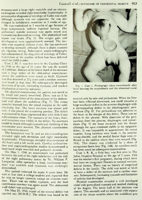
Case 1. C. D., a male infant, was delivered at The Johns Hopkins Hospital by Dr. W. Drummond Eaton on February 8, 1957. In the family history there no other instances of congenital defects. This was his mother’s first pregnancy, during which there had been no recognized infections or unusual environmental exposures. Delivery was uncomplicated, birth weight was 3,170 grams. A large omphalocele immediately apparent (Fig. 1), and it was further noted that the cardiac pulsations were abnormally prominent. The sternum was shortened and the costal arch was elevated and flared. The contour of the heart was visible beneath the skin of the unusually high epigastrium. There was a defect of the musculature of the abdominal wall above the omphalocele; the medial edges of the rectus muscles were widely
From the Department of Surgery, The Johns Hopkins University School of Medicine and The Johns Hopkins Hospital, Baltimore.
separated and inserted on the costal margins at the midclavicular line. The child appeared otherwise normal. No cardiac murmurs were audible.
The child was operated upon (J.R.C.) under vincthene-cther anesthesia. The amniolic sac wasexcised with a small margin of skin (Fig. 2). The liver, which occupied the upper portion of the omphalocele, could then be depressed to reveal a large defect in the ventral portion of the diaphragm (Fig. 3A). The diaphragm arose normally from the posterior and lateral aspects of the rib cage but at a point in line with the insertion of the rectus muscles on the costal margins the diaphragm curved posteriorly, leaving a semilunar defect which was bounded dorsally by a small cufl' of diaphragm on the liver immediately ventral to the inferior vena cava. There was a corresponding defect in the pericardium. The serous linings of the peritoneal and pericardial cavities were continuous across the edge of the diaphragmatic defect. The pleurae were intact. The heart lay somewhat further to the right than normal, and there was an anomalous anterior coronary artery. In the abdomen there was failure of attachment of the mesentery of the small bowel and right colon. '1 he cecum lay in the epigastrium, and the appendix lay within the pericardial cavity.
Attention was first directed to closure of the diaphragmatic defect. The junction of pericardium and peritoneum was incised along the edge of the diaphragmatic defect and the pericardium reflected from the diaphragm. The edge of the diaphragm then sutured to the anterior rib cage (Fig. 3B). It was apparent that the rectus muscles could not be approximated. It was possible, however, to develop a layer of fascia which was continuous with the medial border of the rectus sheath and had the appearance of linea alba (Figs. 4 and 5). The edges of this fascia were brought together in the midline after
made in each
associa-
con-
were
was
was
a long relaxing incision had been anterior rectus sheath.
The child recovered satisfactorily but during his convalescence a murmur was noted over the left second interspace. Mild congestive failure developed which required digitalization. Radiologic study
602
Cantrell cl aL: syndrome or congenital defects 603
demonstrated a large right ventricle and an electrocardiogram revealed right ventricular hypertrophy. A presumptive diagnosis of tetralogy of Fallot was made although cyanosis was not apparent. He was discharged in satisfactory condition at 3 weeks of age.
He was readmitted at 3 months of age because of large symptomatic indirect inguinal hernias. The pulmonary systolic murmur was again noted and cyanosis was detected on crying. The abdominal wall repair was intact (Fig. 6). His weight gain and development were approximately normal. The hernias were repaired without event. He has continued to develop normally although there is slight cyanosis on vigorous crying. Subsequent angiocardiography has substantiated the diagnosis of tetralogy of Fallot. Correction of the cardiac defects has been deferred until the child is older.
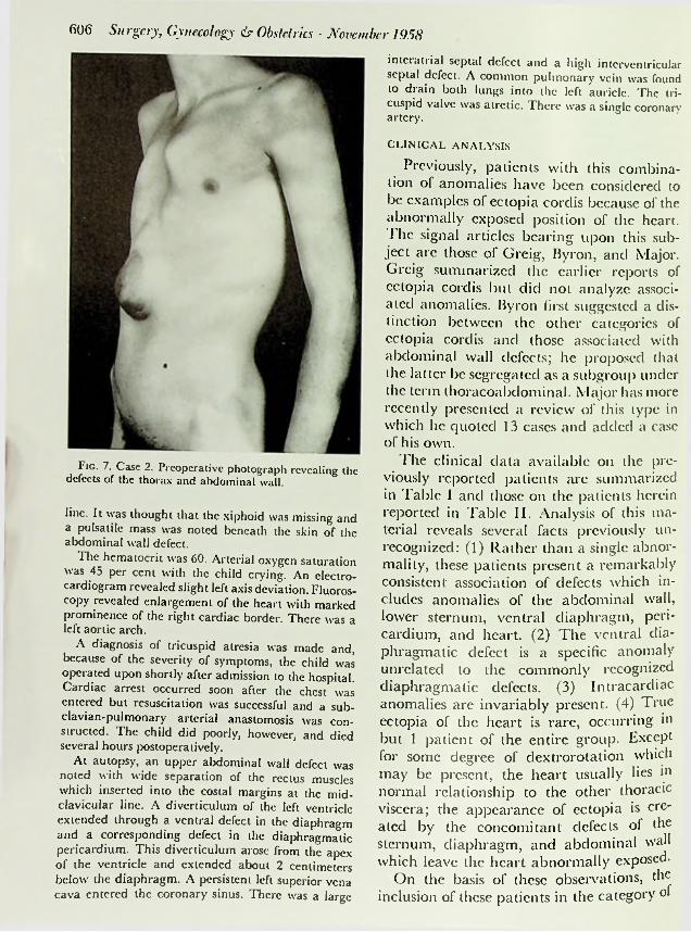
Case 2. M. F. was first seen in the Cardiac Clinicin 1948 at the age of 16 years. He was the second of 3 siblings. Absence of the lower third of the sternum and a large defect of the abdominal musculature above the umbilicus were noted at birth. Cyanosis was first detected at 2]/> years and a cardiac murmur was heard at that time. He subsequently developed shortness of breath, increasing cyanosis, and marked diminution of exercise tolerance.
On physical examination, the patient was noted to be small and poorly nourished. There was an 8 by 10 centimeter defect of the musculature of the abdominal wall above the umbilicus (Fig. 7). The muscles inserted into the costal margins at the mid- clavicular line. The lower sternum was absent and only 4 ribs joined the remaining portion. The defect was covered by hyperpigmented skin with little if any subcutaneous tissue. The contours of the heart, liver, and intestines were visible in the defect. No could be heard although cyanosis and clubbing of the extremities were marked. The physical examination was otherwise normal.
The hematocrit was 76, and an electrocardiogram revealed right axis deviation and right ventricular hypertrophy. Fluoroscopy revealed dextrorotation of the heart and a left aortic arch. Cardiac catheterization and angiocardiographic studies demonstrated a functional single ventricle and pulmonic stenosis.
On May 1, 1948 an anastomosis was performed between the end of the right subclavian and the side of the right pulmonary artery by Dr. William P. Longmirc. After operation a loud, mur was audible and cyanosis was strikingly diminished.
Fig. 1. Case 1. Photograph taken immediately after birth showing the omphalocele and the abnormal costal arch.
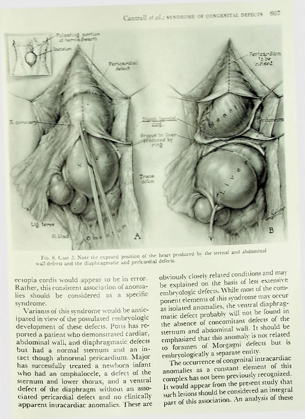
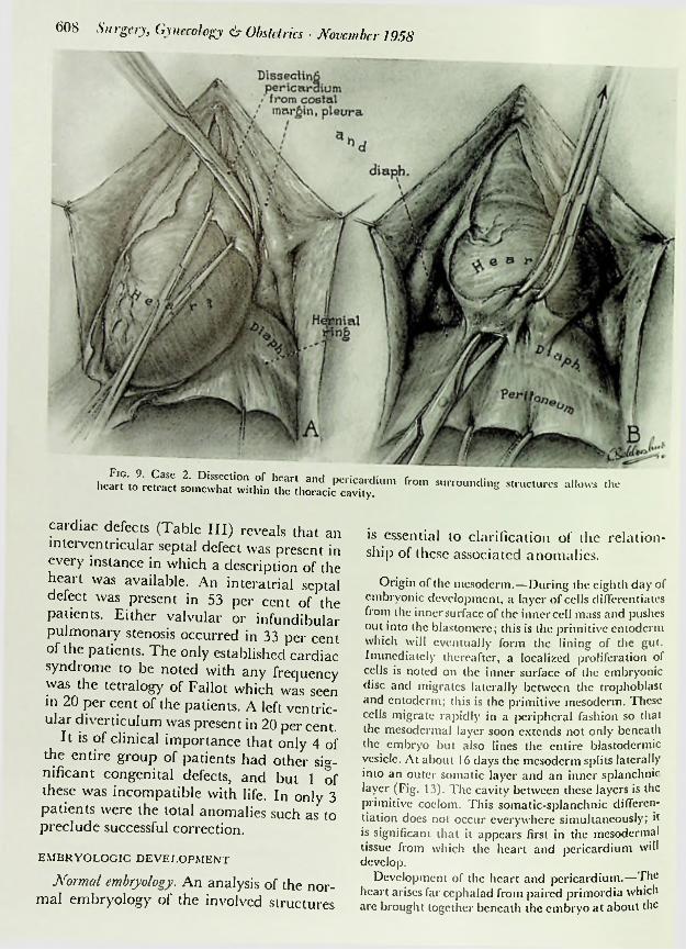
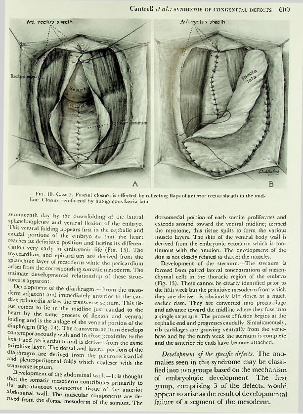
covered only by skin and peritoneum. When the liver had been reflected downward, one could visualize a large semilunar defect in the anterior diaphragm with a corresponding defect in the diaphragmatic pericardium (Fig. 8). The pericardium was adherent to the heart at the margin of this defect. There defect in the pleurae. With dissection of the pericardium from the pleurae, diaphragm, and epicar- dium (Fig. 9) the heart retracted into the thorax although the apex remained visible in the epigastric defect. It was impossible to approximate the rectus muscles. Long incisions were made in the anterior rectus sheaths, and the medial fascial flaps were reflected to the midline where they were sutured (Fig. 10A). This closure was reinforced by a autogenous fascia lata (Fig. 10B). An excellent result has been noted on subsequent examinations.
Case 3. R. B., a little boy, was first seen in The Johns Hopkins Hospital at the age of 2 years. This
his mother’s first pregnancy, during which there had been no recognized illnesses or unusual environmental factors. There was no history of congenital defects in the family. The child was noted at birth to have a defect of the sternum and a cardiac murmur. Subsequently, increasing cyanosis and limitation of activity had developed.
Physical examination revealed a well developed child with generalized cyanosis and marked clubbing of the fingers. The lower half of the sternum was absent. This anomaly and an associated wide separation of the rectus muscles above the umbilicus pro
rectus
was no
murmurs
sheet of
wascontinuous mur-
The patient returned for study 4 years later. Fie was at that time a college student and reported that he was no longer cyanotic except lion. There was no visible cyanosis, and the loud continuous murmur was again noted. The abdominal wall defect was unchanged.
On May 29, 1952, repair of the ventral defect was carried out (M.M.R.). The defect was found to be
on extreme exer-

60-1 Surgery, Gynecology & Obstetrics ■ November 195S
Costal marginApex of heart
Fascia-Fascia--
(lineaalba)
-vrioSffifluid
shin/
Roluxntion incisions
hepatic yv. *Dividedumbilical v.
Closing 3km
f.*'Fig. 5.
and rib cage, and also the exposed position of the heart.Fig. 3. Case 1. Note the defects of the diaphragm,
pericardium, and thorax, and the repair of the diaphragmatic defect.
Fig. 4. Case 1. Closure of the midline defect utilizing fascia medial to the rectus muscles. Note the relaxing incisions in the anterior rectus sheath and lateral undermining of the skin.
Fig. 5. Case 1. Completed closure, illustrating adequate repair of the defect despite the lateral position of the rectus muscles.
l ie. 2. Case 1. Note the wide separation of the muscles which is present, the abnormalities of th rectus
c sternum
duced a large thoracoepigastric defect in which the apex of the heart was palpable beneath the skin. A harsh systolic murmur was audible over the entire precordium, maximal in the second intercostal space.
The hematocrit was 46. An clectrocardio
the heart and a left aortic arch. An angiocardiogram showed marked overriding of the aorta, an interventricular septal defect, and pulmonic stenosis.
It was thought that this patient represented a typical tetralogy of Fallot, and on July 2, 1957 a right subclavian-pulmonary anastomosis was performed (J.A.H.). The clinical response was excellent
grainshowed right axis deviation and right ventricular hypertrophy. Fluoroscopy revealed dextrorotation of
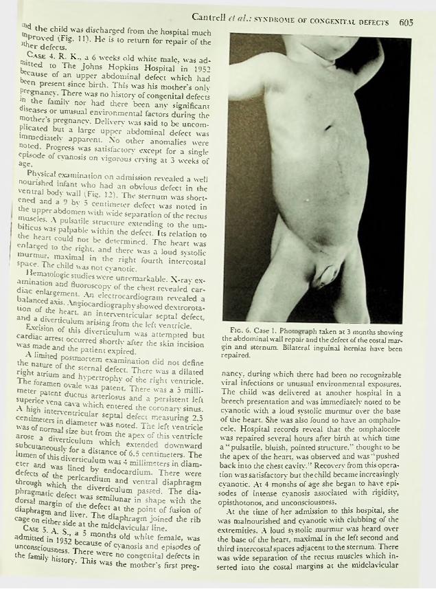
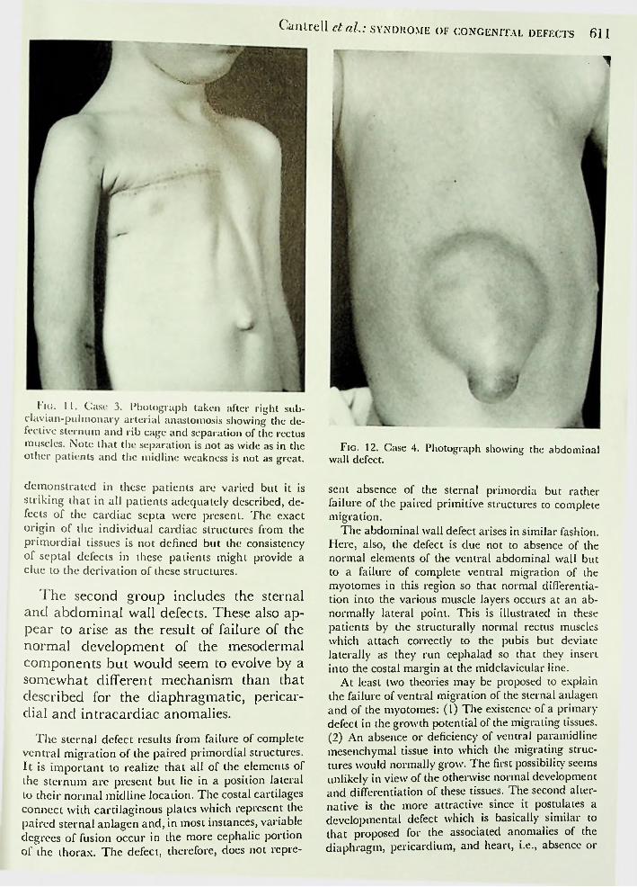
Cantrell ct aI.: SYNDROME OF CONGENITAL DEFECTS 605‘nd the child was discharged from the hospital much ^proved (Fig. 11). He is to return for repair of the ilHer defects. ,
Case 4. R. K.. a 6 weeks old white male, wa^ admitted to The Johns Hopkins Hospital m IX"- becau-e of an uP°er abdominal defect which had been present since birth. This was his mother s only presmanev. There was no history ot congenital defects in the familv nor had there been any significant diseases or unusual environmental factors during the mother's pregnancy. Delivery was said to be uncomplicated but a large upper abdominal delect immediately apparent. No other anomalies "^e noted. Progress was satisfactory except tor a singleepisode of cyanosis on vigorous crying at 3 weeks ol age.
Phvsical examination on admission revealed a well nourished infant who had an obvious defect in the ventral bodv wall (Fig. 12). The sternum was shortened and a 9 by 5 centimeter defect was noted in the upper abdomen with wide separation of the rectus muscles. A pulsatile structure extending to the umbilicus was palpable within the defect. Its relation to the heart could not be determined. The heart was enlarged to the right, and there murmur, maximal in the space. The child was not cyanotic.
Hematologic studies were unremarkable. X-ra> examination and fluoroscopy of the chest revealed cardiac enlargement. An electrocardiogram revealed a balanced axis. Angiocardiography showed dextrorotation of the heart, an interventricular septal defect, and a diverticulum arising from the lelt ventricle.
Excision of this diverticulum was attempted but cardiac arrest occurred shortly after the skin incision was made and the patient expired.
A limited postmortem examination did not define the nature of the sternal defect. There was a dilated right atrium and hypertrophy of the right ventricle. The foramen ovale was patent. There was a 5 millimeter patent ductus arteriosus and a persistent left superior vena cava which entered the coronary sinus.A high interventricular septal defect measuring 2.5 centimeters in diameter was noted. The left ventricle was of normal size but from the apex of this ventricle arose a diverticulum which extended downward subcutaneously for a distance of 6.5 centimeters. The lumen of this diverticulum was 4 millimeters in diameter and was lined by endocardium. There were defects of the pericardium and ventral diaphragm through which the diverticulum passed. The diaphragmatic defect was semilunar in shape with the dorsal margin of the defect at the point of fusion of diaphragm and liver. The diaphragm joined the rib cage on either side at the midclavicular line.
Case 5. A. S., a 5 months old white female, was admitted in 1952 because of cyanosis and episodes of unconsciousness. There were no congenital defects in the family history. This was the mother's first
was
a loud systolic right fourth intercostal
was
Fig. 6. Case 1. Photograph taken at 3 months showing the abdominal wall repair and the defect of the costal gin and sternum. Bilateral inguinal hernias have been repaired.
mar-
nancy, during which there had been no recognizable viral infections or unusual environmental exposures. The child was delivered at another hospital in a breech presentation and was immediately noted to be cyanotic with a loud systolic murmur over the base of the heart. She was also found to have an omphalocele. Hospital records reveal that the omphalocele was repaired several hours after birth at which time a "pulsatile, bluish, pointed structure,'' thought to be the apex of the heart, was observed and was "pushed back into the chest cavity." Recovery from this operation was satisfactory but thechild became increasingly cyanotic. At 4 months of age she began to have episodes of intense cyanosis associated with rigidity, opisthotonos, and unconsciousness.
At the time of her admission to this hospital, she was malnourished and cyanotic with clubbing of the extremities. A loud systolic murmur was heard over the base of the heart, maximal in the left second and third intercostal spaces adjacent to the sternum. There
wide separation of the rectus muscles which inserted into the costal margins at the midclavicularwas
preg-
Cm Su rgery, Gynecology & Obstetrics • November 1958
interatrial septal defect and a high interventricular septal defect. A common pulmonary vein was found to drain both lungs into the left auricle. The tricuspid valve was atretic. There was a single coronary artery.
CLINICAL ANALYSIS
Previously, patients with this combination of anomalies have been considered to be examples of ectopia cordis because of the abnormally exposed position of the heart. The signal articles bearing upon this subject arc those of Greig, Byron, and Major. Greig summarized the earlier reports of ectopia cordis but did not analyze associated anomalies. Byron first suggested a distinction between the other categories of ectopia cordis and those associated with abdominal wall defects; he proposed that the latter be segregated as a subgroup under the term thoracoabdominal. Major has more recently presented a review of this type in which he quoted 13 cases and added a case of his own.
The clinical data available on the previously reported patients arc summarized in Table 1 and those on the patients herein reported in Table II. Analysis of this material reveals several facts previously unrecognized: (1) Rather than a single abnormality, these patients present a remarkably consistent association of defects which includes anomalies of the abdominal wall, lower sternum, ventral diaphragm, pericardium, and heart. (2) The ventral diaphragmatic defect is a specific anomaly unrelated to the commonly recognized diaphragmatic defects. (3) Intracardiac anomalies are invariably present. (4) True ectopia of the heart is rare, occurring m but 1 patient of the entire group. Except for some degree of dextrorotation which may be present, the heart usually lies in normal relationship to the other thoracic viscera; the appearance of ectopia is created by the concomitant defects of the sternum, diaphragm, and abdominal wall which leave the heart abnormally exposed-
On the basis of these observations, the inclusion of these patients in the category o
Fig. 7. Case 2. Preoperative photograph revealing the defects of the thorax and abdominal wall.
line. It was thought that the xiphoid was missing and a pulsatile mass was noted beneath the skin of the abdominal wall defect.
The hematocrit was 60. Arterial oxygen saturation was 45 per cent with the child crying. An electrocardiogram revealed slight left axis deviation. Fluoroscopy revealed enlargement of the heart with marked prominence of the right cardiac border. There left aortic arch.
was a
A diagnosis of tricuspid atresia was made and, because of the severity of symptoms, the child operated upon shortly after admission to the hospital. Cardiac arrest occurred soon after the chest entered but resuscitation was successful and a subclavian-pulmonary arterial anastomosis structed. The child did poorly, however, and died several hours postoperatively.
At autopsy, an upper abdominal wall defect noted with wide separation of the rectus muscles which inserted into the costal margins at the mid- clavicular line. A diverticulum of the left ventricle extended through a ventral defect in the diaphragm and a corresponding defect in the diaphragmatic pericardium. This diverticulum arose from the apex of the ventricle and extended about 2 centimeters below the diaphragm. A persistent left superior cava entered the coronary sinus. There was a large
was
was
was con-
was
vena
reriiir- -be
r.5s*i
ihe sxernal and abdominalof xhc heart produced bvFig. 8. Case 2. Note the exposed position wall defects and the diaphragmatic and pericardial delect*.
obviously closely related conditions and may be explained on die basis of less extensive embryologic defects. While most of the component elements of this syndrome may occur as isolated anomalies, the ventral diaphragmatic defect probably will not be found in the absence of concomitant defects of the sternum and abdominal wall. It should be
phasized that this anomaly is not related to foramen of Morgagni defects but is embryologically a separate entity.
The occurrence of congenital intracardiac anomalies as a constant element of this complex has not been previously recognized. It would appear from the present study that such lesions should be considered an integral
of this association. An analysis of these
ectopia cordis would appear to be in error. Rather, this consistent association of anomalies should be considered as a specific syndrome.
\ ariants of this syndrome would be anticipated in view of the postulated embryologic development of these defects. Potts has reported a patient who demonstrated cardiac, abdominal wall, and diaphragmatic defects but had a normal sternum and an intact though abnormal pericardium. Major has successfully treated a newborn infant who had an omphalocele, a defect of the sternum and lower thorax, and a ventral defect of the diaphragm without an associated pericardial defect and no clinically apparent intracardiac anomalies. These are
cm
part
60S Surgery, Gynecology & Obstetrics ■ November 195S
Fie. 9. Case 2. Dissection of heart heart to retract somewhat within the f''°m """“““"S Struc,urcs all°"s thc
cardiac defects (Table III) reveals that an interventricular septal defect was present in every instance in which a description of the heart defect
is essential to clarification of thc relationship of these associated anomalies.
Origin of thc mesoderm.—During thc eighth day of embryonic development, a layer of cells differentiates from the inner surface of the inner cell mass and pushes out into the blastomcre; this is the primitive entoderm which will eventually form the lining of die gut. Immediately thereafter, a localized proliferation of cells is noted on thc inner surface of thc embryonic disc and migrates laterally between thc trophoblast and entoderm; this is thc primitive mesoderm. These cells migrate rapidly in a peripheral fashion so that the mesodermal layer soon extends not only beneath the embryo but also lines the entire blastodermic vesicle. At about 16 days the mesoderm splits laterally into an outer somatic layer and an inner splanchnic layer (Fig. 13). Thc cavity between these layers is thc primitive coelom. This somatic-splanchnic differentiation does not occur everywhere simultaneously; it is significant that it appears first in the mesodermal tissue from which the heart and pericardium will develop.
Development of the heart and pericardium.—The heart arises far cephalad from paired primordia which arc brought together beneath thc embryo at about the
was available. An interatrial septal was present in 53 per cent of the
patients. Either valvular or infundibular pulmonary stenosis occurred in 33 per cent of the patients. Thc only established cardiac syndrome to be noted with any frequency was the tetralogy of Fallot which in 20 per cent of the patients. A left ular diverticulum
was seen ventric-
was present in 20 per cent, of clinical importance that only 4 of
the entire group of patients had other significant congenital defects, and but 1 of these was incompatible with life. In only 3 patients were the total anomalies such preclude successful correction.
It is
as to
EMBRYOLOGIC DEVELOPMENT
Normal embryology. An analysis of the mal embryology of the involved structures
nor-
Cantrell cl al.: syndrome of congenital defects 609
Anl rectus sheathAnt. rectus sheath
l ie. 10. Case 2. Fascial closure is effected by reflecting flaps of anterior rectus sheath to the mid- hne. Closure reinforced by autogenous fascia lata.
seventeenth day by the downfolding of the lateral splanchnoplcure and ventral flexion of the embryo. This ventral folding appears first in the cephalic and caudal portions of the embryo so that the heart reaches its definitive position and begins its differentiation very early in embryonic life (Fig. 13). The myocardium and epicardium are derived from the splanchnic layer of mesoderm while the pericardium arises from the corresponding somatic mesoderm. The intimate developmental relationship of these structures is apparent.
Development of the diaphragm. —From the mesoderm adjacent and immediately anterior diac primordia arises the
dorsoinesial portion of each somite proliferates and extends around toward the ventral midlinc; termed the myotome, this tissue splits to form the various muscle layers. The skin of the ventral body wall is derived from the embryonic ectoderm which is continuous with the amnion. The development of the skin is not closely related to that of the muscles.
Development of the sternum.—The sternum is formed from paired lateral concentrations of mesenchymal cells in the thoracic region of the embryo (Fig. 15). These cannot be clearly identified prior to the fifth week but the primitive mesoderm from which they arc derived is obviously laid down at a much earlier date. They arc converted into precartilage and advance toward the midlinc where they fuse into a single structure. The process of fusion begins at the cephalic end and progresses caudallv. Simultaneously, rib cartilages are growing ventrally from the vertebrae and by the ninth week the sternum is complete and the anterior rib ends have become attached.
Development of the specific defects. The anomalies seen in this syndrome may be classified into two groups based on the mechanism of embryologic development. The first group, comprising 3 of the defects, would appear to arise as the result of developmental failure of a segment of the mesoderm.
to the car-transversc septum. This tis
sue comes to lie in the midline just caudad to the heart by the same process of flexion and ventral folding and is the anlage of the ventral portion of the diaphragm (Fig. 14). The transverse septum develops contemporaneously with and in close proximity to the heart and pericardium and is derived from the primitive layer. The dorsal and lateral portions of the diaphragm are derived from the pleuropericardial and pleuroperitoneal folds which coalesce with the transverse septum.
Development of the abdominal wall.—It is thought that the somatic mesoderm contributes primarily to the subcutaneous connective tissue of the anterior abdominal wall. The muscular components are derived from the dorsal mesoderm of the somites. The
same

610 Surgery, Gynecology & Obstetrics • November 1958
table l—analysis of defects noted in previouslySternal
Lower M
REPORTED PATI ENTSAbdominal wall Eventration
Diaphragmatic Ventral defect
Cardiac Pericardial OtherAncnccplialy
Absent left lung None
Prochaska (1734)
Pinclli Sandifort
(1772)Wilson
(1798) O'Connor
(1861) Francois-Franck
0 877)
?
Lower 7i OmphaloceleOmphalocele
+ ? ?? Ventral defect IASD, IVSD +Xiphoid Omphalocele Ventral defect IASD, IVSD ? NoneLower M Omphalocele Ventral defect + + NoneLower \i Eventration Ventral defect ? ? None
Holt ? Omphalocele Ventral defect Tetralogy of Fallot ? None(1897)Abbott
(1898)Greifenberg
(1908)
Lower pi Diastasis with eventration
Eventration
Ventral defect ? ? None
Xiphoid ? IVSD + NoneFoy Lower Pi Diastasis Ventral defect IASD, IVSD, left
SVC+ None(1909)
Welch(1910)
Holmes(1919)
Blatt and Zcldcs (1941)
Byron’(1948)
Ehrenhaft
Lower Pi Diastasis with hernia
Diastasis with hernia
Ventral hernia
Ventral defect ? ? None
Lower Pi Ventral defect IASD, IVSD + None
Xiphoid Ventral defect IASD, IVSD, tricuspid atresia
Tetralogy of Fallot, ectopia cordis
Diverticulum left ventricle, IASD, IVSD, truncus arteriosus, pulmonary stenosis IVSD, PDA
? None
Bifidsternum
Omphalocele ? + Malrotationintestines
None+ Diastasis with hernia
Ventral defect +
Johnson + Diastasis with hernia
+ None+
TABLE II.—ANALYSIS OF DEFECTS NOTED IN PRESENTLY REPORTED PATIENTS Sternal
LowerAbdominal wall
OmphaloceleDiaphragmatic
Ventral defectOtherCardiac
Tetralogy of FallotPericardial
1. C.D. Malrotationintestines
+2. M.F. Lower Pi Diastasis with
herniaDiastasis with
herniaDiastasis with
hernia
Ventral defect IASD, IVSD, pulmonary stenosis
IASD, IVSD, pulmonary stenosis
IASD, IVSD, PDA, diverticulum left ventricle
IASD, IVSD, diverticulum left ventricle
None+3. J.B. Lower Pi + None?
4. R.K. Xiphoid Ventral defect None+5. A.S. + Omphalocclc Ventral defect None+
The diaphragmatic defect results from total arises from the somatic mesoderm immediately adjacent to that region of the same layer from which the transverse septum is derived. Defective development of one of these structures without a corresponding defect in the other requires a highly specific loss of somatic mesoderm and is seen only in the uncommon variants in which the diaphragmatic defect is incomplete. Coexisting defects arc much more probable and are seen in the great majority of patients.
The intracardiac lesions noted are the result of faulty development of the epimyocardium which is derived from the splanchnic mesoderm corresponding to that portion of the somatic mesoderm from which the pericardium is derived. The cardiac anomalies
or partial failure of the transverse septum to develop. When complete, the defect corresponds exactly to that portion of the definitive diaphragm which is derived from the transverse septum; it is a ventral defect extending laterally to the region of the pleuroperitoneal folds and dorsally to the point of attachment of liver and diaphragm (Fig. 14). Less extensive defects represent partial rather than total loss of the transverse septum. This anomaly is not related to defects of the foramen of Morgagni.
The pericardial defect is closely related to faulty development of the transverse septum. That portion of the pericardium which is to lie on the diaphragm
Cantrell ct al.: SYNDROME OF CONGENITAL DEFECTS 611
1"V'nW4
1'icj. 11. Case 3. Photograph taken after right subclavian-pulmonary arterial anastomosis showing the defective sternum and rib cage and separation of the rectus muscles. Note that the separation is not as wide as in the other patients and the midline weakness is not as great.
demonstrated in these patients are varied but it is striking that in all patients adequately described, defects of the cardiac septa were present. The exact origin of the individual cardiac structures from the primordial tissues is not defined but the consistency of septal defects in these patients might provide a clue to the derivation of these structures.
The second group includes the sternal and abdominal wall defects. These also appear to arise as the result of failure of the normal development of the mesodermal components but would seem to evolve by a somewhat different mechanism than that described for the diaphragmatic, pericardial and intracardiac anomalies.
The sternal defect results from failure of complete ventral migration of the paired primordial structures. It is important to realize that all of the elements of the sternum are present but lie in a position lateral to their normal mid line location. The costal cartilages connect with cartilaginous plates which represent the paired sternal anlagen and, in most instances, variable degrees of fusion occur in the more cephalic portion of the thorax. The defect, therefore, does not repre-
Fig. 12. Case 4. Photograph showing the abdominal wall defect.
sent absence of the sternal primordia but rather failure of the paired primitive structures to complete migration.
The abdominal wall defect arises in similar fashion. Here, also, the defect is due not to absence of the normal elements of the ventral abdominal wall but to a failure of complete ventral migration of the myotonies in this region so that normal differentiation into the various muscle layers occurs at an abnormally lateral point. This is illustrated in these patients by the structurally normal rectus muscles which attach correctly to the pubis but deviate laterally as they run cephalad so that they insert into the costal margin at the midclavicular line.
At least two theories may be proposed to explain the failure of ventral migration of the sternal anlagen and of the myotonies: (1) The existence of a primary defect in the growth potential of the migrating tissues. (2) An absence or deficiency of ventral paramidline mesenchymal tissue into which the migrating structures would normally grow. The first possibility seems unlikely in view of the otherwise normal development and differentiation of these tissues. The second alternative is the more attractive since it postulates a developmental defect which is basically similar to that proposed for the associated anomalies of the diaphragm, pericardium, and heart, i.e., absence or

612 Surgery, Gynecology & Obstetrics • November 1958Ectoderm mediately after the differentiation of the
primitive intraembryonic mesoderm into its splanchnic and somatic layers since derivatives of both these layers are involved. This localizes the time of origin at about 14 to 18 days of embryonic life. The basic etiology of this mesodermal maldevelop- ment remains obscure.
Dorsal aorln
MXSomatic
mesoderm rij
Splanchnicmesoderm
( EndodermEndocardial
primordiaATREATMENTForegol
Splanchnic . . mesoderm
Somaiic^^^ \ mesoderm Pericardial coelom
HindgulIn view of the multiplicity of problems
posed by these patients, it is not practical to formulate a specific plan of attack which will be optimal for all patients but, in general, treatment should consist of immediate surgical correction of as many of the defects as possible. Repair of the cardiac lesions should be deferred, however, since diagnosis in the neonatal period is difficult and inexact. Furthermore, total correction will require an open heart procedure in most or all of these patients and this technique is at present unsatisfactory in the newborn. The cardiac lesions noted in
AmnionEctoderm
"Transverse oeplum ^ Sgfk
5Fig. 13. A, Diagrammatic reconstruction of cross sec
tion of human embryo at about 16 days just after differentiation of the mesoderm into somatic and splanchnic layers. B, Longitudinal section at about 17 days after the cardiac anlagen have fused. Note the intimate relation of the developing pericardium and transverse septum.
defective development of the paramidline mesoderm. While the mechanism is therefore different in the two groups, it would appear probable that the basic defect is identical.
This explanation demands that the development of the mesoderm be altered at a very early period in embryonic life. The abnormality must occur prior to or im-
the reported patients were not so severe as to require immediate correction. A possible exception to this generalization is the repair of ventricular diverticula which may be amenable to immediate correction.
Some of the sternal defects will not require correction. Minor abnormalities ol the lower sternum are not cosmetically or physiologically serious. Should there be an extensive defect, however, immediate repair must be strongly considered since the correction of large defects becomes progressively more difficult with advancing age.
In those cases in which an omphalocele is present, immediate surgical intervention is, of course, mandatory and it would seem logical to attack any other reparable defects at that time. Certainly in such cases there can be no justification for deferring repanof the diaphragmatic defect. Should the
that
Derived from Irans- \verse septum
Ap Esophageal hiatus
Vena -cavahiatus
Derived from pleuroperitoneal
membrane,
Aortichiatus
omphalocele be sufficiently large s° primary repair is difficult or impossible, necessitating simple closure of the skin ovei the viscera, it would seem doubly important to close the diaphragmatic defect to avoic
Fig. 14. The adult diaphragm, indicating the cm- bryologic origin of the various portions. Note the similarity between the portion derived from the transverse septum and the defect noted in this syndrome.

fI5C*l ntr
the exertion of uncne pressure on ihe incra- it ora tic con; dominal viscera inrougn mi? deieci m.c me pericardium. Repair of even xhe larges; diaphragmatic tiefeci? of the Type seen :n ihese patients can be accomplished in the newborn a? m our L .use i by brinpir.c me remaining tiiaph: ing it to the at costal martin. This > easily none since the remaining diaphragm -> structurally normal and permits the necessary anterior displacement without undue tension. This maneuver simultaneously obliterates the pericardial defect.
Should the patient have an intact skin covering over the defect in the muscular portion of the abdominal wall, repair might be deferred. There is little to be gained by such delay, however, since the ability of the newborn to withstand operation is well known and will not be significantly augmented until well past the neonatal period. It is generally conceded that most congenital defects do not become more amenable to repair with increasing age but rather that correction becomes more difficult.
The technique of abdominal wall closure in such patients is worthy of specific comment. The lateral displacement of the rectus muscles precludes their approximation. In our Case 1. it was possible to develop a fascial structure which extended ventrally from the medial border of the rectus muscles and resembled linea alba. A strong closure of the abdominal wall defect was thus obtained using this local tissue. Linear relaxing incisions in the anterior rectus sheath are
?
,;s and herniation of an
il amerioriv and attachthoracic wall andulterior
F: c-. '. 5. The developxneni of the human 5*c: r.um showing progressive fusion of the lateral aniagea beginning at the cephalic end.
important in relieving tension on this closure.The increasing difficulty of repair at
tendant upon delay in correction is illustrated by Case 2. This patient was operated upon at 20 years of age. at which time it was impossible to isolate the fascial structure described in Case 1 and closure of the abdominal wall was accomplished only with difficulty by swinging flaps of anterior rectus sheath medially and the insertion of autogenous fascia lata as reinforcement. Exten-
dissection of the pericardium from the heart and surrounding structures was necessary to accomplish the repair. Closure of the
not considered
sive
diaphragmatic defect feasible. In all probability, a much simpler procedure would have been possible in infancy.
was
table iu.—analysis of congenital cardiac LESIONS IN* 15 ADEQUATELY TIEXTS
SUMMARY
The consistent association of a group of congenital anomalies involving the abdominal wall, lower sternum, ventral diaphragm, pericardium, and heart has been studied in 21 patients. 5 of whom are herein reported. This complex should be considered
specific syndrome. It is inadequate and inaccurate to consider such patients as examples of ectopia cordis.
DESCRIBED PA-
.Yo. of casesInterventricular septal defectInteratrial septal defect.........Pulmonary stenosis..................Tetralogy of Fallot..................Left ventricular diverticulumPatent ductus arteriosus.........Anomalous venous return (caval) Tricuspid atresia..Truncus arteriosus.Anomalous venous
15S5333 as a3ii
(pulmonary)return 1
614 Surgery, Gynecology & Obstetrics • November 1958
The entire group of anomalies would appear to be closely related in embryologic development, arising as the result of defective formation and differentiation of the ventral mesoderm at about 14 to 18 days of embryonic life.
The unusual ventral defect of the diaphragm seen in these patients results from failure of the transverse septum to develop.This specific anomaly has failed to gain proper recognition in the classification of congenital diaphragmatic defects. Case 1 is the first recorded instance in which repair of this defect has been accomplished.
The consistent association of congenital intracardiac lesions has not been previously noted. An interventricular septal defect would appear to be invariably present.
The coexistence of serious unrelated anomalies appears to be rare.
Treatment should consist of immediate surgical repair except for the intracardiac abnormalities. Correction of the latter is probably better deferred until more exact diagnosis and safer surgical intervention are possible.
REFERENCES1. Abbott, F. G. Congenital abnormality of the ster
num and the diaphragm. Tr. Path. Soc., Lond., 1898, 49: 57.
2. Blatt, M. L., and Zeldes, M. Ectopia cordis. Am. J. Dis. Child., 1942, 63: 515.
3. Byron, F. Ectopia cordis. J. Thorac. Surg., 1948, 17: 717.
4. Eiireniiaft, J. L. Personal communication.5. Foy, G. Ectopic cardiaquc par malformation
stcrnalc. Bull. Soc. anat. Paris, 1909, 11: 446.6. Francois-Franck. Recherchcs sur un cas d’cctopie
congcnitalc du coeur. C. rend. Acad, sc., 1877, 85: 165.
7. Greifenberg, M. Ectopia cordis subthoracica bei lebenden Kindc. Zschr. Gcburtsh., 1908, 62: 453.
8. Greig, D. M. Cleft sternum and ectopia cordis. Edinburgh M.J., 1926, 33: 480.
9. Holmes, J. B. Ectopia cordis (with a report of a case in a 15 month oid infant). Johns Hopkins Mosp. Rep., 1919, 18: 287.
10. Holt, L. E. A remarkable case of cctocardia. Med. News, New York, 1897, 71: 769.
11. Johnson, M. L. Personal communication.12. Major, J. W. Thoracoabdominal ectopia cordis. J.
Thorac. Surg., 1953, 26: 309.13. O’Connor, D. C. A case of monstrosity. Tr. Cork
M. & Surg. Soc., Feb. 27, 1861.14. Pi Nelli. Quoted by Blatt and Zeldes (2).15. Potts, W. J., DeBoer, A., and Johnson, F. R.
Congenital diverticulum of the left ventricle. Surgery, 1953, 33: 301.
16. Prociiaska. Quoted by Blatt and Zeldes (2).17. Sandifort, E. Quoted by Greig (8).18. Welch, J. E. Report of a case of exstrophy of the
heart. Bull. N. York Lying-in-IIosp., 1910, 7: 78.19. Wilson, J. On a very unusual formation of the
human heart. Tr. R. Soc. London, 1798, 88: 332.