Embed Size (px)
Citation preview
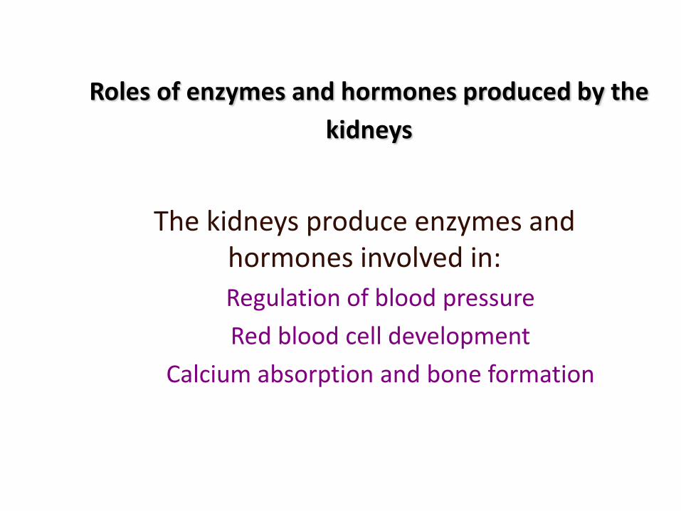
Roles of enzymes and hormones produced by the
kidneys
The kidneys produce enzymes and hormones involved in:
Regulation of blood pressure
Red blood cell development
Calcium absorption and bone formation
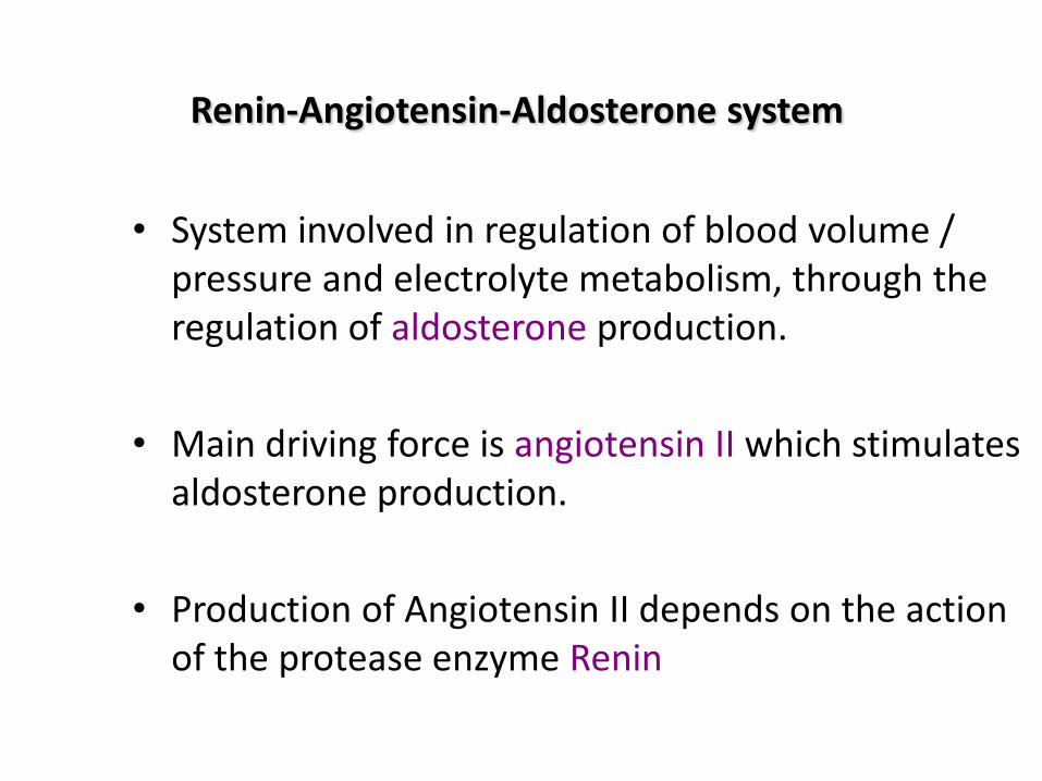
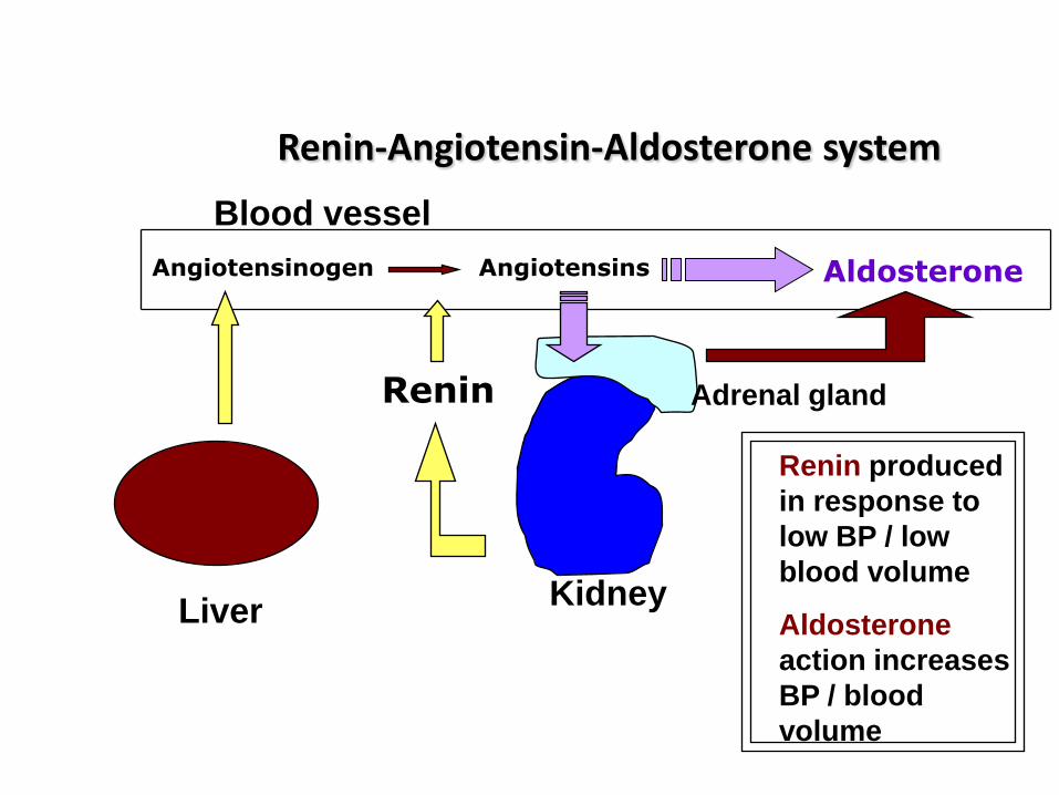
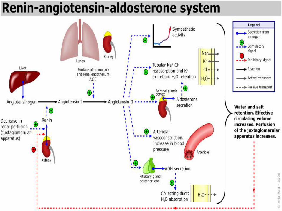
Renin-Angiotensin-Aldosterone system
• System involved in regulation of blood volume / pressure and electrolyte metabolism, through the regulation of aldosterone production.
• Main driving force is angiotensin II which stimulates aldosterone production.
• Production of Angiotensin II depends on the action of the protease enzyme Renin
Renin-Angiotensin-Aldosterone system
Blood vessel
LiverKidney
Adrenal gland
Renin produced
in response to
low BP / low
blood volume
Aldosterone
action increases
BP / blood
volume
Angiotensinogen Angiotensins
Renin
Aldosterone
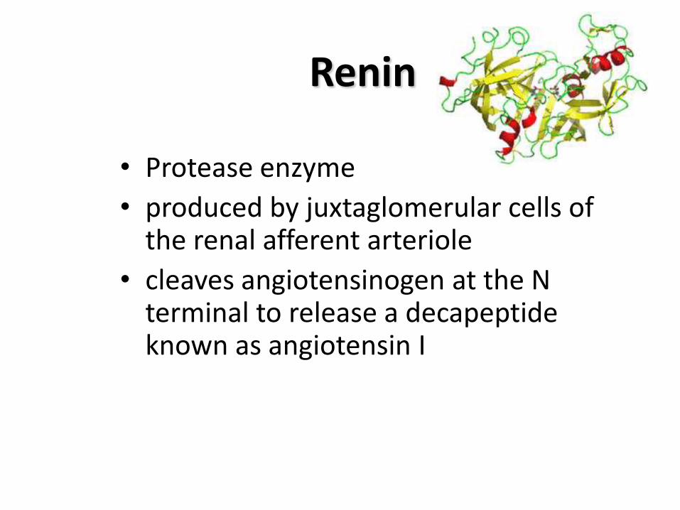
Renin
• Protease enzyme
• produced by juxtaglomerular cells of the renal afferent arteriole
• cleaves angiotensinogen at the N terminal to release a decapeptide known as angiotensin I
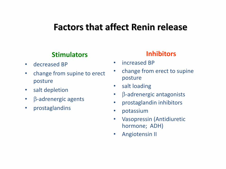
Factors that affect Renin release
Stimulators• decreased BP
• change from supine to erect posture
• salt depletion
• -adrenergic agents
• prostaglandins
Inhibitors• increased BP
• change from erect to supine posture
• salt loading
• -adrenergic antagonists
• prostaglandin inhibitors
• potassium
• Vasopressin (Antidiuretic hormone; ADH)
• Angiotensin II
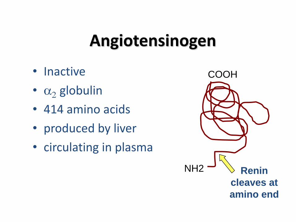
Angiotensinogen
• Inactive
• globulin
• 414 amino acids
• produced by liver
• circulating in plasma
NH2
COOH
Renin
cleaves at
amino end
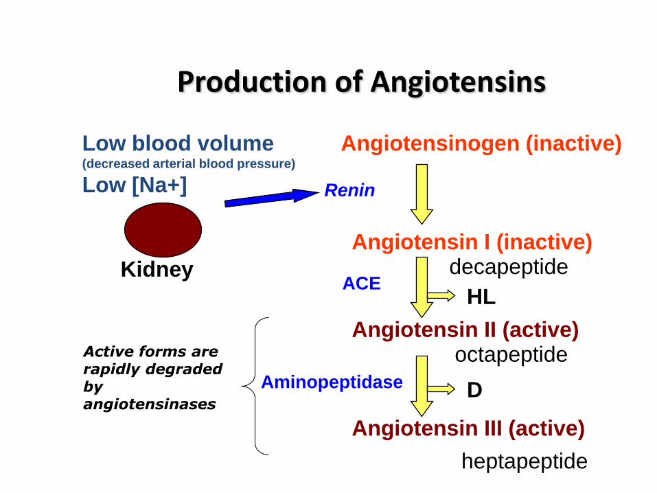
Production of Angiotensins
Kidney
Low blood volume(decreased arterial blood pressure)
Low [Na+] Renin
Angiotensinogen (inactive)
Angiotensin I (inactive)
Angiotensin II (active)
Angiotensin III (active)
decapeptide
octapeptide
heptapeptide
ACE
Aminopeptidase D
HL
Active forms are rapidly degraded by angiotensinases
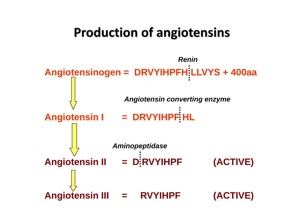
Production of angiotensins
Angiotensinogen = DRVYIHPFH LLVYS + 400aa
Angiotensin I = DRVYIHPF HL
Angiotensin II = D RVYIHPF (ACTIVE)
Angiotensin III = RVYIHPF (ACTIVE)
Renin
Angiotensin converting enzyme
Aminopeptidase
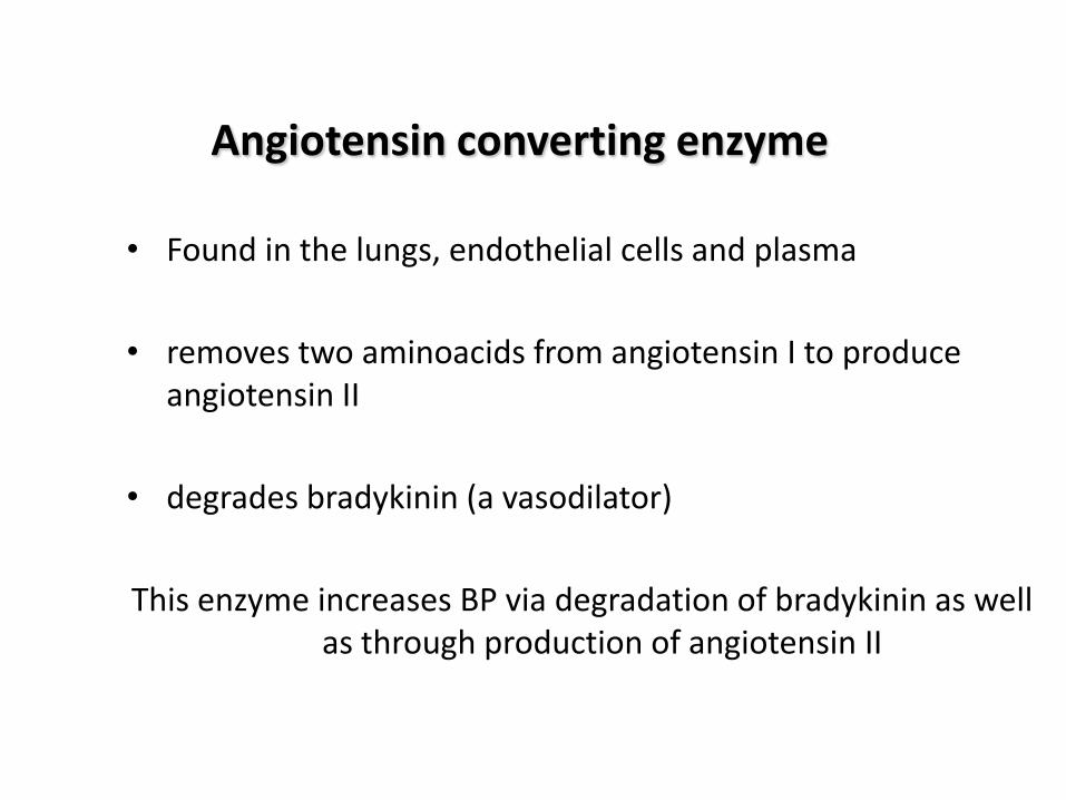
Angiotensin converting enzyme
• Found in the lungs, endothelial cells and plasma
• removes two aminoacids from angiotensin I to produce angiotensin II
• degrades bradykinin (a vasodilator)
This enzyme increases BP via degradation of bradykinin as well as through production of angiotensin II
ACE inhibitors
These are analogues of angiotensin I used as competitive inhibitors of ACE in the treatment of Renin-
dependent hypertension
Aminopeptidase
• Converts angiotensin II to angiotensin III through the removal of a single aminoacid
• Not present in all species
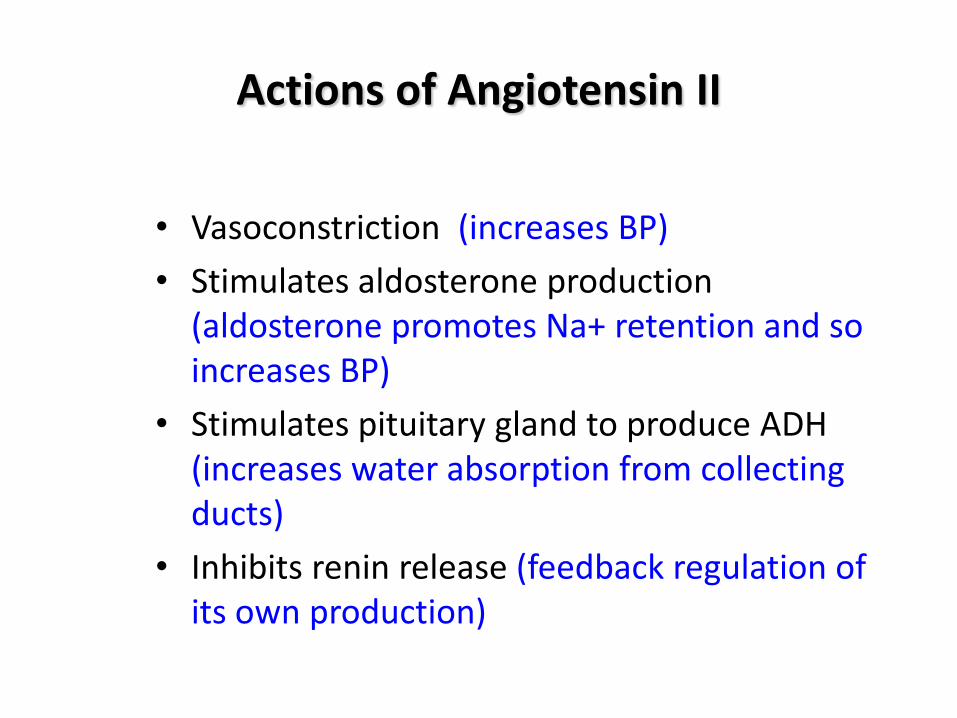
Actions of Angiotensin II
• Vasoconstriction (increases BP)
• Stimulates aldosterone production (aldosterone promotes Na+ retention and so increases BP)
• Stimulates pituitary gland to produce ADH(increases water absorption from collecting ducts)
• Inhibits renin release (feedback regulation of its own production)
Aldosterone is a mineralocorticoid
• Mineralocorticoids are 21-carbon steroids
• Made in zona glomerulosa
• Primary action: promote Na+ retention and K+
excretion (particularly in kidney)
Aldosterone is the most potent mineralocorticoid
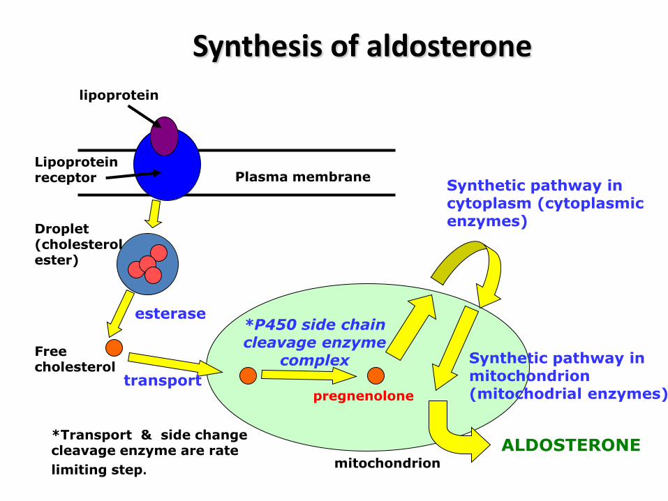
Synthesis of aldosterone
Droplet (cholesterol ester)
Plasma membrane
lipoprotein
Lipoprotein receptor
Free cholesterol
mitochondrion
pregnenolone
*P450 side chain cleavage enzyme
complex
Synthetic pathway in cytoplasm (cytoplasmic enzymes)
Synthetic pathway in mitochondrion (mitochodrial enzymes)
*Transport & side change cleavage enzyme are rate
limiting step.
ALDOSTERONE
esterase
transport
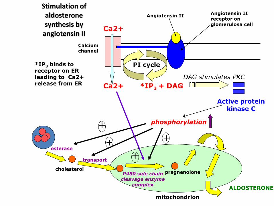
Stimulation of aldosterone synthesis by
angiotensin II
mitochondrion
pregnenoloneP450 side chain cleavage enzyme
complex
esterase
transport
ALDOSTERONE
Angiotensin II receptor on glomerulosa cell
Angiotensin II
Ca2+
Ca2+
Active protein kinase C
Calcium channel
PI cycle
*IP3 + DAG
*IP3 binds to receptor on ER leading to Ca2+ release from ER
+
+
phosphorylation
+
cholesterol
+
DAG stimulates PKC
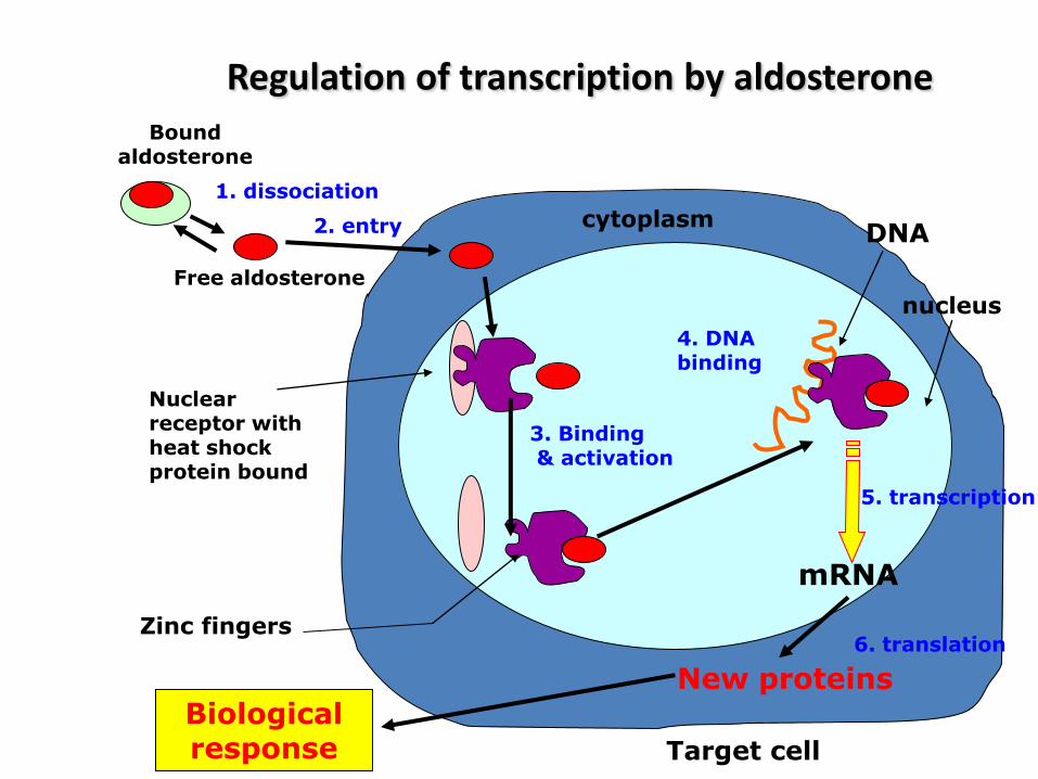
Regulation of transcription by aldosteroneBound
aldosterone
Free aldosterone
2. entry
Nuclear receptor with heat shock protein bound
3. Binding& activation
Zinc fingers
DNA
nucleus
cytoplasm
4. DNA binding
5. transcription
mRNA
New proteinsBiological response
6. translation
1. dissociation
Target cell
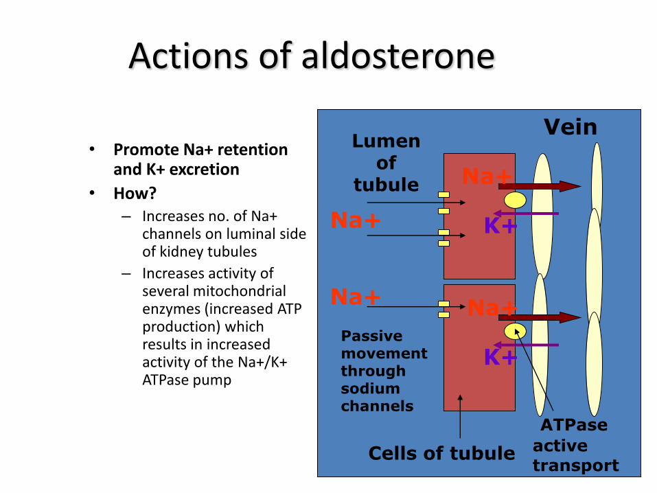
Actions of aldosterone
• Promote Na+ retention and K+ excretion
• How?– Increases no. of Na+
channels on luminal side of kidney tubules
– Increases activity of several mitochondrial enzymes (increased ATP production) which results in increased activity of the Na+/K+ ATPase pump
VeinLumen
of tubule
Cells of tubule
Na+
Passive movement through sodium channels
Na+
K+
Na+
K+
ATPase
active transport
Na+
The kidney and red blood cell formation
The kidney produces erythropoietin (Epo) which stimulates
erythropoiesis (red blood cell formation) in humans.
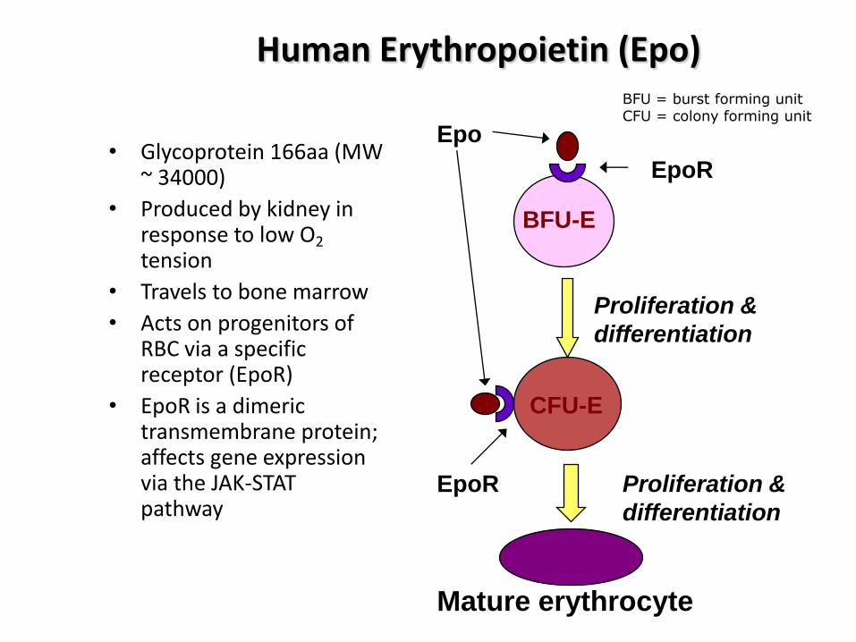
Human Erythropoietin (Epo)
• Glycoprotein 166aa (MW ~ 34000)
• Produced by kidney in response to low O2
tension
• Travels to bone marrow
• Acts on progenitors of RBC via a specific receptor (EpoR)
• EpoR is a dimeric transmembrane protein; affects gene expression via the JAK-STAT pathway
EpoR
Epo
BFU-E
CFU-E
EpoR
Proliferation &
differentiation
Proliferation &
differentiation
Mature erythrocyte
BFU = burst forming unitCFU = colony forming unit
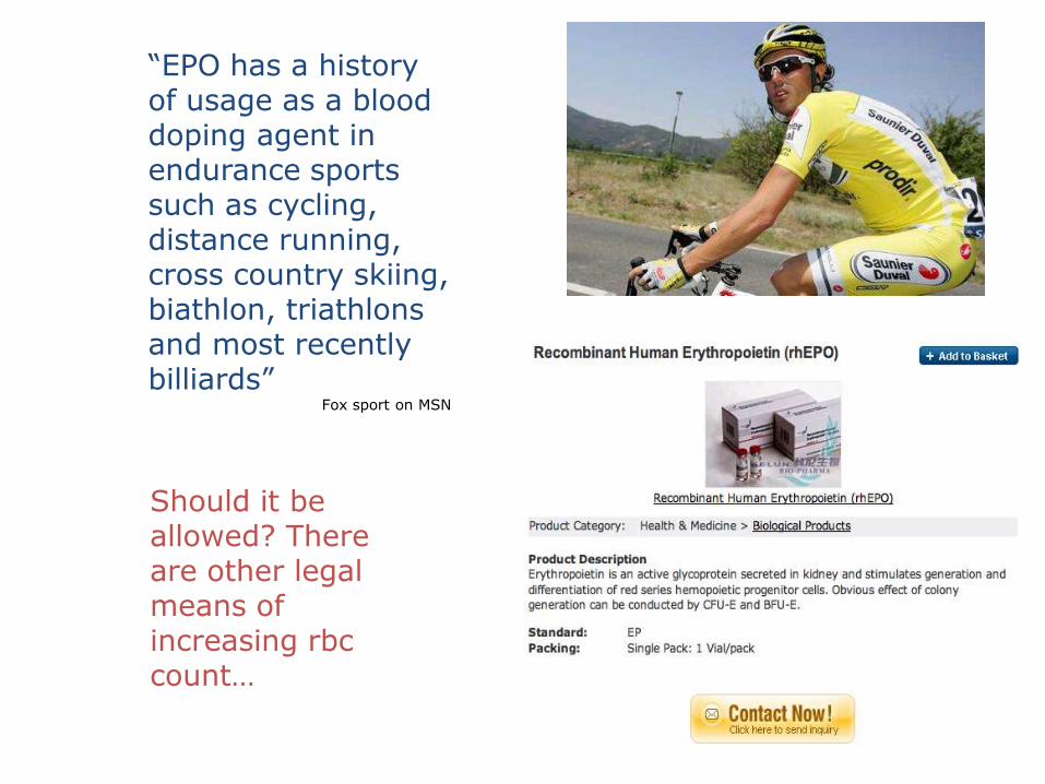
“EPO has a history of usage as a blood doping agent in endurance sports such as cycling, distance running, cross country skiing, biathlon, triathlons and most recently billiards”
Fox sport on MSN
Should it be allowed? There are other legal means of increasing rbc count…
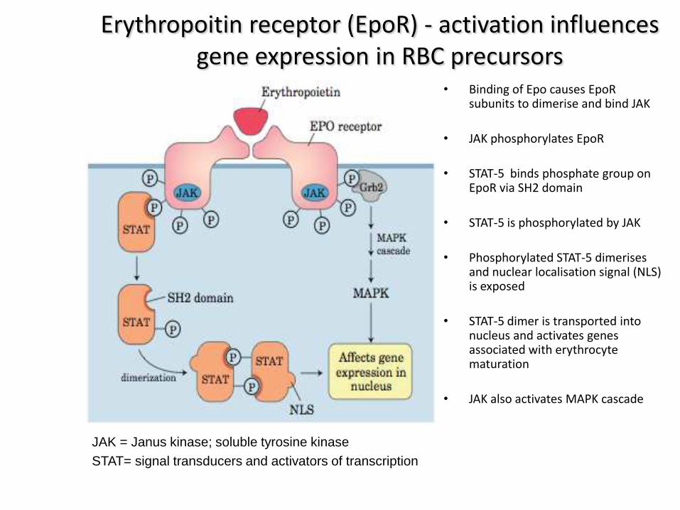
Erythropoitin receptor (EpoR) - activation influences gene expression in RBC precursors
• Binding of Epo causes EpoR subunits to dimerise and bind JAK
• JAK phosphorylates EpoR
• STAT-5 binds phosphate group on EpoR via SH2 domain
• STAT-5 is phosphorylated by JAK
• Phosphorylated STAT-5 dimerises and nuclear localisation signal (NLS) is exposed
• STAT-5 dimer is transported into nucleus and activates genes associated with erythrocyte maturation
• JAK also activates MAPK cascade
JAK = Janus kinase; soluble tyrosine kinase
STAT= signal transducers and activators of transcription
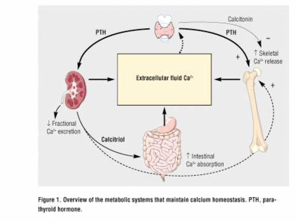
The Kidneys and Calcium Metabolism
• The kidney produces enzymes involved in the conversion of Vitamin D3
(cholecalciferol) to 1,25-dihydrovitamin D3
(1,25-dihydrocholecalciferol)
• 1,25-dihydrocholecalciferol effects increased Ca2+ uptake in the gut
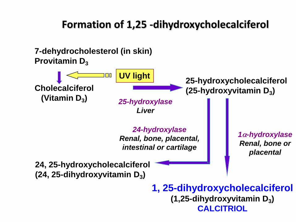
Formation of 1,25 -dihydroxycholecalciferol
7-dehydrocholesterol (in skin)
Provitamin D3
Cholecalciferol
(Vitamin D3)
UV light25-hydroxycholecalciferol
(25-hydroxyvitamin D3)
24, 25-hydroxycholecalciferol
(24, 25-dihydroxyvitamin D3)
1, 25-dihydroxycholecalciferol(1,25-dihydroxyvitamin D3)
CALCITRIOL
25-hydroxylase
Liver
1 -hydroxylase
Renal, bone or
placental
24-hydroxylase
Renal, bone, placental,
intestinal or cartilage
Calcitriol
Hormone of the steroid / thyroid class
Also known as:–1,25-dihydroxycholecalciferol–1,25-dihydroxyvitamin D3
–1,25(OH)2-D3
Calcitriol
Binds intracellular receptor and stimulates expression of genes involved in:
• transfer of Ca 2+ and PO4 3- across the intestinal mucosa
(increases absorption).
• excretion of calcium (reduces excretion by stimulating reabsorption in the distal renal tubules
Provides the proper balance of calcium and phosphorus to support mineralization of bone
Ricketts
• May be caused by:– Vitamin D deficiency– Defect in conversion of 25-hydrocholecalciferol to
calcitriol– Non-functional calcitriol receptor
• Characterised by:– low plasma calcium and phosphorus levels – poorly mineralised bones– skeletal deformities

Renal stones
• Kidney stones (renal stones/renal calculi)– gravel-like collections of chemicals that may appear in any area of the
urinary system, from the kidney to the bladder.
• They may be small or large, single or multiple.
• Four main types:– Calcium stones
– Uric acid stones
– Struvite stones
– Cystine stones
Check http://www.herringlab.com/photos/for other photos including stone from a Mummy (800AD)!
Calcium stones
– Most common type; 95% of all renal stones are calcium
– Caused by:• Defective kidney function which allows too much calcium in the
urine, or excessive calcium absorbed from the stomach and intestines.
• an excess of oxalate, present in many foods; binds easily with calcium to form a stone.
– Risk of calcium stone formation is increased in hyperparathyroidism and inflammatory bowel disease
Other less common types of renal stones
– Uric acid stones• common in people with gout
• may be pure uric acid, calcium containing or a mixture
– Struvite stones• Also called “infection stones”
• Composed of ammoniomagnesium phosphate
• Bacteria convert to ammonium which complex with magnesium and phosphate in the urine to form stones. As the stones form bacteria are trapped within them.
• More common in women than men
– Cystine stones• very rare; almost always diagnosed during childhood
• Genetic defect (autosomal recessive); kidneys do not adequately reabsorb cystine (cystinuria).
Diet and kidney stones
• Change in diet can reduce risk of forming stones in patients
• Recommendations depend on cause of kidney stones
• Recommendations may include:– Increased
• fluid intake
• Magnesium intake
– Reduced • calcium (e.g. milk and other dairy products)
• sodium
• oxalate (e.g. nuts, chocolate, green leafy vegetables, Vitamin C [excess converted to oxalate])
• Foods high in purines (degraded to uric acid) eg. meats