Embed Size (px)
Citation preview
214
THE AETIOLOGY AND PATHOMORPHOLOGY OF RESPIRATORY DISEASES IN CALVES
T. Järveots, T. Suuroja, E. Lepp
ABSTRACT. The aetiology and pathomorphology of respiratory diseases in calves. The aim was to study the possible relation of pathomorphological changes in the respiratory tract and lungs of calves to isolated germs. The study is based on 30 calves aged less than a month from 1997 to 1999. In addition, there was studied the autopsy reports of 408 calves aged less than one month that had been dissected from 1978 to 1988. Histological methods were used to find out the pathomorphological changes in lungs. Virological and bacteriological studies were carried out on 22 calves dissected.
Acute catarrhal pneumonia was diagnosed in 23 calves. Two calves had purulent pneumonia. Research findings show that pneumonia in stillborn and less than 30-day-old calves was mostly caused by various bacteria: E. coli K99, E. coli O78, E. coli O101, Salmonella dublin, Streptococcus pyogenes, Streptococcus pneumoniae, Pasteurella multocida; and viruses: bovine adenovirus, bovine respiratory syncytial virus, and bovine herpesvirus-1.
Keywords: calves, lungs, pneumonia, pathomorphology, aethiology.
Introduction Respiratory diseases in calves may result in death, which makes them a topical problem both in Estonia
and other countries where animal husbandry is highly developed (Ishino et al., 1979; Durham, 1991). Previous research shows that respiratory diseases in calves may be caused, for example, by viruses,
microbes, mycoplasmas, chlamydiae, and thyroid pathology (Saar, 1994; Lepp, 1996; Koslov et al., 1994). In Estonia respiratory diseases are a major cause of disease and death in calves after diseases of the
digestive organs (Koslov et al., 1989; 1990). In a healthy human and animal the respiratory tract below the larynx is sterile because the mucociliary
system, secretory immunoglobulin A, alveolar macrophages, and lymphocytes help to neutralize the inhaled foreign particles and microorganisms. The inhaled foreign particles are discharged during coughing and sneezing (Green, 1997; Leesik, 1993).
Microorganisms may find their way into the lungs by inhaled air, by aspiring potentially pathogenic microbes and anaerobes from the pharynx, haematogenously from some infectious focus, and by contact (Juhkam, 1998).
Cold, viral infections and optionally pathogenic microbes of the upper airway, and poor feeding and keeping conditions contribute to the disorder of the defensive mechanisms of the respiratory organs and the development of pneumonia is facilitated by (Olsson et al., 1993).
Autopsies usually reveal bronchopneumonia and rarely croup or lobar pneumonia. Optionally pathogenic microorganisms are often responsible for pneumonia (Koslov et al., 1989; 1990). These authors claim that infectious diseases, including viral diseases and often associated by a bacterial infection, often cause pneumonia.
Aim of the study The aim was to study the possible relation of pathomorphological changes in the respiratory tract and
lungs of calves to isolated germs.
Material and methods The study is based on thirty calves aged less than a month that were dissected at the Laboratory of
Pathological Anatomy at the Department of Morphology of the Estonian Agricultural University from 1997 to 1999 (Table 1). During the autopsies some samples were taken from the lungs for further pathohistological study. The samples for histological study were fixed in a 10 per cent solution of neutral formalin, embedded in paraffin, and cut into histological stretches 5 (µm) in thickness, which were stained with haematoxylin-eosin and haematoxylin-picrofuchsin.
The samples for microbiological and virological studies were obtained at autopsies, and they were studied at the Laboratory of the Department of Infectious Diseases of the Estonian Agricultural University.
The majority of the dissected calves came from the farms located in Tartu County, but some calves came from Jõgeva County as well.
The aetiology and pathomorphology of respiratory diseases in calves 215
Table 1. Distribution of the studied calves by age, gender, and breed Tabel 1. Uuritud vasikate jaotumine vanuse, soo ja tõu järgi
Sex/Sugu Breed/Tõug Age of calves Vasika vanus
No of calves Vasikate arv male
pullik female lehmik
EH EP
Sb* 3 3 3 1 day/päev 2 2 2
2 days/päeva 1 1 1 4 days/päeva 1 1 1 5 days/päeva 4 1 3 3 1 6 days/päeva 2 2 27 days/päeva 4 3 1 2 2
10 days/päeva 4 3 1 2 214 days/päeva 2 1 1 1 115 days/päeva 2 1 1 219 days/päeva 1 1 130 days/päeva 4 3 1 2 2
In all / Kokku 30 18 12 19 11
Sb* – stillborn/surnultsündinud; EH – Estonian Holstein breed / eesti holstein; EP – Estonian Red breed / eesti punane
In addition, we studied the autopsy reports of 408 calves aged less than one month that had been dissected at the Laboratory of Pathological Anatomy at the Department of Morphology of the Estonian Agricultural University from 1978 to 1988.
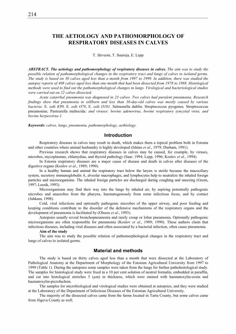
Results Acute catarrhal pneumonia was diagnosed in 23 calves (Figure 1). Inflammatory processes were more
frequently observed in the apical and additional lobes. Two calves had purulent pneumonia. Pneumonia was diagnosed even in some newborn calves. Enteritis was most frequently localized in the small intestine.
The investigated calves were divided into five age groups: stillborn calves, calves aged 1–5 days, 6–10, 11–15, and 16–30 days. The stillborn calves revealed pathological changes mostly in the intestines. The 1-to-5-day-old calves revealed inflammatory changes in the intestines, abomasum, and the lungs. The liver, too, revealed some dystrophic changes. All the 6-to-10-day-old calves revealed signs of intestinal inflammation. In addition, pathological changes were observed in the lungs and the abomasum. The liver of eight calves and the kidneys of six calves showed signs of dystrophy. The 11-to-15-day-old calves had more frequently pneumonia, enteritis, and abomasitis, but enteritis and pneumonia were more common in 16-to-30-day-old calves. The liver of three and the kidneys of three calves showed some dystrophic changes.
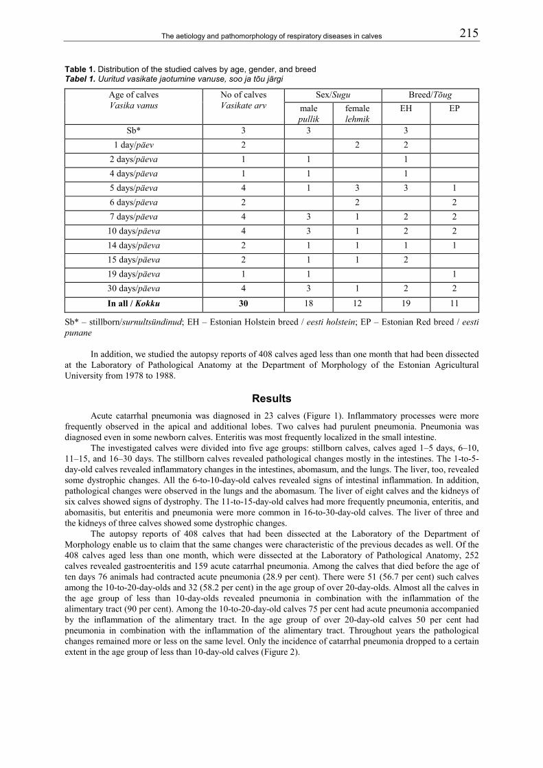
The autopsy reports of 408 calves that had been dissected at the Laboratory of the Department of Morphology enable us to claim that the same changes were characteristic of the previous decades as well. Of the 408 calves aged less than one month, which were dissected at the Laboratory of Pathological Anatomy, 252 calves revealed gastroenteritis and 159 acute catarrhal pneumonia. Among the calves that died before the age of ten days 76 animals had contracted acute pneumonia (28.9 per cent). There were 51 (56.7 per cent) such calves among the 10-to-20-day-olds and 32 (58.2 per cent) in the age group of over 20-day-olds. Almost all the calves in the age group of less than 10-day-olds revealed pneumonia in combination with the inflammation of the alimentary tract (90 per cent). Among the 10-to-20-day-old calves 75 per cent had acute pneumonia accompanied by the inflammation of the alimentary tract. In the age group of over 20-day-old calves 50 per cent had pneumonia in combination with the inflammation of the alimentary tract. Throughout years the pathological changes remained more or less on the same level. Only the incidence of catarrhal pneumonia dropped to a certain extent in the age group of less than 10-day-old calves (Figure 2).
T. Järveots, T. Suuroja, E. Lepp 216
Figure 1. Lung of the 15 days old calf. Acute catarrhal pneumonia Joonis 1. 15 päeva vanuse vasika kops. Äge katarraalne kopsupõletik
Figure 2. Percentage of acute catarrhal pneumonia in less than 10-day-old calves in 1978–1998 Joonis 2. Ägeda katarraalse kopsupõletiku protsentuaalne esinemissagedus alla 1 kuu vanustel vasikatel vanuserühmade lõikes aastatel 1978–1998
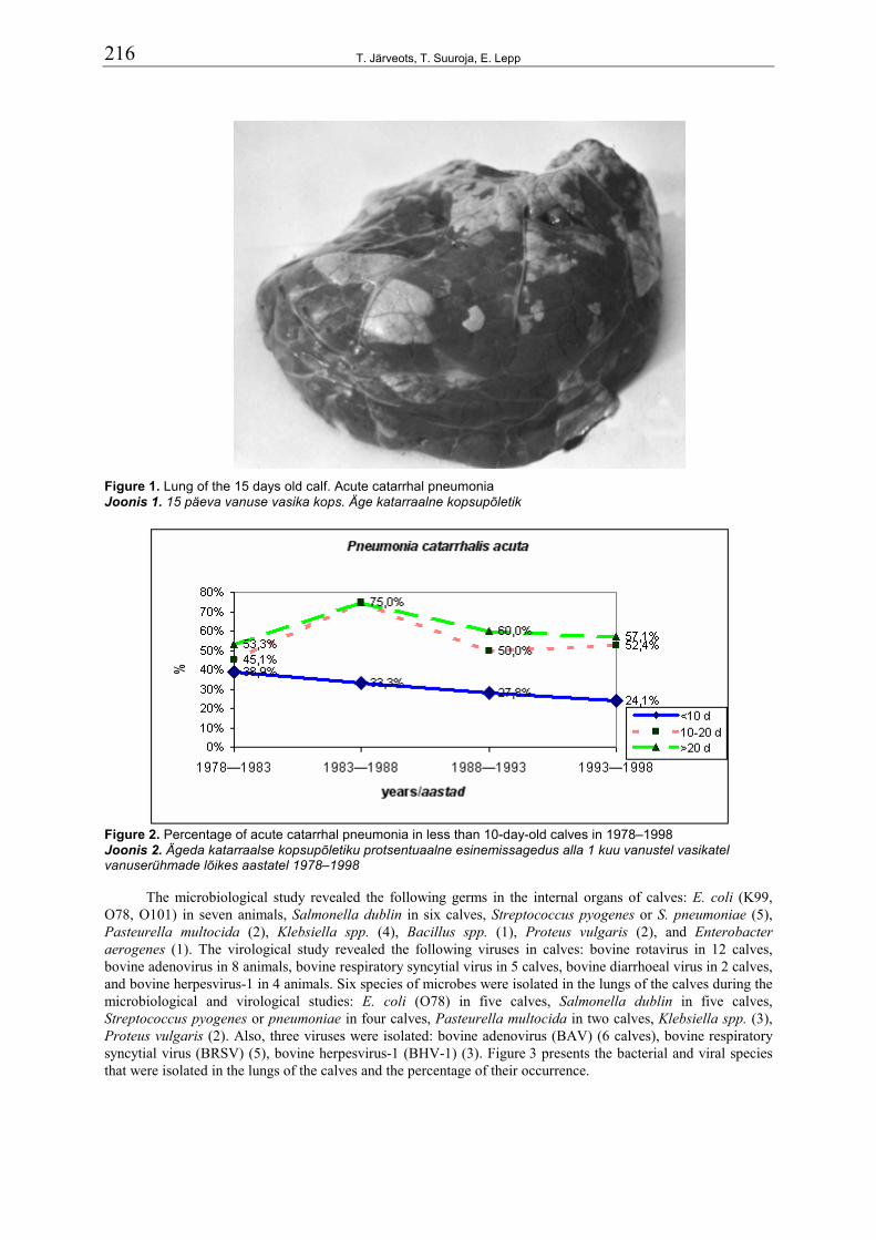
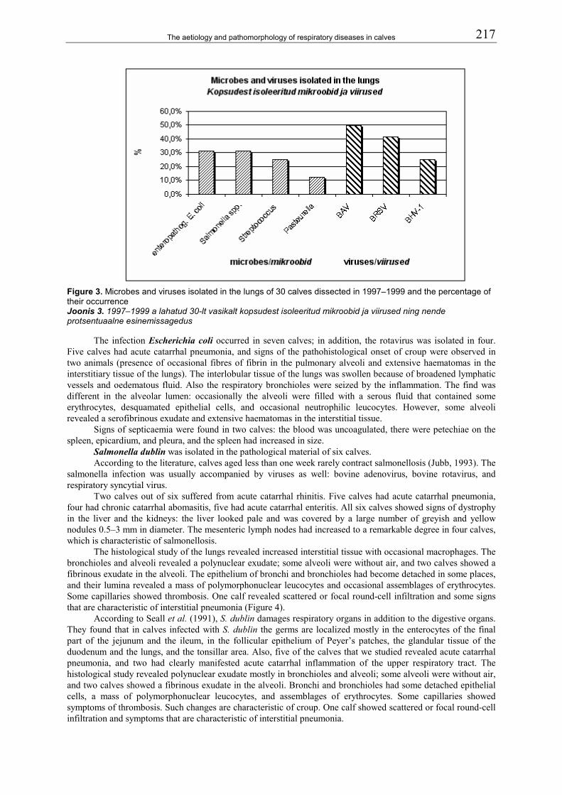
The microbiological study revealed the following germs in the internal organs of calves: E. coli (K99, O78, O101) in seven animals, Salmonella dublin in six calves, Streptococcus pyogenes or S. pneumoniae (5), Pasteurella multocida (2), Klebsiella spp. (4), Bacillus spp. (1), Proteus vulgaris (2), and Enterobacter aerogenes (1). The virological study revealed the following viruses in calves: bovine rotavirus in 12 calves, bovine adenovirus in 8 animals, bovine respiratory syncytial virus in 5 calves, bovine diarrhoeal virus in 2 calves, and bovine herpesvirus-1 in 4 animals. Six species of microbes were isolated in the lungs of the calves during the microbiological and virological studies: E. coli (O78) in five calves, Salmonella dublin in five calves, Streptococcus pyogenes or pneumoniae in four calves, Pasteurella multocida in two calves, Klebsiella spp. (3), Proteus vulgaris (2). Also, three viruses were isolated: bovine adenovirus (BAV) (6 calves), bovine respiratory syncytial virus (BRSV) (5), bovine herpesvirus-1 (BHV-1) (3). Figure 3 presents the bacterial and viral species that were isolated in the lungs of the calves and the percentage of their occurrence.
The aetiology and pathomorphology of respiratory diseases in calves 217
Figure 3. Microbes and viruses isolated in the lungs of 30 calves dissected in 1997–1999 and the percentage of their occurrence Joonis 3. 1997–1999 a lahatud 30-lt vasikalt kopsudest isoleeritud mikroobid ja viirused ning nende protsentuaalne esinemissagedus
The infection Escherichia coli occurred in seven calves; in addition, the rotavirus was isolated in four. Five calves had acute catarrhal pneumonia, and signs of the pathohistological onset of croup were observed in two animals (presence of occasional fibres of fibrin in the pulmonary alveoli and extensive haematomas in the interstitiary tissue of the lungs). The interlobular tissue of the lungs was swollen because of broadened lymphatic vessels and oedematous fluid. Also the respiratory bronchioles were seized by the inflammation. The find was different in the alveolar lumen: occasionally the alveoli were filled with a serous fluid that contained some erythrocytes, desquamated epithelial cells, and occasional neutrophilic leucocytes. However, some alveoli revealed a serofibrinous exudate and extensive haematomas in the interstitial tissue.
Signs of septicaemia were found in two calves: the blood was uncoagulated, there were petechiae on the spleen, epicardium, and pleura, and the spleen had increased in size.
Salmonella dublin was isolated in the pathological material of six calves. According to the literature, calves aged less than one week rarely contract salmonellosis (Jubb, 1993). The
salmonella infection was usually accompanied by viruses as well: bovine adenovirus, bovine rotavirus, and respiratory syncytial virus.
Two calves out of six suffered from acute catarrhal rhinitis. Five calves had acute catarrhal pneumonia, four had chronic catarrhal abomasitis, five had acute catarrhal enteritis. All six calves showed signs of dystrophy in the liver and the kidneys: the liver looked pale and was covered by a large number of greyish and yellow nodules 0.5–3 mm in diameter. The mesenteric lymph nodes had increased to a remarkable degree in four calves, which is characteristic of salmonellosis.
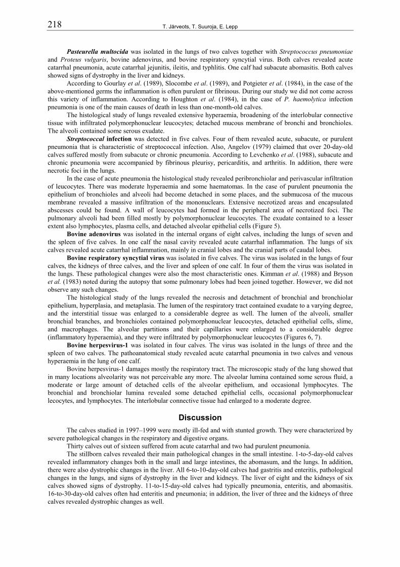
The histological study of the lungs revealed increased interstitial tissue with occasional macrophages. The bronchioles and alveoli revealed a polynuclear exudate; some alveoli were without air, and two calves showed a fibrinous exudate in the alveoli. The epithelium of bronchi and bronchioles had become detached in some places, and their lumina revealed a mass of polymorphonuclear leucocytes and occasional assemblages of erythrocytes. Some capillaries showed thrombosis. One calf revealed scattered or focal round-cell infiltration and some signs that are characteristic of interstitial pneumonia (Figure 4).
According to Seall et al. (1991), S. dublin damages respiratory organs in addition to the digestive organs. They found that in calves infected with S. dublin the germs are localized mostly in the enterocytes of the final part of the jejunum and the ileum, in the follicular epithelium of Peyer’s patches, the glandular tissue of the duodenum and the lungs, and the tonsillar area. Also, five of the calves that we studied revealed acute catarrhal pneumonia, and two had clearly manifested acute catarrhal inflammation of the upper respiratory tract. The histological study revealed polynuclear exudate mostly in bronchioles and alveoli; some alveoli were without air, and two calves showed a fibrinous exudate in the alveoli. Bronchi and bronchioles had some detached epithelial cells, a mass of polymorphonuclear leucocytes, and assemblages of erythrocytes. Some capillaries showed symptoms of thrombosis. Such changes are characteristic of croup. One calf showed scattered or focal round-cell infiltration and symptoms that are characteristic of interstitial pneumonia.
T. Järveots, T. Suuroja, E. Lepp 218
Pasteurella multocida was isolated in the lungs of two calves together with Streptococcus pneumoniae and Proteus vulgaris, bovine adenovirus, and bovine respiratory syncytial virus. Both calves revealed acute catarrhal pneumonia, acute catarrhal jejunitis, ileitis, and typhlitis. One calf had subacute abomasitis. Both calves showed signs of dystrophy in the liver and kidneys.
According to Gourlay et al. (1989), Slocombe et al. (1989), and Potgieter et al. (1984), in the case of the above-mentioned germs the inflammation is often purulent or fibrinous. During our study we did not come across this variety of inflammation. According to Houghton et al. (1984), in the case of P. haemolytica infection pneumonia is one of the main causes of death in less than one-month-old calves.
The histological study of lungs revealed extensive hyperaemia, broadening of the interlobular connective tissue with infiltrated polymorphonuclear leucocytes; detached mucous membrane of bronchi and bronchioles. The alveoli contained some serous exudate.
Streptococcal infection was detected in five calves. Four of them revealed acute, subacute, or purulent pneumonia that is characteristic of streptococcal infection. Also, Angelov (1979) claimed that over 20-day-old calves suffered mostly from subacute or chronic pneumonia. According to Levchenko et al. (1988), subacute and chronic pneumonia were accompanied by fibrinous pleurisy, pericarditis, and arthritis. In addition, there were necrotic foci in the lungs.
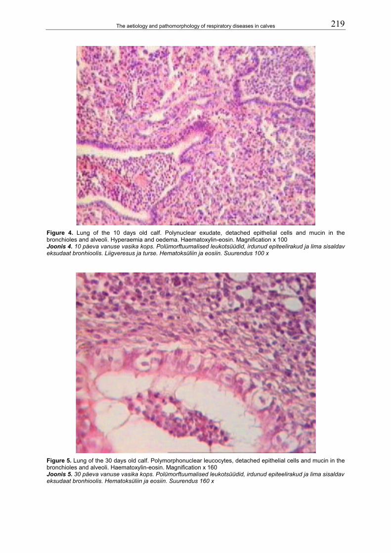
In the case of acute pneumonia the histological study revealed peribronchiolar and perivascular infiltration of leucocytes. There was moderate hyperaemia and some haematomas. In the case of purulent pneumonia the epithelium of bronchioles and alveoli had become detached in some places, and the submucosa of the mucous membrane revealed a massive infiltration of the mononuclears. Extensive necrotized areas and encapsulated abscesses could be found. A wall of leucocytes had formed in the peripheral area of necrotized foci. The pulmonary alveoli had been filled mostly by polymorphonuclear leucocytes. The exudate contained to a lesser extent also lymphocytes, plasma cells, and detached alveolar epithelial cells (Figure 5).
Bovine adenovirus was isolated in the internal organs of eight calves, including the lungs of seven and the spleen of five calves. In one calf the nasal cavity revealed acute catarrhal inflammation. The lungs of six calves revealed acute catarrhal inflammation, mainly in cranial lobes and the cranial parts of caudal lobes.
Bovine respiratory syncytial virus was isolated in five calves. The virus was isolated in the lungs of four calves, the kidneys of three calves, and the liver and spleen of one calf. In four of them the virus was isolated in the lungs. These pathological changes were also the most characteristic ones. Kimman et al. (1988) and Bryson et al. (1983) noted during the autopsy that some pulmonary lobes had been joined together. However, we did not observe any such changes.
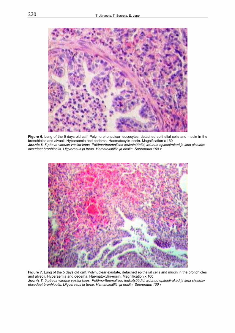
The histological study of the lungs revealed the necrosis and detachment of bronchial and bronchiolar epithelium, hyperplasia, and metaplasia. The lumen of the respiratory tract contained exudate to a varying degree, and the interstitial tissue was enlarged to a considerable degree as well. The lumen of the alveoli, smaller bronchial branches, and bronchioles contained polymorphonuclear leucocytes, detached epithelial cells, slime, and macrophages. The alveolar partitions and their capillaries were enlarged to a considerable degree (inflammatory hyperaemia), and they were infiltrated by polymorphonuclear leucocytes (Figures 6, 7).
Bovine herpesvirus-1 was isolated in four calves. The virus was isolated in the lungs of three and the spleen of two calves. The pathoanatomical study revealed acute catarrhal pneumonia in two calves and venous hyperaemia in the lung of one calf.
Bovine herpesvirus-1 damages mostly the respiratory tract. The microscopic study of the lung showed that in many locations alveolarity was not perceivable any more. The alveolar lumina contained some serous fluid, a moderate or large amount of detached cells of the alveolar epithelium, and occasional lymphocytes. The bronchial and bronchiolar lumina revealed some detached epithelial cells, occasional polymorphonuclear lecocytes, and lymphocytes. The interlobular connective tissue had enlarged to a moderate degree.
Discussion The calves studied in 1997–1999 were mostly ill-fed and with stunted growth. They were characterized by
severe pathological changes in the respiratory and digestive organs. Thirty calves out of sixteen suffered from acute catarrhal and two had purulent pneumonia. The stillborn calves revealed their main pathological changes in the small intestine. 1-to-5-day-old calves
revealed inflammatory changes both in the small and large intestines, the abomasum, and the lungs. In addition, there were also dystrophic changes in the liver. All 6-to-10-day-old calves had gastritis and enteritis, pathological changes in the lungs, and signs of dystrophy in the liver and kidneys. The liver of eight and the kidneys of six calves showed signs of dystrophy. 11-to-15-day-old calves had typically pneumonia, enteritis, and abomasitis. 16-to-30-day-old calves often had enteritis and pneumonia; in addition, the liver of three and the kidneys of three calves revealed dystrophic changes as well.
The aetiology and pathomorphology of respiratory diseases in calves 219
Figure 4. Lung of the 10 days old calf. Polynuclear exudate, detached epithelial cells and mucin in the bronchioles and alveoli. Hyperaemia and oedema. Haematoxylin-eosin. Magnification x 100 Joonis 4. 10 päeva vanuse vasika kops. Polümorftuumalised leukotsüüdid, irdunud epiteelirakud ja lima sisaldav eksudaat bronhioolis. Liigveresus ja turse. Hematoksüliin ja eosiin. Suurendus 100 x
Figure 5. Lung of the 30 days old calf. Polymorphonuclear leucocytes, detached epithelial cells and mucin in the bronchioles and alveoli. Haematoxylin-eosin. Magnification x 160 Joonis 5. 30 päeva vanuse vasika kops. Polümorftuumalised leukotsüüdid, irdunud epiteelirakud ja lima sisaldav eksudaat bronhioolis. Hematoksüliin ja eosiin. Suurendus 160 x
T. Järveots, T. Suuroja, E. Lepp 220
Figure 6. Lung of the 5 days old calf. Polymorphonuclear leucocytes, detached epithelial cells and mucin in the bronchioles and alveoli. Hyperaemia and oedema. Haematoxylin-eosin. Magnification x 160 Joonis 6. 5 päeva vanuse vasika kops. Polümorftuumalised leukotsüüdid, irdunud epiteelirakud ja lima sisaldav eksudaat bronhioolis. Liigveresus ja turse. Hematoksüliin ja eosiin. Suurendus 160 x
Figure 7. Lung of the 5 days old calf. Polynuclear exudate, detached epithelial cells and mucin in the bronchioles and alveoli. Hyperaemia and oedema. Haematoxylin-eosin. Magnification x 100 Joonis 7. 5 päeva vanuse vasika kops. Polümorftuumalised leukotsüüdid, irdunud epiteelirakud ja lima sisaldav eksudaat bronhioolis. Liigveresus ja turse. Hematoksüliin ja eosiin. Suurendus 100 x
The aetiology and pathomorphology of respiratory diseases in calves 221
The research findings show that pathological changes are rather typical of the corresponding germs. Thus, 31 per cent of the calves with E. coli (K99, O78, O101) were suffering from acute catarrhal pneumonia. In the case of the infection Salmonella dublin 31 per cent of the dissected animals revealed acute catarrhal pneumonia. Sreptococcus pyogenes or S. pneumoniae caused acute catarrhal pneumonia in 25 per cent of the calves. Pasteurella multocida was the cause for acute catarrhal pneumonia in 13 per cent of the calves. In the case of bovine adenovirus 42 per cent of the animals were suffering from acute catarrhal pneumonia. Bovine respiratory syncytial virus caused acute catarrhal pneumonia in 42 per cent of the calves; bovine diarrhoeal virus caused acute catarrhal pneumonia in 8 per cent of the calves. Bovine herpesvirus-1 was responsible for 25 per cent of acute catarrhal pneumonia.
Conclusions 1. Among the 408 stillborn and less than 10-day-old calves, dissected at the Laboratory of Pathology of the
Department of Morphology of the Estonian Agricultural University in 1978–1997, 90 per cent of the animals suffered from pneumonia together with inflammatory processes in the alimentary tract.
2. Over the years the incidence of acute catarrhal pneumonia has decreased recently among less than 10-day-old calves.
3. The study indicated that acute catarrhal pneumonia, acute catarrhal enteritis, and acute catarrhal abomasitis were the commonest diseases in calves.
4. Our research findings show that pneumonia in stillborn and less than 30-day-old calves was mostly caused by various bacteria: E. coli K99, E. coli O78, E. coli O101, Salmonella dublin, Streptococcus pyogenes, Streptococcus pneumoniae, Pasteurella multocida. Also, bacterioses and viruses by the following viruses could be found: bovine adenovirus, bovine respiratory syncytial virus and bovine herpesvirus-1.
5. There were usually severe pathological changes in the lungs of stillborn and less than one-month-old calves due to combined or mixed infections.
6. In the case of Salmonella, Streptococcus, and Pasteurella infections were usually accompanied by pneumonia with necrotic or serofibrinous changes.
7. Differently from the data to be found in the literature, no severe pathological changes were found in the lungs in the case of such germs as Pasteurella, respiratory syncytial virus, and bovine herpesvirus-1.
References Angelov, A. 1979. Morphological changes in diplococcosis in calves. – Vet Med Nauki, 16(8), 35–41. Bryson, D. G., McNulty, M. S., Logan, E. F., Cush, P. F. 1983. Respiratory syncytial virus pneumonia in young
calves: clinical and pathologic findings. – Am. J. Vet. Res., 44(9), 1648–1655. Durham, P. J. K., Hassard, L. E., Donkersgold, J. 1991. Serological studies of infectious bovine rhinotracheitis,
parainfluenza-3, bovine viral diarrhea and bovine respiratory syncytial viruses in calves following entry to a bull test station. – Can. Vet. Journal, 32, 427–429.
Gourlay, R. N., Thomas, L. H., Wyld, S. G. 1989. Experimental Pasteurella multocida pneumonia in calves. – Research in veterinary science, Sep, 47(2), 185–189.
Green, G. M., Jakab, G. J., Low, R. B., Davis, G. S. 1977. Defence mechanisms of the respiratory membrane. – Am. Rev. Respir. Dis., 115(3), 479–514.
Houghton, S. B., Gourlay, R. N. 1984. Bacteria associated with calf pneumonia and their effect on gnotobiotic calves. – Research in veterinary science, Sep, 37(2), 194–198.
Ishino, S., Oka, M., Terui, S., Ikeda, S. 1979. Pathological and microbiological studies on calf pneumonia occurring in mass rearing facilities. – Natl. Inst. Anim. Health Q (Tokyo), Fall, 19(3), 91–103.
Jubb, K. V. F, Kennedy, P. C., Palmer, N. C. 1993. Pathology of Domestic Animals. – Academic Press, Inc. San Diego, California, 4th ed., Vol. 2, 52–225.
Juhkam, A. 1998. Bakteriaalnakkustest noorloomadel. – Eesti Loomaarstlik Ringvaade, Tartu, 4, 116–118. Kimman, T. G., Straver, P. J., Zimmer, G. M. 1988. Pathogenesis of naturally acquired bovine respiratory
syncytial virus infection in calves: morphologic and serologic findings. – Am. J. Vet. Res., 50(5), 684–693.
Koslov, N. 1994. Patoloogilisanatoomilistest muutustest ning nende etioloogiast aborteerunud, surnultsündinud, lõpnud ja hädatapetud vasikatel Eestis. – Veterinaarmeditsiin, Tartu, 110–142.
Koslov, N., Lepp, E., Song, J. 1989. Vasikate väljalangemise põhjuste analüüs ja uurimine ning profülaktika abinõude väljatöötamine. – Lepinguline uurimistöö nr. 72/85 aruanne 1989. a kohta, Tartu, EPA. – 121 lk.
Koslov, N., Lepp, E., Song, J., Jaanson, H. 1990. Vasikate väljalangemise põhjuste uurimine ja analüüs vabariigi mõnedes kolhoosides ja sovhoosides 1989–1990. – Eesti Loomaarstlik Ringvaade, 3, 3–15.
Leesik, H. 1993. Äge pneumoonia. – Tartu, lk 6–8. Lepp, E. 1996. Kilpnäärme patoloogiast vasikatel. – Eesti Loomaarstlik Ringvaade, 7/8, 302–304.
T. Järveots, T. Suuroja, E. Lepp 222
Levchenko, Zajarnjuk: Левченко В. И., Заярнюк В. П. 1988. Желудочно-кишечные болезни новорожден-ных телят. – Белая Церков, 11–49.
Olsson, S. O., Viring, S., Emanuelsson, U., Jacobsson, S. O. 1993. Calf diseases and mortality in Swedish dairy herds. – Acta Veterinaria Scandinavica, 34(3), 263–269.
Potgieter, L. N., McCracken, M. D., Hopkins, F. M., Walker, R. D., Guy, J. S. 1984. Experimental production of bovine respiratory tract disease with bovine viral diarrhea virus. – Am. J. Vet. Res., Aug, 45(8), 1582–1585.
Saar, T., Aaver, E. 1994. Veiste viirushaiguste diagnoosimisest ja nende esinemise sagedusest. – Veterinaarmeditsiin, Tartu, 102–109.
Segall, T., Lindberg, A. A. 1991. Experimental oral Salmonella dublin infection in calves. A bacteriological and pathological study. – Zentralbl Veterinarmed [B], 38(3), 169–185.
Slocombe, R. F., Derksen, F. J., Robinson, N. E. 1989. Comparison of pathophysiologic changes in the lungs of calves challenge exposed with Escherichia coli-derived endotoxin and Pasteurella haemolytica, alone or in combination. – Am. J. Vet. Res., 50(5), 701–707.
Vasikate hingamiselundite haiguste etioloogiast ja patomorfoloogiast T. Järveots, T. Suuroja, E. Lepp
Kokkuvõte
Käesoleva töö eesmärgiks oli uurida hukkunud vasikate hingamisteedes ja kopsudes esinevate patomorfo-
loogiliste muutuste võimalikku seost isoleeritud haigustekitajatega. Uurimismaterjaliks olid ajavahemikul 1997. a kuni 1999. a EPMÜ morfoloogia õppetooli patoloogilise anatoomia laboratooriumis lahatud 30 alla ühe kuu vanust vasikat (tabel 1). Vasikad lahati ja edasiseks patohistoloogiliseks uurimiseks võeti materjali kopsust. Histoloogiliseks uurimiseks võetud materjalitükid fikseeriti 10% neutraalse formaliini lahuses, sisestati parafiini ja nendest lõigati 5 µm paksused histoloogilised lõigud, mis värviti hematoksüliin-eosiini ja hematoksüliin-pikrofuksiiniga. Mikrobioloogilisteks ja viroloogilisteks uurimisteks võeti lahangul proovid kopsudest ja need uuriti EPMÜ Nakkushaiguste õppetooli laboratooriumis. Lisaks ülaltoodule, uuriti ka EPMÜ morfoloogia õppetooli patoloogilise anatoomia laboratooriumis aastatel 1978–1988 lahatud 408 alla ühe kuu vanuse vasika lahanguprotokolle.
Ägedat katarraalset kopsupõletikku diagnoositi 30 uuritud loomast 23 vasikal. Põletikulisi protsesse kopsus täheldati sagedamini kopsude tipp ja lisasagarates. Kahel vasikal esines mädane kopsupõletik. Kopsupõletikku diagnoositi isegi vastsündinud vasikatel. Mikrobioloogilisel ja viroloogilisel uurimisel isoleeriti vasikate kopsudest 6 mikroobiliiki: E. coli (O78) 5-l vasikal, Salmonella dublin 5-l vasikal, Streptococcus pyogenes või S. pneumoniae 4-l vasikal, Pasteurella multocida 2-l vasikal, Klebsiella spp. (3), Proteus vulgaris (2) ja 4 viirust: veiste adenoviirus (6 vasikal), veiste respiratoorsüntsütsiaalviirus (5), veiste infektsioosse rinotrahheiidi viirus (3) (joonis 3).
Uurimistulemustest selgus, et aastate lõikes (1978–1998) on alla 10 päevaste vasikate hulgas viimasel ajal langenud ägeda katarraalse kopsupõletiku esinemissagedus (joonis 2). Kõige rohkem esines vasikatel ägedat katarraalset kopsupõletikku; ägedat katarraalset soolepõletikku ja ägedat katarraalset libedikupõletikku. Surnultsündinud ja alla 30 päevaste vasikate kopsupõletike peamiseks põhjuseks on meie uurimistöö andmetel mitmesugustest bakteritest: E. coli K99, E. coli O78, E. coli O101, Salmonella dublin, Streptococcus pyogenes, Streptococcus pneumoniae, Pasteurella multocida ja viirustest nagu veiste adenoviirus, veiste respiratoorsünt-sütsiaalviirus ja veiste infektsioosse rinotrahheiidi tekitaja poolt põhjustatud bakterioosid ja viroosid. Surnult-sündinud ja alla 1 kuu vanuselt surnud vasikatel täheldatud patoloogilised muutused kopsudes olid enamuses raskekujulised kuna tegemist oli kombineeritud e segainfektsiooniga. Salmonella, Streptococcus ja Pasteurella infektsioonide korral täheldati enamasti nekrootiliste ja serofibrinoossete muutustega kopsupõletikke. Erinevalt kirjanduse andmetest ei täheldanud meie mõnede haigusetekitajate nagu Pasteurella, respiratoorsüntsütsiaal-viiruse ja veiste viirusdiarröaviirusnakkuse puhul kopsus eriti raskekujulisi patoloogilisi muutusi.