Embed Size (px)
Citation preview

THE AMERICAN JOURNAL
OF PATHOLOGYVOLUME IV MARCH, I928 NUMBER 2
SOME GENERAL ASPECTS OF PATHOLOGICAL CONDITIONSCAUSED BY FILTERABLE VIRUSES *
T. M. RIVERS, M.D.
(From the Hospital of The Rockefeller Institutefor Medical Research, New York City)
In a previous paper an attempt was made to summarize and tocorrelate in a general way knowledge concerning filterable viruses.At that time, facts and hypotheses regarding the nature and char-acteristics of the agents themselves were primarily considered. Dur-ing the discussion, however, it was shown that in the majority ofvirus diseases a close relationship exists between the etiological agentand cells of the host. In view of this intimate type of parasitism, itseems desirable at the present time to examine carefully and, if pos-sible, to correlate information regarding the reaction of host cells toviruses. Moreover, a knowledge of the pathological conditions pro-duced by viruses is essential to the study of this group of etiologicalagents, because their existence cannot be determined, nor can theiridentification be established in any manner other than by the evi-dences of their activity exhibited in some host.In spite of the fact that many viruses appear incapable of multi-
plying in the absence of suitable living host cells, it is not definitelyknown whether their reproduction occurs intra- or extracellularly.Nevertheless, these agents have a profound influence upon cells andproduce within them remarkable changes. This influence most likelyaccounts for the fact that in lesions produced by many viruses theintracellular changes are sufficiently characteristic to be spoken of asinclusion bodies. In this respect a number of virus diseases differfrom those caused by ordinary bacteria.
* Gross Lecture given before the Philadelphia Pathological Society, November Io,I927.
Received for publication November 26, 1927.9'

The inclusions have attracted the attention of many workerswhose ideas concerning their nature have led to numerous discus-sions. It is undoubtedly true that these bodies are interesting andplay an important rOle in experimental and diagnostic work, yetthere are other pathological phenomena that are just as interestingas, and perhaps more important than the inclusions themselves, in-asmuch as a better knowledge of them may lead to a clearer under-standing of the nature of the action of viruses on cells and conse-quently to an explanation of why certain diseases exhibit significantinclusions while others do not. In view of the fact, however, thatinclusion bodies are thought of immediately when one mentions thepathology of virus diseases, a summary of the knowledge concerningthese structures will be given first and then some of the other inter-esting features of the pathological conditions induced by viruses willbe discussed.
INCLUSION BODIES
Inclusion bodies have been seen in cells of plants, insects, fish,birds, and mammals affected by virus diseases. In some of thediseases the bodies are intranuclear, e. g., in varicella and in poly-hedral diseases of caterpillars; in others they are found in the cyto-plasm, e. g., in vaccinia and in mosaic diseases of some plants; instill others they occur both in the nucleus and in the cytoplasm, e. g.,in smallpox and in paravaccinia. Many of the inclusions described,however, cannot be accepted as specific or characteristic and it isthese that detract from the significance of the ones well establishedand accepted by numerous critical observers. In Table I are listedthe majority of diseases in which inclusions of one kind or anotherhave been described. The diseases are grouped according to the loca-tion of the inclusions in affected cells.
In spite of the chaos suggested by Table I, no worker familiar withthe microscopic pathology of virus diseases doubts the importanceand significance of Guarnieri bodies in vaccinia, Negri bodies inrabies, Bollinger bodies in fowl-pox, polyhedral bodies in certaindiseases of insects, and the nudearinclusions seen in varicella, herpes,and several other virus diseases. Since some of the diseases in thetable exhibit inclusions of significance while others do not, I havemade a selection of the pathological conditions in which the inclu-sions appear sufficiently characteristic to be of importance. The
92 RIVERS

FILTERABLE VIRUSES
TABLE I
A List of th1 Majority of Diseases in which Intracellular Inclusions hate been Described.The Diseases are Grouped According to the Location of the Described
Inclusions within Affected Cells.
A. CYTOPLASm
B. NucLxus
C. CYTOPLASM AND
NucLEus
Mosaic disease of certain plantsSheep-poxContagious epithelioma (fowl-pox)Molluscum contagiosumLymphocystic disease of fishRabiesDistemper of dogsFowl plagueLethargic encephalitisTrachoma and inclusion blenorrheaHog choleraSouth African horse sicknessRickettsia diseasesMeaslesScarlet feverCancer (malignant growths)Kurloff bodies (guinea pigs)Todd bodies (frogs)Grahamella (moles)Bartonella bacilfiformis (verruga peruviana and
Oroya fever)Bartonella muris (splenectomized rats)Protozoan-like bodies in white blood cells of
fowls in Palestine and NigeriaPolyhedral disease of certain caterpillarsFoot-and-mouth diseaseVesicular stomatitisBorna diseaseVirus III infection of rabbitsHerpes zosterSalivary gland disease of guinea pigsEpithelioma of fishCarp-poxWarts of Discoglossus pictus (frog)WartsCondyloma acuminatumPsoriasisProtozoan-like bodies observed in human visceral
lesions of unknown etiologySmallpox (and alastrim)Cow-pox (vaccinia)ParavacciniaChicken-poxInfectious myxomatosis of rabbitsSymptomatic herpes
93

selected diseases have been placed in Table II and grouped accordingto the location of the significant inclusions within affected cells. Itis not unlikely that other diseases will be added to the table, andthat some now included may in the future be omitted. In Plates 22and 23 the significant inclusions of the diseases listed in Table II aregraphically portrayed.
NATuRE OF INCLUSIONS
Various ideas are held concerning the origin and nature of indu-sion bodies, and, in a general way, they may be divided into threegroups. By some investigators they are considered merely as prod-ucts of degeneration, but by others they are believed to be the virusitself, while by yet others they are thought of as virus surroundedby a mantle of altered cellular material.
Inclusions as Virus Itself: The idea that inclusions represent thevirus itself is not absolutely irrational, inasmuch as bacteria andprotozoa are frequently found within cells. Moreover, some pro-tozoa are obligate parasites and multiply only in the cytoplasm oronly in the nucleus of suitable host cells, while others reproduce bothin the cytoplasm and in the nudeus of such cells. In fact, observa-tions concerning coccidia, malarial parasites, and the more recentlydescribed organism of Wright and Craighead have in the main beenresponsible for the idea that indusions are parasites.
Inclusions as Virus Surrounded by Altered Cellular Material:Upon the discovery that the etiological agents of the diseases underdiscussion pass through earthenware filters, a group of workersimmediately realized that inclusion bodies probably do not repre-sent virus alone, inasmuch as many inclusions (2-I5 microns indiameter) are sufficiently large to render such a possibility unlikely.To adapt theories to facts, von Prowazek then described his hy-pothesis concerning the nature and development of viruses and theirrelation to inclusions. According to this worker, extracellular formsof a virus, "elementary bodies," are from 0.25 to i.o micron indiameter and are able to pass through filters. Upon entering a cellthe "elementary bodies" become "initial bodies" which immedi-ately begin to reproduce by division, thus forming a colony of para-sites within its host. The cell then reacts to the presence of theseminute organisms around which a mantle of altered cellular materialis thrown. In this manner von Prowazek's Cklamydozoa, mantled
94 RIVERS

FILTERABLE VIRUSES
TABLE II
A List of the Filterable Virus Diseases in whisch Intracellular Changes are SuficientlyCharacteristic to be of Significance. The Diseases are Grouped According
to the Location of the Significant Changes wathin the Cells.
Mosaic disease of certain plantsSheep-poxCow-pox (vaccinia)Contagious epithelioma (fowl-pox)
A. CYTOPLASM Molluscum contagiosumRabiesLymphocystic disease of fish (no reports on
filtration)Infectious myxomatosis of rabbits
r Polyhedral disease of certain caterpillarsSymptomatic herpesHerpes zoster No reports on
B. NUCLEUS Chicken-pox J filtration. Inclusions areVirus III infection of rabbits acidophilicSalivary gland disease of guinea pigsBorna disease
C. CYTOPLASM AND f Smallpox (and alastrim)NUCLEuS Paravaccinia (no reports on filtration)
Rickettsia diseases are not included in the table because the evidence is in favor ofthe idea that there is a distinct difference between rickettsiae and the inclusions dis-cussed in this paper.
After further study, carp-pox, epithelioma of fish, and warts of Discoglossus pictus(frog) may be placed in Group B in view of the inclusions (probably acidophilic) de-scribed in nuclei of affected cells.
Evidence is increasing in favor of the idea that the nuclei of cells affected by wartsand condyloma acuminatum show certain characteristic changes- basophilic massesor "chromophane" masses of Lipschiitz.
Trachoma and inclusion blenorrhea are omitted from the table awaiting furtherobservations concerning the nature and significance of the cytoplasmic inclusions ob-served in affected cells.
After further study some of the diseases in the table may be removed or new onesmay be added.
95

animals or inclusion bodies, are formed. The host cell finally rup-tures freeing the parasites which again become "elementary bodies."In general, von Prowazek's ideas are in accord with those of Lip-schiitz, who suggests for the small bodies without mantles the nameStrongyloplasmen, rounded bits of protoplasm. The ideas of thesemen are plausible enough, yet in most instances it is difficult eitherto prove or to disprove condusively whether they correctly portraythe actual facts concerning viruses.
Inclusions as Products of Cellular Degeneration: At present, num-erous investigators believe that inclusions do not consist of virusand that at least a major portion of the bodies comprises productsof cellular degeneration. These workers frankly admit, however,that in most cases, it is very difficult to establish the fact that thevirus is not enveloped by the products of cellular reaction. In oneinstance only has it been possible to show that active virus is notstructurally related to the inclusions characteristic of the diseasein which they occur. This was accomplished by Glaser, who foundthat polyhedral bodies observed in virus diseases of caterpillars canbe separated from active incitant and when freed from it are in-capable of producing disease in normal larvae.Although many workers consider inclusions as products of cellular
degeneration, there is no unanimity of opinion regarding the mannerin which they arise and the cellular constituents they comprise. Thenudear inclusions seen in several diseases, e. g., varicella, herpes,and Virus III infection of rabbits, resemble each other so closelythat a differentiation of the diseases one from another by means ofthe appearance of the inclusions alone is impossible. More speci-ficity, however, is observed concerning cytoplasmic inclusions, in-asmuch as no two virus diseases exhibit absolutely identical changesin the cytoplasm of affected cells. The marked degree of specificitydisplayed by these inclusions is believed by Cowdry and others to bedue to the fact that the cytoplasm, by virtue of its composition andposition in the cell, may respond more readily and more character-istically to different kinds of stimuli arising either intra- or extra-cellularly. In spite of the tremendous variations exhibited by inclu-sions of different diseases, the constancy of their size, form, stainingreactions, location in cells, and components such as proteins, fats,and lipoids, in any one disease under similar conditions is very strik-ing. This constancy, however, is by no means dependent upon a
96 RIEVERS

FILTERABLE VIRUSES
homogeneity of the indusions, for the majority of them compriseseveral kinds of constituents. A complete review of the ideas con-cerning the origin and structure of characteristic indusions is notpossible at the present time. Nevertheless, a few opinions will becited for the purpose of emphasizing the radical manner in whichviews of competent workers differ.
Vaccine Bodies:* Guarnieri (I892) believed vaccine bodies to beprotozoa, and, since he thought of them as possessing a peculiarpower of devouring the cytoplasm of cells, thus creating a holewithin which they lie, gave them the name Cytoryctes vacciniae.Although Huckel (I898) considered Guarnieri bodies specific forvaccinia, he believed that they are not the virus itself, but arise en-tirely within the cytoplasm of affected cells. According to him,under the stimulus of the virus, a portion of the cytoplasm undergo-ing colloid degeneration near the nucleus becomes cyanophilic (blue-staining). Later the erythrophilic (red-staining) cytoplasm in theimmediate neighborhood of the blue mass undergoes hyaline degen-eration and separates from the rest of the cell. The two masses, theblue in the center surrounded by the red, are situated within a"hole" in the cytoplasm and constitute a vaccine body. Ewing(I904-05) believes that Guarnieri bodies are altered portions of thecytoreticulum into which nuclear material has diffused. Accordingto Cowdry (I922) these bodies are the result of a stimulation of cellsby vaccine virus leading to an increase of a substance present in smallamounts in normal cells.Molluscum Bodies: Molluscum corpuscles were first described in
I84I by Paterson, who thought of them as parasites. In I892,Macallum (Plate 24, Fig. 5) stated that molluscum bodies are merelymigrated plasmosomes or modified chromatin arising as a result ofhyperplasia and hyperchromatosis. According to him, one mightdassify the disease as a neoplasm. Lipschuitz (I9II) believes thatthree abnormal substances are found in the cytoplasm of cells af-fected by molluscum virus; migrated nuclear substance, products ofa keratin-like degeneration, and a mass of virus, elementary bodiesor Strongyloplasma hominis (Plate 22, Fig. 24). In i9i8, Sanfelice,using Mann's stain in the study of molluscum bodies, described thefollowing steps in their formation. iNormal cells swell. Nucleoli re-
* Weigert, I874, first described and portrayed inclusions in cells affected by smallpoxvirus. (Plate 23, Fig. 8.)
97

tain the red instead of the blue stain and migrate into the cytoplasmwhere they again stain blue. In the cytoplasm the nucleoli becomevacuolated and granular, and, as they enlarge, assume the appear-ance of typical molluscum bodies filled with fine red-staining gran-ules. More recently, 1927, Goodpasture expressed his ideas concern-ing molluscum bodies in the following manner: "The bodies are notderived from extruded nucleoli, nor from any formed cytoplasmicconstituent.... The minute bodies develop about, and later within,cytoplasmic vacuoles which may be regarded as the cellular responseto the presence of a living foreign body . . . elementary bodies ofLipschutz...."
Bollinger Bodies.* Bollinger, in I873, described in tissues affectedby fowl-pox or epithelioma contagiosum a hyperplastic condition ofepithelial cells, many of which were greatly swollen, 25 microns indiameter, and contained near their nuclei large bodies with fat-likeappearances. These bodies were considered by Bollinger to be para-sites. Michaelis (I904) also found evidences of proliferation andswelling of cells in this disease. The inclusions, according to him,are made up of albuminous and fatty substances. Although he wasunable to determine whether the disease is parasitic or not, he was,nevertheless, of the opinion that the inclusions themselves do notrepresent parasites. Borrel (1904), using Loeffler's flagella stain inthe study of material from the lesions of fowl-pox, found myriads ofminute coccoid bodies about each of which appeared some kind ofcapsule. Burnet (I906) thought that the small bodies described byBorrel represent the incitant of the disease and are closely associ-ated with the specific inclusions. Ludford and Findlay (I926) statethat the inclusions of fowl-pox appear only in epidermal cells, anoccurrence which is probably related to the process of keratinization.They state further (i) that the earliest indication of the activity ofthe virus on cells is the formation in their cytoplasm of small vacu-oles, to the periphery of which minute granules adhere, (2) thatmitosist is usually seen in cells containing such vacuoles, (3) that
* Rivolta, i869, was probably the first investigator to describe inclusions in fowl-pox.
t In Fig. 7, Plate 24, mitosis is taking place, and, although Ludford and Findlaysay nothing regarding the matter, one gets the impression from the picture that thevirus body has divided also and that a daughter body will go with each daughter cell.This phenomenon has attracted practically no attention in virus diseases of animals,yet it is a well-known fact (Kunkel and others) that inclusions in mosaic diseases of
98 RINIERS

FILTERABLE VIRUSES
the vacuoles increase in size, and coincidentally with the enlarge-ment become endosed by a lipoidal lining and exhibit a granularappearance internally, (4) that many cells become hypertrophiedand at an early stage in the disease show a complete reversal of theGolgi apparatus, and finally (5) that the inclusions are not the virusitself. (See Plate 24, Figs. 6-9.)
Negri Bodies: In nerve cells injured by the virus of rabies Negri(I903) discovered certain inclusions which he regarded as protozoa.Levaditi and his coworkers (r926) still believe in the parasitic natureof Negri bodies and propose that they be named Glugea lyssae. Ac-cording to Acton and Harvey, however, these structures are notparasitic, but arise as a result of an interaction between the cyto-plasm of cells and particles of nuclear matter which have been ex-truded through catabolic changes induced by the action of the virus.In Goodpasture's opinion (1925), Negri bodies represent the resultsof a slow necrobiosis of nerve cells and originate in a degenerativechange in mitochondria producing vacuoles with small bodies withinthem, "about which through a partial disintegration of neurofibrillarmaterial a capsule is formed."
Nuclear Inclusions: Ideas of the nature and origin of acidophilicnuclear inclusions have also led to numerous discussions. Loewen-thal believes that they are parasites. Lipschiitz, Goodpasture, andothers are of the opinion that the bodies, although not necessarilyconsisting entirely of virus, are in some manner intimately associ-ated with it. Luger and Lauda, however, contend that these nuclearchanges are due to oxychromatic degeneration (Plate 22, Figs. 8-I5) and have no genetic affinity with the virus.
Sufficient examples have been cited to show that able investiga-tors frequently disagree radically in their views concerning the in-clusions of each virus disease. Furthermore, from what has beensaid it is obvious that the intracellular structures described in vari-ous morbid conditions may not be of a similar character. Therefore,in discussing intracellular pathology, one should be careful not tomake general statements based on observations limited to one virus
certain plants frequently divide when the host cells do and that a daughter inclusionbody goes with each daughter cell. This phenomenon is interesting, but it does notnecessarily imply that the inclusion is an autonomous parasite, inasmuch as the Golgiapparatus and at times mitochondria (Cowdry) divide during cell division, and a por-tion of each structure goes with each daughter cell.
99

RIVERS
TABLE III
A List Indicating Hypotheses Concerning the Nature and Origin of IndcusionsObserved in Cells Affected by Virues
A. CYTOPLASMC INCLUSIONSI. Bacteria.2. Fungi.3. Protozoa.4. Products of cellular degeneration or cellular reaction to the viruses. According
to Lipschatz and others, the etiological agents are visible; they are calledstrongyloplasms, initial bodies, or elementary bodies. According to Prowazekand others, inclusion bodies consist of viruses surrounded by products of cellularreaction; they are called chlamydozoa (mantled animals).
S. Products of cellular degeneration; agent unseen or not identified; no proof ofgenetic relation between inclusion and virus.
(a) Engulfed or phagocyted cells undergoing degeneration.(b) Nucleoli or necrotic nuclear derivatives extruded into the cytoplasm.(c) Extruded nudear material mixed with cytoplasmic elements.(d) Central body or archoplasmic structures undergoing degeneration.(ce) Degeneration of cytoplasm and cytoplasmic structures.(I) Degeneration of daughter nuclei in cells in which amitotic division of the
nuclei occurs.(g) Results of secretory activity of affected cells.(h) Inclusions consist of substances normally present in small amounts in
the cytoplasm. Under the influence of viruses, however, these substancesare greatly increased.
-B. NuCLEAR INCLUSIONSz. Protozoa.2. Similar to No. 4 under Cytoplasmic Inclusions.3. Nucleoli undergoing degeneration.4. Results of nuclear degeneration (oxychromatic degeneration). Inclusions are
not genetically related to the viruses.S. Similar to (h) under No. S (Cytoplasmic Inclusions) except that the changes
occur in the nucleus instead of in the cytoplasm.
disease. For convenience, a summary of the ideas regarding themode of origin and the nature of inclusions is given in Table III.
CHIANGES PRODUCED IN INCLUSIONS BY EXPERINMNTALPROCEDURES
The relation of smallpox to vaccinia furnishes an interesting topicfor discussion. In most respects these diseases behave differently:one is highly contagious, the other is spread only by direct inocula-tion; in one, generalized lesions regularly occur, in the other, this
KIoo
FILTERABLE VIRUSES
happens only rarely; in cells affected by smallpox, inclusions are seenin the nucleus as well as in the cytoplasm, in cells injured by vaccinia,significant inclusions are found only in the cytoplasm. Smallpoxvirus, however, passed through several calves, becomes vaccinevirus. Furthermore, it remains vaccine virus, even though it be re-turned to human beings. Thus, smallpox virus passed from man tolower animals, excepting monkeys, is so altered that apparently adifferent disease is caused by its action. Not only is the characterof the disease altered but the intracellular pathology is also changed.Observations of a similar nature have been made in regard to rabies.When "street virus" is passed through a large number of rabbits a"fixed virus " is obtained which no longer produces typical inclu-sions but causes atypical Negri, lyssa, or passage bodies. Thesefacts are extremely interesting, and certainly, so far as smallpox andvaccinia are concerned, no one has been able to determine just whatoccurs when smallpox virus becomes vaccine virus. Nor is it knownwhy the intracellular pathology under these conditions is also al-tered.
INCLUSIONS IN MALIGNANT GROWTHS
Paterson saw molluscum bodies in I84I, Bollinger described in-clusions in contagious epithelioma in i873, and Guarnieri observedvaccine bodies in I892. In spite of these facts, the chief interest ininclusion bodies during the latter part of the nineteenth and the firstpart of the twentieth centuries centered around peculiar intracellularstructures observed in cells of neoplasms. Some of this interest mayhave been due to the fact that many workers considered molluscumcontagiosum and epithelioma contagiosum as tumors. Aside fromthat, however, great interest was evidenced regarding the so-calledcancer indusions described by Plimmer, Feinberg, von Leyden,Russell, Schuiller, Thoma, Darier, and others.
Practically all inclusions described in tumor cells have been situ-ated in the cytoplasm. Many of them have been portrayed as hav-ing definite and characteristic structures, e. g., Plimmer bodies. Hy-potheses regarding the nature of cancer inclusions are numerous,and strange to say, with a few exceptions, are similar to the theoriesin Table III concerning cytoplasmic inclusions in virus diseases.Interest in these structures in cancer resulted in little or no progressin the recognition of the cause of newgrowths. Furthermore, the
IOI
irregularity of their occurrence, and the difficulty encountered infinding them, rendered these bodies of little value as a diagnosticaid. Consequently, cancer inclusions are rarely mentioned at thepresent time.
Concurrently with a decreasing interest in cancer bodies and abetter understanding of the nature of such diseases as contagiousepithelioma and molluscum contagiosum the inclusions in virusdiseases received an increasing amount of attention. The regularitywith which characteristic structures are found in cells affiected bycertain morbid conditions render them of value in experimental anddiagnostic work, e. g., in rabies and in smallpox. Jackson and Good-pasture's descriptions of nuclear changes observed in duct cells ofguinea pigs' salivary glands induced Cole and Kuttner to search fora virus. Their investigations resulted in the discovery of a new fil-terable virus which injected into susceptible pigs produces nuclearchanges identical with those described by Jackson and Goodpasture.Moreover, all work with polyhedral diseases of caterpillars is con-trolled by the presence or the absence in blood cells of characteristicnuclear changes. Consequently, it is unlikely that the inclusions invirus diseases will cease to be of interest as quickly as did those oftumors. It is true that the nature of the intracellular bodies has notbeen definitely determined. Nevertheless, in spite of the ignoranceconcerning their nature, inclusions have held and will continue tohold an important position in the study of this group of diseases.Many attempts to produce significant indusions by artificial
means have been unsuccessful. It is interesting, however, to notethat, when intracellular changes resembling indusions of virus dis-eases have been experimentally induced, they have followed the useof such agents as arsenic, tetanus toxin, and diphtheria toxin. Someof the experimentally induced changes closely resemble those seenin virus diseases, yet they are not identical. Therefore, under prop-erly controlled conditions, the presence of inclusions, accepted assignificant, will undoubtedly in the majority of instances be indica-tive of the presence of a virus in the immediate vicinity.
I02 RIVERS

FILTERABLE VIRUSES
PATHOLOGICAL CHANGES OTHER THAN INCLUSION BODIES
In the foregoing section of the paper, facts and conjectures regard-ing inclusion bodies have been discussed. While these intracellularstructures play an important role in the study of virus diseases, they,nevertheless, constitute a minor part of the reaction in the host tothe infectious agents. Furthermore, if no inclusions are found incells affected by certain disease processes, or if the intracellularchanges described have not been generally accepted as significant,one is not warranted in concluding that the morbid conditions arenot caused by viruses. This statement is emphasized by the factthat significant inclusions have not been described in measles,poliomyelitis, and fowl plague, diseases generally believed to becaused by filterable viruses. Therefore, one is justified in raising thequestion as to whether inclusions constitute the only characteristicresponse of cells to viruses. Moreover, one would like to knowwhether, in addition to specific or characteristic responses, there arealso reactions similar to those induced by substances of bacterial orother origin.
INFLAMMATION
Although the majority of pathologists interested in virus diseaseshave devoted most of their energy to the study of inclusions, thereis a group of workers who either ignore the existence of such struc-tures or believe that they are of no significance, and contend that thepathology of certain virus diseases is not essentially different fromthat observed in inflammatory processes produced by ordinary bac-teria. Furthermore, as one reads the reports of work recorded in theliterature, one is impressed by the fact that many investigatorsassume that inflammation caused by bacteria is thoroughly under-stood. Of course, such is not the case, inasmuch as it is not knownwhether certain responses of the host are due to the direct action ofbacteria and their products, whether they are dependent upon thepresence of substances derived from host cells that have been in-jured or killed by bacterial activity, or whether they are caused bythe action of an injurious agent formed or liberated when infectiousorganisms (antigen) unite with their specific antibody.The fact that inflammation occurs in many virus diseases cannot
be denied, and, despite the acute nature of some of the diseases, if
I03
secondary infections do not intervene, the inflammatory process isusually characterized by an infiltration of mononuclear cells. Thequestion whether the inflammation is a primary or secondary phe-nomenon, however, has in certain instances led to lengthy discus-sions. Weigert (I874) looked upon the primary changes caused bythe virus of smallpox as non-inflammatory and considered them to benecrobiotic or diphtheroid in nature. According to him, when theprimary degenerative changes in the epidermis have reached a cer-tain stage in their development, the inflammatory reaction appearsas a secondary phenomenon and consists of exudation of fluid intothe area of degenerated cells, of proliferation of epidermal cells withgiant-cell formation around the necrotic tissue, and of infiltrationof various kinds of leucocytes. Auspitz, Unna, Renaut, Ledingham,and others have regarded the variolous changes in the skin from thebeginning as merely the expression of an acute inflammatory process.Furthermore, Unna and Ledingham have raised the questionwhether the virus of smallpox must necessarily cause necrosis ofepidermal cells and whether the necrobiosis frequently observed isonly accidental or secondary to an inflammatory reaction whichbegins in the corium. According to this view, the degenerativechanges in epidermal cells and the formation of pocks result frompressure caused by the accumulation of exudate just beneath theepidermis.
In a number of diseases, particularly those involving the skin, itis difficult to determine precisely the location and the character ofthe primary injury or to follow accurately the development of alesion. Tbis difficulty is experienced because of the complex natureof the skin and the rapidity with which the lesions of such diseasesas measles, smallpox, and varicella develop. If the inciting agent isborne into the skin by the blood, it is highly probable that the initialinjury is in or around small blood vessels just beneath the epidermis,provided the cells in these tissues are susceptible to the virus.
So far as the discussion in the present paper is concerned, it is notso important to determine the point of the primary lesion caused bythe virus as it is to ascertain the nature of the injury. Frequentlyconditions are unfavorable for such investigations. In some diseases,however, and under certain conditions in others, information con-cerning this question may be obtained. For instance, in variolous orvaccinal lesions of a rabbit's cornea definite and characteristic
I04 RIVERS

FILTERABLE VIRUSES
changes are observed in the epithelial cells before any evidence ofinflammation in the form of cellular or other exudate is seen. Thelesions of molluscum contagiosum occur within the epidermis, andaccording to Benda,* Unna,* and Goodpasture (1927) little or no in-flammatory reaction is observed in the corium. Furthermore, if in-flammation occurs accidentally through secondary infection or asthe result of treatment, the lesions heal promptly (Henderson, I841).Goodpasture (I92,5) working under experimental conditions, foundthat the first evidences of injury caused by rabic virus are observed"within ganglion cells, not in the surrounding tissue," and that"these cells may undergo complete necrosis without cellular or otherexudate about them." He also found that Negri bodies frequently" occur in great numbers within the ganglion cells entirely in the ab-sence of evidences of inflammation in the form of cellular or otberexudation." Finally, the pathological picture presented by mosaicdisease in plants is one of necrosis and hyperplasia, and that observedin bacteriophagy is unlike the morbid processes usually associatedwith inflammatory diseases.
DEGENERATION AND PROLIFERATIONIf inflammation, as it appears to be, is a secondary phenomenon
in many virus diseases, what, then, are the primary changes pro-duced in cells by these active agents? In all probability, they areeither degenerative or proliferative in character. In fact, both typesof changes are usually seen, and it is difficult at times to determinedefinitely whether degeneration precedes proliferation or whetherthey occur in the reverse order.In certain diseases, particularly the ones producing vesicles in the
skin, the degenerative changes have attracted most attention andthe majority of workers consider that they precede evidences ofproliferation. Opinions as to the type of degeneration, however, arenumerous and many names have been used in its description, e. g.,coagulation necrosis, reticulating colliquation, and diphtheroid,fibrinoid, colloid, hyaline, parenchymatous, ballooning, or reticulat-ing degeneration. Be that as it may, the type of degeneration incertain virus diseases is somewhat different from that ordinarily ob-served, inasmuch as the " reticulum " in the vesicles is formed by the
* Cited by White and Robey.
I05

remains of swollen degenerated cells that have lost their nuclei andfrom which the major portion of the cytoplasm has disappeared(Plate 26, Figs. i-8).To investigate the relation of degeneration to proliferation in
tissues infected with vaccine virus or smallpox virus, one should ob-serve the phenomena as they occur in the cornea of rabbits. Withina short time after the cornea is inoculated, 3 to 6 hours, changes areseen in the immediate vicinity of the point infected; the epithelialcells are larger and stain less intensely than usual, mitotic figuresand amitotic giant cells begin to appear. Within 6 to 24 hours,vaccine bodies are frequently found in affected cells. Small, yetmacroscopic nodules are observed on the surface of the cornea 24 to48 hours after inoculation. Examination of these nodules reveals, inaddition to a hypertrophy of individual cells, a definite increase inthe number of cells as compared with the findings in control areas. Atthis time, 48 hours after inoculation, evidences of degeneration andinflammation appear. Guarnieri, in I892, von Wasielewski, in I9OI,and Paul, in I9I6, graphically recorded many of the changes just de-scribed (Plate 25, Figs. I-4). Moreover, von Wasielewski stated thatthe first influence of vaccine virus is to produce increased nutritionand enlargement of cells. Therefore, as far as vaccinal and variolousinfections in cornea of rabbits are concerned, one seems justified inconcluding that the pathological changes occur in the followingorder: (i) stimulation and proliferation, (2) degeneration, (3) in-flammation.At this point it is interesting to note that in sheep-pox, a disease
similar to vaccinia or cow-pox, Bosc and others have described re-markable proliferative changes in the skin, lungs, and other organsof the body. The proliferation in the lungs (Plate 25, Fig. 5) is evi-denced by minute translucent nodules, "sheep-pox adenomas,"which represent alveoli completely filled with peculiar, pale-stainingcells with vesicular nuclei. Bosc believed the cytoplasmic inclusions(Plate 22, Fig. 22) observed in sheep-pox to be parasites, and, inview of the fact that hypertrophy and hyperplasia of cells are quiteevident in infected tissues, he proposed for the inciting organism thename bryocyte, an agent causing cells to proliferate.Mosaic disease in certain plants, frequently spoken of in Germany
as "Pockenkrankheit," resembles in many respects the vesiculareruptions observed in animals. This similarity is particularly notice-
IO6 RIVERS

FILTERABLE VIRUSES
able when fruits are attacked (Plate 27, Figs. i-8). The pathologicalpicture presented by mosaic is said to be characterized by necrosisand hyperplasia occurring in the order mentioned. In view of thefact, however, that mosaic inclusion bodies are found in living cells,some of which are undergoing division, and in the light of what isknown concerning other virus diseases, it is possible that furtherstudies concerning the relation of necrosis to hyperplasia in mosaicconditions may reveal some interesting facts.
In molluscum contagiosum the pathological changes in the epi-dermal cells are evidenced by hypertrophy and hyperplasia (Macal-lum) which are followed by degenerative activities (Plate 24, Fig. 5).Somewhat the same course of events is observed in warts and con-dyloma acuminatum. Contagious epithelioma of fowls, or fowl-pox, is a disease with two names, each of which suggests a patho-logical process different from that indicated by the other. At onetime this morbid condition was believed to be neoplastic in nature,but at present the tendency is to consider it closely allied to otherpock diseases. In spite of the presence of warty growths that char-acterize the disease, one should remember that a destruction of tis-sue with vesicle formation also occurs (Plate 26, Fig. 8).
In lesions of infectious myxomatosis of rabbits first described bySanarelli, the subepidermal tumor-like masses have attracted mostattention. Interesting changes, however, have been observed also inthe epidermis where a marked swelling and then a complete dissolu-tion of the cells take place. In this disease it appears that prolifera-tive phenomena predominate in the corium and subcutaneous tissuesto such an extent that tumor-like masses are formed, while in theepidermis retrograde changes with veside formation prevail (Plate26, Figs. I-3).The virus of vesicular stomatitis injected in the pads of guinea
pigs causes rapid destruction of epithelial cells, and within i8 to 24hours after inoculation, vesicles are already well developed. While-at this time many swollen cells are still present in the lesions, mitoticfigures, amitotic giant cells, and other evidences of proliferation areeither absent or present in small numbers (Plate 26, Fig. 7). I havenot had the opportunity of examining lesions earlier than i8 hoursafter inoculation, consequently I do not know whether an apprecia-ble amount of proliferation precedes so rapid a destruction of cells asthat observed in this disease. In the light of what is now known con-
I07

cerning the bacteriophage, it is conceivable, however, that such aproliferation may occur.The lysis * of bacteria by bacteriophage, as the name applied to
the phenomenon suggests, has been considered the most importantfeature of the action of this agent on micro6rganisms. D'Herelle,Hadley, and others, however, have spoken of an initial stimulationof bacteria as evidenced by an increase in rate of their multiplica-tion. It has also been observed repeatedly that the size of individualcells increases considerably under the influence of phage. The con-stancy or the importance of this swelling as a factor in the disap-pearance of bacteria has not been generally accepted. Moreover, ithas been suggested that swollen bacteria are very resistant to disso-lution and disappear, if at all, very slowly.The extent of swelling of individual bacteria, the relative propor-
tion of swollen cells, as well as the actual relation between the swell-ing and the lysis, are difficult to establish by the usual methods ofobservation. Consequently, Dr. Bronfenbrenner, with the assistanceof Mtr. Rosenberger, investigated the matter cinematographically.The following is a brief description of their findings: After a shortperiod of lag, bacilli under the influence of phage began to multiplyat a rate noticeably exceeding that of normal organisms photo-graphed under similar conditions. Many cells failed to completetheir division and filaments having a length of from Io to 20 timesthat of normal bacteria were frequently observed. By the end of thefirst hour of growth, occasional cells had already begun to swell, andby the end of the third hour the majority of bacteria in the fieldappeared more or less swollen. The swelling continued slowly untilabout the fifth hour, when, one by one, the bacteria suddenly andquicidy disappeared, leaving little, if any, evidence of their formerexistence (Plate 28, Figs. I-3).
Bronfenbrenner has also shown that in stained preparations thecytoplasm of swollen bacteria takes the dye less intensely and lessevenly than does the cytoplasm of normal cells, in consequence ofwhich it frequently appears segmented or beaded (Plate 28, Fig. 2).These observations were substantiated by photographs of unstainedbacteria made by means of ultra-violet light. The rapid melting
* The description of the pathological picture observed in bacteria undergoing lysisis taken from Bronfenbrenner, Muckenfuss, and Hetler's paper, "The study of intimatemechanism of the lysis of bacteria by bacteriophage," Am. J. Path., 1927, iii, 562.
I08 RIlVERS

FILTERABLE VIRUSES
away of the bacteria recorded cinematographically together withobservations on the appearance of swollen bacteria in stained andunstained preparations probably indicates that the cytoplasm ofbacteria under the influence of phage is liquefied within the cellsprior to the disappearance of their membranes.From the observations recorded it seems that in lesions produced
by the majority of viruses, phenomena related to stimulation, pro-liferation, and degeneration of cells, as well as those connected withinflammation, occur. In some cases, all of the phenomena do notappear, because under the existing conditions it is impossible forcertain of them to take place. For instance, in diseases that attacknerve cells, as in rabies, no proliferation of the involved cells hasbeen described. In such diseases the first response of the affectedcells is probably an alteration in their metabolism or a necrobiosis.Nor does inflammation in the form of cellular exudate always occur,either because certain tissues are involved, e. g., the epidermis inmolluscum contagiosum, or because certain hosts, e. g., plants andbacteria, cannot respond to injury in this manner. Finally, in dis-eases in which all the phenomena occur, one frequently experiencesdifficulty in determining the order of their occurrence. In some dis-eases, however, and under certain conditions in others, the patho-logical changes apparently take place in the following order: (i) hy-pertrophy and hyperplasia,* (2) degeneration or necrobiosis, and(3) inflammation in the form of cellular or other exudation.
RELATION OF PROLIFERATION, DEGENERATION, AND INFLAMMATIONTO THE FO ATION OF INCLUSION BODIES
Inasmuch as typical inclusion bodies are frequently observed incells before any evidences of inflammation in the immediate neigh-borhood are discernible, it appears that these structures are not di-rectly related to the products of inflammation, e. g., engulfed leuco-cytes.Although the question is still open as to whether the incitants of
many virus diseases are structurally related to their significant in-dusions (enveloped by them), and in spite of the contention of cer-tain workers that inclusions represent parasites, it seems most likely
* Some workers may contend that these changes are the first responses to manykinds of injury and constitute evidences of degeneration. Be that as it may, in anyevent they are evidences of stimulation and proliferation.
lOg

that at least the major portion of the constituents of these peculiarbodies are in some manner related to the phenomena of proliferationand degeneration of host cells. Furthermore, in the majority of in-stances they appear to be more closely connected with hypertrophyand hyperplasia of cells than with retrograde changes. In rabies, adisease in which little or no proliferation of affected cells occurs, de-generative processes or altered cell metabolism may be responsiblefor the formation of inclusions. In morbid conditions, however,where proliferation precedes, or at least goes hand in hand with de-generation, and where inclusions are found in actively growing anddividing cells (Plate 22, Figs. i and 3; Plate 23, Figs. i and 8; Plate24, Fig. 7) it seems not unlikely that the cellular activity induced bythe viruses may lead to the formation of the inclusions either throughthe overproduction or the modification of some substance or sub-stances normally present in cells, or in consequence of the retentionof material ordinarily excreted. These views receive further supportfrom observations concerning the size of cells affected by viruses. Incertain instances their diameter increases from 7 to 400 microns(Plate 24, Figs. I-4). Such an enhancement of size forces one to ad-mit that the changes giving rise to it cannot result from degenerationalone and can take place only in living cells with a fairly activemetabolism.
According to these views, inclusion bodies constitute the visiblemanifestations of a series of activities taking place in living and fre-quently in growing cells under the stimulating and degrading influ-ences of certain viruses. In most instances the structural relation ofthe incitants to their specific inclusions is an open question. Thedistinctive differences observed in inclusions may be dependent uponthe species of host, the type of cell and its portion affected, and thenature of the stimulus in the form of virus or its activity. It is truethat proliferation and degeneration of cells are observed in somevirus diseases and also in diseases other than those caused by viruseswithout the occurrence of typical inclusions in affected cells. Thenature of the stimulus which determines the type and extent ofchanges taking place in these diseases probably accounts for theabsence of inclusions under these conditions.
IIO RIVERS

FILTERABLE VIRUSES III
SUMMARYThere are diseases caused by certain peculiar incitants, viruses,
that produce in their hosts pathological changes not entirely unlikethose found in other diseases, yet sufficiently different from them inregard to phenomena related to proliferation and degeneration towarrant placing such agents in a group by themselves. If prolifer-ative phenomena predominate, pathological conditions such aswarts, molluscum contagiosum, and tumors result. If destructiveor retrograde changes prevail, diseases such as varicella, vesicularstomatitis, and lysis of bacteria are the consequence.
Cells affected by many of the diseases in the group exhibit intra-cellular changes sufficiently characteristic to be spoken of as inclu-sion bodies. These structures are probably closely related to theproliferative and degenerative phenomena induced in cells by theaction of viruses.The views expressed in the present paper concerning the patho-
logical conditions observed in virus disease are consistent with livingor with lifeless incitants multiplying either intra- or extracellularly.The pathological changes, however, as well as other phenomena,emphasize the fact that in virus diseases an intimate type of parasit-ism exists.
BIBLIOGRAPHY
GENERAL DIscussIoNsBorrel, A. Epithelioses infectieuses et epitheliomas, Ann. Inst. Pasteur, I903,
xvii, 8i.Findlay, G. M., and Ludford, R. J. The ultra-microscopic viruses. I. Cell in-
clusions associated with certain ultra-microscopic diseases- a picto-graphic review, Brit. J. Exp. Path., I926, Vii, 223.
Lipschiitz, B. Ueber Chlamydozoa-Strongyloplasmen. Die Rolle der Strongylo-plasmen als Erreger von Infektionskrankheiten, Wien. klin. Woch., I9I9,xxxii, 851.
Lipschiitz, B. Ueber Chlamydozoa-Strongyloplasmen. Ueber den Bau und dieEntstehung der "Zelleinschliisse," Wien. klin. Woch., I919, XXXii, II27.
Lipschiitz, B. Der Zeilkern als Virustriiger. (Die Karyooikongruppe derChlamydozoa-Strongyloplasmen), Centr. Bakt., I. Abt., Orig., I92I-22,lxxxvii, 303.
Lipschiitz, B. Kritik und Diagnose der "Zelleinschlussbildung," Centr. Bakt.,I. Abt., Orig., 1925, xcvi, 222.
von Prowazek, S. Chlamydozoa. I. Zusammenfassende tbersicht, Arch.Protistenk., I907, X, 336.
Rivers, T. M. Filterable viruses: A critical review, J. Bact., I927, xiv, 2I7.

112 RIVERS
BORNA DISEASE
Joest, E. Untersuchungen uiber die pathologische Histologie, Pathogenese undpostmortale Diagnose der seuchenhaften Gehim-Ruickenmarksentziindung(Borna schen Krankheit) des Pferdes. Ein Beitrag zur vergleichendenPathologie des Zentralnervensystems, Deutsch. Z. Nervenheilk., igii, xlii,293.
Joest, E. Enzootische Gehirn-Ruickenmarksentzuindung (Bornasche Krank-heit) des Pferdes, in Kolle, W., and von Wassermann, A., Handbuch derpathogenen Mikroorganismen, Jena, Fischer, 2nd ed., I9I3, Vi, 25I.
Joest, E. Die enzootische Enzephalomyelitis (Borna sche Krankheit) desPferdes, Ergebn. allg. Path. u. path. Anat., I9I5, xviii, i. Abt., 359.
Nicolau, S., and Galloway, I. A. Preliminary note on the experimental studyof enzootic encephalo-myelitis (Borna disease), Brit. J. Exp. Path., I927,viii, 336.
Zwick, W., and Seifried, 0. Uebertragbarkeit der seuchenhaften Gehirn- undRuickenmarksentzuindung des Pferdes (Borna'schen Krankheit) auf kleineVersuchtiere (Kaninchen), Berl. tierLirztl. Woch., I925, xli, 129.
COCCIDIOSISHeidenhain, R. Beitriige zur Histologie und Physiologie der Duinndarmschleim-
haut, Arch. ges. Physiol., i888, xliii, Suppl. Hft., i.Pfeffer, E. Untersuchungen fiber die Gregarinen im Darm der Larve von
Tenebrio molitor, Arch. Protistenk., I9I0, xix, I07.Reichenow, E. Die Coccidien, in von Prowazek, S., Handbuch der pathogenen
Protozoen, Leipzig, Barth, I92I, Lief. 8, II36.Schaudinn, F. Studien fiber krankheitserregende Protozoen. I. Cyclospora
caryolytica Schaud., der Erreger der pemiciosen Enteritis des Maulwurfs,Arb. k. Gsndhtsamte, I902, xviii, 378.
Simond, P.-L. L'evolution des sporozoaires du genre coccidium, Ann. Inst.Pastetr, I897, xi, 545.
Steinhaus, J. Karyophagus salamandrae. Eine in den Darmepithelzellkernenparasitisch lebende Coccidie, Arch. path. Anat., I889, cxv, I76.
Steinhaus, J. Cytophagus tritonis. Eine in den Darmepithelzellen parasitischlebende Coccidie, Centr. Bakt., I89I, ix, 50.
Tyzzer, E. E. Coccidium infection of the rabbit's liver, J. Med. Res., I902, Vii,235.
DISEASES OF FISH AND FROGS
Joseph, H. Untersuchungen fiber Lymphocystis Woodc., Arch. Protistenk., I9I8,xxxviii, 155.
Keysselitz, G. tJber ein Epithelioma der Barben, Arch. Protistenk., I908, xi,326.
Loewenthal, W. Einschlussartige Zell- und Kernverinderungen in der Karpfen-pocke, Z. Krebsforsch., I907, v, I97.
Plehn, M. Ueber Geschwiilste bei Kaltbluitem, Z. Krebsforsck., I906, iv, 525.Sanfelice, F. Ueber einige nach der Mannschen Methode fairbbare und Para-
siten vortauschende Gebilde kernigen Ursprungs bei einer Hauterkrankungdes Discoglossus pictus, Centr. Bakt., x. Abt., Orig., I913, lxx, 345.
Weissenberg, R. Lymphocystiskrankheit der Fische, in von Prowazek, S.,Handbuch der pathogenen Protozoen, Leipzig, Barth, 192I, Lief. 9, I344.

FILTERABLE VIRUSES 113
DISTEMPER OF DoGsGoldberg, S. A., and Volgenau, R. H. A clinical and pathological study of the
nervous form of canine distemper, Cornell Veterinarian, 1925, XV, i8i.Kantorowicz, R., and Lewy, F. H. Neue parasitologische und pathologisch-
anatomische Befunde bei der nervosen Staupe der Hunde, Arch. wissensch.u. prakt. Tierheilk, 1922-23, xlix, 137.
Lentz, 0. tSber spezifische Verinderungen an den Ganglienzellen wut- undstaupekranker Tiere, Z. Hyg. u. Infektionskrankh., I909, lxii, 63.
Roman, B., and Lapp, C. M. Pathological changes in the central nervous sys-tem in canine distemper, J. Am. Vet. Med. Assn., I924-25, lxvi, 6I2.
HOG CHoIERAHimmelberger, L. R. Cell inclusions in hog cholera, J. Am. Vet. Med. Assn.,
I9I5-I6, xlviii, 450.Uhlenhuth, P., and Haendel, L. Schweinepest und Schweineseuche, in Kolle
W., and von Wassermann, A., Handbuch der pathogenen Mikroorganis-men, Jena, Fischer, 1913, 2nd ed., vi, 325.
Uhlenhuth, Haendel, Gildemeister, and Schern, K. Weitere Untersuchungenuiber Schweinepest, Arb. k. Gsndhtsamte, 1914, xlvii, 145
Fowi-PoxApolant, H. Beitrag zur Histologie der Gefluigelpocke, Arch. path. Anal., 1903,
clxxiv, 86.Bollinger, 0. Ueber Epithelioma contagiosum beim Haushuhn und die soge-
nannten Pocken des Geflulgels, Arch. path. Anat., I873, lviii, 349.Borrel, A. Stlr les inclusions de l'epithelioma contagieux des oiseaux (molluscum
contagiosum), Compt. Rend. Soc. Biol., 1904, lvii, 642.Burnet, E. Contribution a l'etude de l'epithelioma contagieux des oiseaux,
Ann. Inst. Pasteur, i9o6, xx, 742.Lipschiitz, B. Gefluigelpocke (Epithelioma contagiosum), in von Prowazek, S.,
Handbuch der pathogenen Protozoen, Leipzig, Barth, 1912, i, 230.Ludford, R. J., and Findlay, G. M. The ultra-microscopic viruses. II. The
cytology of fowl-pox, Brit. J. Exp. Path., I926, Vii, 256.Michaelis, L. Mikroskopische Untersuchungen uber die Taubenpocke, Z.
Krebsforsch., i904, i, I05.
FOoT-AND-MOum DISEASEvon Betegh, L. Beitrage zur Aetiologie der Maul- und Klauenseuche, Centr.
Bakt., z. Abt., Orig., I9II, lx, 86.Gins, H. A. Mikroskopische Befunde bei experimenteller Maul- und Klauen-
seuche, Centr. Bakt., z. Abt., Orig., 1922, l=Vxiii, 265.Huntemuiller. Befunde bei Maul- und Klauenseuche, Centr. Bakt., i. Abt., Orig.,
I912, li, 375-Ruhle, F. tJber die Ginsschen Einschlusskorperchen bei Maul- und Klauen-
seuche, Arch. wissensch. u. prakt. Tierheilk., I926, liv, 197.Terni, C. Contribution a l'etude de la variole et du vaccin et des autres mala-
dies similaires, Centr. Bakt., i. Abt., Orig., 1909, 1, 23.Trautwein, K. Zur Frage der Einschlusskorperchen bei Maul- und Klauen-
seuche, Arch. wissensch. u. prakt. Tierheilk., I925, lii, 475.

II4 RIVERS
SPONTANEOUS ENCEPHALITIS IN RABBITSLevaditi, C., Nicolau, S., and Schoen, R. L'etiologie de l'encephalite epizo-
otique du lapin, dans ses rapports avec l'etude experimentale de l'en-cephalite lethargique. Encephalitozoon cuniculi (nov. spec.), Ann. Inst.Pasteur, 1924, Xxxviii, 65I.
Wright, J. H., and Craighead, E. M. Infectious motor paralysis in young rab-bits, J. Exp. Med., I922, XXXVi, I35.
INFECTIOUS MYXOMiATOSIS OF RABBITSBeaurepaire-Aragao, H. Sobre o microbio do myxoma dos coelhos, Brazil-med.,
I9II, xxV, 471.
Lipschiutz, B. Untersuchungen uber die Aetiologie der Myxomkrankheit desKaninchens, Wien. klin. Woch., 1927, xl, IIOI.
Moses, A. 0 virus do mixoma dos coelhos, Mem. Inst. Oswaldo Cruz, I9II, iii,46.
Rivers, T. M. Changes observed in epidermal cells covering myxomatousmasses induced by Virus myxomatosum (Sanarelli), Proc. Soc. Exp. Biol.and Med., I926-27, Xxiv, 435.
Splendore, A. Il virus mixomatoso di conigli, Rev. Soc. Scient. Sao Paulo, I908,iii, I3.
Splendore, A. Ueber das Virus myxomatosum der Kaninchen, Centr. Bakt., I.Abt., Orig., 1909, xivuli, 300.
HERPETIC INFECTIONS AND VARICELLABertarelli, E. Beitrag zur Aetiologie der Windpocken, Centr. Bakt., I. Abt., Orig.,
I909, 1, I8I.Cowdry, E. V., and Nicholson, F. M. Inclusion bodies in experimental herpetic
infection of rabbits, J. Exp. Med., 1923, xxxviii, 695.Da Fano, C. Herpetic meningo-encephalitis in rabbits, J. Path. and Bact., 1923,
xxvi, 85.Flexner, S., and Amoss, H. L. Contributions to the pathology of experimental
virus encephalitis. I. An exotic strain of encephalitogenic virus, J. Exp.Med., I925, xli, 2I5.
Gins, H. A. tYber histologische Verlinderungen und bisher unbekannte Zell-einschliisse in der mit Windpockenpustelinhalt geimpften Kaninchen-hornhaut, Z. Hyg. u. Infektionskrankh., I9I8, lXXXVi, 299.
Goodpasture, E. W., and Teague, 0. Experimental production of herpeticlesions in organs and tissues of the rabbit, J. Med. Res., I923-24, xliv, I21.
Goodpasture, E. W., and Teague, 0. Transmission of the virus of herpes febrilisalong nerves in experimentally infected rabbits, J. Med. Res., 1923-24,xliv, I39.
Goodpasture, E. W. Nudear changes of ganglion cells in experimental herpeticencephalitis, Am. J. Path., I927, iii, 395.
Keysselitz, G., and Mayer, M. Zur Atiologie der Varicellen, Arch. Protistenk.,1909, xiv, I13.
Kopytowski, W. Zur pathologischen Anatomie des Herpes zoster, Arch. Derma-tol. u. Syph., 1900, Hv, I7.
Kopytowski, W. Zur pathologischen Anatomie des Herpes progenitalis, Arch.Dermatol. u. Syph., 1904, lxviii, 55, 387.

FILTERABLE VIRUSES II5
Lauda, E. Zur Histopathologie der herpetischen Meningoencephalitis desKaninchens, Centr. Bakt., i. Abt., Orig., I923-24, xCi, I59.
Lauda, E., and Luger, A. Klinik und Atiologie der herpetischen Manifesta-tionen (Herpes simplex), Ergebn. inn. Med. u. Kinderheilk., I926, xxx, 377.
Levaditi, C., Harvier, P., and Nicolau, S. Etude experimentale de l'encephalitedite "lethargique," Ann. Inst. Pasteur, I922, xxxvi, 63, 105.
Levaditi, C., and Schoen, R. Recherches sur la morphologie du virus herp6tique,Compt. Rend. Soc. Biol., I927, xcvi, 959.
Lipschtitz, B. Untersuchungen uber die Atiologie der Krankheiten der Her-pesgruppe (Herpes zoster, Herpes genitalis, Herpes febrilis), Arch. Der-matol. u. Syph., Orig., 1921, cxxxvi, 428.
Loewenthal, W. Mikroskopische Befunde bei Herpes, Centr. Bakt., I. Abt., Orig.,1925-26, xcvii, Beihft., *I39.
Luger, A., and Silberstern, E. tiber die oxychromatische Degeneration derKerne der Epithelzellen des Nierenbeckens nach renaler Impfung der Mausmit Herpesvirus nebst Mitteilung eigenartiger Verainderungen des Tu-bulusepithels, Z. ges. exp. Med., 1925, xlvii, 545.
Luger, A., and Lauda, E. t'ber oxychromatische Verainderungen am Zellkern.(Auf Grund von Untersuchungen von Herpes simplex, Zoster, Varizellen,Variola und Karpfenpocke), Med. Klin., Berlin, 1926, xxii, 4I5, 456, 493.
Pfeiffer, L. t'ber Parasiten in Bliischeninhalt von Varicella und von Herpeszoster und uber die Beziehungen derselben zu ihnlichen Parasiten desPockenprozesses, Monatshft. prakt. Dermatol., i887, vi, 589.
Simon, C. E., and Scott, J. M. On the occurrence of cell-inclusions in the rabbitcornea after inoculation with the vesicular contents and naso-pharyngealsecretion of varicella cases, Am. J. Hyg., I924, iv, 675.
Stern, F. Herpes und Encephalitis, Centr. Bakt., I. Abt., Orig., 1925-26, xcvii,Beihft. *94.
Swellengrebel, N. H. 'tber Zelleinschlusse die bei der Hornhautimpfung mitVarizellen auftreten, Arch. Hyg., I9II, lxxiv, I64.
Teague, O., and Goodpasture, E. W. Experimental herpes zoster, J. Med. Res.,I923-24, xliv, I85.
Tyzzer, E. E. The histology of the skin lesions in varicella, J. Med. Res.,I905-o6, xiv, 36I.
Unna, P. G. Histopathologie der Hautkrankheiten, in Orth, J., Lehrbuch derspeciellen pathologischen Anatomie, Berlin, Hirschwald, I894, 154, 634.
LETHARGIC ENCEPHALITISDechaume, J. Inclusions cellulaires et agent pathogene de l'enc6phalite api-
d6mique, Compt. Rend. Soc. Biol., I927, xcvii, 755.Da Fano, C. The histology of the central nervous system in an acute case of
encephalitis presumably epidemic, J. Path. and Bact., I924, xxvii, II.Da Fano, C., and Ingleby, H. Histopathological observations in an unsuspected
case of chronic epidemic encephalitis in a young child, J. Path. and Bat.,1924, xVii, 349.
von Economo, C. Die Encephalitis lethargica, Leipzig and Wien, Deuticke,I9I8.
Lucksch, F. Encephalitis nach Vakzination oder Vakzineencephalitis? Centr.Bakt., z. Abt., Orig., I927, Ciii, 227.
II6 RIVERS
FowL PLAGUEKleine, F. K. Neue Beobachtungen zur Huihnerpest, Z. Hyg. u. Infections-
krankh., I905, li, 177.Ottolenghi, D. Ueber einen besonderen Befund bei der Geflulgelpest, Centr.
Bakt., I. Abt., Orig., I9I2-13, lxvii, 510.Rosenthal, W. Ueber Beziehungen zwischen Huihnerpest und Lyssa, Centr.
Bakt., I. Abt., Orig., i9o6, xl, 204.Schiffmann, J. Zur Histologie der Hulmerpest, Wien. klin. Woch., I906, xix,
'347-LYsis oF BACTERIA BY BACTERIOPHAGE
Bronfenbrenner, J. Changes in viscosity during lysis of bacteria by bacterio-phage, Proc. Soc. Exp. Biol. and Med., I925-26, xxiii, 635.
Hadley, P. Proliferative reaction to stimuli by the lytic principle (bacterio-phage) and its significance, J. Infect. Dis., I925, xxxvii, 35.
d'Herelle, F. Immunity in natural infectious disease, tr. by G. H. Smith, Balti-more, Williams and Wilkins, I924, 360-367.
SALIvARY GLAND DISEASE OF GUINEA PIGSCole, R., and Kuttner, A. G. A filterable virus present in the submaxillary
glands of guinea pigs, J. Exp. Med., I926, xliv, 855.Jackson, L. An intracellular protozoan parasite of the ducts of the salivary
glands of the guinea-pig, J. Infect. Dis., 1920, xxvi, 347.
MALIGNANT GROWTHSApolant, H., and Embden, G. Ueber die Natur einiger Zelleinschltisse in
Carcinomen, Z. Hyg. u. Infectionskrankh., I903, xlii, 353.Borrel, A. Les theories parasitaires du cancer, Ann. Inst. Pasteur, igoi, xv, 49.Borrel, A. Le probleme du cancer, Bull. Inst. Pasteur, I907, v, 497, 545, 592,
64I.Fumagalli, A. Sulla struttura di alcuni epiteliomi, Arch. sc. med., I892, XVi,
389.Greenough, R. B. Cell inclusions in cancer and in non-cancerous tissue, J. Med.
Res., I902, vii, 360.Honda, T. Zur parasitaren Atiologie des Karzinoms, Arch. path. Anat., I903,
clxiv, 96.LeCount, E. R. The analogies between'Plimmer's bodies and certain structures
found normally in the cytoplasm, J. Med. Res., I902, vii, 383.von Leyden, E. Weitere Untersuchungen zur Frage der Krebsparasiten, Z.
Krebsforsch., I904, i, 293.Ludford, R. J. Cell organs during keratinization in normal and malignant
growth, Quart. J. Micr. Sc., I924-25, lxix, 27.Morris, H. The Bradshaw Lecture on cancer and its origin, Brit. Med. J., I903,
ii2, I505.Nichols, E. H. The relation of blastomycetes to cancer, J. Med. Res., I902, Vii,
3I2.
Pianese, G. Beitrag zur Histologie und Aetiologie des Carcinoms, Beitr. path.Anat. u. allg. Path., I896, Suppl. I.

FILTERABLE VIRUSES I17
Plimmer, H. G. On the aetiology and histology of cancer, with special referenceto recent work on the subject, Practitioner, I899, lxii, O.S., 430.
Plimmer, H. G. The parasitic theory of cancer, Brit. Med. J., 1903, ii, IuI.Rivers, T. M., and Pearce, L. Growth and persistence of filterable viruses in a
transplantable rabbit neoplasm, J. Exp. Med., 1925, xnii, 523.Russell, W. An address on a characteristic organism of cancer, Brit. Med. J.,
1890, ii, 1356.Sanfelice, F. Einschlusskorper bei einem Hiihnersarkom, Centr. Bakt., I. Abi.,
Orig., 1927, cmii, 415.Schtiller, M. Ueber die Entwickelungsweise der Parasiten beim Krebs und
Sarkom des Menschen etc., Centr. Bakt., z. Abt., Orig., I906, xl, 463.Shattuck, S. G., and Ballance, C. A. A short record of work done on the pa-
thology of cancer during the last few years, Brit. Med. J., 189I, i, 565.Silberstern, E. Zur Histologie des spontanen Maussarkoms mit einem Beitrag
zur Frage der oxychromatischen Degeneration der Zeilkerne, Z. ges. exp.Med., 1925-26, xlviii, 602.
Sjobring, N. Ein parasitarer protozoaartiger Organismus in Carcinomen,Fortschr. Med., I890, viii, 529.
Steinhaus,J. Ueber Carcinom-Einschllisse, Arch. path. Anat., i89I, cxxvi, 533.Steinhaus, J. Ueber abnorme Einschliusse in den Zellkernen menschlicher
Gewebe, Centr. aUlg. Path. u. path. Anat., i8gi, ii, 593.Steinhaus, J. Weitere Beobachtungen iiber Carcinom-Einschlusse, Arch. path.
Anat., I892, cxxvii, I75.Soudakewitch. Recherches sur le parasitisme intracellulaire et intranucl6aire
chez l'homme, Ann. Inst. Pasteur, I892, vi, I45.Unna, P. G. Ueber Pseudoparasiten der Carcinome, Z. Krebsforsch., 1905, iii,
2I8.Virchow, R. Zur Entwickelungsgeschichte des Krebses, Arch. path. Anat., 1847,
i, 94.
MEASLES, SCARLET FEVER, AND DIPHTHERIADenton, J. The pathology of fatal measles, Am. J. Med. Sc., I925, cl*x, 531.Dohle. Leukocyteneinschluisse bei Sch,arlach, Centr. Bakt., x. Abt., Orig., I912,
li, 63.Duval, C. W. Die Protozoen des Scharlachfiebers, Arch. path. Anat., I905,
clxxix, 485.Ewing, J. The epithelial cell changes in measles, J. Infect. Dis., I909, vi, I.Field, C. W. On the presence of certain bodies in the skin and blister fluid from
scarlet-fever and measles, J. Exp. Med., I905, Vii, 343.Macewen, W. An investigation concerning Dohle's leucocytic "indusion
bodies" in scarlet fever and other diseases, J. Path. and Bact., I913-14,xviii, 456.
Mallory, F. B. Scarlet fever. Protozoon-like bodies found in four cases, J. Med.Res., 1903-04, X, 483.
Mallory, F. B. The pathology of diphtheria, in The bacteriology of diphtheria,ed. by G. H. F. Nuttall and G. S. Graham-Smith, Cambridge, UniversityPress, i9o8, 82.
Mallory, F. B., and Medlar, E. M. The skin lesion in measles, J. Med. Res.,I9I9-20, Xli, 327.

x I8 RIVERS
MOLLUSCUM CONTAGIOSUM
Graham, J. E. Molluscum contagiosum, J. Cutan. and Genito-Urin. Dis., I892,x, 89.
Goodpasture, E. W., and King, H. A cytologic study of molluscum contagio-sum, Am. J. Path., 1927, iii, 385.
Henderson, W. Notice of the molluscum contagiosum, Edinb. Med. and Surg. J.,I84I, lVi, 213.
Herzog, H. tJber einen neuen Befund bei Molluscum contagiosum, Arch. path.Anat., 1904, clxxvi, 5I5.
Kingery, L. B. The histogenesis of molluscum contagiosum, Arch. Dermatol.and Syphilol., I920, ii, I44.
Lipschuitz, B. Weitere Beitrage zur Kenntnis des Molluscum contagiosum,Arch. Dermatol. u. Syph., I9II, cvii, 387.
Macallum, A. B. The histology of molluscum contagiosum, J. Cutan. andGenito-Urin. Dis., I892, X, 93.
Paterson, R. Cases and observations on the molluscum contagiosum of Bate-man, with an account of the minute structure of the tumors, Edinb. Med.and Surg. J., I84I, lVi, 279.
Sanfelice, F. Recherches sur la genese des corpuscules du molluscum con-tagiosum, Ann. Inst. Pasteur, I9I8, xxxii, 363.
Virchow, R. Ueber Molluscum contagiosum, Arch. path. Anat., I865, xxxiii,'44.
White, C. J., and Robey, WV. H., Jr. Molluscum contagiosum, J. Med. Res.,1902, Vii, 255.
MosAic DISEASES
Doolittle, S. P. The mosaic disease of cucurbits, U. S. Dept. Agric. Bull., No.879, I920.
Gardner, M. W. Hyperplastic crushing of the tracheal tubes in mosaic tomatostems, Phytopathology, I925, xv, 759.
Gardner, M. W. Necrosis, hyperplasia, and adhesions in mosaic tomato fruits,J. Agric. Res., I925, xxx, 87I.
Iwanowski, D. tYber die Mosaikkrankheit der Tabakspflanze, Z. Pflanzen-krankh., I903-04, xiii, I.
Kunkel, L. 0. Further studies on the intracellular bodies associated with cer-tain mosaic diseases, Bull. Exp. Sta. Hawaiian Sugar Planters' Assn., Bot.Ser., 1924, iii, I08-iI4.
McKinney, H. H., Eckerson, S. H., and Webb, R. WV. The intracellular bodiesassociated with the rosette disease and a mosaiclike leaf mottling of wheat,J. Agric. Res., I923, xxvi, 605.
Palm, B. T. De mosaikziekte van de tabak een chlamydozoonose? VoorloppigeMededeeling, Bull. van het Deliproefstat. te Medan-Sumatra, No. I5, 1922.Abst. in Centr. Bakt., 2. Abt., I923-24, lX, 215.
Rawlins, T. E., and Johnson, J. Cytological studies of the mosaic disease oftobacco, Am. J. Bot., 1925, xii, I9.
Sorokin, H. Phenomena associated with the destruction of the chloroplasts intomato mosaic, Phytopathology, 1927, xvii, 363.

FILTERABLE VIRUSES II9
NUCLEAR CHANGES IN VISCERAL DISEASES OF UNKNIowN ETIOLOGYGoodpasture, E. W., and Talbot, F. B. Concerning the nature of "protozoan-
like" cells in certain lesions of infancy, Am. J. Dis. Child., 192I, xxi, 415.Jesionek and Kiolemenoglou. Ueber einen Befund von protozoenartigen Gebil-
den in den Organen eines hereditar-luetischen Fotus, Munch. med. Woch.,1904, li, 1905.
de Lange, C. t'ber einen merkwiirdigen Nierenbefund, Arch. path. Anat., 1922,CCXXXvII, 276.
Lowenstein, C. Ueber protozoenartige Gebilde in den Organen von Kindern,Centr. adig. Path. u. path. Anat., 1907, xviii, 513.
Miiller, J. tber die protozoenartigen Gebilde in den Harnkanilchen-EpithelienNeugeborener, Arch. path. Anat., 1922, ccxxxviii, 48I.
Ribbert. Ueber protozoenartige Zellen in der Niere eines syphilitischen Neuge-borenen und in-der Parotis von Kindern, Centr. allg. Path. u. path. Anat.,1904, xv, 945-
VonGlahn, W. C., and Pappenheimer, A. M. Intranuclear inclusions in visceraldisease, Am. J. Path., I925, i, 445.
PARAVACCINIALipschitz, B. Untersuchungen uiber Paravakzinia, Arch. Dermatol. u. Syph.,
Orig., I9T9-20, CXXVii, 193.von Pirquet, C. F. Die Paravaccine, Z. Kinderheilk., I9I6, xiii, 309.
VIRUS III INFECTION OF RABBITSAndrewes, C. H., and Miller, C. P., Jr. A filterable virus infection of rabbits.
II. Its occurrence in apparently normal rabbits, J. Exp. Med., I924, xl,789.
Miller, C. P., Jr., Andrewes, C. H., and Swift, H. F. A filterable virus infectionof rabbits. I. Its occurrence in animals inoculated with rheumatic fevermaterial, J. Exp. Med., I924, xl, 773.
Rivers, T. M., and Tillett, W. S. Further observations on the phenomena en-countered in attempting to transmit varicella to rabbits, J. Exp. Med.,1924, xxxix, 777-
Rivers, T. M., and Tillett, W. S. The lesions in rabbits experimentally infectedby a virus encountered in the attempted transmission of varicella, J. Exp.Med., 1924, xl, 28I.
POLYHEDRAL DISEASES OF CATERPILLARSGlaser, R. W. Wilt of gipsy-moth caterpillars, J. Agric. Res., I915, iv, IOI.Glaser, R. W. Studies on the polyhedral diseases of insects due to filterable
viruses, Ann. Entomol. Soc. Am., I927, x, 3I9.Paillot, A. Contribution a l'etude des maladies a virus filtrant chez les inseltes.
Un nouveau groupe de parasites ultramicrobiens: les Borrellina, Ann. Inst.Pasteur, 1926, xi, 314.
Prowazek, S. Chlamydozoa. II. Gelbsucht der Seidenraupen, Arch. Protistenk.,I907, X, 358.
von Prowazek, S. Untersuchungen uber die Gelbsucht der Seidenraupen, Centr.Bakt., I. Abt., Orig., 19I2-13, IXvii, 268.

120 RIVERS
RABIES
Acton, H. W., and Harvey, W. F. The nature and specificity of Negri bodies,Parasitology, IgII, iV, 255.
Acton, H. W., and Harvey, W. F. The fixation of rabies virus in the monkey(Macacus rhesus) with a study of the appearance of Negri bodies in thedifferent passages, Parasitology, 1912-13, V, 227.
d'Amato, L., and Faggella, V. Negrische Korper, Lentzsche Kcrper und Veran-derungen der nervosen Zentren in der Wutkrankheit, Z. Hyg. u. Infektions-krankh., 1910, lxv, 353.
Aujeszky, A. Ueber eine neue Infektionskrankheit bei Haustieren, Centr. Bakt.,z. Abt., Orig., I902, XXiI, 353.
Babes, V. Sur certains caractares des l6sions histologiques de la rage, Ann. Inst.Pasteur, I892, Vi, 209.
Babes, V. Atlas der pathologischen Histologie des Nervensystems, Berlin,Hirschwald, I896, Lief. vii, Taf. iv.
Babes, V. Untersuchung uiber die Negrischen K6rper und ihre Beziehung zudem Virus der Wutkrankheit, Z. Hyg. u. Infektionskrankh., 1907, lvi, 435.
Goodpasture, E. W. A study of rabies, with reference to a neural transmissionof the virus in rabbits, and the structure and significance of Negri bodies,Am. J. Path., I925, i, 547.
Jastrembsky, D. Zur Frage tiber die Negrischen Kdrperchen, Centr. Bakt., z.Abt., Orig., I9I2-13,l1xii, 65.
Koch, J., and Rissling, P. Studien zur Atiologie der ToUwut, Z. Hyg. u. In-fektionskrankh., I910, lxv, 85.
Kraus, R., and Clairmont, P. Ueber experimentelle Lyssa bei Vogeln, Z. Hyg.u. Infectionskrankh., I900, xxxiv, I.
Lentz, 0. Ein Beitrag zur Farbung der Negrischen Korperchen, Centr. Bakt., I.Abt., Orig., I907, xliv, 374.
Levaditi, C., Nicolau, S., and Schoen, R. Recherches sur la rage, Ann. Inst.Pasteur, I926, xl, 973.
Manouelian, Y. etude des corpuscules de Negri et des formations sp6ciales ala rage a virus fixe, Ann. Inst. Pasteur, 19I2, Xxvi, 973.
Manouelian, Y., and Viala, J. "Encephalitozoon rabiei " parasite de la rage,Ann. Inst. Pasteur, 1924, XXXVlI, 258.
Negri, A. Beitrag zum Studium der Aetiologie der Tollwuth, Z. Hyg. u. In-fectionskrankh., 1903, xliii, 507.
Negri, A. Zur Aetiologie der Tollwuth. Die Diagnose der Tollwuth auf Grundder neuen Befunde, Z. Hyg. u. Infectionskrankh., 1903, xliv, 519.
Negri, A. tJber die Morphologie und den Entwicklungszyklus des Parasiten derTollwut. (Neuroryctes hydrophobiae Calkins), Z. Hyg. u. Infektions-krankh., I909, lxiii, 421.
Pinzani, G. Ueber das Vorkommen der Lentzschen Passagewutkorperchen undihre Spezifizitat, Centr. Bakt., I. Abt., Orig., I909, li, 522.
Watson, E. M. The Negri bodies in rabies, J. Exp. Med., I93, xvii, 29.Williams, A. W., and Lowden, M. M. The etiology and diagnosis of hydro-
phobia, J. Infect. Dis., I906, iii, 452.

FILTERABLE VIRUSES I2I
SMALLPOX, SHEEP-POX, AND VACCINIAAldershoff, H., and Broers, C. M. Contribution a l'6tude des corps intra-
6pith6liaux de Guarnieri, Ann. Inst. Pasteur, I906, xx, 779.Andervont, H. B. Is the formation of Guarnieri bodies an exclusively mam-
malian response to infection with the vaccine virus? Am. J. Hyg., 1926, vi,6i8.
Auspitz, H., and Basch, S. Untersuchungen zur Anatomie des BlatternprozessesArch. path. Anat., I863, xxvii, 337.
Boing, W. Untersuchungen uber Vaccine, Arb. Reichsgsndhtsamte, I920, lii, 6I5.Bonc, F. J. Les epith6liomas parasitaires. La davelee et l'epith6lioma claveleux,
Centr. Bakt., I. Abt., Orig., I903, xxxiv, 413, 517, 666.Calkins, G. N. The life history of Cytoryctes variolie, Guarnieri, J. Med. Res.,
I904, , 136.
Councilman, W. T., Magrath, G. B., and Brinckerhoff, W. R. The pathologicalanatomy and histology of variola, J. Med. Res., I904, xi, 12.
Cowdry, E. V. The supravital staining of vaccine bodies, J. Exp. Med., 1922,vi, 667.
Ewing, J. Comparative histology of vaccinia and variola, J. Med. Res., I904,xii, 509.
Ewing, J. The structure of vaccine bodies in isolated cells, J. Med. Res.,I904-05, Xnl, 233.
Gins, H. A. tJber experimentelle Vaccine und Vaccineimmunitat, Z. Hyg. u.Infektionskrankh., I9I6, lxxxii, 89.
Guarnieri, G. Ricerche sulla patogenesi ed etiologia dell' infezione vaccinica evaiolosa, Arch. sc. med., I892, xvi, 403.
Hiickel, A. Die Vaccinek6rperchen. Nach Untersuchungen an der geimpftenHornhaut des Kaninchens, Beitr. path. Anat. u. allg. Path., I898, Suppl. ii, I.
Ledingham, J. C. G. The reaction of the skin to vaccinia virus, Brit. J. Exp.Path., 1924, V, 332.
Ledingham, J. C. G. The Harben Lectures, I925, Lecture III, Studies on van-ola, vaccinia, and avian molluscuin, J. State Med., 1926, xxxiv, 125.
Ledingham, J. C. G. The r6le of the reticulo-endothelial system of the cutis inexperimental vaccinia and other infections: Experiments with indian ink,Brit. J. Exp. Path., I927, viii, I2.
van der Loeff, A. tJber Proteiden in dem animalischen Impfu'ngsstoffe, Monat-shft. prakt. Dermatol., I887, vi, I89.
van der Loeff, A. Ueber Proteiden oder Amoben bei Variola vere, Monatshft.prakt. Dermatol., I887, vi, 447.
Morosow, M. Beitrag zur Frage der Variolavakzine, Centr. Bakt., x. Abt., Orig.,1927, Ciii, 2I7.
Paschen, E. Was wissen wir uiber den Vakzineerreger? Munch. med. Woch.,I906, liii, 239I.
Paschen, E. Vergleichende Untersuchungen von Varizellen, Variola, Scharlach,Masern und Roteln, Deutsch. med. Woch., I917, xliii, 746.
Paul, G. Zur Differentialdiagnose der Variola und der Varicellen. Die Erschei-nungen an der variolierten Hornhaut des Kaninchens und ihre friihzeitigeErkennung, Centr. Bakt., i. Abt., Orig., I9I4-I5, lxxv, 5i8.
Paul, G. Ueber Biologie u'nd Histologie der spezifischen Epithelverainderungenauf der variolierten Kornea des Kaninchenauges, Ber. XL. Versammi.ophth. Ges. Heidelberg, I9I6, 357.

122 RIVERS
Pfeiffer, L. Ein neuer Parasit des Pockenprozesses aus der Gattung Sporozoa(Leuckart), Monatshft. prakt. Dermatol., I887, vi, 435.
Prowazek, S. Untersuchungen iiber die Vaccine. I, Arb. k. Gsndhtsamte, I905,xxii, 535.
von Prowazek, S. Untersuchungen uber den Erreger der Vaccine. II, Arb. k.Gsndhtsamte, I906, xxiii, 525.
von Prowazek, S. Untersuchungen iiber die Vaccine. III, Arb. k. Gsndhtsamte,1907, xxvi, 54.
von Prowazek, S., and Yamamoto, J. Experimentelle und morphologischeStudien uiber das Vakzinevirus, Munch. med. Woch., I909, lvi, 2627.
von Prowazek, S. Vaccine, Handbuch der pathogenen Protozoen, Leipzig,Barth, 19I2, i, 122.
von Prowazek, S. Variola, Handbuch der pathogenen Protozoen, Leipzig, Barth,I9I2, 1, I39.
Renaut, J. Nouvelles recherches anatomiques sur la prepustulation et la pustu-lation varioliques, Ann. Dermatol. et Syphiligr., i88i, ii, Series 2, I.
Schrumpf, P. tJber die als Protozoen beschriebenen Zelleinschliisse bei Variola,Arch. path. Anat., igo9, clxxix, 46I.
Schiutz, V. Beitrige zur Kenntnis der Guarnierischen K6rperchen, Z. Hyg. u.Infektionskrankh., I925-26, cv, i.
Sikorsky. De la nature des corpuscules de Guarnieri, abst. in BuU. Inst. Pasteur,1903, i, 34.
Tyzzer, E. E. The etiology and pathology of vaccinia, J. Med. Res., I904, xi,i8o.
Ungermann, E., and Zuelzer, M. Beitrage zur experimentellen Pockendiagnose,zur 'Histologie des cornealen Impfeffekts und zum Nachweis der Guar-nierischen K6rperchen, Arb. Reichsgsndhtsamte, I920, lii, 4I.
Unna, P. G. Die Histopathologie der Hautkrankheiten, in Orth, J., Lehrbuchder speciellen pathologischen Anatomie, Berlin, Hirschwald, I894, 699.
Volpino, G. Corpitscoli mobili, specifici dell' infezione vaccica nell'epiteliocorneale dei conigli, Riv. ig. e. san. pubb., 1907, xviii, 737.
von Wasielewski. Ueber die Form und Fiirbbarkeit der Zelleinschlisse beiVaccineimpfungen (Cytoryctes vaccinae Guarnieri), Centr. Bakt., i. Abt.,I897, xxi, 9go.
von Wasielewski. Beitrage zur Kenntniss des Vaccine-Erregers, Z. Hyg. u. In-fectionskrankh., I901, XXXViii, 2I2.
Weigert, C. Anatomische Beitrage zur Iehre von den Pocken, Breslau, Cohnand Weigert, I874-75: Die Pocken-Efflorescenz der aussern Haut, Hft. i,I874; Ueber pockenahnliche Gebilde in parenchymat6sen Organen undderen Beziehung zu Bakteriencolonien, Hft. 2, I875.
TRAcu1omA AND INCLUSION BLENORRHEA
Halberstaedter, L., and von Prowazek, S. tJber Zelleinschluisse parasitarerNatur beim Trachom, Arb. k. Gsndhtsamte, I907, xxvi, 44.
Halberstaedter, L., and von Prowazek, S. Zur Aetiologie des Trachoms,Deutsch. med. Woch., I907, xxxii, I285.
Halberstaedter, L., and von Prowazek, S. Ueber die Bedeutung der Chlamy-dozoen bei Trachom und Blennorrhioe, Berl. klin. Woch., I9IO, xlvii, 66i.
FILTERABLE VIRUSES I 23
Halberstaedter, L. Trachom und Chlamydozoenerkrankungen der Schleim-hiiute, in von Prowazek, S., Handbuch der pathogenen Protozoen, Leipzig,Barth, I912, i, I72.
Heymann, B. Ueber die Fundorte der Prowazek' schen K65rperchen, Berl. Iin.Woch., I9IO, xlvii, 663.
Lindner, K. Zur FIirbung der Prowazekschen Emschliisse, Centr. Bakt., i. Abt.,Orig., I9IO, lv, 429.
Nicolle, C., and Lumbroso, U. Seconde contribution a la connaissance de laconjonctivit6 granuleuse naturelle du lapin, Arch. Inst. Pasteur Tunis,I927, xvi, i86.
Noguchi, H. Experimental production of a trachoma-like condition in monkeysby means of a micro-organism isolated from American Indian trachoma,J. A. M. A., 1927, lxxxix, 739.
Trapezontzewa, C. Des inclusions intracellulaires de l'epithelium conjunctivalparticulierement dans le trachome. Quelques reflexions sur leur origine etleur nature, Arch. Inst. Pasteur Tunis, 1927, XVi, 271.
Wolbach, S. B., and McKee, S. H. The nature of trachoma bodies, J. Med. Res.,I9I1, XXiV, 259.
WARTS, CoNDmomA ACUMNATum, AND PSORIASIS
Fiori, P. Ueber einen besonderen Befund von Zelleinscbltissen bei dem Condy-loma acuminatum, Centr. Bakt., z. Abt., Orig., I914, lxxiv, 580.
Kyrle, J. Vorlesungen uber Histo-Biologie der menschlichen Haut und ihrerErkrankungen, Wien and Berlin, Springer, 1925, i, I57 and I67.
Lipschiitz, B. tJber Chlamydozoa-Strongyloplasmen. IX. Cytologische Un-tersuchungen uiber das Condyloma acuminatum, Arch. Dermatol. u. Syph.,I923-24, cxlvi, 427.
Lipschuitz, B. Ueber Chlamydozoa-Strongyloplasmen. X. Beitrag zur Kenntnisder Aetiologie der Warze (Verruca vulgaris), Wien. klin. Woch., 1924,XXXVu, 286.
Lipschuitz, B. Untersuchungen uiber Psoriasis vulgaris. Zweiter Teil: tJbercytologische Befunde bei Psoriasis vulgaris, Arch. Dermatol. u. Syph., 1926,Cd, I95.-
Sangiorgi, G. Ueber einen Befund in der Warze (Verruca Porro), Centr. Bakt.,I. Abt., Orig., I9I5, lxxvi, 257.
MISCELLANEOUS
Adler, S. A disease of fowls in Palestine characterized by leucocyte inclusions,Ann. Trop. Med. and Parasitol., 1925, xix, I27.
Bender, L. Klurloff bodies in the blood of guinea pigs, J. Med. Res., I923-24,xliv, 383.
Dutton, J. E., Todd, J. L., and Tobey, E. N. Concerning certain parasiticprotozoa observed in Africa, Ann. Trop. Med. and Parasitol., 1907-08, i,287.
Graham-Smith, G. S. A new form of parasite found in the red blood corpusclesof moles, J. Hyg., I905, v, 453.
Kuhn, P. Ergebnisse von Untersuchungen der siidafrikanischen Pferdesterbe,Centr. Bakt., I. Abt., Ref., I9II, 1, Beil. *3I.

124 RIVERS
Macfie, J. W. S. Notes on some blood parasites collected in Nigeria, Ann. Trop.Med. and Parasitol., I914-I5, viii, 439.
von Prowazek, S. (Todd bodies) Beitrage zur Kenntnis der Protozoen und ver-wandter Organismen von Sunatra (Deli), Arch. Protistenk., I9I2, XXV!,250.
von Prowazek, S. (Todd bodies) Zur Parasitologie von Westafrika, Centr. Bakt.,I. Abt., Orig., I913, lxx, 32.
Schilling-Torgau, V. Ueber die feinere Morphologie der Kurloff-Ktrper undihre Aehnlichkeit mit Chlamydozoen-Einschliissen etc. II, Centr. Bakt., I.Abt., Orig., I9I3, lxix, 412.
Wolbach, S. B. A new type of cell inclusion, not parasitic, associated with dis-seminated granulomatous lesions, J. Med. Res., I9II, xxiv, 243.

DESCRIPTION OF PLATES
PLATE 22
FIGS. I and 2. Epithelial cells affected by the virus of contagious epithelioma;the pink-staining masses in the cytoplasm represent Bollinger bodies.Fig. i also shows the result of amitotic division of the nucleus. Zenker'sfixative; eosin and methylene blue stain. X I700.
FIGS. 3 and 4. Cells, probably endothelial leucocytes, in the interstitial tissueof a rabbit's testicle affected by Virus III. The inclusions are representedby the pink intranuclear masses. Fig. 3 represents an amitotic giant cell.Zenker's fixative; eosin and methylene blue stain. x 1700.
FIGS. 5-7. Human epithelial cells injured by the virus of varicella. The pinkmasses in the nuclei are the characteristic inclusions. Fig. 7 represents anamitotic giant cell. Zenker's fixative; eosin and methylene blue stain.X I700.
FIGS. 8-II. These figures represent a, b, e, and g respectively of Abb. 8 inLauda and Luger's article on herpes simplex, Ergbn. inn. Med. u. Kinder-heilk., I926, xxx, 377, and show the initial stages of oxychromatic nucleardegeneration.
FIGS. 12 and I3. These figures represent a and b respectively of Abb. 9 inLauda and Luger's article on herpes simplex (see above), and show oxy-chromatic nuclear degeneration fully developed.
FIGS. I4 and I5. These figures represent 2 cells of Abb. I0 in Lauda andLuger's article on herpes simplex (see above), and show the end stages ofoxychromatic nuclear degeneration.
FIGS. i6 and 17. Vaccine bodies- Guarnieri bodies- in the cytoplasm ofcorneal epithelial cells of a rabbit, 48 hours post inoculation. Zenker'sfixative; eosin and methylene blue slain. x 1700.
FIG. i8. Represents one cell from Abb. i8 of Lipschiitz's article on the herpesgroup of diseases, Arch. Dermatol. u. Syph., Orig., 1921, cxxxvi, 428. Thecell, corneal epithelial cell of a rabbit inoculated with herpes simplex virus,shows an acidophilic nuclear inclusion. Sublimate alcohol fixation; hama-laun eosin stain; Zeiss I/i2 im. and oc. 4.
FIG. I9. Represents an amitotic giant cell in human genital herpes. FromAbb. io in Lipschutz's article (see above). Sublimate alcohol fixation;Giemsa's stain; Zeiss I/I2 im. and oc. 4.
FIG. 20. Represents a human epithelial cell affected by the virus of herpeszoster. From Abb. i in Lipschuitz's article (see above). The characteristiccha,pge is the acidophilic mass in the nucleus. Sublimate alcohol fixation;Giemsa's stain; Zeiss im. and oc. 4.
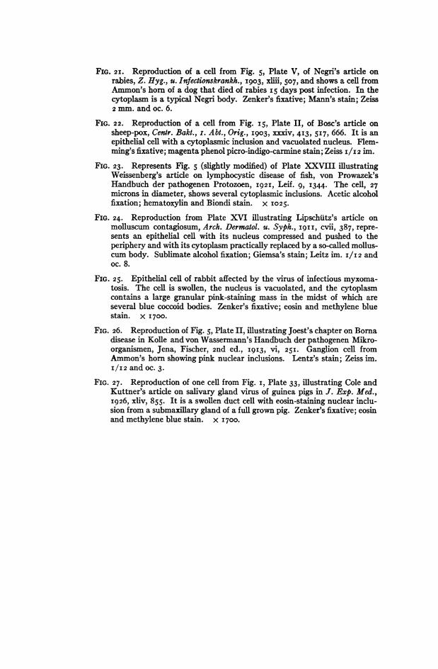
FIG. 2I. Reproduction of a cell from Fig. 5, Plate V, of Negri's article onrabies, Z. Hyg., u. Infectionskrankh., 1903,, xiii, 507, and shows a ceU fromAmmon's horn of a dog that died of rabies I5 days post infection. In thecytoplasm is a typical Negri body. Zenker's fixative; Mann's stain; Zeiss2 mm. and oc. 6.
FIG. 22. Reproduction of a cell from Fig. I5, Plate II, of Bosc's article onsheep-pox, Cenir. Bakt., i. Abt., Orig., 1903, Xxxiv, 4I3, 5I7, 666. It is anepithelial cell with a cytoplasmic inclusion and vacuolated nucleus. Flem-ming's fixative; magenta phenol picro-indigo-carmine stain; Zeiss I/I2 im.
FIG. 23. Represents Fig. 5 (slightly modified) of Plate XXVIII imustratingWeissenberg's article on lymphocystic disease of fish, von Prowazek'sHandbuch der pathogenen Protozoen, I92I, Leif. 9, I344. The cell, 27microns in diameter, shows several cytoplasmic inclusions. Acetic alcoholfixation; hematoxylin and Biondi stain. x I025.
FIG. 24. Reproduction from Plate XVI imlustrating Lipschiitz's artide onmolluscum contagiosum, Arch. Dermatol. u. Syph., 1911, CVii, 387, repre-sents an epithelial cell with its nucleus compressed and pushed to theperiphery and with its cytoplasm practically replaced by a so-called mollus-cum body. Sublimate alcohol fixation; Giemsa's stain; Leitz im. I/[2 andoc. 8.
FIG. 25. Epithelial cell of rabbit affected by the virus of infectious myxoma-tosis. The cell is swollen, the nucleus is vacuolated, and the cytoplasmcontains a large granular pink-staining mass in the midst of which areseveral blue coccoid bodies. Zenker's fixative; eosin and methylene bluestain. x I700.
FIG. 26. Reproduction of Fig. 5, Plate II, mlustrating Joest's chapter on Bornadisease in Kolle and von Wassermann's Handbuch der pathogenen Mikro-organismen, Jena, Fischer, 2nd ed., I9I3, Vi, 25I. Ganglion cell fromAmmon's horn showing pink nuclear inclusions. Lentz's stain; Zeiss im.I/I2 and oc. 3.
FIG. 27. Reproduction of one cell from Fig. i, Plate 33, mlustrating Cole andKuttner's article on salivary gland virus of guinea pigs in J. Exp. Med.,I926, xliv, 855. It is a swollen duct cell with eosin-staining nuclear inclu-sion from a submaxillary gland of a full grown pig. Zenker's fixative; eosinand methylene blue stain. x 1700.
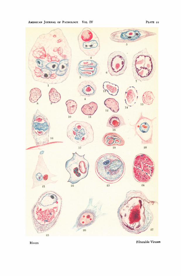
AMERICAN JOURNAL OF PATHOLOGY. VOL. IV
10~~~~~~~~~1
19 20
23
I,
III..I;;w
Filterable Viruses
PLATE 2 2
Rivers
kw*4,.

PLATE 23
FIG. 1. Tobacco mosaic. "Young palisade cell containing striate materialradiating from the nucleus and a crescent-shaped body (A) adjacent tonucleus. " (After Rawlins and Johnson, Am. J. Bot., I925, Xii, I9.)
FIG. 2. "Leaf cell containing mitotic figure, crescent-shaped body (A), andstriate material." (After Rawlins and Johnson.)
FIG. 3. " Cell of a short glandular trichome containing a body (A) of the sametype shown in Fig. 4." (After Rawlins and Johnson.)
FIG. 4. " Cell of a short glandular trichome showing striate material and vacu-olate body (A) containing a dark-staining granule." (After Rawlins andJohnson.)
FIG. 5. Intracellular body (marked X) in rosette-diseased wheat. (AfterMcKinney, Eckerson, and Webb, J. Agric. Res., I923, xxvi, 605.)
FIG. 6. Shows nuclear and cytoplasmic inclusions in paravaccinia. Reproduc-tion of Fig. I2, Plate III, in Lipschiitz's article on paravaccinia, Arch. Der-matol. u. Syph., Orig., I9I9-20, CXXVii, 193.
FIG. 7. Reproduction from Guarnieri's article on vaccinia in I892 showing avaccine body in the cytoplasm.
FIG. 8. Reproduction from Weigert's article on smallpox in I874 showingamitotic giant cells and bodies in cytoplasm of affected cells similar to theones later described by Guarnieri.
FIGS. 9-12. Cells showing nuclear and cytoplasmic inclusions in smallpox.(After Councilman, Magrath, and Brinckerhoff, J. Med. Res., 1904, xi, 12.)
FIGS. 13 and I4. Polyhedral bodies in jaundice of silkworms. (After vonProwazek, Cenir. Bakt., i. Abt., Orig., I9I2-I3, lXvii, 268.)

AM1ERICAN JOURNAL OF PATHOLOGY. VOL. IV- , ~~~~~~~~~~~~~~~~~~~~~~~~~~~~...... ... .... PLATE 23
_-
L. .....!
.i
6
... ...
|~f.r ::.
\.. ...
C72
....l:?.. ...'
Filterable VirusesRivers
Ift,'
t1
.3p, zz
;e

PLATE 24
FIGS. I-4. Illustrations by Joseph (igi8) showing the remarkable increase insize of cells affected by the virus of lymphocystic disease of fish. In Fig. i,there are three small dark cells showing the early effect of the virus, andtwo large ones exhibiting the picture presented at a later stage. Fig. 3 pre-sents a cell going to pieces. Fig. 4 shows new cells filling the space left byone degenerated cell. Fig. i = X 250; Fig. 2 = x 650; Fig. 3 = x 200;Fig. 4 = X 200.
FIG. 5. Reproduction from Macallum's (I892) article showing the changesthat take place in epidermal cells under the influence of molluscum con-tagiosum virus.
FIGS. 6-9. Reproductions from Ludford and Findlay's article (I926) on fowl-pox. Fig. 6 shows small vacuole with minute adherent granules. Fig. 7represents a cell with a mitotic figure and two virus bodies. Fig. 8 showsreversal of Golgi apparatus. Fig. 9 presents a well developed virus body ina greatly swollen cell.

AMERICAN JOURNAL OF PATHOLOGY. VOL. IV
a
Filterable Viruses
PLATE 24
Rivers
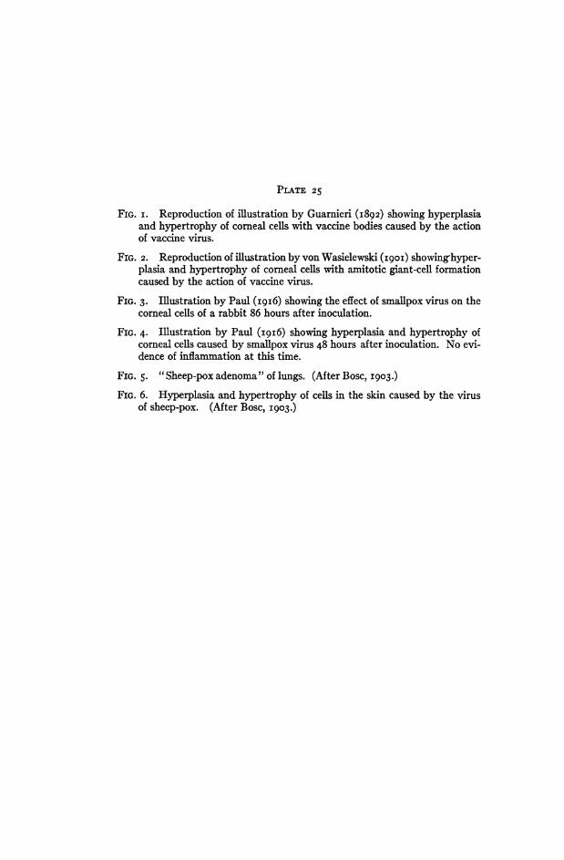
PLATE 25
FIG. i. Reproduction of illustration by Guarnieri (I892) showing hyperplasiaand hypertrophy of corneal cells with vaccine bodies caused by the actionof vaccine virus.
FIG. 2. Reproduction of imustration by von Wasielewski (I9OI) showinghyper-plasia and hypertrophy of comeal cells with amitotic giant-cell formationcaused by the action of vaccine virus.
FIG. 3. Illustration by Paul (I9I6) showing the effect of smallpox virus on thecorneal cells of a rabbit 86 hours after inoculation.
FIG. 4. Illustration by Paul (I9I6) showing hyperplasia and hypertrophy ofcorneal cells caused by smallpox virus 48 hours after inoculation. No evi-dence of inflammation at this time.
FIG. 5. "Sheep-pox adenoma" of lungs. (After Bosc, 1903.)FIG. 6. Hyperplasia and hypertrophy of cells in the skin caused by the virus
of sheep-pox. (After Bosc, I903.)

AmiskicAN JOURNAL OF PATHOLOGY. VOL. IV
....
''Of?
i\\ >.ik.AV . i.
-F a!f;i
J} llif
7ito e-;
ira
.. vc,) -
A~~~~~~~~~~~~~~~~~~~~~~~~~~~~~~~~~~~~~~~~~~~~~~- ~ ..
lob-
tobit5 - -:-Y Ced
-. ...4 ,
............-
Filterable Viruses
.:;
PLATE 25
,.: .-- .: ...m-:k ...11 .;' "
1.:..
i ..-A ),
loll"I)j, Sod
Rivers
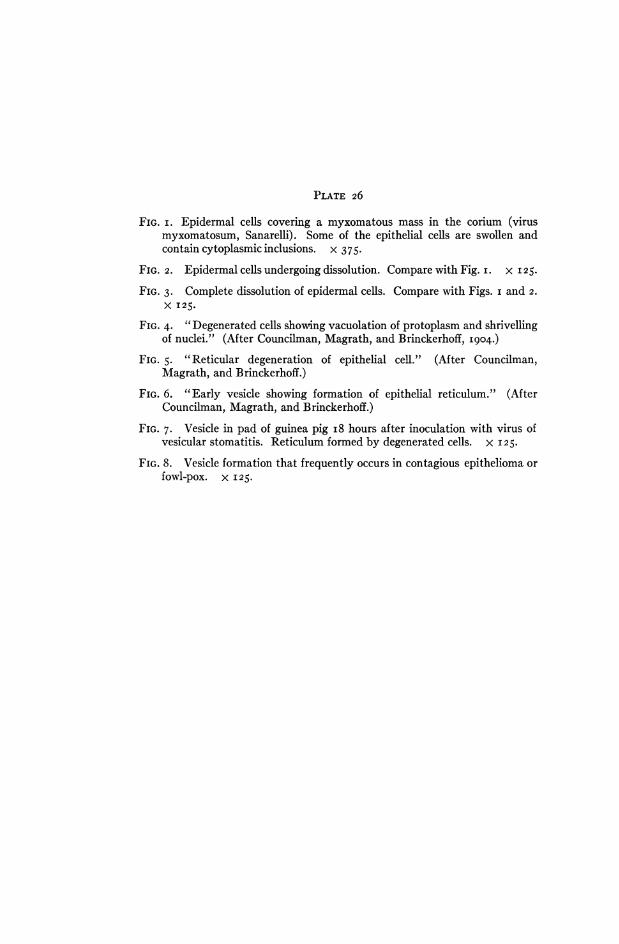
PLATE 26
FIG. i. Epidermal cells covering a myxomatous mass in the corium (virusmyxomatosum, Sanarelli). Some of the epithelial cells are swollen andcontaincytoplasmicinclusions. x 375.
FIG. 2. Epidermal cells undergoing dissolution. Compare with Fig. i. X I 25.
FIG. 3. Complete dissolution of epidermal cells. Compare with Figs. i and 2.X I25.
FIG. 4. "Degenerated cells showing vacuolation of protoplasm and shrivellingof nuclei." (After Councilman, Magrath, and Brinckerhoff, 1904.)
FIG. 5. "Reticular degeneration of epithelial cell." (After Councilman,Magrath, and Brinckerhoff.)
FIG. 6. "Early vesicle showing formation of epithelial reticulum." (AfterCouncilman, Magrath, and Brinckerhoff.)
FIG. 7. Vesicle in pad of guinea pig i8 hours after inoculation with virus ofvesicular stomatitis. Reticulum formed by degenerated cells. x I25.
FIG. 8. Vesicle formation that frequently occurs in contagious epithelioma orfowl-pox. x I25.

AMERlCAN JOURNAL OF PATHOLOGY. VOL. IV
Filterable Viruses
PLATE 26
Rivers

PLATE 27
FIG. I. "Crown tissue from rosette-diseased plant" (wheat). "Note the cellscontaining intracellular bodies in addition to the nuclei and also the granu-lar nature of the cells which have become necrotic." (After McKinney,Eckerson, and Webb, J. Agric. Res., I923, xxvi, 605.)
FIG. 2. "Crown tissue from healthy plant." Compare with Fig. i. (AfterMcKinney, Eckerson, and Webb.)
FIG. 3. " Section of an early stage of a fruit blister showing the brown necrosisof the epidermal cells and the hyperplasia of the subepidermal cells."(After Gardner, J. Agric. Res., I925, xxx, 87I.)
FIG. 4. " Section of a pericarp lesion resulting from necrosis and collapse of anarea of subepidermal cells and hypertrophy of the cells immediately be-neath the necrotic area." (After Gardner.)
FIG. 5. "Surface blisters on a mosaic tomato." (After Gardner.)
FIG. 6. Mosaic leaf of a gourd. (After Doolittle, U. S. Dept. Agric. !3ull.,No. 879, I920.)
FIG. 7. "Mosaic fruit of pumpkin, showing large dark-green swellings on a* yellow background." (After Doolittle.)
FIG. 8. Mosaic disease of Summer Crookneck squash. (After Doolittle.)

AMERlCAN JOURNAL OF PATHOLOGY. VOL. IV
Filterable Viruses
PLATE 2 7
Rivers

PLATE 28
FIGS. I and 3. Fig. 3 is an enlargement of Fig. i. At the left side of Fig. 3 arenormal bacteria. The large organisms in the center of the picture show theswelling induced by the action of bacteriophage. Pictures supplied byJ. J. Bronfenbrenner.
FIG. 2. Stained preparation of bacteria undergoing lysis. The protoplasm isbeaded and segmented. Picture supplied by J. J. Bronfenbrenner.

AMERICAN JOURNAL OF PATHOLOGY. VOL. IV
'U
I I .
if
. *Mt.t
'o
Filterable Viruses
1ajNL..
i:-- .' ...e: . ..*.::.:.B:. * .k 3
N-
PLATE 28
Rivers